Leukemia Research 28 (2004) 773–774
Letter to the Editor
Imatinib-mesylate for all patients with hypereosinophilic syndrome?
Some recent papers have focused on the activity of imatinib-mesylate, a selective inhibitor of tyrosine kinase, in idiopathic hypereosinophilic syndrome (HES) [1–4]. In this setting, a possible therapeutic target was identified by Cools et al. [2], who described the fusion tyrosine-kinase gene FIP1L1/PDGFRA as the result of an interstitial dele-tion within chromosome 4 in nine out of sixteen (56%) patients affected by HES. Of interest, although in this
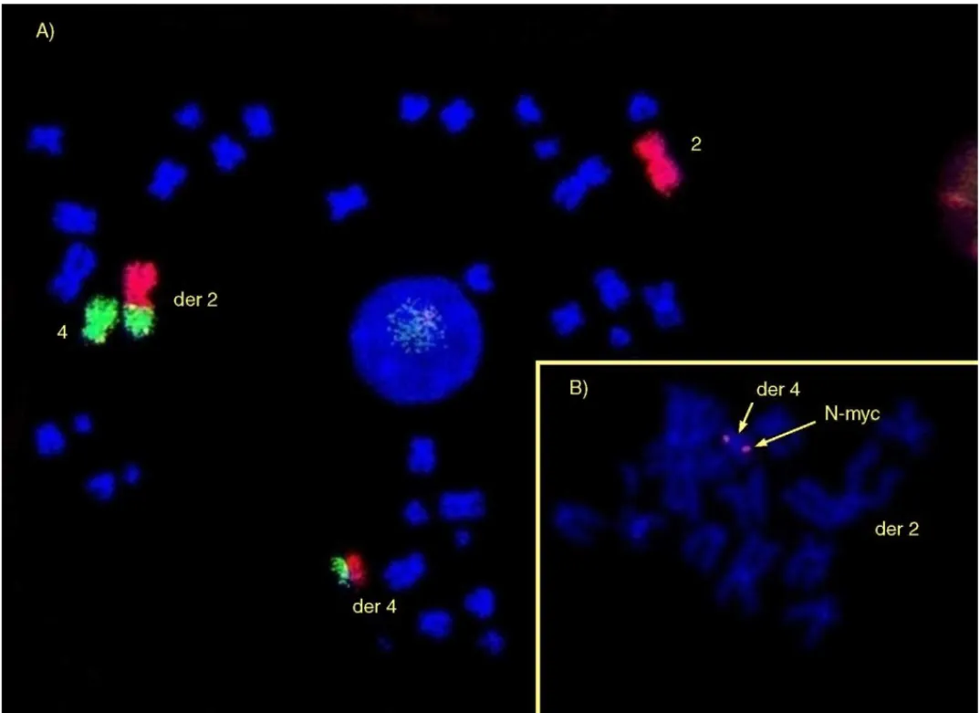
Fig. 1. Panel (A) shows the results of fluorescence in situ hybridization (FISH) with Cy3-labelled (red) chromosome 2 and fluorescein-labelled (green) chromosome 4 libraries (dual-color painting), confirming the cytogenetic evidence of t (2;4) (p24;q12). In panel (B), FISH analysis using a single locus Spectrum-Orange-labelled (red) probe for N-myc shows the translocation of this oncogene from its normal site on chromosome 2 (p24), to the derivative chromosome 4, in a region adjacent to where PDGFRA is located (q12). This strongly suggests the possibility of a N-myc-PDGFRA fusion gene.
study the response to imatinib was strictly correlated with the presence of FIP1L1/PDGFRA rearrangement (all pa-tients with such a molecular lesion treated with imatinib responded), only five out of nine responding patients ev-idenced the abnormal transcript [2]. Among the possible alternative mechanisms for the activation of the PDGFRA tyrosine-kinase domain, these authors suggested there may be a different fusion gene.
We have recently observed a t (2;4) (p24;q12) recipro-cal translocation in 64-year-old male affected by HES with complete clinical, hematological and cytogenetic response
0145-2126/$ – see front matter © 2004 Elsevier Ltd. All rights reserved. doi:10.1016/j.leukres.2003.11.014
774 Letter to the Editor / Leukemia Research 28 (2004) 773–774
to imatinib (100 mg per day), currently maintained after ten months of therapy with a dose of 100 mg given every other day. A preliminary molecular screening performed accord-ing to the previously described procedures[2]failed to de-tect FIP1L1/PDGFRA in this patient. Cytogenetic and FISH analyses (Fig. 1A) suggested the possibility of a molecu-lar lesion different from FIP1L1/PDGFRA rearrangement. Indeed, the fusion of PDGFRA with the oncogene N-myc could be hypothesized (Fig. 1B).
A possible heterogeneity in the molecular pathogenetic mechanisms inducing HES could justify the wide spectrum of response observed in four consecutive patients we re-cently treated with imatinib, which ranged from an impres-sive and durable response to low doses in the patient with t (2;4), to the need of administering higher doses of the drug (400 mg per day) to achieve response, until to the evidence of progressively acquired or primary resistance to doses up to 800 mg per day[5].
Since not all HES patients respond to imatinib, a rele-vant aspect to consider is the fact that clonal populations of CD3−/CD4+ Th2 lymphocytes may secrete large amounts of interleuk5 (IL-5) and other cytokines, possibly in-volved in the pathogenesis of at least some cases of HES [6,7]. Although serum levels of IL-5 do not seem to represent a surrogate marker of response to imatinib in this disease [3], it is intriguing to note that all cases of HES responding to imatinib so far reported did not evidence the presence of T cell clones expansion, when tested[1,3]. By contrast, one patient described by Pardanani et al. [3] and the only our patient in whom an occult T cell clone could be detected, did not respond to imatinib at all. It is conceviable that “T cell-correlated” HES may have a different pathogenesis, not including constitutive activation of tyrosine-kinases. There-fore, it may be expected these forms are not responsive to imatinib.
Imatinib-mesylate is certainly a promising treatment for HES. However, the clinical and biological profile of patients who are potential responders needs to be better defined.
References
[1] Gleich GJ, Leiferman KM, Pardanani P, Tefferi A, Butterfield JH. Treatment of hypereosinophilic syndrome with imatinib mesylate. Lancet 2002;359:1577–8.
[2] Cools J, De Angelo D, Gotlib J, Stover EH, Legare RD, Cortes J, et al. A tyrosine kinase created by fusion of the PDGFRA and FIP1L1 genes as a therapeutic target of imatinib in idiopathic hypereosinophilic syndrome. New Engl J Med 2003;348:1201–14.
[3] Pardanani A, Reeder T, Porrata LF, Li CY, Tazelaar HD, Baxter EJ, et al. Imatinib therapy for hypereosinophilic syndrome and other eosinophilic disorders. Blood 2003;101:3391–7.
[4] Cortes JE, Ault P, Koller C, Thomas D, Ferrajoli A, Wierda W, et al. Efficacy of imatinib mesylate in the treatment of hypereosinophilic syndrome. Blood 2003;101:4714–6.
[5] Musto P, Falcone A, Sanpaolo G, Bodenizza C, Perla G, Minervini MM. Heterogeneity of response to imatinib-mesylate (Glivec) in hypereosinophilic syndrome: implications for dosing and pathogenesis. Leuk Lymphoma 2004, in press.
[6] Roufosse F, Schandene L, Sibille C, Willard-Gallo K, Kennes B, Efira A, et al. Clonal Th2 lymphocytes in patients with the idiopathic hypereosinophilic syndrome. Br J Haematol 2000;109: 540–8.
[7] Bain BJ. Hypereosinophilia. Curr Opin Hematol 2002;7:21–5.
Pellegrino Musto∗ Gianni Perla Maria Marta Minervini Angelo Michele Carella
Unit of Hematology and Stem Cell Transplantation IRCCS “Casa Sollievo della Sofferenza” 71013 S. Giovanni Rotondo, Italy ∗Corresponding author. Tel.:+39-0882-411389;
fax:+39-0882-411389.
E-mail address: [email protected] (P. Musto).
Francesco Lo Coco Gianfranco Catalano
Chair of Hematology, “Tor Vergata” University, Rome, Italy