POLITECNICO DI MILANO
Master of Science in Nuclear Engineering
Department of Energy
COMBINING MAGNETIC HYPERTHERMIA
WITH HADRONTHERAPY:
AN IN-VITRO STUDY ON PANCREATIC
TUMOR CELLS
Relator: Prof. Stefano Luigi Maria GIULINI CASTIGLIONI
AGOSTEO
Correlator: Prof. Daniela BETTEGA (INFN)
Master Thesis of:
Maria Claudia MANSI
ad Adriana e Chiara,
donne eroiche
Contents
Abstract………I Estratto………...II Riassunto……….III
Introduction………..1
Chapter 1: The Importance of Radiotherapic Technique in Cases of Pancreatic Cancer………3
1.1 Introduction to Carcinogenesis………..3
1.1.1 Cell-division Cycle……….5
1.1.2 Neoplastic Transformation………..7
1.1.3 Pancreatic Adenocarcinoma………...9
1.2 Fundamentals of Ionizing Radiation and Human Body Interaction………11
1.2.1 History………11
1.2.2 Fundamentals of Radiobiology……….13
Absorbed Dose………..15
Densely and Sparsely Ionizing Radiations & Linear Energy Transfer…………16
DNA Damages………22
Radiolysis of Water……….24
Loss of Reproductive Capacity & Surviving Curves………...27
Relative Biological Effectiveness………..30
Oxygen Enhancement Ratio………36
Chapter 2: Conventional Radiotherapy and Hadrontherapy………..…38
2.1 Principles of radiation treatment ………...39
Local Control Probability……….40
Fractionation of Dose……….42
Target Volume………43
Types of Radiation used to Treat Cancer……….45
2.2 Conventional Radiotherapy……….47
2.2.1 Mechanisms of Photonic Interaction with Matter………48
2.3 Hadrontherapy………54
2.3.1 History………54
Bragg Peak and Spread-Out Bragg Peak………..58
Bethe Equation………..59
2.3.3 Systems to obtain Spread-Out Bragg Peak………60
2.3.4 Problems of Fragmentation of Ions after the Interaction with Body and Lateral Dispersion………64
Differences between Heavy Ions and Light Ions……….66
Chapter 3: Magnetic Hyperthermia………70
3.1 Hyperthermia for Cancer Treatment……….71
3.1.1 History……….71
3.1.2 Hyperthermia to treat cancer………..72
3.2 Magnetic Fluid Hyperthermia Therapy: use of Nanoparticles……….74
3.2.1 Principles of Magnetic Nanoparticles……….74
3.2.2 Magnetic Nanoparticles in Medical Care………..81
3.2.3 Magnetic Nanoparticles in Hyperthermia Therapy………83
3.2.4 Uptake of Magnetic Nanoparticles in Cell Population………..87
3.2.5 Biological Response to the combined treatment of Magnetic Hyperthermia and Irradiation……….88
Chapter 4: Experimental Procedure………..91
4.1 Characterization of Cell Line BxPC3………..91
4.1.0 Defrosting and Pre-Planting of Cell Population………91
4.1.1 Generation Time & Growth Curve………92
4.1.2 Efficiency of Planting (E.P.)……….93
4.1.3 Growth Curve by Counting of Colonies……….95
4.1.4 Survival Curve of the Cell Line BxPC3 with Photons……….96
4.2 Cytotoxicity due to Nanoparticles………98
4.2.1 Properties of Nanoparticles (NPs).……….98
4.2.2 E.P. & Survival of Cell Population with uptake of NPs………..100
4.3.a Test of Magnetic Hyperthermia on Cell Pellet with uptake of NPs……….…103
4.3.b Cytotoxicity due to Nanoparticles and Hyperthermia………107
4.4 Treatment of Cell Population with Hadronic Radiation: Experiment at CNAO…..108
4.4.1 Survival Curve of the Cell Line BxPC3 with Hadronic Radiation………108
4.4.2 Survival Curve of the Cell Line BxPC3 with Hadronic Radiation & NPs...110
4.4.3 Survival Curve of the Cell Line BxPC3with Hadronic Radiation, NPs and Hyperthermia……….110
Chapter 5: Experimental Results………..112
5.1 Characterization of Cell Line BxPC3………113
5.1.1 Generation Time and Efficiency of Planting: Growth Curve……….113
5.1.2 Growth Curve by Counting of Colonies………..…114
5.1.3 Cytotoxicity due to Photon Radiation………..115
5.2 Cytotoxicity due to Nanoparticles……….116
5.2.1 Efficiency of Plating & Survival of Population of BxPC3 with uptake of Nanoparticles………116
5.2.2 Efficiency of Plating & Survival of Population of BxPC3 cells as a function of Nanoparticles Time Uptake………..118
5.3 Hyperthermic Effect due to Nanoparticles………..120
5.4 Cytotoxicity due to Hadronic Radiation: Results of Experiment at CNAO………….121
5.4.1 Survival Curve of the Cell Line BxPC3 with Hadronic Radiation…………..122
5.4.2 Survival Curve of the Cell Line BxPC3 with Hadronic Radiation & NPs...123
5.4.3 Survival Curve of the Cell Line BxPC3 with Hadronic Radiation, NPs and Hyperthermia……….124
Conclusions………..129
List of Figures
1.1 Main phases of the cell cycle………..6
1.2 Radiosensitivity variation in time………..…7
1.3 Incidence of pancreatic cancer all over the world. ………9
1.4 Imaging from electronic microscope of density of BxPC3………..10
1.5 Portrait of Wilhelm Conrad Röntgen and his discovery………..12
1.6 Cyclotron at Lawrence Radiation Laboratory, Berkley. ………..13
1.7 Injuries to DNA due to high and low LET. ……….18
1.8 Collective Distributions for several radiations of Absorbed Dose in function of LET in water………..20
1.9 Tracks produced by protons at different energies in water vapor, in case of Ionizations and Excitation………..21
1.10 Schematic view of different particle tracks against different biological targets………..22
1.11 Schematic view of DNA and damages induced by ionizing radiation to DNA………….22
1.12 Nature of Reactive Species produced in water by ionizing radiations………..24
1.13 The influences of LET on the following radiation effects on biological tissues…………27
1.14 Survival curves in case of high and low LET. ……….28
1.15 Cell survival curves and RBE definition for a 10% survival level. ………..31
1.16 Survival of CHO-K1 cells as a function of X-ray and 11 MeV/u Carbon ions and RBE values corresponding to different dose-effect levels. ……….32
1.17 RBE trend from experimental data collected for different types of ions from the Heidelberg Ion Beam Therapy center. ………..32
1.18 RBE as a function of the LET for a fixed atomic number Z value and a schematic comparison of RBE values for different types of hadron particles. ………34
1.19 Survival curves for a single irradiation and two subsequent irradiations. ………35
1.20 Distributions represented cells under aerated and hypoxia conditions.……….36
1.21 OER trend, decreasing with the increase of LET. ………37
2.1 Dose – Effect curve for Tumor Control and Healthy Tissues Complications. ………41
2.2 Expected Survival Rate after Fractionation of dose during the radiation treatment…..42
2.3 Schematic showing the different volumes used in three-dimensional treatment planning. ………44
2.4 Dose deposition of electrons, photons and beams of hadrons, depending on the depth of penetration in water. ……….47
2.5 Dose-depth curves in water for photon beams having maximum energies in the range of 6 to 25 MeV. ………48
2.6 The exponential transmission curve of γ-Rays measured under “good geometry” condition. ………..………..49
2.7 Phenomenon of Photoelectric Effect. ………..…….…………50
2.8 Phenomenon of Compton Effect. ………51
2.9 Phenomenon of Pairs Production. ……….…52
2.10 Development of cross sections, in cases of Photoelectric Effect, Compton Scattering and Production of Pairs as a function of the incident photon energy. ……….53
2.11 Loss of energy for different ions of interest in the therapeutic field as a function of the initial kinetic energy. ………..57
2.12 Variation of the specific energy loss in air vs. energy of charged particles shown…….58
2.13 The specific energy loss along an alpha track. ………..58
2.14 Representation of two Bragg peaks: protons and carbon ions………..59
2.15 Schematization of passive scanning for SOBP. ……….62
2.16 Scheme of active irradiation for SOBP..………63
2.17 Comparison of dose distributions with depth related to protons and carbon ions of different energy. ……….65
2.18 Comparison of the angular dispersion experienced by photons, protons and carbon ions as the water penetration distance varies..………..66
2.19 Dose transmission curves with the depth that are obtained with a controlled variation of the energy of the proton beam, Carbon ions and Neon ions..………..67
2.20 Dose deposited in water due to a carbon ion beam of 270 MeV/u..………..69
3.1 Hysteresis curve of ferromagnetic materials. ………..76
3.2 Illustration of the relationship between coercivity and size in small particles………76
3.3 Hysteresis curves of MNPs measured at T = 300 and 5 K for HADROCOMBI experiment. ………..77
3.4 Evolution of the magnetic energy with the tilt angle between the easy axis ………78
3.5 Mechanisms of the relaxation of Brown and Nèel. ………80
3.6 Hypothetical system for magnetic drug delivery……….82
3.7 Schematic representation of a multifunctional magnetic nanoparticle………..83
3.8 The increase of temperature registered on a water dispersion of MNPs at field HAC=16.2 kA/m and frequency f =110 kHz. ………..85
3.9 Extracellular and intracellular uptake in function of Iron concentration in experiment of glioblastoma cells T98G. ………..88
3.10 BxPC3 cells uptake after 72 h of incubation using ICP-OES……….………89
3.11 The increase of temperature registered on cell pellet of BxPC3 (cells incubated for 48 h with 100 μg/ml concentration of MNPs) at field HAC=16.2 kA/m and frequency f =110 kHz. ……….……….90
4.1 Laminar flow hood where cellular experiments take place. ……….92
4.3 Fixation of cell population. ………..………..95
4.4 Colonies visible to naked eye and ready to clonogenic assay. ……….96
4.5 SAR Values in function of applied field on samples of 3mg/ml in liquid, gel at 0.5% and gel at 2%.……….99
4.6 Setup for Magnetic Hyperthermia experiments. ……….104
4.7 Display of treatment irradiation with Ions C. ………109
4.8 Apparatus of Hyperthermia……….111
5.1 Eppendorf of 0.2 ml with cell pellet and uptake of nanoparticles………..119
5.2 Bright field images of fixed BxPC3 cells after 24 h of incubation with 10 g/ml and 25 g/ml. ………..119
5.3 20x phase contrast optical images of fixed BxPC3 cells after 24 h of incubation with: 10 μg/ml and 25 μg/ml. ………119
List of Tables
1.1 Electromagnetic and corpuscular types of radiation………..14
1.2 Modalities of ionization and correspondent type of radiation……….…….15
1.3 Effects due to phenomenon of ionization. ………..16
2.1 Definitions of volumes involved in a radiotherapy treatment………..44
4.1 Number of initial inocula of cells at each time………..……92
4.2 Inocula of cells depending on the concentration of irradiation doses……….97
4.3 Inocula of cells depending on the concentration of nanoparticles to be inserted…..…100
4.4 Inocula of cells depending on the concentration of nanoparticles to be inserted……..101
4.6 Inocula of cells depending on the concentration of nanoparticles to be inserted, for the experiment of magnetic hyperthermia……….106
4.7 Inocula of cells depending on the concentration of irradiation doses………..…109
4.8 Inocula of cells depending on the concentration of uptake of NPs and irradiation doses………...110
4.9 Inocula of cells depending on the concentration of uptake of nanoparticles, irradiation doses and treatment of hyperthermia………..110
5.1 Results of E.P. and Survival in cases of different nanoparticles concentration…………117
5.2 Results of E.P. and Survival for uptake time of 24 hours……….…118
5.3 Results of E.P. and Survival for uptake time of 48 hours……….……118
5.4 Results of cell survival in the case of Ions C alone, in function of dose; Values of the exponential fit of survival curve and its parameters………....122
5.5 Results of cell survival in the case of Ions C and NPs, in function of dose; Values of the exponential fit of survival curve and its parameters……….…...123
5.6 Results of cell survival in the case of Ions C, NPs and Hyperthermia, in function of dose; Values of the exponential fit of survival curve and its parameters………..124
5.7 Results of Survival normalized at the control in the case of Ions C and NPs……….126
5.8 Results of Survival normalized at the control in the case of Ions C, NPs and Hyperthermia…..……….…126
List of Graphs
5.1 Cellular Growth Curve of BxPC3 cells as a function of time after plating……….113 5.2 Average number of cells / colony as a function of time after plating………...….114 5.3 Survival Curves for photonic radiation determined in the experiment done at
Fondazione IRCCS (Istituto Nazionale dei Tumori) of Milan……….…..115 5.4 Comparison of cell survival and E.P. in two as a function of nanoparticles
concentration………..……..117 5.5 Survival Curves for Ions C alone, determined in the experiment done at CNAO of Pavia………...122 5.6 Survival Curves for Ions C and NP, determined in the experiment done at CNAO of Pavia………...123 5.7 Survival Curves for Ions C + NP + Hyperthermia, determined in the experiment done at CNAO of Pavia……….….124 5.8 Survival Curves for Ions C alone, Ions C and NP, and Ions C + NP + Hyperthermia, determined in the experiment done at CNAO of Pavia………..125 5.9 Survival curves of Ions C alone, Ions C and NPs, and Ions C, NPs and Hyperthermia, given by the normalization at control of each case……….……….126
Abstract
The aim of this project is to implement the survival curves of a population of pancreatic tumor cells subjected to hadrontherapeutic treatment in combination with magnetic hyperthermia. This combined procedure involves the insertion into the in vitro cell samples of magnetite nanoparticles that, once the uptake is performed, can release heat through the external action of an alternating magnetic field. The increase in temperature would represent a radiosensitizing factor which, in addition to irradiation by hadronic particles (carbon ions), would enhance the action of such radiotherapy, bringing damage difficult to repair to the tumor area. The experiments included a preliminary phase of characterization of the BxPC3 cells line, the study of cytotoxicity as a function of different concentrations of nanoparticles and in the presence of hyperthermic treatment, so as to choose the most suitable protocol for the final experiment: irradiation with bundles of carbon ions on cell samples already sensitized by the presence of nanoparticles, and consequent heating by the hyperthermia system. The results allowed to obtain survival curves with the same decreasing exponential trend, but gradually increasing slope in relation to the addition of radiosensitizers: this shows that the combination of nanoparticles, hyperthermia and irradiation with such densely ionizing particles brings an improvement in efficiency of the hadrontherapy, even if in percentages not too high. In fact, at the same dose, there is an increase in mortality (and therefore a decrease in survival) of about ten percent, a good result that can be improved with further research. The next step will be to use the same protocol for photonic radiation, applicable to conventional radiotherapies.
Estratto
Lo scopo di questo progetto è implementare le curve di sopravvivenza di una popolazione di cellule tumorali pancreatiche sottoposta a trattamento adroterapico in combinazione con l’ipertermia magnetica. Questa procedura combinata prevede l’inserimento nei campioni cellulari in vitro di nanoparticelle di magnetite che, una volta effettuato l’uptake, sono in grado di rilasciare calore tramite l’azione esterna di un campo magnetico alternato. L’innalzamento della temperatura rappresenterebbe un fattore radiosensibilizzante che, in aggiunta all’irraggiamento da parte di particelle adroniche (ioni carbonio), potenzierebbe l’azione di tale radioterapia, portando danni difficilmente riparabili alla zona tumorale. Gli esperimenti hanno previsto una fase preliminare di caratterizzazione della linea cellulare BxPC3, lo studio della citotossicità in funzione di diverse concentrazioni di nanoparticelle ed in presenza di trattamento ipertermico, di modo da poter scegliere il protocollo più idoneo per l’esperimento finale: irraggiamento con fasci di ioni carbonio su campioni cellulari già sensibilizzati dalla presenza di nanoparticelle, e conseguente riscaldamento tramite il sistema di ipertermia. I risultati hanno consentito di ottenere curve di sopravvivenza con stesso andamento esponenziale decrescente, ma pendenza via via crescente in relazione all’aggiunta di radiosensibilizzanti: ciò dimostra che la combinazione di nanoparticelle, ipertermia e irradiazione con particelle così densamente ionizzanti porta un miglioramento dell’efficienza del trattamento adroterapico, anche se in percentuali non troppo elevate. A parità di dose si ha infatti un incremento della mortalità (e quindi un decremento della sopravvivenza) di circa il dieci per cento, risultato buono e potenziabile con ulteriori ricerche. Infatti, il prossimo passo sarà utilizzare lo stesso protocollo per la radiazione fotonica, applicabile alle radioterapie convenzionali.
Riassunto
L’adroterapia è una tecnica radioterapica che sfrutta le proprietà fisiche delle particelle adroniche (principalmente i protoni e gli ioni carbonio) per ridurre la presenza di un’area tumorale ed impedirne la crescita cellulare. Infatti, gli adroni sono in grado di rilasciare gran parte della propria energia alla fine del percorso in un mezzo, in corrispondenza del picco di Bragg, consentendo di effettuare un piano di trattamento in modo tale da trasferire tutta (gran parte) dell’energia nella regione in cui è presente il tumore. Inoltre, trattandosi di radiazione di alto LET (Linear Energy Transfert, i.e. trasferimento di energia per unità di percorso) il danno causato sulla popolazione cellulare è maggiore rispetto alle tecniche radioterapiche convenzionali (in cui si utilizzano raggi gamma e x – radiazioni di basso LET), poiché non permette il riparo delle strutture biologiche danneggiate dalla ionizzazione della radiazione incidente. Questa ultima caratteristica può dunque essere impiegata nella cura dei tumori radioresistenti, come il tumore del pancreas.
Lo scopo di questo lavoro di tesi è la valutazione della citotossicità indotta in una popolazione di cellule tumorali pancreatiche (BxPC3), dovuta all’irraggiamento da parte di particelle adroniche, e nello specifico di fasci di ioni Carbonio. Poiché le curve di sopravvivenza di popolazioni bombardate da radiazione ad alto LET evidenziano la diminuzione esponenziale della proliferazione cellulare, vi sono importanti vantaggi riguardo la probabilità di controllo del tumore durante i trattamenti radioterapici. Infatti, si è pensato di introdurre due agenti radiosensibilizzanti con l’intento di apportare maggiore mortalità alla popolazione sotto indagine: è il caso delle
nanoparticelle di magnetite e dell’ipertermia magnetica (i.e. innalzamento della
temperatura fino ad un valore di circa 42°C). Va da se che per ottenere l’andamento della sopravvivenza della popolazione in vitro (e quindi la mortalità promossa dagli agenti) è stato necessario effettuare diversi test sperimentali, di modo da identificare:
o l’andamento di crescita della popolazione in esame in condizioni simili a quelle dell’organismo biologico (temperatura di 37 °C, no agenti esterni introdotti),
o la sopravvivenza della popolazione all’irraggiamento da parte di una radiazione di basso LET (fotoni di 6 MV, con valori di dose 0, 0.5, 1, 2, 3, 5, 7 Gy),
o la sopravvivenza della popolazione all’inserimento di nano particelle di magnetite Fe3O4 (con concentrazioni di 0, 10, 25, 50, 100 g/ml e un tempo di assorbimento (o uptake) di 24 o 48 ore),
o la funzionalità del sistema di ipertermia magnetica, e quindi la valutazione del corretto innalzamento della temperatura all’interno della popolazione (opportunamente propagata e tenuta in provette da 1 o 0.2 ml), sia senza sia con l’uptake delle nano particelle,
o la sopravvivenza della popolazione al bombardamento da parte di fasci di Ioni Carbonio (esperimento effettuato al Centro Nazionale di Adroterapia Oncologica (CNAO) di Pavia con accelerazione delle particelle adroniche tramite il Sincrotrone),
o la sopravvivenza della popolazione al bombardamento da parte di fasci di Ioni Carbonio, con precedente uptake (48 ore) della concentrazione di nano particelle più ottimale (50 g/ml, ovvero la concentrazione che dà una bassa percentuale di sopravvivenza della popolazione, circa 30 %, valore molto simile a 100 g/ml, quantità che avrebbe richiesto dell’utile spreco a parità di risultato),
o la sopravvivenza della popolazione al bombardamento da parte di fasci di Ioni Carbonio, con precedente uptake di 48 ore di 50 g/ml di nano particelle di magnetite e conseguente trattamento di ipertermia.
È chiaro che il progetto in questione ha visto una lunga fase preliminare, durante la quale si sono ricercati i risultati più confacenti, così da creare un protocollo per l’esperimento complessivo che coinvolgesse i due fattori radiosensibilizzanti e l’irraggiamento con radiazione di alto LET. Inoltre, ogni test ha richiesto i tempi biologici necessari affinché la popolazione in esame potesse propagare per avere un campione contenente sufficiente numero di cellule, soprattutto nel caso di trattamento ipertermico; infatti, per la popolazione BxPC3 ed un numero dell’ordine del milione di cellule il tempo stimato per la propagazione e la crescita è di circa dieci giorni.
L’utilizzo delle nano particelle con proprietà magnetiche, combinato al trattamento ipertermico, è stato pensato per via dei meccanismi di rilascio termico conseguenti all’applicazione di un campo magnetico alternato (AMF). Infatti, nella magneto-fluido ipertermia (MFH), l’inibizione della crescita della popolazione tumorale è ottenuta attraverso l'inserimento delle nano particelle (ferro-fluido) nella zona da scaldare (nel nostro caso nella provetta o nelle flasks). L'energia necessaria a provocare il surriscaldamento del campione è dunque generata dall'esposizione delle nanoparticelle al AMF, la cui intensità deve essere sufficiente a raggiungere gli scopi del trattamento (campo di 16,2 kA/m alla frequenza di 110 kHz), ma non è troppo elevata per evitare danni ai tessuti sani se sperimentata durante un ipotetico trattamento in vivo.
L’esperimento al CNAO (Centro Nazionale di Adroterapia Oncologica) ha permesso di ottenere i dati per la realizzazione di tre curve di sopravvivenza:
o Curva di Sopravvivenza in funzione della dose dei soli Ioni Carbonio
o Curva di Sopravvivenza in funzione della dose degli Ioni Carbonio e uptake
delle Nanoparticelle
o Curva di Sopravvivenza in funzione della dose degli Ioni Carbonio, uptake delle
Nanoparticelle e conseguente Ipertermia.
La pendenza della curva di sopravvivenza relativa al trattamento combinato ioni carbonio e nanoparticelle e quella relativa al trattamento combinato ioni carbonio, nanoparticelle e ipertermia è rispettivamente maggiore del 13 e 18 % di quella relativa al trattamento con i soli ioni carbonio. I risultati dimostrano che l’ultima delle curve citate mostra un andamento più ripido e induce maggior citotossicità alla popolazione in esame, così da confermare l’ipotesi del potenziamento apportato dall’ipertermia magnetica.
1
Introduction
Among the most serious and obscure questions of the individual nowadays are those concerning the onset of cancers. In Italy, about 280,000 patients undergo radiation treatment every year in the 187 radiotherapy centers operating in the national territory
[1] and in the last fifteen years, the increase in cases of pancreatic cancer has reached
almost 60 percent.
How can we prevent and combat the occurrence of this disease? They are manifested in various forms and have different cure depending on the organ and the stage reached at the time of diagnosis. One of the most difficult forms to treat is cancer of the pancreas; in fact, it does not give any sign at an early stage, but in an advanced state, when survival possibilities are low, and dimensions have reached considerable levels. These conditions do not allow successful surgical removal, which would cause serious damage to the organ and the surrounding tissues, but as a viable alternative, radiotherapy treatment is proposed.
Hadrocombi is a project financed by the INFN (Istituto Nazionale di Fisica Nucleare)
to make a significant contribution to refining the parameters involved in such techniques so that they can successfully cure those pathologies, involving several groups at national level, which have carried out tests and experiments to pursue their goal for the overall project. The main goal is to investigate the co-operative action of Hadrontherapy (radiotherapy using hadronic radiation) and Magnetic Fluid Hyperthermia, considering both interesting alternative techniques in those cases where conventional techniques fail. As a valid comparison with this proposed technique we have used an X-ray radiotherapy control combined with Magnetic Fluid Hyperthermia. The group I took part in developing this master thesis project was responsible for the response of cultures of BxPC3 pancreatic tumour cells in vitro in relation to introduction of nanoparticles, the magnetic hyperthermia and the irradiation, and the
2
resulting cytotoxicity due to these factors, single or combined. Along with other groups involved in the project, we carried out several tests to find out what the best choice of convoluted parameters was to achieve the best result of cellular mortality. Specifically, we had changed the uptake time and the concentration of nanoparticles, the radiation dose, the size of the test tubes used, and we evaluated the cytotoxicity induced by a clonogenic assay.
We finally found the standard of values that gives more mortality to the specific cell population, hoping that this result could be used in vivo as a valid support in vivo for Hadrontherapy.
3
Chapter 1
The Importance of Radiotherapic Techniques in Cases of
Pancreatic Cancer
1.1
Introduction to Carcinogenesis
It is well known that our body is made up of so many compartments capable of performing specific tasks to maintain a regular functioning of our vital functions. In general, such compartments are the organs, which in turn are made up of tissues composed of eukaryotic cells. These cells have different composition depending on their function, and we can classify them into four categories, depending on tissue that they constitute: epithelial, connective, muscular and nervous.
For example, the connective tissue is composed by macrophage blood cells such as lymphocytes (which are part of the immune system), as well as all blood cells, but also adipocytes, cells that collect and preserve lipids (fat), or bone cells like osteoblasts. To study biological systems from the most elementary physical entities (molecules and atoms inside cells), we need to look at the phenomena that underlie the structure and functions of living organisms: these include atomic and molecular motions, molecular aggregations and chemical reactions. The interactions among atoms, molecules and supramolecular structures are the basis of the main functions that determine the natural processes that give rise to inorganic, organic and biological matter. Current knowledge of the nature of such interactions is constantly developing and provide descriptions useful to understand the functionality of cellular and tissue substrates. It is therefore normal to think that mistakes in the homeostasis, that is the balance between proliferation and programmed cell death maintained by strictly regulating both processes to ensure the integrity of organs and tissues, could lead to abnormalities in small scales (DNA mutations), which can then be transposed into a series of tissue or systemic complications.
4
The process of neoplastictransformation finds its birth: normal cells become cancerous due to the subsequent accumulation of genetic mutations that govern cell proliferation and it could lead tissues to apoptosis or necrosis.
The alterations leading to this malignant transformation can be grouped according to some essential features:
o the ability to proliferate independently of the presence of growth factors (oncogene activation);
o the ability to escape apoptotic signals, which favor cell death;
o the insensitivity to antiproliferative signals (inactivation of antithorogens); o the capacity of unlimited cell divisions;
o the potential to colonize sites away from the onset (metastasis);
o the ability to stimulate the production of new blood vessels (neoangiogenesis). The acquisition of these characteristics, each one capable of conferring the cell on a selective growth advantage, proceeds both through staged phases and by alternative chronological sequences, not only in the different types of tumors, but within the same tumor type.
This phenomenon is called Carcinogenesis, term that derived from greek “καρκίνος” or from latin “cancer”, which means “crab” and with the suffix “-genesis”, from greek “Γένεσις”, with means “genesis”; the entire word actually means "that raises cancer”. The growth of tumor mass is different depending on the type of tissue, and hence the organ involved in the neoplastic transformation; in fact, we know that there are low and high cellular turn-over organs, defined as life cycle of the cells, then the speed of tissue regeneration.
The choice to treat pancreatic cancer cells lies in this fundamental difference: it is a high activity proliferating organ, which is therefore able to regenerate its cells quickly, unlike systems like the nervous system that have the same cells, neurons, throughout the life cycle.
This feature of rapid regeneration leads to the fast development and expansion of the cancerous area, up to the formation of metastasis and secondary tumors, preventing from noticing any signs at an early stage but in an advanced state, when survival possibilities are now low, and dimensions have reached considerable values.
5
These conditions do not allow for successful surgical removal, which would cause serious damage to the organ and surrounding tissues, but as a valid alternative radiotherapy treatment is proposed. For a good understanding of how radiation therapy sensitizes the affected tumor area and promotes mass reduction, it is important to understand first what are the basal characteristics of a cell, its normal functioning, and possible neoplastic transformations; then we will discuss the basis of the interaction between ionizing agents and those biological components.
1.1.1 Cell-division Cycle
“The dream of every cell is to become two cells”
François Jacob
The cell is the fundamental unit of every living organisms, which can be distinguished in unicellular (such as bacteria or protozoa) and pluricellular ones, a category which, without a doubt, humans belong to.
Unicellular organisms are composed of cells with morphologic characteristics usually uniform; by increasing the number of cells in an organism, however, they differ in form, size and specialized functions, up to the constitution of tissues, organs and apparatus.
It is therefore important that within each cell there is a regular exchange of nutrients and waste materials, to ensure homeostasis and so proper cell survival.
Obviously after a while, each cell will run out of its own life cycle depending on its characteristics and functions; for example, pancreatic cells have a life cycle of about two days, but it is different for other cells, as neurons that are the same for the entire human life.
We can therefore give a general definition of a cell cycle: sequence of events between two successive cell divisions; such events, better defined as cellular phases, place the cell under varying conditions. In fact, for example, during the Synthesis phase, the cell is organized to perform a synthesis of its genetic heritage, to ensure the division into two daughter cells with the same genetic information.
6
Fig 1.1 Main phases of the cell cycle. [3]
Interphase: G0, G1 (Pre-Synthesis), S (DNA Synthesis), G2 (Pre-Mitosis). Mitosis: M.
Each component involved in the cell cycle will have a different evolution depending on its role within the cellular environment. For example, microtubules are composed of proteins and they play an essential role in cell division because of their responsibility for producing mitotic spindle; without them the cell would not be able to make the cell fit for the division of its membrane.
In radiobiology it is important to understand at which time the cell's life cycle is present, since it is in a condition of greater or less radiosensitivity.
In general, some factors influence this propensity: for example, high-rate division tissue has more probability to be radiosensitive. Moreover, the radiosensitivity of a tissue depends on the percentage of mitotic activity and the inverse of the degree of cell differentiation; cells are more sensitive in G2-phase (pre-mitosis) and M-phase (mitosis).
In an actively proliferating population each cell progresses in its cycle independently of the others and hence the fraction of cells occupying the different phases is proportional to the duration of the phases.
The loss of proliferative activity could be attributed to two factors: chemical agent and radiation; the first one has a dose threshold, but the second one has an unfavorable behavior and an asymptotic behavior.
7
Fig. 1.2 Radiosensitivity variation in time. [4]
1.1.2 Neoplastic Transformation
As mentioned above, a normal cell can become carcinogenic due to spontaneous genetic mutations or induced by chemical agents or ionizing radiation.
In our case, it is interesting to understand the general concept of neoplasia, as the project involves studies on an already tumoural population, for which it is desired to have a high mortality rate because of high LET radiation incidence; it will break DNA bonds, giving apoptosis and/or necrosis of the affected area and thus reducing tumor mass.
The problem of radiation-induced neoplastic transformation is only related to the chance that irradiation will strike healthy tissue around the tumoural area that you want to reduce.
Now let us make a general definition of neoplasia: it is an "abnormal mass of tissue that grows excessively and in an uncoordinated manner with respect to normal tissues, and which persists in this state after cessation of the stimuli that have induced the process"
8
Mutations in the genetic heritage of some cells alter tissue homeostasis, and continued proliferation promotes the formation of a significant tumor mass.
Moreover, it is important to say that cancer cells grow in multilayer, so we could distinguish them to the normal ones.
The rate of growth of a neoplasia is influenced by three fundamental factors:
o Growth Fraction: the percentage of cells in a neoplastic population that is included in the proliferative compartment.
o Growth rate of the tumor, and hence with which neoplastic cells complete the cell cycle;
o The ability of the mass to ensure sufficient trophic contribution through neoangiogenesis, and therefore the position within the tissue.
The growth of the tumor mass may have an exponential trend, for which the volume increases with a constant fraction in equal intervals of time. The formula describing this situation is as follows:
𝑉(𝑡) = 𝑉
0𝑒
0,693𝑇𝑑 𝑡
(1.1) where Td is the time of tumor duplication, for which the tumor grows slower when it has reached asignificant size. Instead, non-exponential growth may be due to cellular loss, to passage to a non-proliferating layer, and to increased duplication time. We can also consider a relationship between the number of cell divisions and the tumor mass achievable, assuming that:
o Cells grow exponentially according to the law : 𝑁(𝑡) = 2𝑡/𝑇𝑑 (1.2),
o The mass of the single cell is in the order of 10-9 g, o There is no cell loss.
For example, for forty divisions it will have 1012 cells and then 103 g of tumor mass; therefore, above the thirtieth divisions the tumor is detectable (m=1 g).
Neoplastic cells divide into p - cells, q - cells, differentiated cells, and dying cells. This distinctionis due to the alternation between highly proliferating cells and those that are part of the necrotic tumor tissue. Specifically, dying cells are found in the tumoural area with insufficient vascularization, so they go against necrosis (cell death) due to lack of oxygen supply. In contrast, "p" cells are part of the growth fraction of the cancerous
9
population, and are highly proliferating. Among proliferating cells are also those defined as "q", quiescent cells that activate if necessary to give tumor repopulation. The category that does not cause problems because it is not able to divide is that of differentiated cells.
The loss of some cells, due to the entry of differentiated ones or the lack of oxygen-free division, is regulated by growth, if there is no excess of proliferation.
1.1.3 Pancreatic Adenocarcinoma
The final goal of this project is to improve hadrontherapy in case of pancreatic cancer. This choice comes from the seriousness of this kind of pathology: in fact, pancreas is a high-rate of proliferation organ, so the increasing of tumoural mass is rapid and it is often no possible to identify it until the most critical states.
The incidence of pancreatic cancer varies greatly across regions and populations: incidence rates for pancreatic cancer in 2012 were highest in Northern America (7.4 per 100000 people) and Western Europe (7.3 per 100000 people), followed by other regions in Europe and Australia/New Zealand (equally about 6.5 per 100000 people).
[5]
Fig.1.3 Incidence of pancreatic cancer all over the world. [6]
he lowest rates (about 1.0 per 100000 people) were observed in Middle Africa and South-Central Asia. For now, during the 2017 about 54000 cases were estimated, of
10
which about 3% of new diagnosis. The estimated deaths during the 2017 were about 43000 and survival percentage is almost 8%. [7]
Clearly, this type of cancer brings several problems and significant mortality, so this project focuses on the choice of experimenting about one of the most common stabilized cellular line: BxPC3 (cell dimension about nine µm). This line is capable to produce important colonies of cells in not so long time, so it is easy to implement experiments.
11
1.2
Fundamentals of Ionizing Radiation and Human Body Interaction
1.2.1 History
Nowadays it is common knowledge to consider radiation as an energy source that, if incident on the human body, causes alterations in genetic heritage and therefore significant immediate or tardive damages.
Thus, this destructive capability can be exploited to eliminate tumoural masses from the body, which is the role of oncology radiotherapy; in the next paragraphs we will see the properties and the consequences induced by different types of radiation, and hence the different results obtained by conventional radiotherapy (taking advantage of the physical characteristics of photonic radiation) and by Hadrontherapy (where the radiation involved is the hadronic one).
Before that it is best to remember what the first discoveries were about the interaction between the body and the radioactivity.
In the Autumn of 1895 the German physicist and mechanical engineer Wilhelm Conrad Röntgen observed a strange and penetrating radiation (called X-Rays) came from his vacuum tube, without immediately knowing the great potential of his discovery. That evening, concluded daily experiments on cathode ray, he had left the Crookes tube covered with black paper sheets on the table, a few meters away from a sheet of fluorescent material. Exiting the laboratory lights, as he came out, he observed an intense fluorescence of green colour emitted by the barium platinum cyanide crystals, of which the plate was made.
Thus, began the study of the radiation that Röntgen called "X" to indicate that it was still of unknown type. The name remained, although many of his colleagues suggested calling it "Röntgen Rays" in his honour.
The characterization of these rays led to the knowledge of high frequency electromagnetic waves, that is, highly energy and low wavelengths, capable of penetrating the bodies and to "see inside".
After that, in the Spring of 1896 the French physicist Antoine Henri Becquerel discovered the natural radioactivity and, two years later, the Polonium and Radio were
12
discovered by the French physicist and spouses Pierre Curie and Maria Sklodowska Curie.
Fig. 1.5 Portrait of Wilhelm Conrad Röntgen and his discovery. [9]
Although the biological effects of ionizing radiation were not yet well-known at the time, it was understood that exposure to it would cause cutaneous burns, so the idea of treating the tumours using these mysterious radiations was soon accomplished and some surface tumours were successfully treated by applying a radioactive source directly in contact with the skin.
These pioneering treatments marked the birth radiotherapy (RT). Unfortunately, however, a problem arisen immediately which is still today one of the biggest challenges in the radiotherapy field: to really have effectiveness must be achieved so that radiation reaches only the tumour mass, saving as much as possible the surrounding healthy tissues.
The idea of using accelerators in the medical field to focus the beam radiation came to American physicists Ernest Lawrence and Stan Livingston, who in 1931 built the first cyclotron.
At first, patients with salivary gland tumours were irradiated, and neutron beams were used for accelerated deuton collisions up to 5 MeV with a Beryllium target. Considering that such neutrons produce nuclear fragments, these treatments can be considered the first use of ions for cancer treatment.
Subsequently, he was the American physicist Robert Rathbun Wilson who in 1946 had the idea of curing the tumours using charged particles and published his famous article "Radiological Use of Fast Protons". He was called by Harvard University in the same
13
year to lead the team that would have to design and build the new 160-MeV cyclotron, thus commencing collaboration with Ernest Lawrence at Berkley.
From this work the idea of ion-beam therapy (IBT) was born: during cyclotron shielding studies, Wilson realized that the protons in the matter released energy in a totally different way from a beam of X-rays.
We can now justify this fact with the knowledge of the Bragg peak, high energy release at the end of the typical pathway of the defined hadronic particles.
Fig. 1.6 Cyclotron at Lawrence Radiation Laboratory, Berkley (California). [10]
In the next paragraph, there will be an array of radiobiology basics to understand what is the mechanism that causes cancer cell deterioration due to radiotherapy techniques and what are the different modes for more efficient treatments.
1.2.2 Fundamentals of Radiobiology
Radiobiology is the branch of Physics that deals with the interaction of ionizing radiation with biological matter; its study contributes to the development of new methods of radiotherapy and of the prediction of the most suitable treatment for the individual patient, and provides knowledge of the processes underlying the tumour and normal tissue responses.
The interaction between radiation and human body is manifested by the transfer of energy through processes of excitation and ionization of the atoms and molecules involved in the tissue crossed by radiation.
14
The excitation process, typical of the interaction between optical radiation and matter, consists in the absorption of an amount of energy such as to bring an atomic electron to a higher energy level, usually followed by de-excitation: the electron returns to its fundamental state which results in the emission of photons (quantum of electromagnetic energy) with energy equal to the difference between the energy states involved.
We speak of ionization when the energy absorbed by the material is higher than the typical binding energy of the electrons in the material (ex. the binding energy for the water molecule is about 0.8 eV).
The kinetic energy that characterizes the released electron is given by the difference between the energy transferred by radiation to matter and its binding energy; thus, this could be released, breaking bonds and damaging the matter.
Types of Radiation
Electromagnetic Quantum of Energy X-Rays γ-Rays Corpuscolar Charged or Neutral
Particles Protons Electrons Neutrons α particles Ions
Tab.1.1 Electromagnetic and corpuscular types of radiation.
The incident radiation can ionize directly or indirectly due to energy absorption mode in matter; in fact, the following tables show types of ionizing radiation and how the ionization process takes place.
15
Tab.1.2 Modalities of ionization and correspondent type of radiation.
Absorbed Dose
The biological, chemical or physical effects induced in the biological matter by ionizing radiations are therefore related to the physical characteristics of the radiation field and are manifested only because of a transfer of energy to matter.
So, in order to solve the problem intuitively, it is appropriate to introduce a magnitude, the absorbed dose, which represents the ratio between the imparted energy dEa actually absorbed in the mass volume dm, and the infinitesimal mass dm. Ea represents the sold energy average to a certain volume; it is a stochastic quantity and may also have large fluctuations if small volumes or modest fluxes of particles are considered, but if we consider the mean value no longer considered stochastic.
𝐷 =
𝑑𝐸𝑎𝑑𝑚 (1.3)
with
𝐸
𝑎= 𝑅
𝑖𝑛+ 𝑅
𝑜𝑢𝑡+ ∑ 𝑄(𝐽)
(1.4)which means that the energy absorbed into a volume V of interaction in matter is the difference between the amount of energy delivered by the directly and indirectly ionizing radiation, which “enters” into the volume considered and the one that exits,
Modalities of Ionization
Directly
Radiation can produce directly chemical and biological alterations in matter. Protons Ions Electrons Indirectly
Interacting with matter, give up energy to
release charged particles, such as electrons and ions. These are secondary
products are responsible for biological damage. Neutrons X-rays γ-rays
16
plus the sum of the energies liberated minus the sum of those consumed in each transformation of nuclei and elementary particles in that volume.
The term ΣQ expresses the mass conversion processes in energy and vice versa: it considers, for example, the fact that within the volume considered can occur phenomena such as the production of pairs. In the first case this term gives a positive contribution to the absorbed energy, on the contrary, the contribution is negative. The unit of measurement in the International System is J / kg, defined as Gray (Gy). Can we understand if 1 Gy is a significant value and what is the absorbable limit of a human before going to a catastrophe?
If we take the example of raising 1 ° C of a litre of water, we know that it is possible if we supply 1000 cal of energy, which corresponds to 4160 Gy; so, Gray is a small unit of measurement, but this is true only if the energy is absorbed as the heat of the mass. In fact, damage mechanisms are triggered by ionization and not by heat, so a dose of 1 Gy is a high dose.
If we consider that the lethal dose for an average man is 4 Gy, and we make a calculation to find the corresponding energy in cal, we find a ridiculous value comparable to a cup of coffee.
The big difference is that the energy is deposited locally and breaks the bonds into the most important molecule of our organism, causing local or systemic damage.
Thus, if an average person weighs 70 kg and the absorbed dose is 4 Gy, we find an energy value of about 67 cal, much higher than the chemical bond energies between biologically important atoms.
To be able to characterize biological effects, it is important to consider the absorbed dose but also the time frame at which it is administered: The Dose Rate 𝑑𝐷
𝑑𝑡 is then
introduced.
The absorbed dose is an important magnitude when dealing with ionizing radiation, such as it describes the energy transfer (power supply) at the macroscopic level; however, to describe more completely the effects of ionizing radiation on biological matter, it is important to introduce other quantities, such as the Linear Energy Transfer
17
(LET), Relative Biological Effectiveness (RBE), and the Oxygen Enhancement Ratio (OER). These quantities will be better defined in the following paragraphs.
Densely and Sparsely Ionizing Radiation & Linear Energy Transfer
To understand how radiation interacts with the human body and bring about considerable damage such as carcinogenesis, it is necessary to ask immediately what area is affected by the interaction. Specifically, the incident radiation on an organism interferes with and deposits energy with the fundamental part of the genetic cell: DNA (Deoxyribonucleic Acid).
It is a biological macromolecule made up of nucleic acids (biopolymers composed by monomers, which are nucleotides made of three components: a 5-carbon sugar, a phosphate group and a nitrogenous base) and consisted in turn by nitrogenous bases (adenine, guanine, cytosine, thymine). This molecule has a double helix structure: the nitrogenous bases of a DNA helix combine to the other of the complementarity rule: Adenine joins Thymine and Guanine joins Cytosine.
The sequence of these components gives the DNA the gene information that gives the corresponding cell its function and allows it to transfer that information to the two daughter cells. The ionizing radiation incident breaks the bonds of such molecules, altering the gene information of DNA and thus leading to the mutation of the cells themselves.
For a better understanding of how important this interaction and its consequences are, we must distinguish radiations that deposit locally small energy from those that instead deposit them several, causing hence damages.
So, it is important to define the Linear Energy Transfer (LET) and make a distinction between densely and sparsely ionizing radiation.
Together with time, radiosensitizing substances and phases of the cell cycle, LET represents one of the modulating factors for cellular response to ionizing radiation; it is a term used in dosimetry and describes how much energy an ionizing radiation transfers to the material traversed per unit distance.
18
Fig. 1.7 Injuries to DNA due to high and low LET. [11]
By definition, LET is a positive quantity and depends on the nature of the radiation as well as on the material traversed:
𝐿𝐸𝑇 =
𝑑𝐸𝑑𝑥
(1.5)
where
dE
represents the average energy transferred locally to matter anddx
the infinitesimal displacement of the particle inside the tissue crossed.For a LET estimate, local energy deposition events must be calculated within a generic dm mass element due to both the primary particle (in the case of directly ionizing radiation) and any secondary particle produced.
The LET calculation is complicated for several reasons: first, radiation is never monochromatic, i.e. characterized by particles or photons all with the same energy. Since LET depends on energy, if radiation has an energy spectrum, it will also be characterized by a spectrum of LET.
Second, the energy sold during the transfer events made by the various particles or photons that constitute the radiation is never the same; secondary particles are then generated at different speeds, expanding the spectrum of the LET.
19
An average value of LET can be calculated from empirical bases, measuring the energy transferred along the path and mediating the results. So, typically the unit of measurement used is the keV/μm.
For energy release to be considered local, secondary particles must possess kinetic energy such that their path is within the mass element dm chosen; by convention it is considered that the release is local for electrons produced with energies of up to 100 eV, which can move away from the production site up to a maximum of 5 nm.
Thus, LET defined is indicated by LETΔ, where Δ is an energy limit of 100 keV, that is therefore a maximum range for secondary electrons, above which the power supply is no longer considered local; if an energy limit is set so high that all energy losses are considered local, the LET would coincide with the loss of energy for collisions (Stopping Power).
In the case of the LET∞, the total transfer events will be included in the calculation of the energy released, so that the LET∞ will actually be identical to the
−
𝑑𝐸𝑑𝑥
described by
20
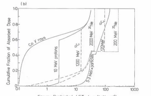
Fig. 1.8 Collective Distributions for several radiations of Absorbed Dose in function of LET in
water. (a) Unrestricted LET distribution for spectrum of charged particles from γ-rays of Co60, monoenergetic neutrons of 14.6 and 1.8 MeV for short paths of monoenergetic heavy ions tracks. (b) Energy-Restricted LET distributions with threshold energy for δ-rays of ∆=100 eV,
for slowdown spectrum of charged particles from γ-rays of Co60, for α-particles tracks of 5.3 MeV, and for short paths of tracks of some heavy ions with energy of 10 or 100 MeV/u. [12]
Since secondary particles have different speeds and different charges they are characterized by different LET values, so an average value of the LET is calculated, though it does not fully describe the properties of energy deposition events at the microscopic level, it is widely used to characterize the quality of radiation.
Secondary loads in motion may have sufficient energy to construct traces separate from those of the primary charge particle (δ-rays) and thus produce ionization at a distance from it, or they can form only a few ion groups near the primary track itself, if their energy is modest.
The value of this path is different between primary radiation and secondary tracks due to charging and different velocities; for example, if we consider secondary electrons (secondary particles released from indirectly ionizing radiation) and we get a power limit of 100 eV, we find that it is ionized every 5 nm. Considering that the double helix of the DNA has a width of about 2 nm, depending on the value of the distance between
21
ionization events it is possible to distinguish two types of radiation: low and high LET radiation.
In the first case, we have a Sparsely Ionizing Radiation, so the average ionization distance is about 100 nm. In fact, for electromagnetic radiation, LET is calculated from the kinetic energy of secondary produced electrons, which for X and γ is in the order of 0.1-1 MeV, bringing to LET values between 0.3 and 2.5 keV/μm. For example, LET of secondary electrons from γ-rays of Co60 is about 0,23 keV/µm, so the average distance between two ionization events is almost 200 nm.
In the second case, however, the track of primary radiation will be denser of ionization events, and this inevitably leads to greater damage inflicted on the crossed material (and thus on DNA); such radiation is also called Densely Ionizing and includes charged particles, ions and alpha particles, for which the transferred energy values are greater than 50-100 keV/μm. For example, LET of α-particles with energy is 43 keV/µm, so the distance between two ionization events is almost 1,4 nm, with the production of “track core”. For carbon ions the LET varies in the range between 15 and 170 keV/μm, which is 100 times greater than in the case of conventional photon beams. [12]
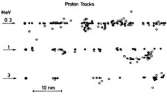
When energy deposition events overlap along the path of the particle they create a “continuum” of ionizations on the track core. The track will also be characterized by a region called “track penumbra”, which corresponds to region crossed by δ-rays that move away from the primary trace; the radius of the penumbra € will equal the maximum range of delta electrons. The energy released in the center of the track or in the penumbra is noticeably different. By leaving the core, the deposited energy decreases approximately as r-2. The sparsely ionizing radiations on the contrary have a trace structure characterized by distinct and separate events in space.
Fig. 1.9 Tracks produced by protons at different energies in water vapor, in case of Ionizations
22
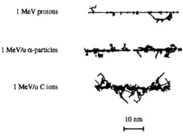
Fig. 1.10 Schematic view of different particle tracks against different biological targets. [14]
So, LET value doesn’t completely describe properties of energy deposit at microscopic level, but because of it is a simple quantity it is used to characterize radiation quality.
DNA Damages
As mentioned earlier, the underlying problems arising from the interaction of ionizing radiation with biological matter are the consequent damages to DNA. The fact that the genome is unique within the cell makes it essential that the base sequence be preserved unaltered and that the DNA molecule is not damaged in order to guarantee the proper maintenance of cellular functions.
Fig 1.11 (a) Schematic view of DNA, with focus of components of nitrogenous bases; (b)
Damages induced by ionizing radiation to DNA. [4]
23
For indirect ionization refers to the phenomenon whereby a generally low LET and sparsely ionizing moves secondary electrons (delta ray radiation) that will be the protagonists of the ionization of the medium atoms.
In the case of direct ionization, however, this is a radiation (generally) of high LET or densely ionizing that can directly break the bonds established in biologically important molecules such as DNA and water (of which it is composed of 80% (control) our body). The phenomenon of water radiolysis involves the release of hydrates electrons and highly reactive radicals, such as ROS (Reactive Oxygen Species), capable of producing poisonous molecules to the cellular environment; this topic will be discussed in the next section.
As for DNA damages, if we assume that a deposition event of energy leads to split atomic bonds, the action of a secondary electron energy thus brings the ionization and/or excitation of atoms of the nucleic acids DNA, causing injuries of various degree of danger.
Damage with the most drastic consequences, that could lead the cell to death or mutations which are likely to lead to carcinogenesis, is the Double Strand Break, a phenomenon that breaks both helix chains. If the breaks are close, the repair of the damage is not enough to prevent such disastrous evolution; if such injuries are far enough away, damage can be repaired, as in the case of the Single Strand Break.
This last one can easily be repaired based on the complementary rule: the intact helix acts as a “mold” to rebuild the damaged helix part.
Clearly, densely ionizing radiation, for which ionizations are close, is more likely to lead to DSB lesions, and therefore carcinogenesis or cell deaths.
Other DNA damage can be related to the loss of nitrogenous bases or to the breaking of the existing bond between sugar and phosphoric acid, quickly fixed and no chain breaks; finally, incorrect nitrogenous bases match can lead to denaturation of certain areas resulting in chain broadening, while intramolecular or protein cross linkings alternating cellular vital functions could be created.
Also, considering that during Mitosis, the chromatin, made up of thickened DNA, tends to form chromosomes, so ionizing radiation can also cause damage to them, such as chromosomal aberrations.
24
From the above, magnitude and type of damage caused by ionizing radiation on DNA depends on the type of radiation, the modality of ionization action, the distinction between DNA in solution and cell DNA and, as we shall now deepen, on the production of hydroxyl radicals due to the radiolysis of water.
Radiolysis of Water
The human body, and therefore the cellular environment, consists mostly of water (> 70% in the body). The incidence of radiation causes ionization and/or excitation of the constituent atoms of this vital molecule, resulting in free radical liberation.
The problem arises when such radicals recombine with each other to give serious chemical damages to the body, such as hydrogen peroxide.
For a good understanding of the mechanism we see what happens to a single molecule of water when it is affected by radiation.
Fig. 1.12 Nature of Reactive Species produced in water by ionizing radiations. (a) Aqueous
electron of solvated electron; (b) Hydrogen radical; (c) Hydrogen ion; (d) Hydroxyl radical; € Hydroxyl ion. [4]
The resulting processes can be:
- Ionization: H2O + rad H2O+ + e- H2O+ + e-hydrate - Excitation: H2O + rad H+ + OH
-and lead to the formation of the four primary radiolysis products, each of which will have a different path and a different mean life time. Specifically, e-hydrate is defined as the “hydrate” electron, because electron in water is never free, but is surrounded by a
25
complex of water molecules; this is possible because the electron interacts electrostatically with the positive pole of the polar molecule of water and creates the typical structure (Fig.1.11).
The average life of hydrated electron is 2 * 10-4 s, much higher than that of the free electron which is of the order of a few picoseconds, because the molecules of water surrounding the electron shield it from the outside, not reacting immediately with the atoms of the surrounding environment. In contrast, for H+ and OH- radicals the average life value is about 1 μs. In the specific case of • OH is such that it can move within a range of 12 to 15 nm, so this radical must be created close to the target in the DNA molecule to be able to spread and interact, considering that the chains are 2 nm and bases 3.4 nm between them. Subsequently, the further interaction of these products between them yields secondary products, as can be seen in the reactions below:
H2O+ H • + • OH e-+ H2O e-hydrate
e- + H2O H2O- H • + OH e- + H+ H •
It is also possible to have recombination, which leads to the formation of innocuous or devastating molecules, such as the above-mentioned hydrogen peroxide:
e-hydrate + e-hydrate + 2H2O H2 + 2OH- H • + H • H2
• OH + • OH H2O2 • OH + H • H2O
26
Tab.1.3 Effects due to phenomenon of ionization.
Hydrogen peroxide H2O2 is an oxidizing agent capable of penetrating into cell membranes by catalyzing metal ions to form • OH radicals.
Alternatively, it can react with Cl- ions to form hydrochloric acid ClOH. Such species can interact with DNA, causing the typical breaks mentioned above, and therefore critical consequences at cellular level.
The presence of O2 favors further reactions with radiolysis radicals. In solutions containing Oxygen there is formation of a greater amount of H2O2, while the Oxygen is partially regenerated.
Effects
due to Ionizing
Radiation
Low LET
High LET
Production of H2O2
Low probability of recombination between hydroxide radicals •OH
because of a major distance between following ionizations
(ionic clusters).
High probability of recombination for smaller distance between
ionizations.
Production of Chromosome
Abnormalities
Low probability because of a requirement of almost two clusters
close enough to involve breaking.
High probability of production for smaller distance between
ionizations.
Inactivation of
Enzymes One single ionization is enough to inactivate enzymes.
Waste of energy, so minor inactivation events.
27
Fig. 1.13 The influences of LET on the following radiation effects on biological tissues:
oxygenated water production, enzyme inactivation and chromosome aberrations. [4]
Loss of Reproductive Capacity & Surviving Curves
At the base of our project lies the concept of the mortality, and therefore the loss of proliferative capacity, caused by the incident radiation on the cellular population in question.
The term "cell survival" expresses an unlimited proliferation capability and the survival curves show a percentage of the population surviving at a given radiation dose value; knowing this trend you can choose the most appropriate dose for optimal hadrontherapy treatment.
Numerous studies have shown that for high-LET radiations the cell-survival curve is an exponential decrease, whereas low-LET radiation, the curve has a shoulder at low doses.
Normally, the curve is represented in semi-logarithmic scale, i.e. the x-axis indicates the dose and axis of the ordinates the logarithm of base 10 of survival.
![Fig. 1.2 Radiosensitivity variation in time. [4]](https://thumb-eu.123doks.com/thumbv2/123dokorg/7522933.106236/23.892.202.658.166.440/fig-radiosensitivity-variation-in-time.webp)
![Fig. 1.4 Imaging from electronic microscope of density of BxPC3. [8]](https://thumb-eu.123doks.com/thumbv2/123dokorg/7522933.106236/26.892.207.686.337.640/fig-imaging-electronic-microscope-density-bxpc.webp)
![Fig. 1.6 Cyclotron at Lawrence Radiation Laboratory, Berkley (California). [10]](https://thumb-eu.123doks.com/thumbv2/123dokorg/7522933.106236/29.892.277.616.356.631/fig-cyclotron-lawrence-radiation-laboratory-berkley-california.webp)
![Fig. 1.7 Injuries to DNA due to high and low LET. [11]](https://thumb-eu.123doks.com/thumbv2/123dokorg/7522933.106236/34.892.226.666.93.432/fig-injuries-dna-high-low-let.webp)


![Fig 1.14 Survival curves in case of high and low LET. [15]](https://thumb-eu.123doks.com/thumbv2/123dokorg/7522933.106236/44.892.223.666.158.479/fig-survival-curves-case-high-low-let.webp)
