DOCTORATE
MOLECULAR MEDICINE AND MEDICAL BIOTECHNOLOGY XXXI CYCLE
Isolation and Characterization of Circulating Tumor Cells from
Patients with Metastatic Breast Cancer
Tutor Candidate
Prof. Bianca Maria Veneziani Maria Rita Carbone
COORDINATOR Prof. Vittorio Enrico
Avvedimento Academic Year 2017/2018
ABSTRACT ... 1
Abbreviations ... 2
1. INTRODUCTION ... 3
1.1 Breast cancer: epidemiological data and risk factors ... 3
1.2 Breast cancer molecular subtypes and standard therapies ... 4
1.3 Metastatic breast cancer ... 6
1.4 Circulating Tumor Cells (CTCs) ... 8
2. AIM ... 13
3. MATERIALS AND METHODS ... 14
3.1 Cell cultures ... 14
3.2 Patient samples and blood collection ... 14
3.3 Sample processing for circulating cells isolation ... 15
3.4 PBMCs isolation ... 16
3.5 Real Time PCR analysis ... 16
3.6 Immunofluorescence ... 17
4. RESULTS ... 18
4.1 RosetteSepTM kit allows the enrichment of mesenchymal cells and pauperizes blood samples of adherent cells ... 18
4.2 Comparison between PBMCs and adherent cells isolated by Ficoll stratification ... 19
4.3 Isolated adherent cells express epithelial and mesenchymal markers .. 20
4.4 Systemic therapy affects the number of different populations of circulating cells ... 21
5. DISCUSSION ... 24
6. CONCLUSION ... 28
7. REFERENCES ... 29
1
Introduction: Circulating tumor cells (CTCs) are cells originating from primary and/or metastatic tumor and circulating freely in the peripheral blood of cancer patients. CTCs can serve as a liquid biopsy for real time monitoring of disease progression and therapy efficacy. Due to CTCs rarity in the blood and the lack of strategies to isolate CTCs entire heterogeneous population, CTCs phenotype has not been fully defined. Commonly used epithelial markers-based isolation approaches failed in detecting epithelial mesenchymal (EMT) transitioned CTCs. The purpose of this study was to isolate and characterize the entire population of viable CTCs from metastatic breast cancer (MBC) patients.
Methods: Peripheral blood samples from 44 locally advanced and MBC patients and 4 healthy donors (HD) were collected in this study. In the first part of the study we isolated CTCs from blood samples of 14 MBC patients through an antibody-mediated enrichment procedure (RosetteSepTM). Then, freshly isolated CTCs from 8 samples were analyzed by immunofluorescence (IF), whereas isolated cells from 6 samples were seeded on cover glass. In the second part of the study we isolated circulating cells from 30 MBC samples and 4 HD by using Ficoll density gradient followed by the seeding of free labeled PBMCs on cover glass. In 6/30 samples mRNA expression levels of CD45, ERα, HER2, CK19 and Vimentin were evaluated by qRT-PCR for both PBMCs and adherent cells. Furthermore, IF experiments were performed on adherent isolated cells, and on samples collected after three months of therapy, for testing the presence of ERα, HER2 and different EMT markers.
Results: CTCs isolated by RosetteSepTM protocol stained positive only for the mesenchymal marker Vimentin, and RosetteSepTM seemed to affect the adherence ability of circulating cells. Interestingly, adherent cells isolated by Ficoll stratification showed reduced mRNA levels of leukocyte marker CD45 and higher mRNA levels of CK19 and Vimentin than PBMCs. Also, Vimentin and CK19 proteins were the most representative markers in adherent cells fraction. Furthermore, therapy reduced the number of CD45- cells and affected the number of different adherent cells subpopulations.
Conclusions: We established a label-free method for the isolation and characterization of circulating cells in MBC, which allowed the depletion of most contaminating leukocytes and the enrichment of circulating cells expressing epithelial and mesenchymal markers. Heterogeneity of adherent cells underlined the importance of further characterize these cells to understand more about their role in metastatic disease and to improve personalized therapeutic approaches.
2 CTCs Circulating tumor cells CK5 cytokeratin 5
CK18 cytokeratin 18 CK19 cytokeratin 19
EGFR Epithelial growth factor receptor EMT Epithelial Mesenchymal Transition EpCAM Epithelial cell adhesion molecule ER estrogen receptor
HD healthy donors
HER2 epidermal growth factor receptor 2 IF immunofluorescence
MBC metastatic breast cancer OS overall survival
PBMCs Peripheral blood mononuclear cells PFS progression free survival
PR progesterone receptor RBCs red blood cells
TNBC triple negative breast cancer WBCs white blood cells
3
1. INTRODUCTION
1.1 Breast cancer: epidemiological data and risk factors
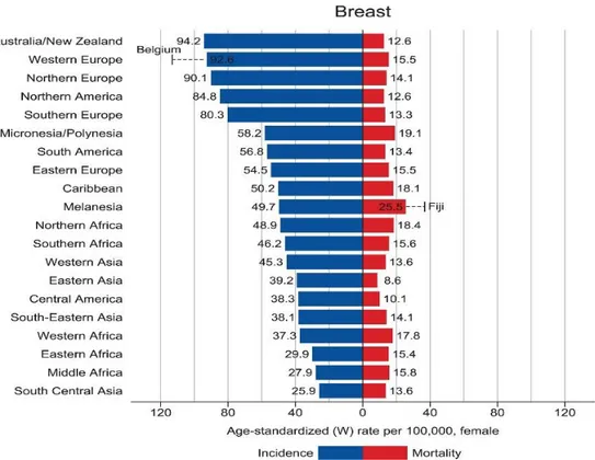
Breast cancer is the second most common cancer for both sexes combined, preceded only by lung cancer. Among women, breast cancer is the most frequently diagnosed cancer with 2,1 million newly diagnosed cancer cases in 2018 (Bray et al. 2018). Also, it represents the leading cause of cancer death among females in over 100 countries. Breast cancer incidence rate is highest in Australia/ New Zeeland, Western and Northern Europe, Northern America, Southern Europe; however, mortality rate compared to incidence is higher in less developed regions, such as Middle / Western Africa and South-Central Asia, than in more developed ones (figure1). This trend relies on different factors related to social and economic development: having few children, obesity, sedentary lifestyle, breast cancer screening and awareness. Indeed, inherited mutations (BRCA1, BRCA2, and other breast cancer susceptibility genes) are responsible for only the 5-10% of breast cancer cases, whereas non-hereditary factors are the main drivers of observed differences in incidence rate among countries (Bray et al. 2018). Non- hereditary risks factors for breast cancer developing include long exposure to estrogen hormones (early menarche, late menopause, nulliparity, late age at first birth, few children) and nutrition (alcohol intake, high fat diet, high weight). On the contrary, low fat diet, breastfeeding and physical activity are considered protective factors (Falkenberry and Legare 2002).
4
Figure 1. Bar chart of region-specific incidence and mortality for breast cancer in 2018 (Bray et al. 2018).
1.2 Breast cancer molecular subtypes and standard therapies
Breast cancer is a highly heterogeneous disease, which is commonly classified in noninvasive or in-situ carcinoma, confined to the ducts and lobules, and infiltrating or invasive carcinoma, in which malignant cells have penetrated through the duct wall into the stroma. In addition, both in-situ and invasive breast carcinomas are defined as ductal or lobular depending on the site of origin of tumor cells. In the last decades, the improving of gene profiling techniques have been allowed a molecular classification of breast tumors based on the expression of hormones receptors, estrogen receptor (ER) and progesterone receptor (PR), and the low expression or amplification of human epidermal growth factor receptor 2 (HER2) gene (Perou et al. 2000). Importantly, this molecular classification of breast tumors is fundamental for therapeutic planning. Indeed, there are four molecular breast cancer subtypes: Luminal A, Luminal B, HER2 positive and Basal like, that are treated with specific therapies.
Luminal A is ER/PR positive, and it accounts for 50% of breast cancers. The Luminal B subtype, representing the 20% of invasive breast tumors, is positive for ER/PR and HER2, has a proliferative index rate (Ki67) and a histological
5
grade higher than Luminal A. HER2 positive subtype accounts for the 15% of invasive breast cancers overexpress HER2; this subtype is characterized by high Ki67 and commonly shows TP53 mutations. The basal like breast cancers, also named triple negative, show a pattern of expression similar to mammary basal epithelial cells and myoepithelial cells. They are ER/PR and HER2 negative, typically express cytokeratins 5/6 (CK5/6) and/or epithelial growth factor receptor (EGFR), have high Ki67 and are frequently TP53 mutated. This last group comprises about 15% of invasive breast tumors and has in general poor prognosis.
Molecular classification of breast cancer subtypes has helped in defining type specific targeted therapy.
Estrogen dependent breast cancers (Luminal subtypes) are treated with inhibitors of the estrogen signaling pathway, such as Tamoxifen. Tamoxifen, a selective estrogen receptor modulators (SERM), is the first approved drug for the treatment of estrogen positive metastatic breast cancer; it reduced recurrence by approximately 40%-50%. In menopausal and post- menopausal women, the estrogen signaling pathway is sustained by aromatase enzyme activity, which consists in synthesizing estrogens by using androgen hormones as a substrate. Aromatase inhibitors (anastrozole, letrozole, exemestane) are the elective therapy for this condition (den Hollander et al. 2013).
As estrogen inhibitor are the best choice for estrogens-drived cancer subtypes, the inhibition of HER2 signaling represents the therapeutic goal in HER2 overexpressing breast tumors. In 1998 the FDA approved the first drug for the treatment of HER2 positive breast cancers, the recombinant antibody trastuzumab (Herceptin) (Pegram et al. 1998). Since then, other agents have been approved, pertuzumab (HER2 inhibitor) and lapatinib (tyrosine kinase inhibitor). In 2013 a boosted version of trastuzumab was approved by FDA for the treatment of HER2 positive breast tumors, the conjugated monoclonal antibody TDM1 (trastuzumab emtansine). TDM1 efficiently vehicles the DM1 drug, a microtubule inhibitor, directly into HER2 positive breast cancer cells to inhibit their growth (Verma et al. 2012).
Triple negative breast cancers (TNBC), which lack hormone receptors and HER2, are conventionally treated by using taxol derivatives and anthracycline chemotherapy.
Besides that, some patients have tumors whose cancer cells overexpress specific proteins, which are fundamental to drive tumor growth. Moreover, there are several drugs targeting these molecules that represent useful tools for breast cancer cure. Everolimus (Afinitor), a m-TOR inhibitor, stops cancer cells from getting energy supply (Steelman et al. 2016), and bevacizumab (Avastin), that acts against the vascular endothelial growth factor (VEGF),
6
which stimulates the formation of new blood vessels, necessary for the filling of oxygen and nutrients (den Hollander et al. 2013).
Despite the big effort in tumor classification and in the development of breast cancer subtype targeted therapy, the 25% of breast cancer patients relapse and about 10-15% of patients develop an aggressive disease with distant metastases within 5 years after diagnosis (Colzani et al. 2014).
1.3 Metastatic breast cancer
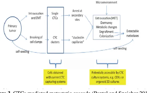
Metastatic breast cancer is a pathological condition in which cancer cells have spread from primary tumor toward distant sites to form secondary tumor, also named metastasis. Metastasis is the main cause of cancer related death. Indeed, breast cancer metastases can occur even ten years after the removal of primary tumor. The common sites of metastases in breast cancer are lymph nodes, bone, liver, lung and brain, but breast cancer can give rise to metastasis at almost any sites. Nowadays the molecular profiling of the primary tumor has helped in designing personalized therapy, and metastatic breast cancer is treated with a combination of chemotherapy and targeted therapy according with the molecular subtype of the primary tumor. Unfortunately, many patients do not respond to systemic treatment due to molecular heterogeneity and dynamic nature of the tumor. Although dissecting the molecular basis of metastasis formation has been the main goal of many research groups in the last decades, how cancer cells acquire the metastatic traits remain unknown.
The metastatic cascade is a very complex biological process and different theories about metastasis development have been proposed. The theory of clonal evolution of cancer supports the idea that metastasis arises from the clonal expansion of preexisting variant cancer cells within the primary tumor mass (Fidler and Kripke 1977). In 2006 Hess and collaborators proposed that drivers mutations confer the metastatic ability to cancer cells (Hess et al. 2006). Besides the need of a metastatic genetic trait, the “seed and soil” theory, enunciated by Paget in 19th century, introduced the relationship between cancer
cells and microenvironment (Paget 1889). Paget affirmed that cancer cells seed metastasis only when they reach a proper microenvironment. Furthermore, there are evidences in the literature that partially support all these theories, making metastasis formation a very articulated process. Indeed, many cancer cells can be released from the bulk of primary tumor, but only few of them are capable to generate metastasis (Gupta and Massagué 2006) (Fidler 2003). To form clinically detectable metastasis, cancer cells must invade the neighboring tissues, entry and survive into the circulation, arrest at secondary site, where they have extravasate and colonize the new region. Once metastatic cells have colonized the secondary organ, they can proliferate to form detectable macrometastasis or, they can form undetectable micrometastasis
7
remaining in a latent state also for years (Vanharanta and Massagué 2013) (figure2).
To face the metastatic cascade, cancer cells need to change their phenotype from a static epithelial to an invasive and migratory one. This occurs because of a biological process called epithelial mesenchymal transition (EMT). Epithelial Mesenchymal Transition, a well-known process involved in embryonic development, wound healing and tissue regeneration, represents a hallmark in metastasis formation and correlates with the acquiring of stem-like features and poorly differentiated phenotype (Micalizzi et al. 2010). During EMT epithelial cells lose cell polarity and cell adhesion and gain invasive and migratory properties. Also, epithelial mesenchymal transitioned cells become resistance to anoikis, substrate-dependent apoptosis, allowing them to survive in the circulation. Generally, cells that have undergone EMT lose or express low levels of epithelial markers, such as cytokeratins 18 (CK18), cytokeratin 19 (CK19), EpCAM and E-Cadherin and express mesenchymal markers, such as N-Cadherin, Alpha-SMA, β-catenin and Vimentin. All these changes are orchestrated by the activation of genetic signatures regulated by specific transcription factors: Snail1, Snail2, Twist, EF1/ZEB1, SIP1/ZEB2 and E47 (Craene and Berx 2013).
Carcinoma cells that have disseminated to distant tissues can persist at the secondary site for a long time as dormant disseminated tumor cells (DTCs), without giving metastasis; because of signaling pathways that sustain DTCs quiescence and survival. The microenvironment contributes to these events, by releasing mediators such as CXCL12 (C-X-C motif chemokine 12), which promotes the activation of SRC and AKT survival pathway in DTCs, or by releasing bone morphogenetic proteins (BMP4/7), which enacts a quiescent program in DTCs that is associated with an ERKlow/p38high signaling state. DTCs dormancy is promoted also by thrombospondin-1 (TSP1), which is present in the basement membrane of blood vessels. Moreover, dormant cells can repress natural killer cell-activating ligand, thus evading NK cells detection (Lambert et al. 2017). The latent state of cancer cells is the responsible for late recurrence in breast cancer.
Blocking the metastatic cascade is fundamental to fight cancer progression. In this scenario, understanding the biology of circulating cancer cells could be the answer to many open questions about the metastatic process and would help in the clinical management of tumor progression.
8
Figure 2. Steps for metastasis formation (Vanharanta and Massagué 2013).
1.4 Circulating Tumor Cells (CTCs)
Circulating tumor cells (CTCs) are cancer cells, that originate from primary and/or metastatic tumor and circulate freely in the peripheral blood of cancer patients (Allard 2004). It has been shown that CTCs resemble cancer stem cells, in terms of self-renewal, tumor initiation capability and invasion at the single cell level (Aktas et al. 2009). Importantly, CTCs are considered the responsible of distant metastasis (figure3). Thus, the study of CTCs could elucidate the molecular basis of metastasis formation and tumor progression. Moreover, CTCs can be found in the bloodstream, thus their sampling can be considered as a “liquid biopsy”, a noninvasive procedure to monitor cancer progression and therapy efficacy.
Importantly, the presence of CTCs in MBC patients has a significant prognostic impact regardless the subtype of breast tumors. Indeed, CTCs count in MBC patients independently predicts progression free survival (PFS) and overall survival (OS).
The first evidence about the prognostic value of CTCs were provided by Cristofanilli et al; they found that patients with CTCs number higher than 5
9
cells per 7,5 mL of blood had a bad prognosis, evaluated by PFS and OS (Cristofanilli et al. 2004).
Figure 3. CTCs mediated metastatic cascade (Pantel and Speicher 2015). Then, the same cut off has been confirmed also by other studies (Hayes et al. 2006; Dawood et al. 2008). Moreover, baseline CTCs number is a predictive marker in patients receiving different type of first-line systemic treatment (Giuliano et al. 2011). Also, many studies have investigated the prognostic value of CTC number, in MBC patients, depending on the subtype of primary tumor. Two studies underlined the prognostic value of CTCs, except for patients with HER2 overexpressing tumors treated with HER2 targeted therapies; thus, it was hypothesized that anti-HER2 treatments eliminate CTCs that freely circulated in the blood of breast cancer patients (Giordano et al. 2012; Pierga et al. 2012). In effect, a large prospective multicenter study, in which only 6,5% of patients received anti-HER2 based therapies, demonstrated the prognostic value of CTCs independently from primary tumor subtype (Wallwiener et al. 2015). Besides the metastatic disease, CTCs enumeration has an important impact also in the prognosis of non-invasive breast cancer. In fact, CTCs were detected also in peripheral blood of early breast cancer patients. In a large study conducted by the German SUCCESS study group (EUDRA-CT No.2005-000490-21, NCT02181101), about 22% of early breast cancer patients had CTCs in their blood. Although all tested cut off ( from 0 to more than 5 CTCs in 30 mL of blood) showed a significant impact on patients outcome, the prognosis deteriorated continuously with increasing CTCs number (Rack et al. 2014). Indeed, in patients with primary
10
breast cancer, who have undergone neoadjuvant or adjuvant therapy, CTCs detection is a prognostic factor for early relapse (Nakagawa et al. 2007; Pierga et al. 2008) (Bidard et al. 2013). All these data underline the importance of CTCs count for monitoring the status of disease in both early and metastatic breast cancer. Despite that, to improve CTCs clinical utility and to develop CTCs based personalized treatment, the enumeration of these cells is not enough. Instead, a complete characterization of CTCs is needed.
Considering CTCs rarity in the blood (1-100 CTCs per 10 9 blood cells), and the lack of unique specific markers to identify them, the isolation of CTCs represents an additional challenge for their molecular characterization. In the last decade many efforts have been focused on the development of techniques to efficiently isolate and characterize CTCs.
CTCs enrichment methods are divided in non-affinity and affinity-based technologies.
Non-affinity isolation strategies distinguish CTCs from surrounding hematopoietic cells according to their physical (size, electric charge, deformability) characteristics. Among these CTCs enrichment techniques, there are methods, such as ISET® (Rarecells, France), ScreenCell® (ScreenCell,
France) and CellSieve (Creatv Microtech, USA), that select CTCs by size-based filtration (Desitter et al. 2011; Adams, Zhu, et al. 2014). Indeed, CTCs have a diameter of 15-25µm, whereas red blood cells and white blood cells measures 5-7µm and 7-15µm of diameters, respectively. These techniques have low sensitivity and specificity, because CTCs are a heterogeneous population; therefore, small CTCs can pass through the filters, while big leukocytes can be trapped, contaminating the sample.
Density gradient centrifugation separates mononuclear cells from other blood cells. When stratified over Ficoll medium cells migrate in different layers according to their density (BOYUM et al. 1991). Despite the contamination by mononuclear leukocytes, this method allows the isolation of a broad population of viable CTCs.
Microfluidic techniques take advantages of physical and biological qualities of CTCs. The most representative of this group are DEPArrayTM (Menarini Silicon Biosystem, ITA) and CTC ichip. The DEPArrayTM identify desired cells by staining them for selected markers; cells are then trapped in cages by using an electric field (dielectrophoresis). The monolithic CTC ichip combines the depletion of leukocytes, by using antibodies targeting CD45, CD16 and CD66b, with a cell sorting system based on cell size (Fachin et al. 2017). The affinity-based technologies isolate CTCs by using markers that are not present in normal blood cells. Among these strategies, the most used is the CellSearch System® (Veridex, USA), which consists in ferrofluidic beads coated with an antibody against the epithelial cells adhesion molecule
11
(EpCAM) for the enrichment of epithelial cells. CTCs are identified as nucleated cells (DAPI positive staining), expressing cytokeratins 8, 18, 19, and negative for leukocyte common antigen CD45. The analysis results are presented as number of cells per 7,5 mL of blood. CellSearch® allows the
isolation of EpCAM positive circulating epithelial cells, losing circulating cells that do not express EpCAM. Indeed, it has been broadly described that epithelial markers, especially EpCAM, are often lost or downregulated during tumor progression. Despite that, so far, the CellSearch® system remains the only method approved by the Food and Drug Administration (FDA) for the enumeration of CTCs in solid tumors (Allard 2004; Cristofanilli et al. 2004). Another affinity-based method to isolate CTCs is the AdnaTest (AdnaGen AG, Germany). It enriches CTCs by using magnetic beads coated with anti-EpCAM antibodies, then isolated labeled cells are subjected to multiplex RT-PCR to evaluate the expression of tumor specific markers: GA773-2, MUC-1 and HER2 (Andreopoulou et al. 2012).
Affinity based technologies include also depletion methods such as EasySepTM and RosetteSepTM depletion kit. Easy Sep kit (StemCell Technologies, CA) uses CD45 antibodies bound to magnetic beads and depletes white blood cells (WBCs) by using a magnetic field (Yang et al. 2009). RosetteSepTM kit
(StemCell Technologies, CA) combines Ficoll density gradient with antibody-mediated depletion of white blood cells (WBCs) and red blood cells (RBCs) (Naume et al. 2004).
Although many companies have invested in the development of innovative strategies to isolate CTCs, currently a method to efficiently enrich the whole population of CTCs from cancer blood samples do not yet exists. This failure relies on the highly heterogeneity of circulating cells, indeed a unique marker that is present only on CTCs and is shared from the entire population of CTCs has not been identified so far. Thus, research community has been focusing in a wide characterization of CTCs to find a panel of markers that can be used as CTCs signature for an efficient isolation and an extensive study of these cells. It has been demonstrated that CTCs express low levels of E-cadherin and high levels of N-cadherin, as expected for their status of circulating cells. Regarding CTCs heterogeneity, it has been found that they can express both epithelial and mesenchymal markers (Yu, Bardia, Ben S. Wittner, et al. 2013). The most studied epithelial markers are EpCAM and cytokeratins (CK8, CK18, CK19), whereas the most observed mesenchymal markers are Vimentin and fibronectin. Furthermore, CTCs resemble cancer stem cells in the expression of stemness markers such as CD44, NOTCH1 and ALDH1 (aldehyde dehydrogenase) (Papadaki et al. 2014). Also, proteins involved in pathways that sustain migration and survival such as EGFR, PI3K/AKT signaling molecules are expressed in CTCs of breast cancer patients (Kallergi et al.
12
2008). Moreover, it has been proposed that CTCs chemotherapy resistance is due to the pre-activation status of checkpoints protein complexes that allow a faster DNA damage response (Gong et al. 2015) . In addition, EMT and stem like phenotype described for CTCs are themselves related to chemoresistance (Mitra et al. 2015).Thus, a better characterization of these cells to find CTCs specific druggable targets needs to be addressed.
The characterization of CTCs is an exciting challenge; as tumor mass is composed by different cancer cells clones, then CTCs consists in different circulating subpopulations of tumor cells. Interestingly, it has been proposed that the identification and molecular profiling of CTCs subpopulations could help not only in simply predict tumor progression but also the site of future metastasis. In addition, a subpopulation of breast cancer CTCs competent for brain metastasis, characterized by the specific signature HER2+/EGFR+/HPSE+/Notch1+ , have been recently identified (Zhang et al. 2013; Boral et al. 2017)
It has been reported that CTCs can circulate in the blood as single cells and clusters. It has been demonstrated in a mammary tumor mouse model that CTCs clusters originate from the bulk of primary tumor, they are enriched of plakoglobin and have higher metastatic potential than single CTCs (Aceto et al. 2014). Additionally, the interactions that CTCs may have in the bloodstream and at the final site of extravasation could influence their behavior and affect the formation of metastasis. CTCs are found in the blood also in cluster with leukocytes. It seems that CTCs interact with circulating macrophages in the bloodstream, and it has been proposed that those macrophages can help tumor cells to reach distant metastatic sites (Adams, Martin, et al. 2014). Additional studies are needed to better define the interactions between CTCs and blood cells in order to reveal whether these interactions can affect metastatic process.
Together these data build a scenario in which the study of CTCs is a crucial opportunity for more understanding the biology of metastatic disease and for improving cancer management in general.
13
2. AIM
Despite recently advances in personalized targeted cancer therapy, 25% of breast cancer patients relapse and about 10-15% of patients develop an aggressive disease with distant metastases within 5 years after diagnosis. Therapeutic failure mainly relies on breast cancer heterogeneity both at the beginning of disease and during its progression. The changes that occur during progression are driven by different subpopulation of cancer cells. Hence, sampling the tumor at multiple time points to analyze cancer cells subpopulations would guide clinicians in planning personalized treatments based on time and stage-dependent tumor characteristics.
CTCs are considered the responsible for the formation of distant metastasis; they are cancer cells originating from primary and/or metastatic tumor and circulating freely in the peripheral blood of cancer patients. The analysis of CTCs isolated from peripheral blood samples represents a non-invasive procedure that could be routinely conducted to monitor “in real time” disease progression and therapy efficacy. Despite this promising evidence, the use of CTCs in the clinical practice is still limited. Indeed, their rarity in the blood (1 CTC in 106-107 leukocytes) and the lack of strategies to isolate the whole CTCs heterogeneous population make their phenotyping a technological challenge. In addition, mainly CTCs isolation methods are epithelial marker-based approaches, which fail in detecting circulating cells with mesenchymal features. Then, a better understanding of CTCs population through an improvement of isolation procedures, and an extensive characterization are required.
In this thesis, we aim to address this need by characterizing MBC circulating cells isolated through non-epithelial-based strategies. Isolated cells features were uncovered by qRT-PCR and a more extensive characterization was conducted by IF. Furthermore, IF experiments were performed also on samples collected after three months of therapy, to enlighten changes eventually occurred in circulating cells subpopulations.
14
3. MATERIALS AND METHODS
3.1 Cell culturesThe breast cancer cell lines MCF-7, MDA- MB-231 (MDA) and BT474 were obtained from the American Type Culture Collection (ATCC; Manassas, VA, USA). Cells were grown in standard medium DMEM (Sigma-Aldrich, USA) supplemented with 2mM glutamine (Sigma-Aldrich, USA), 1% penicillin/streptomycin (Life Technologies, Carlsbad, CA, USA) and 15mM HEPES (Sigma-Aldrich, USA) at 37°C in a 5% CO2 humidified atmosphere. For MCF7 and MDA maintenance medium was supplemented with 5% fetal bovine serum (FBS) (Invitrogen, ITA), whereas for BT474 10% FBS was added. Cell cultures were routinely checked for mycoplasma with Hoechst 33258 (Sigma-Aldrich, USA) staining; mycoplasma-negative cells were used for experiments.
3.2 Patient samples and blood collection
Forty-four metastatic breast cancer (MBC) patients, from the Oncology department of Azienda Ospedaliera Universitaria Federico II di Napoli were enrolled before starting a new line of systemic therapy. Also, blood samples from 19 patients were collected after three months of therapy. In addition, 4 healthy donors (HD) were enrolled in the study. The median age was 61 years (range, 39 to 82 years). The most prevalent histotype was ductal (38 out of 44, 86%) and the most frequent subtype was luminal A (57%). At the time of the first blood sampling, all patients had one or more sites of metastases. The clinical pathological features of enrolled patients are summarized in Table 1. All patients gave their informed consent to participate in the study. Peripheral blood (8 to 21 mL) was collected in BD Vacutainer K3EDTA tubes (BD Biosciences, San Jose, CA, USA) and processed within 4 hours of blood collection.
15
CLINICAL CHARACTERISTIC NUMBER
TOTAL NUMBER OF SAMPLE 44
AGE, YEARS MEDIAN 61 RANGE 39 to 82 HISTOLOGY DUCTAL ORIGIN 38 LOBULAR ORIGIN 6
BREAST CANCER SUBTYPE
ER-PR-Her2- (TNBC) 3
ER+Her2- (ER+) 25
Her2+ 16
NUMBER OF METASTATIC SITES
1 16 2 16 ≥3 12 SITES OF METASTASIS BONE 21 LUNG 7 LIVER 17 BRAIN 16
Table1 Clinical characteristic of the patients enrolled in the study. ER, Estrogen receptor; PR, Progesterone; TNBC, Triple-negative breast cancer. 3.3 Sample processing for circulating cells isolation
CTCs enrichment from peripheral blood samples of 14 MBC patients was performed by using RosetteSepTM kit (Stem Cell Technologies, CA) according
to manufacturer’s instructions. RosetteSepTM consists in a tetrameric antibody complex that recognizes several antigens present on the surface of unwanted cells (i.e. CD45 and glycophorin A, present on leukocytes and red blood cells surface, respectively), allowing their depletion from blood sample. Briefly, the whole blood volume was incubated with an appropriate volume of RosetteSepTM antibody cocktail for 20 minutes, then centrifuged over a Ficoll density gradient (Ficoll-Paque Plus, GE Healthcare, Sweden). Unwanted cells pellet along with red blood cells, and the buffy coat, containing CTCs, was collected and immediately fixed on adhesion slides (ImmunoSelect ®, Squarix Biotechnology, USA) for immunofluorescence (IF) experiments.
Circulating cells from 30 MBC patients were isolated by Ficoll density gradient, then the buffy coat, containing the whole peripheral blood mononuclear cell (PBMCs) population, was seeded on cover glasses. Five days
16
after seeding, cells were fixed for IF analysis and/or collected for RNA extraction.
3.4 PBMCs isolation
PBMCs were isolated from 2 mL of peripheral blood by Ficoll stratification (Ficoll-Paque Plus, GE Healthcare, Sweden). Briefly, whole blood was diluted with an equal volume of phosphate buffered saline (PBS) and carefully layered over 2mL of Ficoll into a 15 mL conical tube. Samples were centrifuged at 2100 rpm for 20 minutes at room temperatures. PBMCs were collected, washed twice in PBS, and stored at -80°C till RNA extraction.
3.5 Real Time PCR analysis
Total RNA was isolated from samples and control cells by using TRIzol Reagent (Invitrogen, USA) according to the manufacturer’s instructions. RNA concentration and purity were determined by measuring the absorbance ratio at 260/280 nm in a NanoDrop 2000 (Thermo Scientific, Germany). The integrity of RNA was assessed on a standard 1% agarose/formaldehyde gel. cDNA was synthesized from 1 µg of total RNA by using the Super Script III reverse transcriptase kit (Invitrogen, USA) according to the manufacturer’s instructions. time PCR amplification was carried out on a Step One Real-Time thermocycler (Applied Biosystems, USA) using the iTaq Universal SYBR Green Supermix (Bio-Rad, USA). The amplification efficiency of primers was assessed by using serial dilutions of cDNA from positive control cells.
Experiments were performed in triplicate for each data point, and the expression of housekeeping beta-2-microglobulin gene (B2M Forward: 5’-GCA GAA TTT GGA ATT CAT CCA AT-3’; Reverse: 5’- CCG AGT GAA GAT CCC CTT TTT-3’) was used for normalization.
17 Name Sequences (5’-3’) Product size (bp) Tm (°C) CD45 F ATGGAAGTGCTGCAATGTGTCATT 114 59 CD45 R GAGGCCTACACTTGACATGCATA 61 ERα F TCTTGGACAGGAACCAGGG 71 59 ERα R TGATGTAGCCAGCAGCATGT 57 HER2 F CCAGCCTTCGACAACCTCTATT 87 66 HER2 R TGCCGTAGGTGTCCCTTTG 60 CK19 F TCAGCGGTATTGAAGCCCAG 117 59 CK19 R GGTAGGTGGCAATCTCCTGC 61 Vimentin F GGCTCGTCACCTTCGTGAAT 113 59 Vimentin R GCAGAGAAATCCTGCTCTCCT 60
Table2. list of primers. 3.6 Immunofluorescence
For immunofluorescence analysis circulating cells isolated by RosetteSepTM were immediately fixed on adhesion slides (ImmunoSelect ®, Squarix Biotechnology, USA), whereas circulating cells isolated by Ficoll density gradient were plated on coverslips and fixed 5 days after the seeding. Fixed cells were stained at 4°C overnight with either one of the following antibodies: anti-ERα (clone 6F11;1:50; Novocastra, UK) anti-HER-2 (clone SP3; 1:50; Thermofisher Scientific, USA), anti- EpCAM(C-10; 1:50; Santa Cruz Biotechnology, USA), anti-E-cadherin(24E10; 1:50;Cell Signaling Technology, USA) CK19 (EPR1579Y; 1:100; Abcam, UK) and anti-Vimentin (IF01; 1:200;Merck, USA), then treated for 30’ with appropriate secondary antibody: goat anti-rabbit IgG Alexa Fluor 594 (1:300; Life Technologies, USA) or goat anti-mouse IgG Alexa Fluor 594 (1:100; Life Technologies, USA).
Then, cells were stained with anti-CD45-Alexa 488 conjugated antibody (HI30; 1:100; Molecular Probes, USA) and the nuclei were colored with 1:1 (v/v) Hoechst 33258 (SigmaAldrich, USA)/DRAQ5TM (Abcam, UK) solution. All immunofluorescence images were acquired by using a Zeiss LSM510 Meta argon/krypton laser-scanning confocal microscope. Results were reported as number of cells positive for each marker per milliliters of blood.
18
4. RESULTS
4.1 RosetteSepTM kit allows the enrichment of mesenchymal cells and pauperizes blood samples of adherent cells
Blood samples from 14 MBC patients were subjected to an antibody-mediated depletion procedure (RosetteSepTM, Stem Cell Technologies) for the
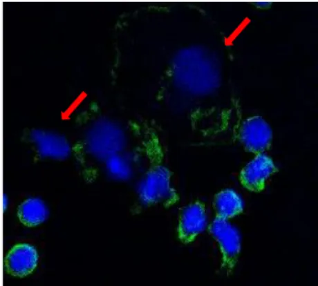
enrichment of nucleated CD45-negative cells (CTCs). In 8 out of 14 samples the phenotype of freshly isolated cells was investigated by immunofluorescence. We successfully isolated CTCs in a variable number, ranging from 10 to 3243 CTCs /7,5 mL of blood, from 7 out of 8 enrolled patients. CTCs were identified as Hoechst/Draq5 + CD45- cells by immunofluorescence (figure 4).
Figure 4. Staining of isolated cells with Hoechst/Draq5 mix (blue) and CD45 antibody (green); red arrows indicate CTCs (63X objective).
Also, freshly isolated CTCs were probed for the expression of the hormone receptor ERα, the growth factor receptor HER2 and EMT markers (CK19, Vimentin). Immunofluorescence results were compared with the clinical pathological features of the primary tumor. Although all patients had ERα- positive primary tumor, we did not isolate any ERα-positive CTCs. In addition, isolated cells were negative for HER2 and CK19 expression. Interestingly, CTCs from 6 out of 7 samples stained positive for the mesenchymal marker Vimentin. Indeed, the 45% of freshly isolated CTCs expressed Vimentin. To inquire the adhesion capability of CTCs isolated by RosetteSepTM, we seeded freshly CTCs from 6 out of 14 MBC patients on cover glasses. After that we looked for nucleated CD45- cells in both the adherent fraction and the supernatant by immunofluorescence. CTCs were found only in the adherent
19
fraction of 2 out of 6 samples; These results suggested that RosetteSepTM
affects CTCs adherence capacity.
4.2 Comparison between PBMCs and adherent cells isolated by Ficoll stratification
The obtained results prompted us to hypothesize that RosetteSepTM pauperizes sample of epithelial adherent cells, thus we decided to eliminate the incubation step with RosetteSepTM antibody cocktail from our isolation protocol. Therefore, the isolation of circulating cells from 6 MBC patients was performed by Ficoll density gradient followed by the seeding of PBMCs on cover glasses for the enrichment of adherent cells. Also, an aliquot of PBMCs from each blood sample was preserved and used for further analysis. To investigate which types of cells are enriched by the seeding of free labeled PBMCs, the mRNA expression levels of CD45, ERα, HER2, CK19 and Vimentin were evaluated by qRT-PCR in both PBMCs and adherent cells. In all examined samples, adherent cells expressed lower levels of the leukocyte marker CD45 and higher levels of CK19 and Vimentin than PBMCs (figure 5).
Figure 5. Comparison of CD45, CK19 and Vimentin mRNA levels between PBMCs and adherent cells from MBC patients.
20
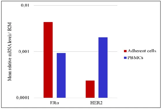
In addition, both PBMCs and adherent cell fraction expressed ERα at variable levels among different patients; on average ERα was higher in adherent cells than in PBMCs (figure 6). In 5 out of 6 samples HER2 was higher in PBMCs than in adherent cells, the mean HER2 mRNA expression levels obtained from these samples is shown in figure 6. These results suggested that the seeding step eliminated most of contaminating leukocytes and allowed the enrichment of adherent cells with both epithelial and
mesenchymal features.
Figure 6. mRNA expression levels of ERα and HER2 in PBMCs and adherent cells from MBC patients.
4.3 Isolated adherent cells express epithelial and mesenchymal markers The seeding of PBMCs after Ficoll stratification eliminated most of leukocytes as indicated by the low levels of CD45 in adherent cells respect to PBMCs as seen in the experiments reported above. To confirm these findings, adherent cells isolated from 30 MBC patients (“before therapy” patients), which have started a new line of systemic therapy right after the blood sample withdrawal, were analyzed by immunofluorescence for the presence of CD45.
21
Interestingly, among the adherent isolated cells, there were more CD45-than CD45+ cells (p< 0,05); on average 9 CD45+ cells/ mL of blood and 48 CD45- cell/mL of blood.
To verify that isolated adherent CD45- cells were CTCs and not cells of hematopoietic origin, we proceeded to their characterization.
To further characterize adherent cells, we stained them with antibody against either one of the following antigens: ERα and HER2 (breast cancer specific markers), CK19, EpCAM, CK18 and E-cadherin (epithelial markers), CK5 and Vimentin (mesenchymal markers).
The number of different cell populations was highly variable between different patients. Among examined samples EpCAM+ and E-cadherin+ cells were the less representative populations (from 0 to 40 positive cells/ mL of blood). We found cells expressing epithelial markers and cells positive for mesenchymal markers in a variable number among different patients. The mesenchymal markers Vimentin and CK5 were the most expressed (from 19 to 115 positive cells/ mL of blood), whereas the most represented epithelial markers were CK18 and CK19, with a number of positive cells ranging from 14 to 112/mL of blood. Also, ERα positive and HER2 positive cells were observed (from 0 to 88 ERα+ and HER2+ cells/mL of blood).
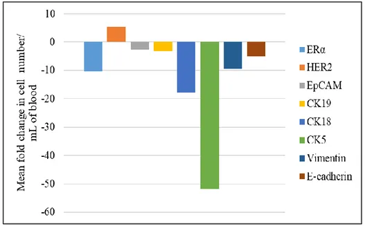
4.4 Systemic therapy affects the number of different populations of circulating cells
We wondered whether the phenotype of adherent cells could change after therapy. To address this question, we collected blood sample from 18 MBC patients after three months of systemic therapy. Circulating cells were isolated and the phenotype of adherent cells was investigated by immunofluorescence. The results were compared with those obtained from the same patients before starting therapy. Importantly, the number of CD45- counted cells/mL of blood significantly decreased after therapy of about 62% (p≤0,05).
Although the number of enrolled patients is not enough to do neither a statistical analysis nor a correlation with treatment response, a specific trend for each investigated marker seemed to emerge. Besides HER2 positive cells, all other cells populations seemed to decrease after therapy (figure 7). In media, CK5+, CK18+ and ERα+ cell populations are the most decreased ones after treatment, followed by Vimentin+ and CK19+ cells. EpCAM+ and E-cadherin+ cells were the lowest representative population before therapy; they further decreased in samples obtained from treated patients.
22
Figure 7. Bar plots showing the mean fold change in the number of different circulating cells populations after therapy.
4.5 Exploiting the utility of PBMCs molecular analysis in MBC management Another aim of this study was to exploit the utility of PBMCs analysis in monitoring MBC progression and therapy efficacy. To this aim, we examined the mRNA expression levels of CD45, ERα, HER2, CK19 and Vimentin in PBMCs derived from blood samples of 10 MBC patients and 3 healthy donors. Figure 8 shows the average expression for each gene. The expression levels of CK19 were significantly higher (p≤ 0,05) in PBMCs from MBC patients than from HD. Also, ERα seemed to be more expressed in patients than in healthy controls (figure 8). Furthermore, we repeated the analysis on 5 patients after three months of systemic therapy; we observed a decrease in ERα mRNA levels in 4 out of 5 samples, whereas the mRNA levels of HER2, CK19 and Vimentin remained almost the same. ERα expression levels in PBMCs seems to discriminate metastatic patients from healthy controls in addition to being modulated by systemic treatments. However, to assess the validity of PBMCs analysis in the management of MBC disease, a larger number of genes and patients needed to be examined.
23
Figure 8. mRNA expression levels in PBMCs from Metastatic breast cancer patients (MBC) and healthy donors (HD). * p value ≤ 0,05
24
5. DISCUSSION
Tumor heterogeneity is the main responsible for the failure of therapeutic strategies. Breast cancer is highly heterogeneous, and different breast cancer subtypes exist. During disease progression cancer generally become more heterogeneous; moreover, in the same patient the molecular profile of primary and the secondary tumor can be different. Targeted therapies can exert a selective pressure on cancer cell clones in favor of specific tumor cell subpopulations (Dagogo-Jack and Shaw 2018). All these aspects make cancer clinical management a challenge, especially in the metastatic disease. In this scenario, the sampling of the tumor at multiple time points to analyze cancer cells subpopulations represents an advantageous tool to overcome tumor heterogeneity problem and to plan personalized treatments based on time and stage-dependent tumor characteristics.
The analysis of CTCs isolated from peripheral blood samples represents a non-invasive procedure that could be routinely conducted to monitor “in real time” disease progression and therapy efficacy. Despite that, the use of CTCs in the clinical practice is still limited because of their rarity in the blood and the lack of strategies to efficiently isolate the whole CTCs heterogeneous population. Indeed, mainly CTCs isolation methods are epithelial marker-based approaches, which fail in detecting circulating cells with mesenchymal features. To boost the use of CTCs in the clinical practice, an improvement of isolation procedures and an extensive characterization of these cells are needed.
To this aim, we characterized circulating cells isolated through non-epithelial-based strategies in MBC blood samples. For the enrichment of CTCs from MBC blood samples two isolation procedures were tested. In the first part of the study we used an antibody-mediated method (RosetteSepTM) for the depletion of WBCs and RBCs from MBC patients enrolled prior a new line of systemic therapy; this method allowed the isolation of mesenchymal Vimentin positive cells, underlining the importance of mesenchymal circulating cells in metastatic condition. Neither epithelial (CK19+ cells) nor ERα+ and HER2+ cells were found. Moreover, RosetteSepTM seemed to affect circulating cells
adhesion capability, as suggested by the fact that only isolated cells from 2/6 samples processed with RosetteSepTM adhered to the cover glasses.
These results prompted us to eliminate the incubation with RosetteSep antibody cocktail and proceeding in the isolation of CTCs by using Ficoll density gradient followed by the seeding of PBMCs on cover glasses.
25
This method (Ficoll density gradient followed by the seeding of PBMCs) allowed an efficient depletion of contaminating leukocytes and the isolation of adherent cells which expressed both epithelial and mesenchymal markers. Indeed, the comparison of PBMCs and adherent cells molecular phenotype showed that adherent cells expressed lower levels of CD45 and higher levels of CK19 and Vimentin than PBMCs. Also, the depletion of WBCs from the samples was confirmed by the low number of CD45+ cells respect to CD45- cells (putative CTCs) investigated by immunofluorescence.
In addition, immunofluorescence experiments revealed that among adherent isolated circulating cells there were cells positive for epithelial markers (CK19, CK18) and cells positive for mesenchymal markers (CK5, Vimentin). These results are consistent with the crucial role of EMT in the spreading of metastatic disease. Mesenchymal CTCs have already been described in breast cancer, and it seems that they are associated with disease progression (Yu, Bardia, Ben S Wittner, et al. 2013). Moreover recent evidence suggests that both epithelial and mesenchymal features are needed for metastasis formation: mesenchymal phenotype confers migrative and invasive properties, whereas epithelial characteristics are required to proliferate at secondary site (Mitra et al. 2015).
Currently, the isolation of CTCs relies on EpCAM-based methods; however, EpCAM is downregulated during tumor dissemination due to EMT process. Hence, EpCAM-based isolation approaches underestimate CTCs number, missing critical subpopulation of circulating cancer cells (Hyun et al. 2016). In agreement with this assumption, in our analysis EpCAM+ and E-cadherin+ cells were the less representative populations in adherent isolated circulating cells. These findings underlined that an improvement of CTCs isolation strategies to study epithelial-mesenchymal transitioned CTCs is indispensable. Recently, it has been recognized the importance of monitoring ERα and HER2 status also in more advanced stages of breast cancer. In our study, both immunofluorescence and qRT-PCR experiments detected the presence of ERα+ and HER2+ cells in the adherent cell fractions isolated from MBC blood samples. In particular, qRT-PCR analysis showed that ERα was expressed at a variable level among patients, whereas HER2 was higher in PBMCs than in adherent cells, probably because of natural killer cells/granulocytes, which may be eliminated by the seeding. Indeed, it has been documented that natural killer cells/granulocytes are the main source of HER2 in PBMCs (You et al. 2008). Consistent with literature data (Onstenk et al. 2015), our experiments demonstrated that ERα and HER2 expression in CTCs differs from ERα and HER2 clinical pathological features of the same patient’s primary tumor. Discrepancy between primary tumor and CTCs profiles have been proposed as a prognostic factor in MBC. In addition, defining the role of ERα+ and HER2+
26
CTCs could be useful to identify metastatic patients that will benefit from receiving specific targeted therapies (Onstenk et al. 2015; Jordan et al. 2016). In our study we also characterized adherent circulating cells isolated from MBC patients who underwent three months of systemic therapy, to exploit changes eventually occurred in CTCs phenotype. The number of CD45- cells significantly decreased in post therapy samples, also the number of different examined populations of circulating cells changed. A considerably decrease was registered for CK5+, CK18+ and ERα+ cell populations; Vimentin+ and CK19+ cells slightly diminished after therapy. Furthermore, EpCAM+ and E-cadherin+ cells, which were the lowest representative population before therapy, further decreased in samples obtained from treated patients. HER2+ population only did not decrease after therapy. We are planning to validate our results in a larger cohort of patients in order to assess correlations between CTCs phenotype and treatment response. The phenotyping of CTCs subpopulations under therapeutic pressure would give a great contribution in the clinical management of cancer in general. It has been demonstrated that in small cell lung cancer patients the increase of Vimentin+ CTCs number after one-treatment cycle correlates with low OS (Messaritakis et al. 2017). Also, a study conducted by Angelaki and collaborators on TNBC patients demonstrated that the presence of HER2+ CTCs after adjuvant chemotherapy is indicative for progression disease (Agelaki et al. 2017). The recent increasing interest in CTC-phenotypization is underlined by the fact that large phase III study, named DETECT III, is ongoing with the aim to exploit utility of CTC phenotype in guiding therapeutic decision. The study will compare standard therapy alone versus standard therapy plus Lapatinib in patients with initially HER2- MBC and HER2+ CTCs.
Our results together with literatures data support the need of a wide characterization of CTCs both before and at different time points during therapy to improve their isolation and to introduce CTCs-based clinical therapeutic decisions.
Despite recent advances in isolation method an efficient exhaustive study of CTCs remains challenging; on the contrary, PBMCs collection is well established and routinely performable. For these reasons the possibility to predict disease progression and therapy response based on PBMCs molecular profile needs to be exploited. To this aim, we examined the mRNA expression levels of CD45, ERα, HER2, CK19 and Vimentin in PBMCs derived from blood samples of 10 MBC patients and 3 HD. Interestingly, CK19 was significantly more expressed in MBC-PBMCs than in HD-PBMCs. To note, CK19 has been the most extensively used markers for the detection of CTCs in the blood of cancer patients. Moreover, the detection of CK19 mRNA before the initiation of adjuvant chemotherapy has been recognized as an independent
27
prognostic factor for disease recurrence and decreased survival in patients with early breast cancer (Stathopoulou et al. 2002; Xenidis et al. 2006).
In our study ERα seemed to be more expressed in patients than in healthy controls. In addition, the analysis on 5 patients after three months of systemic therapy revealed a decrease in ERα mRNA levels in 4 out of 5 samples. ERα expression levels in PBMCs seems to discriminate metastatic patients from healthy controls in addition to being modulated by systemic treatments. Recently, a gene expression profile study has demonstrated that ERα gene is significantly differentially expressed in MBC respect to HD (Hensler et al. 2016). The identification of a PBMCs genes panel revealing the presence of CTCs (CTCS signature) in the blood of cancer patients would help in the management of MBC disease.
28
6. CONCLUSION
In this study we established a label-free method for the isolation and characterization of circulating cells in MBC. This method, consisting in Ficoll density gradient centrifugation followed by the seeding of label free PBMCs on cover glasses, allowed the depletion of most contaminating leukocytes and the enrichment of adherent cells expressing both epithelial and mesenchymal markers, overcoming the loss of mesenchymal CTCs that occurs by using commonly epithelial-based isolation strategies. Adherent cells were also characterized for the expression of different markers (hormone receptor, growth factor and EMT markers), revealing the presence of different circulating cell subpopulations. These subpopulations were investigated also in post therapy samples. So far, the number of enrolled patients was not enough to do any correlation with clinical features and patients’ outcome. Despite that, our results, together with literature data, support the idea that CTCs phenotyping both before and at different time points during therapy is necessary for the improvement of CTCs isolation and for the designing of CTCs-based cancer therapies. Furthermore, this study exploited the possibility to use PBMCs molecular profile as a surrogate of CTCs signature for MBC clinical management.
In conclusion, the analysis of CTCs remains a promising tool in cancer management that needs to be improved to obtain “ad hoc” personalized therapies to contrast the tumor heterogeneity underlying the progression of disease.
29
7. REFERENCES
Aceto N, Bardia A, Miyamoto DT, Donaldson MC, Wittner BS, Spencer JA, Yu M, Pely A, Engstrom A, Zhu H, et al. 2014. Circulating Tumor Cell Clusters Are Oligoclonal Precursors of Breast Cancer Metastasis. Cell. 158:1110–1122. doi:10.1016/j.cell.2014.07.013.
Adams DL, Martin SS, Alpaugh RK, Charpentier M, Tsai S, Bergan RC, Ogden IM, Catalona W, Chumsri S, Tang C-M, et al. 2014. Circulating giant macrophages as a potential biomarker of solid tumors. Proc Natl Acad Sci. 111:3514–3519. doi:10.1073/pnas.1320198111.
Adams DL, Zhu P, Makarova O V., Martin SS, Charpentier M, Chumsri S, Li S, Amstutz P, Tang CM. 2014. The systematic study of circulating tumor cell isolation using lithographic microfilters. RSC Adv. 4:4334–4342. doi:10.1039/c3ra46839a.
Agelaki S, Dragolia M, Markonanolaki H, Alkahtani S, Stournaras C, Georgoulias V, Kallergi G. 2017. Phenotypic characterization of circulating tumor cells in triple negative breast cancer patients. Oncotarget. 8:156. doi:10.18632/oncotarget.14144.
Aktas B, Tewes M, Fehm T, Hauch S, Kimmig R, Kasimir-Bauer S. 2009. Stem cell and epithelial-mesenchymal transition markers are frequently overexpressed in circulating tumor cells of metastatic breast cancer patients. Breast Cancer Res. 11:1–9. doi:10.1186/bcr2333.
Allard WJ. 2004. Tumor Cells Circulate in the Peripheral Blood of All Major Carcinomas but not in Healthy Subjects or Patients With Nonmalignant Diseases. Clin Cancer Res. 10:6897–6904. doi:10.1158/1078-0432.CCR-04-0378.
Andreopoulou E, Yang LY, Rangel KM, Reuben JM, Hsu L, Krishnamurthy S, Valero V, Fritsche HA, Cristofanilli M. 2012. Comparison of assay methods for detection of circulating tumor cells in metastatic breast cancer: AdnaGen AdnaTest BreastCancer Select/DetectTM versus Veridex CellSearchTM system. Int J Cancer. 130:1590–1597. doi:10.1002/ijc.26111.
30
Bidard F-C, Belin L, Delaloge S, Lerebours F, Ngo C, Reyal F, Alran S, Giacchetti S, Marty M, Lebofsky R, et al. 2013. Time-Dependent Prognostic Impact of Circulating Tumor Cells Detection in Non-Metastatic Breast Cancer: 70-Month Analysis of the REMAGUS02 Study. Int J Breast Cancer. 2013:1– 5. doi:10.1155/2013/130470.
Boral D, Vishnoi M, Liu HN, Yin W, Sprouse ML, Scamardo A, Hong DS, Tan TZ, Thiery JP, Chang JC, et al. 2017. Molecular characterization of breast cancer CTCs associated with brain metastasis. Nat Commun. 8. doi:10.1038/s41467-017-00196-1.
BOYUM A, LOVHAUG D, TRESLAND L, NORDLIE EM. 1991. Separation of Leucocytes: Improved Cell Purity by Fine Adjustments of Gradient Medium Density and Osmolality. Scand J Immunol. 34:697–712. doi:10.1111/j.1365-3083.1991.tb01594.x.
Bray F, Ferlay J, Soerjomataram I, Siegel R, Torre L, Jemal A. 2018. Global cancer statistics 2018: GLOBOCAN estimates of incidence and mortality worldwide for 36 cancers in 185 countries. CA A J Clin. doi:10.3322/caac.21492.
Colzani E, Johansson ALV, Liljegren A, Foukakis T, Clements M, Adolfsson J, Hall P, Czene K. 2014. Time-dependent risk of developing distant metastasis in breast cancer patients according to treatment, age and tumour characteristics. Br J Cancer. 110:1378–1384. doi:10.1038/bjc.2014.5.
Craene B De, Berx G. 2013. Regulatory networks defining EMT during cancer initiation and progression. Nat Rev Cancer. doi:10.1038/nrc3447.
Cristofanilli M, Budd GT, Ellis MJ, Stopeck A, Matera J, Miller MC, Reuben JM, Doyle G V., Allard WJ, Terstappen LWMM, et al. 2004. Circulating Tumor Cells, Disease Progression, and Survival in Metastatic Breast Cancer. N Engl J Med. 351:781–791. doi:10.1056/NEJMoa040766.
Dagogo-Jack I, Shaw AT. 2018. Tumour heterogeneity and resistance to cancer therapies. Nat Rev Clin Oncol. doi:10.1038/nrclinonc.2017.166. Dawood S, Broglio K, Valero V, Reuben J, Handy B, Islam R, Jackson S, Hortobagyi GN, Fritsche H, Cristofanilli M. 2008. Circulating tumor cells in
31
metastatic breast cancer: From prognostic stratification to modification of the staging system? Cancer. 113:2422–2430. doi:10.1002/cncr.23852.
Desitter I, Guerrouahen BS, Benali-Furet N, Wechsler J, Jänne PA, Kuang Y, Yanagita M, Wang L, Berkowitz JA, Distel RJ, et al. 2011. A new device for rapid isolation by size and characterization of rare circulating tumor cells. Anticancer Res. 31:427–41.
Fachin F, Spuhler P, Martel-Foley JM, Edd JF, Barber TA, Walsh J, Karabacak M, Pai V, Yu M, Smith K, et al. 2017. Monolithic Chip for High-throughput Blood Cell Depletion to Sort Rare Circulating Tumor Cells. Sci Rep. 7:1–11. doi:10.1038/s41598-017-11119-x.
Falkenberry SS, Legare RD. 2002. Risk factors for breast cancer. Risk factors breast cancer Obstet Gynecol Clin. 29:159–172. doi:10.1136/bmj.309.6969.1662.
Fidler IJ. 2003. The pathogenesis of cancer metastasis: The “seed and soil” hypothesis revisited. Nat Rev Cancer. doi:10.1038/nrc1098.
Fidler IJ, Kripke ML. 1977. Metastasis results from preexisting variant cells within a malignant tumor. Science (80- ). doi:10.1126/science.887927.
Giordano A, Giuliano M, De laurentiis M, Arpino G, Jackson S, Handy BC, Ueno NT, Andreopoulou E, Alvarez RH, Valero V, et al. 2012. Circulating tumor cells in immunohistochemical subtypes of metastatic breast cancer: Lack of prediction in HER2-positive disease treated with targeted therapy. Ann Oncol. 23:1144–1150. doi:10.1093/annonc/mdr434.
Giuliano M, Giordano A, Jackson S, Hess KR, De Giorgi U, Mego M, Handy BC, Ueno NT, Alvarez RH, De Laurentiis M, et al. 2011. Circulating tumor cells as prognostic and predictive markers in metastatic breast cancer patients receiving first-line systemic treatment. Breast Cancer Res. 13:R67. doi:10.1186/bcr2907.
Gong C, Liu B, Yao Y, Qu S, Luo W, Tan W, Liu Q, Yao H, Zou L, Su F, et al. 2015. Potentiated DNA damage response in circulating breast tumor cells confers resistance to chemotherapy. J Biol Chem. 290:14811–14825. doi:10.1074/jbc.M115.652628.
32
Gupta GP, Massagué J. 2006. Cancer Metastasis: Building a Framework. Cell. 127:679–695. doi:10.1016/j.cell.2006.11.001.
Hayes DF, Cristofanilli M, Budd GT, Ellis MJ, Stopeck A, Miller MC, Matera J, Allard WJ, Doyle G V., Terstappen LWWM. 2006. Circulating tumor cells at each follow-up time point during therapy of metastatic breast cancer patients predict progression-free and overall survival. Clin Cancer Res. 12:4218–4224. doi:10.1158/1078-0432.CCR-05-2821.
Hensler M, Vančurová I, Becht E, Palata O, Strnad P, Tesařová P, Čabiňaková M, Švec D, Kubista M, Bartůňková J, et al. 2016. Gene expression profiling of circulating tumor cells and peripheral blood mononuclear cells from breast cancer patients. Oncoimmunology. 5. doi:10.1080/2162402X.2015.1102827. Hess KR, Varadhachary GR, Taylor SH, Wei W, Raber MN, Lenzi R, Abbruzzese JL. 2006. Metastatic patterns in adenocarcinoma. Cancer. 106:1624–1633. doi:10.1002/cncr.21778.
den Hollander P, Savage MI, Brown PH. 2013. Targeted Therapy for Breast Cancer Prevention. Front Oncol. 3:1–15. doi:10.3389/fonc.2013.00250. Hyun K-A, Koo G-B, Han H, Sohn J, Choi W, Kim S-I, Jung H-I, Kim Y-S. 2016. Epithelial-to-mesenchymal transition leads to loss of EpCAM and different physical properties in circulating tumor cells from metastatic breast cancer. Oncotarget. 7:24677–24687. doi:10.18632/oncotarget.8250.
Jordan NV, Bardia A, Wittner BS, Benes C, Ligorio M, Zheng Y, Yu M, Sundaresan TK, Licausi JA, Desai R, et al. 2016. HER2 expression identifies dynamic functional states within circulating breast cancer cells. Nature. 537:102–106. doi:10.1038/nature19328.
Kallergi G, Agelaki S, Kalykaki A, Stournaras C, Mavroudis D, Georgoulias V. 2008. Phosphorylated EGFR and PI3K/Akt signaling kinases are expressed in circulating tumor cells of breast cancer patients. Breast Cancer Res. 10:1– 11. doi:10.1186/bcr2149.
Lambert AW, Pattabiraman DR, Weinberg RA. 2017. Emerging Biological Principles of Metastasis. Cell. 168:670–691. doi:10.1016/j.cell.2016.11.037.
33
Messaritakis I, Politaki E, Kotsakis A, Dermitzaki E-K, Koinis F, Lagoudaki E, Koutsopoulos A, Kallergi G, Souglakos J, Georgoulias V. 2017. Phenotypic characterization of circulating tumor cells in the peripheral blood of patients with small cell lung cancer. Ahmad A, editor. PLoS One. 12:e0181211. doi:10.1371/journal.pone.0181211.
Micalizzi DS, Farabaugh SM, Ford HL. 2010. Epithelial-mesenchymal transition in cancer: Parallels between normal development and tumor progression. J Mammary Gland Biol Neoplasia. 15:117–134. doi:10.1007/s10911-010-9178-9.
Mitra A, Mishra L, Li S. 2015. EMT, CTCs and CSCs in tumor relapse and drug-resistance. Oncotarget. 6:10697–10711. doi:10.18632/oncotarget.4037. Nakagawa T, Martinez SR, Goto Y, Koyanagi K, Kitago M, Shingai T, Elashoff DA, Ye X, Singer FR, Giuliano AE, et al. 2007. Detection of circulating tumor cells in early-stage breast cancer metastasis to axillary lymph nodes. Clin Cancer Res. 13:4105–4110. doi:10.1158/1078-0432.CCR-07-0419.
Naume B, Borgen E, Tøssvik S, Pavlak N, Oates D, Nesland JM. 2004. Detection of isolated tumor cells in peripheral blood and in BM: Evaluation of a new enrichment method. Cytotherapy. doi:10.1080/14653240410006086. Onstenk W, Sieuwerts AM, Weekhout M, Mostert B, Reijm EA, van Deurzen CHM, Bolt-de Vries JB, Peeters DJ, Hamberg P, Seynaeve C, et al. 2015. Gene expression profiles of circulating tumor cells versus primary tumors in metastatic breast cancer. Cancer Lett. 362. doi:10.1016/j.canlet.2015.03.020. Paget S. 1889. THE DISTRIBUTION OF SECONDARY GROWTHS IN CANCER OF THE BREAST. Lancet. 133:571–573. doi:10.1016/S0140-6736(00)49915-0.
Pantel K, Speicher MR. 2015. The biology of circulating tumor cells. Oncogene. doi:10.1038/onc.2015.192.
Papadaki MA, Kallergi G, Zafeiriou Z, Manouras L, Theodoropoulos PA, Mavroudis D, Georgoulias V, Agelaki S. 2014. Co-expression of putative stemness and epithelial-to-mesenchymal transition markers on single
34
circulating tumour cells from patients with early and metastatic breast cancer. BMC Cancer. 14:651. doi:10.1186/1471-2407-14-651.
Pegram BMD, Lipton A, Hayes DF, Weber BL, Baselga JM, Tripathy D, Baly D, Baughman SA, Twaddell T, Glaspy JA, et al. 1998. Phase II Study of Receptor-Enhanced Chemosensitivity Using Recombinant Humanized Anti-p185HER2 / neu Monoclonal Antibody Plus Cisplatin in Patients With Refractory to Chemotherapy Treatment. J Clin Oncol. 16:2659–2671.
Perou CM, Sørile T, Eisen MB, Van De Rijn M, Jeffrey SS, Ress CA, Pollack JR, Ross DT, Johnsen H, Akslen LA, et al. 2000. Molecular portraits of human breast tumours. Nature. 406:747–752. doi:10.1038/35021093.
Pierga J-Y, Bidard F-C, Mathiot C, Brain E, Delaloge S, Giachetti S, de Cremoux P, Salmon R, Vincent-Salomon A, Marty M. 2008. Circulating Tumor Cell Detection Predicts Early Metastatic Relapse After Neoadjuvant Chemotherapy in Large Operable and Locally Advanced Breast Cancer in a Phase II Randomized Trial. Clin Cancer Res. 14:7004–7010. doi:10.1158/1078-0432.CCR-08-0030.
Pierga JY, Hajage D, Bachelot T, Delaloge S, Brain E, Campone M, Diéras V, Rolland E, Mignot L, Mathiot C, et al. 2012. High independent prognostic and predictive value of circulating tumor cells compared with serum tumor markers in a large prospective trial in first-line chemotherapy for metastatic breast cancer patients. Ann Oncol. 23:618–624. doi:10.1093/annonc/mdr263.
Rack B, Schindlbeck C, Jückstock J, Andergassen U, Hepp P, Zwingers T, Friedl TWP, Lorenz R, Tesch H, Fasching PA, et al. 2014. Circulating tumor cells predict survival in early average-to-high risk breast cancer patients. J Natl Cancer Inst. doi:10.1093/jnci/dju066.
Stathopoulou A, Vlachonikolis I, Mavroudis D, Perraki M, Kouroussis C, Apostolaki S, Malamos N, Kakolyris S, Kotsakis A, Xenidis N, et al. 2002. Molecular detection of cytokeratin-19-positive cells in the peripheral blood of patients with operable breast cancer: Evaluation of their prognostic significance. J Clin Oncol. doi:10.1200/JCO.2002.08.135.
Steelman LS, Martelli AM, Cocco L, Libra M, Nicoletti F, Abrams SL, McCubrey JA. 2016. The therapeutic potential of mTOR inhibitors in breast cancer. Br J Clin Pharmacol.:1189–1212. doi:10.1111/bcp.12958.
35
Vanharanta S, Massagué J. 2013. Origins of Metastatic Traits. Cancer Cell. 24:410–421. doi:10.1016/j.ccr.2013.09.007.
Verma S, Miles D, Gianni L, Krop IE, Welslau M, Baselga J, Pegram M, Oh D-Y, Diéras V, Guardino E, et al. 2012. Trastuzumab Emtansine for HER2-Positive Advanced Breast Cancer. N Engl J Med. 367:1783–1791. doi:10.1056/NEJMoa1209124.
Wallwiener M, Hartkopf AD, Riethdorf S, Nees J, Sprick MR, Schönfisch B, Taran FA, Heil J, Sohn C, Pantel K, et al. 2015. The impact of HER2 phenotype of circulating tumor cells in metastatic breast cancer: A retrospective study in 107 patients. BMC Cancer. 15:1–7. doi:10.1186/s12885-015-1423-6.
Xenidis N, Perraki M, Kafousi M, Apostolaki S, Bolonaki I, Stathopoulou A, Kalbakis K, Androulakis N, Kouroussis C, Pallis T, et al. 2006. Predictive and prognostic value of peripheral blood cytokeratin-19 mRNA-positive cells detected by real-time polymerase chain reaction in node-negative breast cancer patients. J Clin Oncol. 24:3756–3762. doi:10.1200/JCO.2005.04.5948.
Yang L, Lang JC, Balasubramanian P, Jatana KR, Schuller D, Agrawal A, Zborowski M, Chalmers JJ. 2009. Optimization of an enrichment process for circulating tumor cells from the blood of head and neck cancer patients through depletion of normal cells. Biotechnol Bioeng. 102:521–534. doi:10.1002/bit.22066.
You F, Roberts LA, Kang SP, Nunes RA, Dias C, Iglehart JD, Solomon NA, Friedman PN, Harris LN. 2008. Low-level expression of HER2 and CK19 in normal peripheral blood mononuclear cells: Relevance for detection of circulating tumor cells. J Hematol Oncol. 1:1–10. doi:10.1186/1756-8722-1-2. Yu M, Bardia A, Wittner BS, Stott SL, Smas ME, Ting DT, Isakoff SJ, Ciciliano JC, Wells MN, Shah AM, et al. 2013. Circulating breast tumor cells exhibit dynamic changes in epithelial and mesenchymal composition. Science (80- ). 339:580–584. doi:10.1126/science.1228522.
Yu M, Bardia A, Wittner BS, Stott SL, Smas ME, Ting DT, Isakoff SJ, Ciciliano JC, Wells MN, Shah AM, et al. 2013. Circulating Breast Tumor Cells Exhibit Dynamic Changes in Epithelial and Mesenchymal Composition.