Proteasome Involvement and Accumulation of Ubiquitinated
Proteins in Cerebellar Granule Neurons Undergoing Apoptosis
Nadia Canu,
1Christian Barbato,
1Maria Teresa Ciotti,
2Annalucia Serafino,
3Laura Dus,
2and
Pietro Calissano
1,21
Dipartimento di Neuroscienze, Facolta` di Medicina e Chirurgia, Universita` di Tor Vergata, 00133 Roma, Italia,
2Istituto di
Neurobiologia, Consiglio Nazionale delle Ricerche (CNR), 00137 Roma, Italia, and
3Area di Ricerca di Roma, Tor Vergata,
CNR, 00133 Roma, Italia
We investigated the potential role of the ubiquitin proteolytic system in the death of cerebellar granule neurons induced by reduction of extracellular potassium. Inhibitors of proteasomal function block apoptosis if administered at onset of this pro-cess, but they do not exert such effect when added 2–3 hr later. The same inhibitors also prevent caspase-3 activity and calpain-caspase-3-mediated processing of tau protein, sug-gesting that proteasomes are involved upstream of the caspase activation. Although the proteasomes seem to play an early primary role in programmed cell death, we found that with progression of apoptosis, during the execution phase, a per-turbation in normal ubiquitin-proteasome function occurs, and high levels of ubiquitinated proteins accumulate in the cyto-plasm of dying cells. Such accumulation correlates with a
progressive decline of proteasome chymotrypsin and trypsin-like activities and, to a lower extent, of postacidic-trypsin-like activity. Both intracytoplasmic accumulation of ubiquitinated proteins and decline of proteasome function are reversed by the pan-caspase inhibitor Z-VAD-fmk. The decline in proteasome func-tion is accompanied by, and likely attributable to, a marked and progressive decline of deubiquitinating activities. The finding that the proteasomes are early involved in apoptosis and that ubiquitinated proteins accumulate during this process prospect granule neurons as a model system aimed at correlating these events with neurodegenerative diseases.
Key words: apoptosis; neurodegeneration; ubiquitin-protein conjugates; proteasome activity; deubiquitinating activity; cer-ebellar granule neurons
The presence of intracellular inclusions of insoluble aggregates of ubiquitin protein conjugates is a hallmark of chronic neurodegen-erative diseases and, to lesser extent, of physiological brain aging (Lowe et al., 1993). To date, the mechanism or mechanisms leading to such aberrant deposits is unknown. Over time, the accumulation of ubiquitinated proteins results in pathological aggregates that perturb the normal physiology of affected neurons and lead to proteotoxicity.
Ubiquitination of proteins is required for rapid degradation of short-lived or of damaged proteins by the ubiquitin-proteasome system. This is the major proteolytic system in the cytosol of eukaryotic cells endowed with multiple activities, referred to as chymotrypsin-like, trypsin-like, and postacidic or caspase-like ac-tivities (Peters, 1994).
In the ubiquitin-proteasome pathway, proteins are first modi-fied by the covalent conjugation to multiple ubiquitin molecules that subsequently tag the conjugates for rapid hydrolysis by the proteasome. This complex hydrolyzes exhaustively target pro-teins releasing small peptides, most of which are further degraded by cellular exopeptidases, with concomitant recycling of ubiquitin molecules by deubiquitinating enzymes (Wilkinson, 1997).
A growing number of studies indicate that ubiquitin-mediated proteolysis plays important roles in a variety of basic cellular processes such as regulation of cell cycle and division, response to cellular stress, morphogenesis of neuronal network, long-term synaptic plasticity, transcriptional regulation of cell surface re-ceptors (Jentsch 1992; Ciechanover 1994; Hochstrasser 1995), and activation of the transcription factor NF-KB (Palombella et al., 1995). Moreover, several observations suggest that a pertur-bation in ubiquitin-proteasome function plays a critical role in the accumulation of misfolded or modified proteins such as paired helical filaments in Alzheimer’s disease (Morishima-Kawashima et al., 1993), nuclear aggregates of huntingtin in Huntington’s disease (DiFiglia et al., 1997), and aggregation ofa-synuclein in Parkinson’s disease (Takeda et al., 1998). The inability of remov-ing ubiquitin conjugates may result from an altered proteolytic degradation or from a modification of damaged proteins that makes them inaccessible to proteolytic machinery.
It has been recently reported that the proteasome system is critically involved in programmed cell death, by acting upstream of caspase activation. Generally, proliferating cells respond to inhibitors of proteasome function by activation of apoptosis, whereas in nondividing cells (sympathetic neurons or T-cells) such inhibitors exert antiapoptotic effects (Grimm et al., 1996; Sadoul et al., 1996; Drexler, 1997). On the other hand, it has been postulated that apoptosis plays a role in the pathogenesis of neurodegenerative diseases (Su et al., 1994; Smale et al., 1995; Thompson, 1995).
In view of the postulated connection of apoptotic death, altered proteasome function, and neurodegenerative diseases, we inves-tigated the role of ubiquitin-proteasome system in an in vitro
Received June 25, 1999; revised Oct. 29, 1999; accepted Nov. 3, 1999.
This work was supported by Telethon-Italy (Grant E855), Progetto Finalizzato Biotecnologie, and Cofinanziamento Ministero dell’Universita´ e della Ricerca Sci-entifica e Tecnologica (40% to P.C.). We thank Dr. Andrea Levi for comments on this manuscript and Marianna De Bernardinis for helpful suggestions on MTT assay.
Correspondence should be addressed to Dr. Nadia Canu, Istituto di Neurobiolo-gia Consiglio Nazionale delle Ricerche, V.le Marx 15, 00137 Roma, Italia. E-mail: [email protected].
model of cerebellar granule cells (CGCs) undergoing massive apoptotic death after removal of depolarizing concentration of potassium (D’Mello et al., 1993). We report that the proteasome system appears twice involved during apoptosis: first, its function is crucial in the early phase, upstream caspase activation, then, in the execution phase, proteasome function declines with conse-quent accumulation of ubiquitinated proteins. Moreover, an im-pairment of deubiquitinating activities accompanies and likely contributes to the proteasome failure.
MATERIALS AND METHODS
The proteasome inhibitors PSI [Z-Ile-Glu (OtBu)-Ala-Leucinal] and Lactacystin and the caspase inhibitor z-VAD-fmk (Benzyloxycarbonyl-Val-Ala-Asp-fluoromethylchetone) were from Calbiochem (La Jolla, CA). The proteasome inhibitor MG132 (N-CBZ-Leu-Leu-Leu-Al), the calpain inhibitor II ALLM (N-acetyl-Leu-Leu-Methioninal), E64d (trans-epoxy succinyl-L-leucylamydo-3-methyl-butane ethyl ester), and leupeptin were purchased from Sigma (St. Louis, MO). Substances were dissolved in dimethylsulfoxide at 10003 concentration. No more than 0.1% solvent was present in culture medium. The fluorogenic substrates for proteasome assay Suc-LLVY-MCA (succinyl-Leu-Leu-Val-Tyr-7-amido-4-methylcoumarin), Boc-LRR-MCA (N-t-boc-Leu-Arg-Arg-7-amido-4-methylcoumarin), and Z-LLE-bNap (N-CBZ-Leu-Leu-Glu-b-naphtylamide) were from Sigma. Multi-ubiquitin chains [Ub4, Ub3 and Ub2, and the monoclonal antibody (mAb) toa-type subunits (PW8195)] were from AFFINITI Research Products Ltd. Ac-DEVD-AMC was from Biomol (Plymouth Meeting, PA). Rabbit anti-ubiquitin was from Dako (Carpinteria, CA).
Neuronal cultures. Cultures enriched in granule neurons were obtained
from dissociated cerebella of 8-d-old Wistar rats (Charles River, Calco, Italy), as described by Levi et al. (1984). Cells were plated in basal medium Eagle (BME; Life Technologies, Gaithersburg, MD) supple-mented with 10% fetal bovine serum, 25 mMKCl, and 2 mMglutamine
(Life Technologies) on dishes (Nunc, Roskilde, Denmark) coated with poly-L-lysine. Cells were plated at 2.53 106per 35 mm dish or 73 106
per 60 mm dish. 1b-Arabinofuranosylcytosine (10 mM) was added to the
culture medium 18 –22 hr after plating to prevent proliferation of non-neuronal cells.
Induction of apoptosis. Cultures at 6 –7 days in vitro (DIV) were washed
two times and switched in serum-free BME containing 5 mM KCl
supplemented with glutamine and gentamicin. Control cells were washed with BME and maintained in serum-free medium containing 25 mMKCl
(D’Mello et al., 1993).
Assessment of neuronal viability. Viable granule neurons were
quanti-fied by counting the number of intact nuclei after lysing the cells in detergent-containing solution by the method of Soto and Sonnenschein (1985) modified by Volonte` et al. (1994) and by the MTT tetrazolium salt assay , as described by Manthorpe et al. (1986). Briefly, MTT tetrazolium salt (0.25 mg/ml) was added to neurons grown in 24-well plates and incubated for 1–2 hr at 37°C. The reaction media were then gently aspirated, and isopropanol containing 0.08 N HCl was added to solubi-lize the blue formazan product. Formazan–isopranol mixtures were then transferred to 96-well plates and quantified using a Multiskan plate reader at 570 nm (Labsystems Multiskan MCC/340).
Immuofluorescence. Cerebellar granule cells were fixed with 4%
para-formaldehyde (w/v in PBS) for 15 min at room temperature, washed in PBS, pH 7.5, and then permeabilized with 0.1%Triton X-100 and Tris-Cl, pH 7.5, for 5 min. The coverslips were treated with polyclonal antibody against ubiquitin (Dako; 1:100) in a moist chamber overnight at 4°C, rinsed in PBS, and stained with FITC-conjugated secondary anti-bodies (Sigma) for 30 min. Nuclei were stained with propidium iodide (Sigma; 5 mg/ml) and RNase (100 mg/ml) in PBS for 5 min at room temperature. Confocal microscopy was performed with a Leica (Nuss-loch, Germany) TCS 4D system, equipped with 1003 1.3–0.6 oil-immersion objective (optical section, 1mM). Images of double-labeled
samples were recorded with simultaneous excitation and detection of both dyes to ensure proper image alignment. Optical sections were stereo-pair and three-dimensional reconstituted. To correct for possible crosstalk resulting from overlapping excitation and emission spectra of the dyes used, when necessary, recorded images were corrected using the MultiColor analysis package software by Leica.
Caspase-3 activity: DEVD-MCA cleavage assay. DEVD-MCA cleavage
activity was measured , as described by Armstrong et al. (1997). After 12 hr
in S-K5, 500,000 granule cells were washed once with PBS and lysed in 100 ml of buffer A (10 mMHEPES, pH 7.4, 42 mMKCl, 5 mMMgCl2, 1 mMDTT,
and 1 mM PMSF, 0.5%
3-[(3-cholamidopropyl)dimethylammonio]-1-propanesulfonic acid (CHAPS), and 1mg/ml leupeptin). Twenty-five micro-liters of lysate was combined with 75ml of buffer B (25 mMHEPES, 1 mM
EDTA, 0.1% CHAPS, 10% sucrose, and 3 mMDTT, pH 7.5) containing 30
mMAc-DEVD-AMC and incubated for 20 min at room temperature.
Fluo-rescence was measured at an excitation of 380 nm and an emission of 460 nm using a Kontron AG (Zurich, Switzerland) SFM spectrofluorometer.
Fluorogenic peptide substrate assay for proteasome activity. CGCs were
lysed in ice-cold homogenization buffer [20 mMTris/HCl, pH 7.2, 0.1 mM
EDTA, 1 mM2-mercaptoethanol, 5 mMATP, 20% (v/v) glycerol, and
0.04% (v/v) Nonidet P-40, as described in Beyette et al. (1998)]. Lysates (10mg) of CGCs were incubated at 37°C with the fluorogenic substrates Suc-LLVY-MCA (50mM), Boc-LRR-MCA (100mM), and Z-LLE-bNap
(400mM) in 100ml of 50 mMHEPES, pH 8, 5 mMEGTA, for 20, 30, and
60 min, respectively. The reaction was stopped by adding 900ml of SDS (1%). Hydrolysis of peptides was measured at 380 nm excitation and 460 emission for MCA and at 335 nm excitation and 410 nm emission for bNap using a Kontron SFM spectrofluorometer.
Western blot analysis. Equal amounts of proteins were subjected to
SDS-PAGE on 8 –15% linear gradient gels (Laemmli, 1970). After elec-troblotting to nitrocellulose (Hybond-C), proteins were visualized using appropriate primary antibodies. All primary antibodies were diluted in 0.5% (w/v) nonfat dry milk and incubated with the nitrocellulose blot overnight at 4°C. Incubation with secondary peroxidase-coupled anti-mouse or anti-rabbit antibodies was performed at room temperature for 45 min. Blots were developed by using the ECL system (Amersham, Arlington Heights, IL). Developments of Western blots were terminated before band intensity was saturated; relative optical densities and areas of bands were quantified using a computerized image analysis system.
Preparation of protein extracts from CGCs and assay for deubiquitinating activity. CGCs were collected and resuspended in ice-cold lysis buffer (20
mMTris-HCl, pH 7.2, 10 mMMgCl2, 1 mMEDTA, 5% glycerol, 1 mM
DTT, 1mg/ml aprotinin, and 1 mg/ml pepstatin) and frozen and thawed three times. The extracts were centrifuged, and supernatants were stored at270°C as 50% glycerol solutions. Assay of deubiquitinating enzyme (DUB) activity was conducted at 22°C in a buffer containing 50 mM
Tris-HCl, pH 7.3, and 5 mMDTT in a total volume of 30ml. Reaction
mixture contained 5 mg of CGC extract and 1 mg of multi-ubiquitin chains. Aliquots of 5ml were removed and separated by 12.5% polyacryl-amide gels, using a Tricine gel system (Schagger and von Jagow, 1987), transferred to Immobilon P membranes (Millipore, Bedford, MA) and analyzed by anti-ubiquitin antibody.
RESULTS
Inhibition of proteasome function rescues granule neurons from K1deprivation-induced cell death
Rat cerebellar granule neurons can be induced to undergo apo-ptosis if the potassium concentration is reduced to 5 mM(K5) and
serum is removed (S2) after a period of initial growth in 25 mM
potassium (K25) and serum (S1) (D’Mello et al., 1993; Galli et al., 1995). Commitment to apoptosis occurs between 2 and 6 hr after K1deprivation (Galli et al., 1995; Schulz et al., 1996; Nardi
et al., 1997) and at 6–8 hr degenerative changes such as chroma-tin condensation, vacuole formation, DNA fragmentation, and neurite retraction become detectable (D’Mello et al., 1993; Schulz et al., 1996; Armstrong et al., 1997). Moreover, during the same period the microtubule-associated protein tau is being degraded by caspase-3 and calpain to a 17 kDa fragment that accumulates in perikarya of dying neurons (Canu et al., 1998), and an amy-loidogenic route leading to an increased secretion ofb-amyloid is activated (Galli et al., 1998).
Because the proteasome system has been implicated as a pos-itive mediator of apoptosis triggered by damaging stimuli in terminally differentiated cell types e.g., by degrading regulatory proteins that normally inhibit the apoptotic pathway or activating proteins that promote cell death likely working upstream the caspase-activation (Grimm et al., 1996; Sadoul et al., 1996), we investigated the involvement of proteasome system in the CGC
model of neuronal apoptosis. To this aim the effects of a panel of proteasome inhibitors on the survival of CGCs were examined. The peptide aldehydes MG132 and PSI [Z-IE(OtBu)AL-CHO] have been shown to reversibly inhibit the proteolytic activity of proteasomes (Figueiredo-Pereira et al., 1994; Rock et al., 1994), whereas Lactacystin, a microbial metabolite, irreversibly blocks the proteasome function (Fenteany et al., 1995). These sub-stances were added before or simultaneously with the induction of apoptosis, and neuronal survival was assessed 12 hr later because at longer incubation times these inhibitors become toxic. Cell viability was analyzed by counting the number of intact nuclei and by the MTT assay, as described in Materials and Methods. Results shown in Figure 1, A and B, provide evidence that MG132 and Lactacystin significantly inhibit apoptosis. MG132 was a more potent inhibitor of cell death, producing .90% inhibition of cell death at 2mMas compared with
Lacta-cystin, which inhibits cell death by 85% at 25mM. In the presence
of these compounds, the cell bodies of neurons and the neurites remained intact for 12–18 hr (data not shown). By contrast, PSI protects CGCs from apoptosis only partially (20%) and at 50–100 mM as assessed by counting the number of intact nuclei. Very
similar results are obtained when viability is assayed with the MTT procedure (Fig. 1B) with a slight difference at high concen-tration of PSI (50mM), indicating that the mitochondrial function
is still partially operative in a portion of neurons. Because MG132 also affects calpain and cathepsin besides proteasomes, we also tested the effect of ALLM (5–50mM), E64d ester (20–100mM),
and leupeptin (20–100mM), which are cell-permeable inhibitors
of calpain and cathepsin. As indicated in Figure 1C, these agents failed to rescue CGCs from apoptosis, suggesting that MG132 elicits its effects by acting primarily on proteasomes.
We found that MG132 must be added within 1 hr after induc-tion to efficiently block apoptosis (Fig. 2), whereas no rescuing effect was noticed when this agent was given 3 hr after the apoptotic stimulus. Identical results were obtained with Lactacys-tin (data not shown). This finding supports the hypothesis that proteasome inhibitors act on the early events of apoptosis (Grimm et al., 1996; Sadoul et al., 1996) and become ineffective after the commitment point that occurs between 2–6 hr after apoptotic stimulus (Galli et al., 1995; Schulz et al., 1996; Nardi et al., 1997).
Inhibition of proteasome function prevents caspase-3 activity and cleavage of the microtubule-associated protein tau
The execution phase of apoptosis is initiated by activation of specific proteases of the caspase family (Steller, 1995). Of the caspases, caspase-3 is that involved in the cleavage of most apo-ptotic substrates (Cohen, 1997). An increase of caspase-3 activity is also present in extracts of granule neurons undergoing apopto-sis (Eldadah et al., 1997; Marks et al., 1998; Bobba et al., 1999). We investigated the role of proteasome with respect to this protease activity. Primary CGCs were induced to die in the absence or in the presence of MG132 (5 mM), Lactacystin (25
mM), and PSI (20 and 50 mM). Twelve hours later caspase-3
activity was assayed by the cleavage of the fluorogenic substrate Ac-DEVD-MCA (Enari et al., 1996). As shown in Figure 3A, neurons undergoing apoptosis exhibited a 12- to 13-fold elevation of caspase-3 activity as compared to controls. Caspase activation induced by apoptosis is partially inhibited by MG132, Lactacys-tin, and PSI to an extent which, at least in part, mirrors their anti-apoptotic effect. To date, only few intracellular substrates of
activated caspase-3 have been identified in CGCs undergoing apoptosis (Nath et al., 1996; Canu et al., 1998). For instance we have reported that in CGCs undergoing apoptosis the microtubule-associated protein tau is a substrate for both
Figure 1. Inhibitors of proteasome rescue cerebellar neurons from
apo-ptosis. Cultures at 6 DIV were washed and maintained in high-potassium and serum-free medium (S-K25) or switched to K5 and serum-free me-dium (S-K5) in the absence or in the presence of different concentrations of proteasome inhibitors. After 12 hr, cell viability was determined by counting the number of intact nuclei (A) or by MTT assay (B), as described in Materials and Methods. C, Effect of calpain and cathepsin inhibitors on cell survival. ALLM, E64d, and leupeptin were added to the medium at the concentrations indicated. Twelve to 15 hr after apoptosis induction, granule neurons were analyzed for survival, as described in Materials and Methods. Results are means6 SD of duplicate determi-nations of three independent experiments.
caspase-3 and calpain with a production of a diagnostic fragment of 17 kDa and concomitant collapse of the microtubule network (Canu et al., 1998). Whole-cell extracts prepared from CGCs incubated in the presence of MG-132 and lactacystin were ana-lyzed by SDS-PAGE, electroblotted, and probed with the anti-tau antibody, mAb Tau-1. As shown in Figure 3B we found that the 17 kDa band, corresponding to the major cleavage product of tau previously described, is not detectable in the presence of protea-some inhibitors. These findings suggest that these agents block proteasome function upstream the activation of proteases such as caspase-3 and calpain, a conclusion also supported by the obser-vation that the same inhibitors prevent the release of cytochrome c (A. Bobba, N. Canu, A. Atlante, E. Marra, and P. Calissano, unpublished observation), an event preceding caspase-3 activa-tion in CGCs undergoing apoptosis (Bobba et al., 1999).
Proteasome activity decreases during apoptosis
The proteasome can cleave peptides on the carboxyl side of hydrophobic, basic, and acid residues (Orlowski, 1990). These proteolytic functions commonly referred to as the chymotryptic, tryptic, and postacidic or caspase-like, can be measured by eval-uating the hydrolysis of the fluorogenic substrates Suc-LLVY-MCA, Boc-LRR-Suc-LLVY-MCA, and Z-LLE-bNap, respectively. We therefore determined whether and which of these specific activi-ties are modulated during neuronal apoptosis. To this aim, con-trol cells and cells undergoing apoptosis were homogenized in a buffer containing 5 mM ATP and 20% glycerol to preserve the
integrity of the 26 S proteasome (Hough et al., 1987), and their supernatant fractions were tested for the ability to hydrolyze these fluorogenic substrates. Assays were performed in a buffer (pH 8.0) containing 5 mMEGTA to inhibit lysosomal peptidases
and calpains. As shown in Figure 4A, the supernatant fractions of CGCs undergoing apoptosis exhibit a progressively reduced hy-drolysis of Suc-LLVY-MCA to;60% of control value at 12 hr of apoptosis. The time course analysis of this activity demonstrates that this decline parallels the number of apoptotic nuclei (Fig. 4D). A similar trend was also observed for trypsin-like and caspase-like activities of proteasome as detected using the fluoro-genic substrates Boc-LRR-MCA and Z-LLE-bNap respectively (Fig. 4B,C), although the decline of caspase like-activity is less pronounced and does not mirror the extent of apoptosis. It is
interesting to note, moreover, that the time course analysis of the three distinct peptidase activities of proteasome revealed, in the early phase of apoptosis, a slight, but consistent increase com-pared to control cells (Fig. 4A–C).
To ascertain if the decline in proteasome activity was attribut-able to a decrease in the actual proteasomes content, equal amounts of proteins from control cells and cells undergoing apoptosis for different times were subjected to SDS-PAGE, elec-troblotted, and probed with a monoclonal antibody directed against alla-proteasome subunits. As shown in Figure 4E, the intracellular level of proteasomes remained unchanged in apo-ptotic granule neurons as compared with control cells, indicating that the observed decline in proteasome activity during apoptosis is attributable to other causes (see Discussion). A similar result was also obtained using a mAb against thea-subunit C9 (data not shown).
It has been reported that MG132, lactacystin, and PSI have
Figure 2. Rescue time by MG132 on granule neurons undergoing
apo-ptosis. Cells were exposed to 5mMMG132 either 1 hr before (,1 hr) or
at different times after apoptotic stimulus. The number of viable cells was determined 12 hr later, as described in Materials and Methods.
Figure 3. Inhibitors of the proteasomes block caspase activity and
pre-vent cleavage of tau. A, Cultures were switched to S-K5 medium in the absence or in the presence of proteasome inhibitors MG132 (5 mM),
Lactacystin (25mM), or PSI (20 or 50mM). Control cultures were switched
to S-K25. After 12 hr, cultures were lysed and assayed for DEVD-MCA cleavage. Accumulation of MCA was assayed fluorometrically after 20 min at room temperature. Data are reported as the percentage of the rate of cleavage in S-K25 extracts (100%). B, Tau cleavage was analyzed by immunoblotting of cell extracts 12 hr after induction of apoptosis trig-gered in the absence or in the presence of 1 mM MG132 or 25mM
Lactacystin (LC). The mouse monoclonal antibody Tau-1 was used to detect tau cleavage (Canu et al., 1998).
different inhibitory potencies on the proteasomal activities (for review, see Lee and Goldberg, 1998). Because they protect CGCs from apoptosis to a different extent (Fig. 1), we determined their effectiveness by incubating granule neurons in their presence and assessing the chymotrypsin, trypsin-like, and postacidic-like ac-tivities of proteasomes with specific substrates. As shown in Figure 5 all compounds inhibit the chymotrypsin-like activity to the same extent (MG132,290 6 6%; PSI, 280 6 10%; lactacys-tin,285 6 10%). Considerable differences, on the contrary, were observed for trypsin-like activity (234 6 10% with MG132, 250 6 5% with Lactacystin, and 270 6 10% with PSI) as well as postacidic-like activity (265 6 5% with MG132, 250 6 7% with Lactacystin, and238 6 11% with PSI). The data reported dem-onstrate that the inhibitors tested are able to enter the cells and to block the proteasome activity to an extent comparable to that previously reported for their antiapoptotic action. Moreover, on the basis of these results we hypothesize that the anti-apoptotic effect is exerted by inhibitors able to inhibit also the postacidic-like activity.
Proteasome failure occurs downstream caspase activation
To determine whether the reduced proteasome activity is a reg-ulated event located downstream the caspase activation, we mea-sured proteasome activities in supernatant fractions from CGCs induced to undergo apoptosis for 12 hr in the presence or absence of the general caspase inhibitor Z-VAD-fmk (100 mM). The
hydrolysis of Suc-LLVY-MCA, Boc-LLR-MCA, and ZLLE b-Nap by CGCs was decreased to 53 6 8%, 58 6 11%, and 73 6 4%, respectively, after the apoptotic stimulus. When CGCs were induced in apoptosis in the presence of 100mMof Z-VAD-fmk,
the chymotrypsin, trypsin, and caspase-like activities were par-tially restored mirroring the antiapoptic effect of this general caspase inhibitor in this model of neuronal apoptosis (Fig. 6).
Figure 4. Profile of proteasome activities and proteasome concentration
during the apoptosis of CGCs. A, Cultures at 6 DIV were washed and maintained in high-potassium and serum-free medium (S-K25) for 12 hr or switched to K5 and serum free-medium (S-K5). At the time indicated after the induction of apoptosis, 10mg of supernatants were incubated with substrates specific for chymotrypsin-like (Suc-LLVY-MCA) (A), trypsin-like (Boc-LRR-MCA) ( B), and caspase-like (Z-LLE-bNap) ac-tivities ( C) at 37°C for 20, 30, and 60 min, respectively, and assayed in triplicate. Specific activities are expressed as the percentage of activities of control cells, which have been given a value of 100. Results are the mean6 SD of experiments from three separate CGC preparations. D, For each time point the corresponding viability was determined, as described in Materials and Methods. E, Western blot analysis of protea-some content in 20 mg of whole extracts, from control cells and cells undergoing apoptosis for different times, performed with a mAb against thea-subunits of 20S proteasome.
Figure 5. Effect of proteasome inhibitors on apoptosis-induced change in
proteasome activity. Extracts of granule neurons deprived of K1 and
serum for 12 hr in the absence or in the presence of proteasome inhibitors were incubated with substrates specific for the chymotrypsin, trypsin, and postacidic-like activities and assayed, as described in Materials and Meth-ods. Data are expressed as the percentage of activities in S-K5. Results are the means6 SD from two separate granule neurons preparations.
Accumulation of ubiquitinated-proteins in cerebellar granule neurons undergoing apoptosis
The progressive failure of proteasomal enzymatic activities prompted a study aimed at ascertaining whether induction of apoptosis was accompanied by changes in protein ubiquitination, as suggested by the marked reduction in the chymotrypsin-like activity of proteasomes. In fact, an impairment of this specific activity is closely related to the accumulation of ubiquitinated proteins (Heinemeyer et al., 1991; Figueiredo-Pereira et al., 1994).
We therefore examined the ubiquitin immunoreactivity using an antibody that is routinely used for immunohistochemical iden-tification of ubiquitin-conjugated filamentous inclusions in neu-rodegenerating brains (Perry et al., 1987).
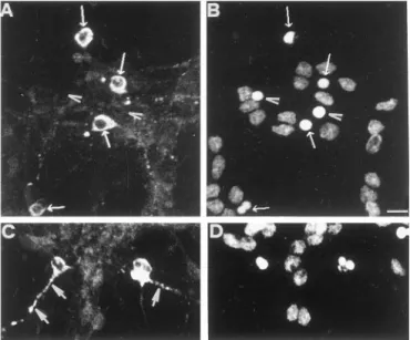
As shown in Figure 7A, neurons undergoing apoptosis and in the execution phase, recognizable as those having a small con-densed or fragmented nucleus strongly stained by propidium iodide (Fig. 7B), are characterized by a prominent ubiquitin immunoreactivity in the cytoplasm (Fig. 7A) and in the degener-ating neurites (Fig. 7C), whereas the nuclear staining is markedly reduced. In contrast, cells with a normal nuclear morphology exhibit a heterogeneous pattern of ubiquitin immunofluorescence barely detectable as a very faint staining of dendrites, perikarya, and nucleus.
We found that at any given time of apoptosis only a small fraction of the whole population of neurons identified by pro-pidium staining demonstrate an high reactivity with the antibody recognizing ubiquitinated proteins. The simplest explanation is that such high positivity is transitory because of the subsequent loss of the cytoplasmatic content. In fact, cell ghosts that have lost membrane integrity are not stained with anti-ubiquitin antibody (Fig. 7A,B) suggesting that ubiquitinated proteins, which
accumu-late in the cytoplasm of dying cells, are subsequently sequestered in the apoptotic blebs.
We next assessed whether such increased and modified intra-cellular distribution was also accompanied by an altered intra-cellular amount of polyubiquitinated proteins. Western blot analysis of cytoplasmatic and nuclear extracts derived from control cells and cells undergoing apoptosis were immunodeveloped with the same anti-ubiquitin antibody used for the immunofluorescence analy-sis. Figure 8A shows that within 6 hr after the apoptotic stimulus, a progressive increase of a heterogeneous population of high molecular weight proteins is detectable in the cytoplasmic ex-tracts. Notice that at 12 hr after the onset of apoptosis, whereas the extent of dying neurons continues to increase and approxi-mates 50% of total neurons, the amount of ubiquitinated polypeptides is lower than that detectable at 6 hr, probably because of the sequestration of proteasome and ubiquitinated proteins in the apoptotic blebs as previously described (Pitzer et al., 1996). A different trend is observed for nuclear proteins, because during the same period a progressive reduction in the ubiquitin immunostaining occurs. It is interesting to note that the actual increase of ubiquitinated polypeptides is a measure of the whole population of dying neurons. Because, as shown in Figure 7, only a small fraction of the total population of apoptotic neurons exhibit a clear cut increase in ubiquitin immunostaining at any given time after the apoptotic stimulus, we are brought to conclude that in these neurons the increase of ubiquitinated proteins is several fold higher than that detected by Western blot. Furthermore, ubiquitinated proteins accumulate within 1 hr, de-spite the slight increase of chymotrypsin-like activity (Figs. 4A, 8A). This apparent discrepancy could be attributable to (1) the
Figure 6. Effect of the general inhibitor, Z-VAD-fmk, on proteasome
activities. CGCs were washed and maintained in high-potassium and serum-free medium (S-K25) for 12 hr or switched to K5 and serum free-medium (S-K5) in the absence or in the presence of 100mM
Z-VAD-fmk. Twelve hours later, 10 mg of supernatants were incubated with substrates specific for chymotrypsin-like (Suc-LLVY-MCA), trypsin-like (Boc-LRR-MCA), and caspase-like (Z-LLG-bNap) activities at 37°C for 20, 30, and 60 min, respectively, and assayed in triplicate. Specific activ-ities are expressed as the percentage of activactiv-ities of control cells, where control cells have been given a value of 100. Results are the mean6 SD of experiments from three separate CGC preparations.
Figure 7. Ubiquitin immunostaining in cerebellar granule neurons
un-dergoing apoptosis analyzed by confocal microscopy. Apoptosis in CGC was induced by potassium and serum withdrawal, as described in Mate-rials and Methods. Double immunostaining of ubiquitin (A, C) and DNA (B, D) in CGCs was performed 6 hr after induction, using a polyclonal anti-ubiquitin antibody and propidium iodide, as described in Materials and Methods. Notice that only few, scattered neurons are heavily stained ( A) compared to the number of neurons present in the same field ( B) and already stained with propidium. Arrows indicate the immunostaining with anti-ubiquitin ( A) and their corresponding nuclei ( B). Arrowheads indi-cate ghost cells that have lost the cytoplasm content (C) and their corresponding nuclei ( D). Large arrows indicate beaded neurites stained very brightly for ubiquitin (C). Scale bar, 7mm.
generation of proteins unable to enter the proteosome cavity because not properly ubiquitinated or edited, (2) the increased amount of ubiquitinated proteins not completely degraded by the slight increment of chymotrypsin activity shown in Figure 4; and (3) the existence of two populations of CGCs, one undergoing fast and another undergoing slow apoptotic death. (Miller and Johnson, 1996).
Proteasome and caspase inhibitors prevent the intracytoplasmatic accumulation of ubiquitinated proteins in apoptotic neurons
Because proteasome inhibitors counteract apoptosis of CGCs, we examined their influence on the intracytoplasmatic accumulation
of ubiquitinated proteins by indirect immunofluorescence analy-sis performed 6 hr after onset of the apoptotic stimulus. As can been seen in Figure 9 the proteasome inhibitor MG132 prevents the diffuse intracytoplasmatic accumulation of ubiquitinated pro-teins, characteristic of apoptotic cells and shown also in Figure 7. Furthermore, the general caspase inhibitor Z-VAD-fmk, was also able to prevent this event, although to a slightly lower extent, indicating that the massive build up of ubiquitinated proteins in degenerating granule neurons is an event located downstream the caspase activation, as also suggested by the finding that the
Figure 8. Accumulation of ubiquitin conjugates in cerebellar granule
cells undergoing apoptosis. A, At 6 DIV cells were induced to apoptosis and after 1, 6, and 12 hr, nuclear (10mg) and cytoplasmatic proteins (20 mg) were collected, resolved by SDS gel electrophoresis (8–15%), and immunoblotted with the polyclonal anti-ubiquitin antibody, as described in Materials and Methods. Molecular weight markers are indicated on the
left. B, Densitometric analysis of Western blot reported above. The
absolute scanning values are given as arbitrary units. C, Time course of neuronal death after onset of apoptosis in sister cultures.
Figure 9. Proteasome and caspase inhibitors affect the
intracytoplas-matic accumulation of ubiquitinated proteins in apoptotic granules. Ap-optosis in CGCs was induced in the absence or in the presence of MG132 (5mM), Z-VAD-fmk (100mM), ALLM (40 mM), and E64d (20 mM).
Ubiquitin immunostainings were examined 6 hr after induction using anti-ubiquitin antibodies, as described in Materials and Methods. Fluo-rescent images were taken in a laser confocal microscope (Leica TCS 4D system, equipped with 1003 1.3–0.6 oil-immersion objective, optical sec-tion 1 mM). Optical sections were stereo-pair and three-dimensional
decline in chymotrypsin-like activity of proteasomes is restored in apoptotic granules treated with Z-VAD-fmk (Fig. 6). On the contrary, treatment with calpain inhibitors (such as E64d or ALLM) did not block the accumulation of ubiquitinated proteins or inhibit apoptosis (Fig. 1C).
Deubiquitinating activities decline during apoptosis
As a first attempt to investigate the possible cause or causes of proteasome failure during neuronal apoptosis we measured the activity of DUB. These enzymes, also referred to as ubiquitin C-terminal peptidases, catalyze the removal of ubiquitin from various cellular adducts, thus playing an important role in the editing of ubiquitination state of proteins and in the recycling of ubiquitin (Wilkinson, 1997). The recycling of free ubiquitin from poly-ubiquitin remnants is required for the continued action of the complete proteolytic system. In fact, polyUb chains, not properly disassembled, bind avidly to and inhibit the 26 S pro-teasome complex, presumably via competition with polyubiquiti-nated substrates (Hadari et al., 1992; Amerik et al., 1997). DUB enzymes are encoded by two gene families: the UCH family (ubiquitin C-terminal hydrolases, with molecular weight of;30 kDa, hydrolyzing small C-terminal derivatives) and UBP family (ubiquitin-specific processing proteases, with molecular weight of ;110 kDa, hydrolyzing large derivatives of ubiquitin).
We measured both activities in cell extracts of CGCs under-going apoptosis using as substrate multi-ubiquitin chains com-posed of a mixture of (Ub)4, (Ub)3, and (Ub)2 oligomers. Such
oligomers are converted by the action of DUB enzymes into monomeric ubiquitin UB1(Wilkinson et al., 1995). These
multi-ubiquitin chains are suitable substrates for measuring the activity of UBP family of DUB enzymes (Wilkinson et al., 1995). More-over, the oligomer (UB)2is also a good substrate for the UCH-L1
(Larsen et al., 1998), which is one of the most abundant enzyme in the brain, comprising up to 2% of total brain proteins (Leroy et al., 1998).
The cleavage of these substrates was monitored by Western immunoblot using anti-ubiquitin antibody , as described in Ma-terials and Methods. As shown in Figure 10A, a striking differ-ence in cleavage rate was observed, this being markedly reduced after 12 hr of apoptosis, as suggested by the finding that the immunoreactivity for (Ub)4, (Ub)3, and (Ub)2 oligomers is
greater in CGCs undergoing apoptosis than in control cells, whereas the immunoreactivity for (UB)1, the end product of
DUB activity, is lower in apoptotic cells.
Notice also that the DUB activity is restored in granule neu-rons undergoing apoptosis in the presence of a general caspase inhibitor Z-VAD-fmk (Fig. 10B) This latter finding suggest that the impairment of deubiquitinating activity is an event that oc-curs downstream of caspase activation. Similar results were also obtained using as substrate fluorogenic substrate Z-RLRGG-MCA (Stein et al., 1995; Dang et al., 1998), which is based on the C terminus of ubiquitin (data not shown). Further experiments are required to clarify, by the use of specific substrate, which family of DUB enzymes, UCH or UBP, is responsible for the decreased deubiquitinating activity detected in apoptotic neurons.
DISCUSSION
Two major findings reported in this paper deserve some com-ments and deal with the demonstration that specific inhibitors of proteasome activities administered at the time of triggering ap-optosis largely prevent this otherwise irreversible program of cell
death, such inhibition being most probably upstream the caspase activation. The second peculiar finding is that within 12 hr of inducing apoptosis, a progressive accumulation of ubiquitinated proteins occurs, likely as a result of a progressive deficit in proteasome chymotrypsin-like activity, such accumulation being downstream of the caspase activation. A corollary of these stud-ies, in line with some previous hypothesis, postulates that such proteasome involvement may have some relevance with neurode-generative diseases. For the sake of clarity, each of these specific issues will be separately discussed.
Previous studies have attributed to the proteasome a critical role in apoptosis triggered by different stimuli in terminally dif-ferentiated cells (Grimm et al., 1996; Sadoul et al., 1996). Our findings, using a panel of proteasomal inhibitors with different inhibitory capabilities against the proteasome, demonstrate that this proteolytic machinery plays also a role in death by apoptosis of CGCs. We have found that not all inhibitors exert the same protective effect. In fact, the most powerful blockers were MG132 and Lactacystin, producing a neuronal survival of 98 and 85%,
Figure 10. Deubiquitinating activity in granule neurons undergoing
ap-optosis. A, Cultures at 6 DIV were washed and switched to K5 and serum free-medium (S-K5) and in K25 and serum free-medium (S-K25) for 1, 3, 6, and 12 hr. At the time indicated after the induction of apoptosis, 5mg of supernatants were incubated with 1 mg of multi-ubiquitin chains, substrates for DUB enzymes, incubated at 22°C for 10 min, and immu-noblotted with anti-ubiquitin antibody. B, Effect of the general caspase inhibitor Z-VAD-fmk on deubiquitinating activities. CGCs were washed and maintained in high-potassium and serum free-medium (S-K25) for 12 hr or switched to K5 and serum free-medium (S-K5) in the absence or in the presence of 100mMZ-VAD-fmk. Twelve hours later deubiquitinating
respectively, after 12 hr of apoptosis. By contrast, another pro-teasome blocker PSI (Z-IE (OtBu) AL-CHO) is a poor inhibitor even at the highest concentration tested (50–100mM). Such weak
inhibitory activity is in contrast with the report dealing with the protective effect of PSI (at concentration ranging from 10 to 100 nM) on sympathetic neurons deprived of NGF (Sadoul et al.,
1996). The simplest explanation is that the composition of the proteasome complex in CGCs and sympathetic neurons is not identical, or that the pathway or pathways linking their function with other intracellular activities are distinct and they therefore exhibit markedly different sensitivity to this inhibitor. For exam-ple, proteasomes involved in antigen presentation differ both in composition and function from proteasomes involved in other processes (Driscoll et al., 1993). On the other side, it is possible that the block of cell death in CGCs requires the simultaneous repression of more than one peptidase activity of proteasome, one of which is of caspase-like nature. In fact, as shown in Figure 5, the most powerful inhibitors are those that also affect more deeply the postacidic activity (MG132 and Lactacystin). This activity could be devoted to cleave a particular substrate that promotes cell death, for example a pro-caspase family member into an active form, or could degrade regulatory proteins that normally control the apoptotic pathway. This hypothesis is strengthened by the observation that proteasomes can cleave after Asp in synthetic peptide substrates (Rivett et al., 1994; Kisselev et al., 1999) and that proteasomes are the major ICE-like proteinase in P19 cells treated with retinoic acid (Kobayashi et al., 1996). On the basis of such considerations and of previous findings, it is tempting to place the proteasome activity in CGCs undergoing apoptosis upstream of the caspase activation, as al-ready shown in sympathetic neurons deprived of NGF (Sadoul et al., 1996) and in thymocytes treated with dexamethasone, g-irradiation, or etoposide (Grimm et al., 1996; Hirsch et al., 1998; Stefanelli et al., 1998). The suggestion that the proteasome complex is acting upstream of the caspases is also based on the finding that the proteasomal inhibitors block caspase-3 activity and prevent the cleavage of tau in granule neurons undergoing apoptosis (Fig. 3). Furthermore, these inhibitors block the early release of cytochrome c from mitochondria (Bobba, Canu, Atlante, Marra, and Calissano, unpublished observations), an event preceding caspase-3 activation (Bobba et al., 1999).
After such early involvement, proteasomes become part of a generalized cellular failure that affects the major activities of the apoptotic neuron, as shown by the finding that CGCs undergoing apoptosis accumulate ubiquitinated-proteins (Figs. 7, 8A). Al-though several mechanisms could account for the building up of ubiquitinated proteins, a decrease of proteasome activity in CGCs undergoing apoptosis could be one of the major causes. We found that after 6 hr of apoptosis there is a significant decline in proteasome activity that further decreases by 12 hr and that it tightly reflects the extent of apoptosis. This decline involves the chymotrypsin-like, trypsin-like and to, a lower extent, the caspase-like activity (Fig. 4A–C). Among them, the chymotrypsin-caspase-like activity has been directly linked to the degradation of ubiquitin conjugates. Defects in the yeast proteasome subunits bearing this catalytic activity result in decreased ability of cells to cope with stress conditions, cause accumulation of ubiquitin conjugates, and reduce protein degradation rates (Heinemeyer et al., 1991). More-over, the inhibition of chymotrypsin-like activity (by the inhibitor PSI) of proteasome in neuronal cells is sufficient to induce accu-mulation of ubiquitinated proteins (Figueiredo- Pereira et al., 1994), and a decrease of the proteasome chymotrypsin-like
activ-ity is observed in transient ischemia (Kamikubo and Hayashi, 1996). Although a decline in proteasome function seems to play a major role, the possibility that during apoptosis the proteolytic machinery may be overwhelmed or incapable of dealing with an increased amount of ubiquitinated proteins cannot be ruled out. It is also possible that the proteasome efficacy is additionally hampered by the generation, during apoptosis, of poor substrates for proteolysis such as cross-linked or aggregated proteins be-cause, for example, of the disruption of the intracellular sulfhy-dryl homeostasis (Figueiredo-Pereira et al., 1998).
The significant decrease in proteasome function is likely attrib-utable to a specific downregulation and not of proteasome con-tent, as suggested by the finding that the amount of proteasome subunits does not change within the first 24 hr of apoptosis. Because we have used a broad range antiserum directed against alla-subunits, we cannot exclude that a specific subunit could be a target of caspases, resulting in reduced activity. Whatever the correct explanation, the impairment in proteasome function is likely correlated and linked to the decline in deubiquitinating activities that also occurs downstream of the caspase activation. It is worth noting that deubiquitinating enzymes facilitate the pro-tein degradation by the proteasome, possibly by supervising the ubiquitinated state of proteins and by preventing accumulation of ubiquitin chains generated as intermediates in substrate degrada-tion (Hadari et al., 1992). These chains may need to be disassem-bled to avoid accumulation to a level that inhibit proteolysis. Disruption of DUB enzymes in yeast leads to a decrease in protein degradation, accumulation of polyubiquitin proteins, and to depletion of cellular ubiquitin pools (Papa and Hochstrasser, 1993; Amerik et al., 1997). Our finding that DUB activity declines downstream of caspase activation suggests that it could be directly or indirectly a target of caspase or caspases. In this regard, it must pointed out that a missense mutation in Parkinson’s disease (Leroy et al., 1998) or intragenic deletion in gad mice (Saigoh et al., 1999) of the deubiquitinating enzyme UCH-L1 cause partial loss of the catalytic activity of this enzyme, which could lead to aberration in the proteolytic pathway and aggregation of proteins. Our data provide evidence that apoptosis in neuronal cells is accompanied by an early involvement of proteasome activities followed by a decrease in its function. To date, two other studies reported the modifications of proteasome activity during dexamethasone-induced apoptosis in thymocytes (Beyette et al., 1998; Hirsch et al., 1998). Beyette et al., (1998) found that dexamethasone-induced apoptosis in rat thymocytes is accompa-nied by a reduction of proteasome activity similar to the extent of apoptosis. Hirsch et al. (1998), on the other hand, reported that, in the same experimental paradigm, the proteasome is activated concurrently with the onset of apoptotic death. Our data mirror partly those of Beyette et al. (1998), showing a decrease, during apoptosis, after a brief and slight increase, of all the activities of proteasome. In addition, our studies, using as a parameter of proteasome failure the accumulation of ubiquitinated proteins, place the decline of proteasome activity downstream of the caspase activation as also confirmed by the finding that the chymotrypsin-like activity is restored in supernatant fraction from CGCs induced to undergo apoptosis in the presence of Z-VAD-fmk.
It is reported that the high levels of ubiquitin immunoreactivity associated with the inclusion bodies characteristic of a range of neurodegenerative disorders is attributable to the inability of the intracellular proteolytic system in lysing these structures. More-over, inappropriate apoptosis is believed to be the underlying
mechanism in the pathogenesis of neurodegenerative diseases. The studies reported in this article define an important role of apoptosis for proteasome activity failure, likely caused by a deficit in deubiquitinating activities, in the accumulation of ubiquiti-nated proteins, giving new insights into the mechanism that generates ubiquitinylated inclusions in many neuropathologies .
The question then arises as to the nature of the mechanism that first involves proteasomes as primary actors of apoptosis, to the extent that their inhibition blocks this event, and subsequently, when their apoptosis-promoting activity has been launched to other intracellular site or sites, these same structures become object of their own message so that their most typical and pre-ferred substrate, ubiquitinated proteins, accumulate in the cyto-plasm of dying cells.
REFERENCES
Amerik AYu, Swaminathan S, Krantz BA, Wilkinson KD, Hochstrasser M (1997) In vivo disassembly of free polyubiquitin chains by yeast Ubp14 modulates rates of protein degradation by the proteasome. EMBO J 16:4826–38.
Armstrong RC, Aja TJ, Hoang KD, Gaur S, Bai X, Alnemri ES, Litwack G, Karanewsky DS, Fritz LC, Tomaselli KJ (1997) Activation of the CED3/ICE-related protease CPP32 in cerebellar granule neurons un-dergoing apoptosis but not necrosis. J Neurosci 15:553–562.
Beyette J, Mason GG, Murray RZ, Cohen GM, Rivett AJ (1998) Pro-teasome activities decrease during dexamethasone-induced apoptosis of thymocytes. Biochem J 332:315–320.
Bobba A, Atlante A, Sgaramella G, Calissano P, Marra E (1999) Early release and subsequent caspase-mediated degradation of cytochrome C in apoptotic cerebellar granule cells. FEBS Lett 457:126–130. Canu N, Dus L, Barbato C, Ciotti MT, Brancolini C, Rinaldi AM, Novak
M, Cattaneo A, Bradbury A, Calissano P (1998) Tau cleavage and dephosphorylation in cerebellar granule neurons undergoing apoptosis. J Neurosci 18:7061–7074.
Ciechanover A (1994) The ubiquitin-proteasome proteolytic pathway. Cell 79:13–21.
Cohen GM (1997) Caspase: the executioners of apoptosis. Biochem J 326:1–16.
Dang LC, Melandri FD, Stein RL (1998) Kinetic and mechanistic studies on the hydrolysis of ubiquitin C-terminal 7-amido-4-methylcoumarin by deubiquitinating enzymes. Biochemistry 37:1868–1879.
DiFiglia M, Sapp E, Chase KO, Davies SW, Bates GP, Vonsattel JP, Aronin N (1997) Aggregation of huntingtin in neuronal intranuclear inclusions and dystrophic neurites in brain. Science 277:1990–1993. D’Mello SR, Galli C, Ciotti T, Calissano P (1993) Induction of apoptosis
in cerebellar granule neurons by low potassium: inhibition of death by insulin-like growth factor I and cAMP. Proc Natl Acad Sci USA 90:10989–10993.
Drexler HC (1997) Activation of the cell death program by inhibition of proteasome function. Proc Natl Acad Sci USA 94:855–860.
Driscoll J, Brown MG, Finley D, Monaco JJ (1993) MHC-linked LMP gene products specifically alter peptidase activities of the proteasome. Nature 365:262–4.
Eldadah BA, Yakovlev AG, Faden AI (1997) The role of CED-3-related cysteine proteases in apoptosis of cerebellar granule cells. J Neurosci 17:6105–13.
Enari M, Talanian RV, Wong WW, Nagata S (1996) Sequential activa-tion of ICE-like and CPP32-like proteases during Fas-mediated apo-ptosis. Nature 380:723–726.
Fenteany G, Standaert RF, Lane WS, Choi S, Corey EJ, Schreiber SL (1995) Inhibition of proteasome activities and subunit-specific amino-terminal threonine modification by lactacystin. Science 268:726–731. Figueiredo-Pereira ME, Berg KA, Wilk S (1994) A new inhibitor of the
chymotrypsin-like activity of the multicatalytic proteinase complex (20S proteasome) induces accumulation of ubiquitin-protein conjugates in a neuronal cell. J Neurochem 63:1578–1581.
Figueiredo-Pereira ME, Yakushin S, Cohen G (1998) Disruption of the intracellular sulfhydryl homeostasis by cadmium-induced oxidative stress leads to protein thiolation and ubiquitination in neuronal cells. J Biol Chem 273:12703–12709.
Galli C, Meucci O, Scorziello A, Werge TM, Calissano P, Schettini G (1995) Apoptosis in cerebellar granule cells is blocked by high KCl,
forskolin, and IGF-1 through distinct mechanisms of action: the in-volvement of intracellular calcium and RNA synthesis. J Neurosci 15:1172–1179.
Galli C, Piccini A, Ciotti MT, Castellani L, Calissano P, Zaccheo D, Tabaton M (1998) Increased amyloidogenic secretion in cerebellar granule cells undergoing apoptosis. Proc Natl Acad Sci USA 95:1247–1252.
Grimm LM, Goldberg AL, Poirier GG, Schwartz LM, Osborne BA (1996) Proteasomes play an essential role in thymocyte apoptosis. EMBO J 15:3835–3844.
Hadari T, Warms JV, Rose IA, Hershko A (1992) A ubiquitin C-terminal isopeptidase that acts on polyubiquitin chains. Role in protein degradation. J Biol Chem 267:719–727.
Heinemeyer W, Kleinschmidt JA, Saidowsky J, Escher C, Wolf DH (1991) Proteinase yscE, the yeast proteasome/multicatalytic-multifunctional proteinase: mutants unravel its function in stress in-duced proteolysis and uncover its necessity for cell survival. EMBO J 10:555–62.
Hirsch T, Dallaporta B, Zamzami N, Susin SA, Ravagnan L, Marzo I, Brenner C, Kroemer G (1998) Proteasome activation occurs at an early, premitochondrial step of thymocyte apoptosis. J Immunol 161:35–40.
Hochstrasser M (1995) Ubiquitin, proteasome, and the regulation of intracellular protein degradation. Curr Opin Cell Biol 7:215–233. Hough R, Pratt G, Rechsteiner M (1987) Purification of two high
mo-lecular weight proteases from rabbit reticulocyte lysate. J Biol Chem 262:8303–8313.
Jentsch S (1992) The ubiquitin-conjugation system. Annu Rev Genet 26:179–207.
Kamikubo T, Hayashi T (1996) Changes in proteasome activity follow-ing transient ischemia. Neurochem Int 28:209–212.
Kisselev AF, Akopian TN, Woo KM, Goldberg AL (1999) The sizes of peptides generated from proteins by mammalian 26 and 20S protea-somes. J Biol Chem 274:3363–3371.
Kobayashi T, Shinozaki A, Momoi T, Arahata K, Tsukahara T (1996) Identification of an interleukin-1 beta converting enzyme-like activity that increases upon treatment of P19 with retinoic acid as the protea-some. J Biochem 120:699–704.
Laemmli UK (1970) Cleavage of structural proteins during the assembly of the head of bacteriophage T4. Nature 227:680–685.
Larsen CN, Krantz BA, Wilkinson KD (1998) Substrate specificity of deubiquitinating enzymes: ubiquitin C-terminal hydrolases. Biochem-istry 37:3358–68.
Lee DH, Goldberg AL (1998) Proteasome inhibitors: valuable new tools for cell biologist. Trends Cell Biol 8:397–403.
Leroy E, Boyer R, Auburger G, Leube B, Ulm G, Mezey E, Harta G, Brownstein MJ, Jonnalagada S, Chernova T, Dehejia A, Lavedan C, Gasser T, Steinbach PJ, Wilkinson KD, Polymeropoulos MH (1998) The ubiquitin pathway in Parkinson’s disease. Nature 395:451–452. Levi G, Aloisi F, Ciotti M T, Gallo V (1984) Autoradiographic
localiza-tion and depolarizalocaliza-tion-induced release of acidic amino acids differen-tiating cerebellar granule cells cultures. Brain Res 290:77–86. Lowe J, Mayer RJ, Landon M (1993) Ubiquitin in neurodegenerative
diseases. Brain Pathol 3:55–65.
Manthorpe M, Fagnani R, Skaper SD, Varon S (1986) An automated colorimetric microassay for neuronotrophic factors. Brain Res 390:191–198.
Marks N, Berg MJ, Guidotti A, Saito M (1998) Activation of caspase-3 and apoptosis in cerebellar granule cells. J Neurosci Res 52:334–341. Miller TM, Johnson Jr EM (1996) Metabolic and genetic analyses of
apoptosis in potassium/serum-deprived rat cerebellar granule cells. J Neurosci 16:7487–7495.
Morishima-Kawashima M, Hasegawa M, Takio K, Suzuki M, Titani K, Ihara Y (1993) Ubiquitin is conjugated with amino-terminally pro-cessed tau in paired helical filaments. Neuron 10:1151–1160. Nardi N, Avidan G, Daily D, Zilkha-Falb R, Barzilai A (1997)
Biochem-ical and temporal analysis of events associated with apoptosis induced by lowering the extracellular potassium concentration in mouse cere-bellar granule neurons. J Neurochem 68:750–759.
Nath R, Raser KJ, Stafford D, Hajimohammadreza I, Posner A, Allen H, Talanian R, Yuen P, Gilbertsen RB, Wang KKW (1996) Non-erythroid a-spectra breakdown by calpain and interleukin 1b-converting-enzyme-like protease(s) in apoptotic cell: contributory roles of both protease families in neuronal apoptosis. Biochem J 319:683–690.
Orlowski M (1990) The multicatalytic proteinase complex, a major ex-tralysosomal proteolytic system. Biochemistry 29:10289–10297. Palombella VJ, Rando OJ, Goldberg AL, Maniatis T (1995) The
ubiquitin-proteasome pathway is required for processing the NF-KB1 precursor protein and the activation of NF-KB. Cell 78:773–785. Papa FR, Hochstrasser M (1993) The yeast DOA4 gene encodes a
deu-biquitinating enzyme related to a product of the human tre-2 oncogene. Nature 366:313–319.
Perry G, Friedman R, Shaw G, Chau V (1987) Ubiquitin is detected in neurofibrillary tangles and senile plaque neurites of Alzheimer disease brains. Proc Natl Acad Sci USA 84:3033–3036.
Peters JM (1994) Proteasome:protein degradation machines of the cell. Trends Biochem Sci 19:377–382.
Pitzer F, Dantes A, Fuchs T, Baumeister W, Amsterdam A (1996) Removal of proteasomes from the nucleus and their accumulation in apoptotic blebs during programmed cell death. FEBS Lett 394:47–50. Rivett AJ, Savory PJ, Djaballah H (1994) Multicatalytic endopeptidase
complex: proteasome. Methods Enzymol 244:331–350.
Rock KL, Gramm C, Rothstein L, Clark K, Stein R, Dick L, Hwang D, Goldberg AL (1994) Inhibitors of the proteasome block the degrada-tion of most cell proteins and the generadegrada-tion of peptides presented on MHC class I molecules. Cell 78:761–771.
Sadoul R, Fernandez PA, Quiquerez AL, Martinou I, Maki M, Schroter M, Becherer JD, Irmler M, Tschopp J, Martinou JC (1996) Involve-ment of the proteasome in the programmed cell death of NGF-deprived sympathetic neurons. EMBO J 15:3845–3852.
Saigoh K, Wang Y-L, Suh J-G, Yamanishi T, Sakai Y, Kiyosawa H, Harada T, Ichihara N, Wakana S, Kikuchi T, Wada K (1999) Intra-genic deletion in gene encoding ubiquitin carboxy-terminal hydrolase in gad mice. Nat Genet 23:47–51.
Schagger H, von Jagow G (1987) Tricine-sodium dodecyl sulfate-polyacrylamide gel electrophoresis for the separation of proteins in the range from 1 to 100 kDa. Anal Biochem 166:368–379.
Schulz JB, Weller M, Klockgether T (1996) Potassium deprivation-induced apoptosis of cerebellar granule neurons: a sequential
require-ment for new mRNAand protein synthesis, ICE-like protease activity, and reactive oxygen species. J Neurosci 16:4696–4706.
Smale G, Nichols NR, Brady DR, Finch CE, Horton WEJ (1995) Evi-dence for apoptotic cell death in Alzheimer’s disease. Exp Neurol 133:225–230.
Soto AM, Sonnenschein C (1985) The role of estrogen on the prolifer-ation of human breast tumor cells (MCF-7). J Steroid Biochem 23:87–94.
Stefanelli C, Bonavita F, Stanic I, Pignatti C, Farruggia G, Masotti L, Guarnieri C, Caldarera CM (1998) Inhibition of etoposide-induced apoptosis with peptidealdehyde inhibitors of proteasome. Biochem J 332:661–665.
Stein RL, Chen Z, Melandri F (1995) Kinetic studies of isopeptidase T: modulation of peptidase activity by ubiquitin. Biochemistry 34:12616–12623.
Steller H (1995) Mechanism and genes of cellular suicide. Science 267:1445–1449.
Su JH, Anderson AJ, Cummings BJ, Cotman CW (1994) Immuno-histochemical evidence for apoptosis in Alzheimer’s disease. Neuro-Report 5:2529–2533.
Takeda A, Mallory M, Sundsmo M, Honer W, Hansen L, Masliah E (1998) Abnormal accumulation of NACP/alpha-synuclein in neurode-generative disorders. Am J Pathol 152:367–372.
Thompson C (1995) Apoptosis in the pathogenesis and treatment of disease. Science 267:1456–1462.
Volonte` C, Ciotti MT, Battistini L (1994) Development of a method for measuring cell number: application to CNS primary neuronal culture. Cytometry 17:274–276.
Wilkinson K (1997) Regulation of ubiquitin-dependent process by deu-biquitinating enzymes. FASEB J 11:1245–1246.
Wilkinson KD, Tashayev VL, O’Connor LB, Larsen CN, Kasperek E, Pickart CM (1995) Metabolism of the polyubiquitin degradation sig-nal: structure, mechanism, and role of isopeptidase T. Biochemistry 34:14535–14546.