Alma Mater Studiorum – Università di Bologna
DOTTORATO DI RICERCA IN
BIOLOGIA CELLULARE E MOLECOLARE
Ciclo XXIX
Settore Concorsuale SSC 05/E2
Settore Disciplinare SSD BIO/11
Titolo tesi:
“Approaches to new generation vaccines
against pertussis and identification
of new virulence factors”
Presentata da:
Gianmarco Gasperini
Coordinatore Dottorato
Prof. Giovanni Capranico
Relatore:
Prof. Vincenzo Scarlato
Correlatore:
Dott.ssa Beatrice Aricò
2
Because getting your dreams
it's strange, but it seems
a little - well - complicated.
There's a kind of a sort of cost,
there's a couple of things that get lost,
There are bridges you cross
you didn't know you crossed
3
Fundings
This study was sponsored by GSK Vaccines (formerly Novartis Vaccines).
4
Table of contents
1. Introduction...5
1.1. Pertussis disease...5
1.1.1. Bordetella pertussis...7
1.1.2. Pertussis vaccines and resurgence of pertussis...12
1.2. Outer Membrane Vesicles...15
2. Chapter one: OMV-based and proteomic-driven antigen selection
for a new generation vaccine against pertussis...17
2.1. Results...18
2.2. Discussion and conclusions...38
3. Chapter two: contribution of naturally released Outer Membrane
Vesicles from Bordetella pertussis to bacterial physiology and
pathogenesis...44
3.1. Results and discussion...45
3.2. Conclusions...57
4. Experimental procedures...58
4.1. Bacterial strains and growth conditions...58
4.2. Generation of E. coli strains expressing heterologous antigens....59
4.3. Cell lines and growht conditions ...60
4.4. OMV isolation...61
4.5. SDS-PAGE gel analysis...61
4.6. Nanoparticle tracking analysis...62
4.7. Generation of mouse immune sera...62
4.8. Adhesion assay...63
4.9. Adhesion inhibition assay...64
4.10. Proteomic
analysis
by
LC-MS/MS,
data
analysis
and
bioinformatics... 65
4.11. Flow citometry...67
4.12. Recombinant antigen production...67
4.13. Mouse aerosol challenge model...68
4.14. Electron microscopy...68
4.15. Confocal microscopy...69
4.16. Western Blotting...70
4.17. PT intoxication assay...70
4.18. Growth assay for iron delivery...70
5
1. Introduction
1.1. Pertussis disease
Pertussis is a highly contagious, acute respiratory disease whose etiological agent is the Gram-negative bacterium Bordetella pertussis. The name pertussis takes its origins from Latin (per, intensive, and tussis, cough) and describes the most consistent and prominent clinical feature of the illness. The common name for the illness, “whooping cough”, comes from the characteristic inspiratory sound made at the end of an episode of paroxysmal coughing.
B. pertussis infection results in colonization and rapid multiplication of the bacteria on
the mucous membrane of the respiratory tract. Histopathological studies of fatal pertussis cases documented large airway disease as the principal component of B. pertussis infection and showed large numbers of bacteria on cilia lining trachea and
bronchi, causing local damage (Mallory and Hornor 1912). More recently, electron microscopy studies have demonstrated that B. pertussis adheres exclusively to the tufts of ciliated epithelial cells of the upper respiratory tract; no attachment to non-ciliated cells was observed (Tuomanen and Hendley 1983).
Transmission
B. pertussis is a strict human pathogen and no other animal or environmental reservoir
is known. It is transmitted from human to human through aerosolized respiratory droplets expelled via coughing and sneezing, as further demonstrated in a recent study in a baboon model (Warfel, Beren et al. 2012). Adults, who often lack classic symptoms, have a primary role in the spread of the disease, as they serve as important
6
reservoirs of B. pertussis, transmitting pertussis to infants and children, who are at greater risk for morbidity and mortality (Bisgard, Pascual et al. 2004, Wendelboe, Njamkepo et al. 2007).
Clinical manifestations
Clinical manifestations of the infection may show substantial variation, depending on several factors, such as clinical condition and age of the patient, previous vaccination or infection, presence of passively acquired antibodies (Mattoo and Cherry 2005). Pertussis can result in asymptomatic infection, mild cough illness or classic illness. Classic illness often occurs in primary infection in unimmunized children between 1 and 10 years of age (Cherry 1999), lasts 6 to 12 weeks or longer and is divided into three stages: catarrhal, paroxysmal and convalescent phase (Fig.1). After an incubation period of 7 to 10 days, illness begins with the catarrhal phase, which lasts 1-2 weeks and is usually characterized by nonspecific symptoms including rhinorrhea, lacrimation, progressive cough, reduced activity, loss of appetite and occasional low-grade fever. At the end of this phase, most children have frequently a leukocytosis with absolute lymphocytosis, which is absent in adults (Postels-Multani, Schmitt et al. 1995, Mattoo and Cherry 2005, Melvin, Scheller et al. 2014). During the subsequent paroxysmal stage, lasting 1-6 weeks or longer, the cough becomes more persistent and severe, with
paroxysms, consisting of repetitive series of forceful coughs during a single expiration. A paroxysm is often followed by a forced inspiration during which the characteristic whoop occurs and is often associated with tenacious mucus, cyanosis and post-tussive vomiting. The patient usually shows normal airway function between attacks. Infants under 6 months of age may not have the strength to have a whoop, so the disease may manifest with spells of apnea, with consequent bradycardia, cyanosis and unresponsiveness (Christie and Baltimore 1989). The transition to the last convalescent
7
phase is associated with a gradual decrease in the frequency and in the severity of the
paroxysms before the patient returns to normal. This stage usually lasts 2-3 weeks but it is often prolonged. Most of the asymptomatic infections or mild illnesses occur in previously immunized or infected subjects, both adolescents and adults. The predominant symptom may be a persistent cough, that is often associated with nonspecific symptoms of more common viral upper respiratory tract infections (Cornia, Hersh et al. 2010). Pertussis is most severe in young infants, but it occurs in all age groups. Most deaths due to pertussis occur in infants, for which the infection is particularly severe: in neonatal infection the risk of death correlates with frequent episodes of apnea, often resulting in seizures due to hypoxia (Christie and Baltimore 1989), whereas in young infants is related directly with lymphocytosis, which positively correlates with intractable pulmonary hypertension, respiratory failure and death (Paddock, Sanden et al. 2008).
1.1.1. Bordetella pertussis
Bordetella pertussis was first isolated in pure culture by Bordet and Gengou in 1906. It
is a small (approximately 0.5 x 1 µm) encapsulated bacterium, strict aerobe, non-motile and non-sporulating.
Following the inhalation of infected aerosol, B. pertussis enters the upper respiratory tract and adheres to ciliated epithelial cells in the nasopharynx and trachea. Once attached to the mucosal surface, B. pertussis produces a wide array of virulence factors, including adhesins and toxins. The concerted expression of these factors prevents rapid clearance of the bacteria and enables replication and dissemination to the lower areas of the respiratory tract (de Gouw, Diavatopoulos et al. 2011).
8
The expression of the majority of these virulence factors is under the control of a two-component regulatory system encoded by the bvgAS locus (Arico, Miller et al. 1989). BvgS is a 135-kDa transmembrane sensor kinase consisting of several domains involved in phosphorylation cascade, while BvgA is a 23-kDa response regulator protein, with a receiver domain at its N-terminus and a DNA-binding domain at its C-terminus (Cotter and Jones 2003). Under inducing signals BvgS autophosphorilates and initiate a phopshorelay that leads to BvgA phosphorylation and activation (Uhl and Miller 1996). Activated BvgA dimerizes and binds to specific DNA sequences to positively or negatively regulate transcription (Decker, James et al. 2012). In response to environmental conditions, BvgAS controls the expression of at least three distinct phenotypic phases, each characterized by maximal expression of some genes and minimal expression of others (Lacey 1960). The signals to which BvgS responds in vivo are still unknown. Bvg+ phase occurs when bacteria are grown under permissive conditions. In this phase BvgAS is active and promotes the expression of virulence-activated genes (vag-genes, divided in class 1 and class 2 genes), while represses the expression of virulence-repressed genes (vrg-genes, or class 4 genes) by the repressor protein BvgR, whose gene is located downstream of the bvgAS locus and is activated by BvgA (Merkel, Barros et al. 1998). Vag-genes encode adhesins and toxins that play a key role in the respiratory tract colonization in vivo (Merkel, Stibitz et al. 1998). Bvg- phase occurs when bacteria are grown under modulating conditions, such as growing at low temperature (25°C) or in the presence of nicotinic acid or MgSO4 (Melton and
Weiss 1993). During this phase, BvgAS is inactive and is unable to repress vrg-genes, which are therefore maximally expressed, while no expression of vag-genes occurs. Since these genes are expressed when bacteria are grown under limiting conditions, they may be involved in B. pertussis survival in the ex vivo environment. Bvgi phase occurs when bacteria are grown under submodulating conditions (low concentration of
9
chemical modulators). In this phase bacteria display a phenotype that is intermediate between Bvg+ phase and Bvg- phase, since BvgAS is partially active: vrg-genes and class 1 genes (e.g. PT, ACT) are not expressed, while class 2 genes (e.g. FHA) and the genes specific of this phase (class 3 genes) are maximally expressed (Deora, Bootsma et al. 2001). This phenotype may be important for transmission between hosts, but this remains to be elucidated. The complexity of this system may allow multiple levels of control of gene expression that could be important in the infection cycle.
B. pertussis virulence factors include several adhesins and toxins that contribute to its
ability to cause the disease. The major virulence factors and antigenic components expessed during the virulent Bvg+ phase, are schematically reported in Table 1 and Fig. 1. Among them, Filamentous hemagglutinin, Pertactin and Pertussis Toxin, are the antigens included in three-component acellular pertussis vaccine.
10
Virulence Determinant Description
Filamentous hemagglutinin (FHA)
220 kDa surface-associated and secreted protein. Dominant adhesin, promotes attachment to respiratory epithelium. Immunomodulatory properties.
Pertactin (PRN) 69 kDa outer membrane protein. Autotransporter. Mediates eukaryotic cells binding in vitro. Role in resistance to neutrophil-mediated clearance.
Fimbriae (FIM) Filamentous cell surface structures. Two serologic types (type 2 and 3). Role in mediating adherence to ciliated respiratory epithelium. Potential immunomodulatory activity.
Pertussis toxin (PT) 105 kDa ADP-ribosylating A-B toxin. Responsible of pertussis-associated leukocytosis with lymphocytosis, hypoglycemia and sensitization to histamine. Important immunomodulatory activity. Strong adjuvant.
Adenylate cyclase toxin (ACT)
Calmodulin-activated toxin with adenylate cyclase and hemolysin activity. Anti-inflammatory and antiphagocytic factor. Possible contribution to local tissue damage in the respiratory tract.
Dermonecrotic toxin (DNT) 160 kDa heat-labile cytoplasmic toxin. Activates Rho. Induces necrosis in vivo and in vitro. Possible role in bacterial survival and local tissue damage in the respiratory tract.
Tracheal cytotoxin (TCT) 921 Da disaccharide-tetrapeptide monomer of peptidoglycan. Not regulated by BvgAS. Causes disruption of tight junctions, damage to cilia with ciliary stasis, IL-1 and NO production.
LOS Envelope toxin. Often referred to as lipooligosaccharide (LOS) because it lacks O-antigen. Endotoxin activities. Role in antibiotic and serum resistance. Cause of whole cell vaccines reactogenicity.
Type III secretion system (T3SS)
Translocate effector proteins into host cells. Required for persistent tracheal colonization. Immunomodulatory activity. Activates ERK1/2.
Autotransporters
(BrkA, TcfA, Vag8, SphB1)
Surface-associated or secreted. Putative roles in mediating adherence, serum resistance, evasion of antibody-mediated clearance, proteolytic processing of other surface proteins.
11
FIG. 1. B. pertussis pathogenesis: transmission and disease progression Adapted from (Tozzi, Celentano et al. 2005). Bordetella pertussis engages multiple strategies in order
to create a niche for colonization of the mucosal epithelium. From left to the right: several virulence factors mediate adhesion to respiratory cells (FHA, 69K, Fimbriae), colonization and dissemination. Bordetella pertussis evades the immune system and the complement mediated killing (BrkA and Vag8) and invades epithelial cells. Tracheal cytotoxin (TCT) and lipo-oligosaccharide (LOS) synergistically evoke ciliary damage to the respiratory epithelium. Pertussis Toxin (PT), Adenylate cyclase toxin (ACT) and the type III secretion system (T3SS) subvert intraepithelial signaling pathways leading to cytotoxicity.
12
1.1.2. Pertussis vaccines and resurgence of pertussis
In the pre-vaccine era, pertussis was an endemic and epidemic disease mainly affecting young children, with high morbidity and mortality (Cherry 1999). Given the severity of the disease, vaccines were introduced soon after the isolation of the etiological agent, to treat and prevent the disease. Whole-cell pertussis (wP) vaccines, that consist of suspensions of killed B. pertussis organisms (Xing, Markey et al. 2014), were first introduced in the 1940s and mass vaccination programs resulted in a rapid reduction in both the incidence of pertussis and the number of death caused by the infection in all age groups. However, the high reactogenicity of wP vaccines due to the presence of endotoxin, including an elevated risk of fever (Barkin and Pichichero 1979) and rare cases of convulsions, motivated the development in the 1980s of acellular pertussis (aP) vaccines. aP vaccines, consisting of one to five purified antigens (PT, FHA, PRN and FIM 2/3), are less reactogenic, thanks to the removal of the LPS, and starting from the late 1990s progressively replaced wP vaccines in many developed countries. Since then, in the last three decades a steady increase in the number of pertussis cases was observed in various countries following aP vaccination programs (Black, Cousens et al. 2010, Chiappini, Stival et al. 2013, Ausiello and Cassone 2014, Mills, Ross et al. 2014), confirming a reemergence of pertussis.
There are several causes that may contribute to the resurgence of pertussis; one of these is the emergence of B. pertussis strains that have undergone variations in the antigens included in the vaccine, as consequence of immune selective pressure. Indeed, it has been reported in the past decade the appearance of B. pertussis strains with increased production of pertussis toxin and strains pertactin-deficient (de Gouw, Hermans et al. 2014, Mooi, Van Der Maas et al. 2014). Antigenic divergence could affect both
13
memory recall and the efficacy of antibodies, while higher levels of pertussis toxin may increase suppression of the innate and acquired immune system.
A second possible reason is the waning immunity. Although initially induced at a sufficiently protective level, the acquired pertussis-specific immunity is progressively lost with time. Estimates indicate that infection-acquired immunity against pertussis disease wanes after 4–20 years and protective immunity after vaccination with wP or aP vaccines wanes after 4–12 years. (Wendelboe, Van Rie et al. 2005, Klein, Bartlett et al. 2012). There is also evidence that some wP vaccines induce longer lasting immunity than aP vaccines (Mooi, Van Der Maas et al. 2014). Further research into the rate of waning of vaccine-acquired immunity will help to determine the optimal timing and frequency of booster immunizations and their role in pertussis control.
Another explanation for the increasing incidence of pertussis related to the switch to aP vaccines lies in the difference between the mechanisms of both humoral and cellular immunity induced by natural infection or vaccination with a wP vaccine versus the vaccination with aP vaccine. Studies in murine models have shown that innate immune response help to control bacterial infection, while the complete clearance of bacteria requires cellular immunity mediated by T cells that secrete IFN- γ (T-helper type 1, Th1) and IL-17 (T-helper type 17, Th17) (Higgs, Higgins et al. 2012, Ross, Sutton et al. 2013). Complementary studies have shown that natural infection with B. pertussis or immunization with wP vaccines, which contain killed bacteria, promote naïve T-cell differentiation into Th1 and Th17 types, which promote bacterial killing by activation of macrophages and neutrophils and induction of IgG2a opsonizing antibodies. On the contrast, current aP vaccines, containing three to five purified antigens, promote naïve T-cell differentiation into Th17 cells, which activate neutrophils, and Th2 cells, which promote eosinophil activation, with no known protective role, and induction of
toxin-14
neutralizing IgG1 antibodies (Higgs, Higgins et al. 2012). These findings were also confirmed by a recent study with a baboon model, in which animals showed the same type of immunity response (Warfel, Zimmerman et al. 2014). Importantly, baboons immunized with aP vaccines were protected from pertussis-associated symptoms but not from colonization and were able to transmit the disease to unimmunized animals. By contrast, immunization with wP vaccines resulted in a more rapid B. pertussis clearance.
The observation that aP vaccines, which induce a different immune response from natural infection, fail to prevent colonization and transmission, in addition to waning immunity and the emergence of strains with mutated antigens, provides a plausible explanation of pertussis resurgence and suggests that control of the disease requires the development of improved vaccines (Clark, Messonnier et al. 2012).
15
1.2. Outer Membrane Vesicles
Outer Membrane Vesicles (OMV) are blebs of the outer membrane that are spontaneously released by virtually any Gram-negative bacteria (Haurat, Elhenawy et al. 2015, Schwechheimer and Kuehn 2015). Although the observation of OMV release was made over 40 years ago, only in the past 10 years their study has become focused on their functions and biologica roles, especially as they relate to bacterial pathogenesis. This phenomenon happens in very different settings, including planktonic cultures, surface-attached biofilm communities, solid and liquid media, and natural environments such as fresh and salt water or mammalian hosts (Beveridge, Makin et al. 1997, Beveridge 1999, Hellman, Loiselle et al. 2000, Biller, Schubotz et al. 2014). OMV are spherical and bilyered structure and they range from 50 to 250 nm in size. Clearly, being liberated from the outer membrane, OMV contain lipopolysaccharide (LPS), outer membrane proteins and phospholipids, along with periplasmic proteins in their lumen (McBroom, Johnson et al. 2006).
To better understand the mechanisms that are involved in OMV biogenesis it is important to consider the peculiar architecture of the bacterial cell envelope. Indeed, the budding and detachment of OMV depends on speficic interactions between the bacterial outer membrane and the underlying peptidoglycan layer (Braun and Wolff 1975, Cascales, Bernadac et al. 2002). In particular, specific crosslinks determine envelope stability, give bacteria their shape and confer protection from osmotic changes and sheer stress. OMV biogenesis therefore must rely on the dissociation of the outer membrane from the peptidoglycan layer in specific areas were attachments are depleted or missing. The budding vesicles then bulge outwards until the membrane undergoes fission and detaches (Schwechheimer and Kuehn 2015).
16
OMV are largely described in literature for their multifaceted roles in bacterial physiology and pathogenesis. The various functions attributed to OMV can be classified in two main groups: response to physical and chemical stress and delivery of biomolecules. Therefore, bacteria can utilize OMV either to improve their chances for survival or to induce changes in their environment, including both the host and the surrounding bacterial community.
Within the first group of biological roles we can list resistance to antibiotics and antimicrobial peptides and protection from antibodies and bacteriophages (Manning and Kuehn 2011). Also, OMV can act to remove toxic compounds from bacterial cells, such as misfolded proteins during temperature stress (McBroom and Kuehn 2007).
On the other hand, within the second group of described functions, we can list different types of OMV cargo, ranging from proteins to saccharides and nucleic acids. OMV are described to stimulate the immune system by presenting antigenic outer membrane components and LPS (Alaniz, Deatherage et al. 2007), to deliver autolysins to competing bacterial species, and to be involved in horizontal DNA transfer (Dorward, Garon et al. 1989). OMV can serve as sources of carbon and nitrogen (Biller, Schubotz et al. 2014), and can carry enzymes able to digest complex biomolecules during nutrient stress (Toledo, Coleman et al. 2012). They were also involved in iron and zinc acquisition, providing bacteria with access to essential metals (Dashper, Hendtlass et al. 2000, Smalley, Byrne et al. 2011, Prados-Rosales, Weinrick et al. 2014). Finally, they were shown to assist in quorum sensing and biofilm formation (Mashburn-Warren, Howe et al. 2009). Most interestingly, when they are released from Gram-negative pathogens, OMV have been found to be enriched with active toxins and other virulence factors that enables them to interact with host cell and contribute to intoxication (Fiocca, Necchi et al. 1999, Kesty, Mason et al. 2004, Chatterjee and Chaudhuri 2011).
17
2. Chapter one: OMV-based and proteomic driven
antigen selection for a new generation vaccine against
pertussis
Despite high vaccination coverage world-wide, whooping cough is recently increasing in occurrence suggesting that novel vaccine formulations targeted at the prevention of colonization and transmission should be investigated. In order to identify new candidates for inclusion in the acellular formulation, we used spontaneously released outer membrane vesicles (OMV) as a potential source of key adhesins. The ability of anti-OMV serum to inhibit the adhesion of B. pertussis to lung epithelial cells in vitro was demonstrated. We employed a proteomic approach to quantify proteins in OMV purified from bacteria in the Bvg+ and Bvg- phase, thus comparing the outer membrane protein pattern of this pathogen in its virulent or avirulent state. Six of the most abundant outer membrane proteins were selected as candidates to be evaluated for their adhesive properties and vaccine potential. We generated E. coli strains singularly expressing the selected proteins and assessed their ability to adhere to lung epithelial cells in vitro. Four out of the selected proteins conferred adhesive ability to E. coli. Three of the candidates were specifically detected by anti-OMV mouse serum suggesting that these proteins are immunogenic antigens able to elicit an antibody response when displayed on the OMV. Anti-OMV serum was able to inhibit only BrkA-expressing E. coli adhesion to lung epithelial cells. Finally, stand-alone immunization of mice with recombinant BrkA resulted in significant protection against infection of the lower respiratory tract after challenge with B. pertussis. Taken together, these data support the inclusion of BrkA and possibly further adhesins to the current acellular pertussis vaccines to improve the impact of vaccination on the bacterial clearance.
18
2.1. Results
OMV purification, visualization, enumeration and sizing
In order to identify possible Bvg-regulated outer membrane adhesins, we compared the protein composition of OMV from two B. pertussis strains, the Tohama I-derivatives BP536 and BP537 representative of the Bvg+ and Bvg- phases, respectively.
OMV were purified using a detergent-free method from cell-free culture supernatants through ultracentrifugation. Equal amounts with respect to total protein quantities from both preparations were loaded onto SDS-PAGE gel (Fig. 2A) showing significant differences between the two samples representing Bvg+ and Bvg- phases. Several high molecular weight protein bands appear to be exclusive to Bvg+ OMV, as well as two major bands with apparent molecular weight of 42 and 32 kDa, while one intense protein band of apparent molecular weight around 40 kDa seems to be majorly expressed in Bvg- OMV. The 40 kDa protein band was excised, destained and digested with trypsin for peptide mass fingerprinting (PMF) identification: outer membrane porin protein BP0840 was identified with a sequence coverage of 42% (data not shown).
To evaluate the actual number and the size of vesicles we used nanoparticle tracking analysis (NTA), a method for direct, real-time visualization of nanoparticles in liquids through the tracking of the brownian movement of individual particles (Olaya-Abril, Prados-Rosales et al. 2014). OMV samples from BP536 and BP537, normalized to the same protein concentration of 1 mg/mL, contained 6 x10¹¹/mL and 6.3 x 10¹¹/mL OMV-particles, respectively. The size distribution of OMV isolated from both strains was in the 70–230 nm diameter range with the majority of the OMV particles at 115 nm (Fig. 2B). These results indicated that, despite the dramatic difference in protein
19
composition of the two OMV samples, 1 mg of proteins from each OMV preparation as measured with the Lowry assay, corresponds to the same number of vesicles (around 6 x 10¹¹ blebs).
FIG. 2. SDS-PAGE and nanoparticle tracking analysis of OMV samples from BP536 and BP537.
(A) SDS-PAGE and Coomassie blue staining of 10 µg (protein content) of OMV samples from B.
pertussis BP536 Bvg+ (lane 1) and its Bvg- derivative BP537 (lane 2). (B) Nanoparticle tracking analysis
measurement of OMV preparation (1 mg protein/mL) showing the sizes and total number of OMV per mL.
20 Inhibition of B. pertussis adhesion with anti-OMV serum
We checked whether antibodies raised against antigens present in Bvg+ OMV could have anti-adhesive properties on B. pertussis. We tested the ability of anti-OMV sera to inhibit adhesion of BP536 to A549 respiratory epithelial cells. Fluorescent bacteria were pre-incubated with mouse pooled sera in a range of four dilutions and A549 cells were infected as described previously (Zanaboni, Arato et al. 2016). Mouse anti-sera were raised to Bvg+ OMV and whole bacteria adsorbed to aluminum hydroxide and collected after the third immunization together with control sera from mice immunized with adjuvant only. We found that anti-OMV serum conferred a substantial reduction of adhering bacteria even at low serum concentrations. Interestingly, sera raised against whole bacteria, therefore targeting the entire membrane antigen repertoire, showed a comparable kinetic of inhibition. Anti-alum serum was included in the experiments as negative control and, as expected, did not induce any inhibition of B. pertussis adhesion (Fig. 3).
21
FIG. 3. Impact of anti-OMV serum on B. pertussis adhesion to A549 cells. Sera from 10 mice
immunized with either OMV, whole bacteria or aluminum hydroxide as control were pooled, serially diluted in infection medium and incubated with labeled wild-type B. pertussis BP536 for 1 h. A549 cells were then infected with the bacteria/sera mixtures for 1 hour and, after extensive washes to remove unbound bacteria, cell-associated bacteria were quantified by fluorescence reading at Ex/Em 485/535 nm. Results represent mean ± SD of one representative of three independent experiments each performed in triplicates.
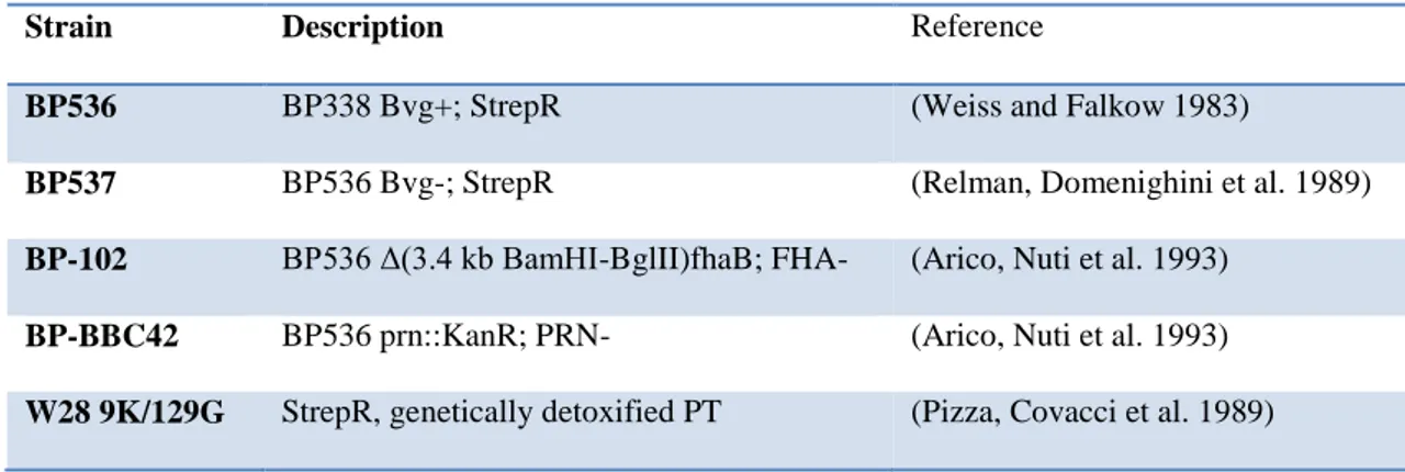
22 Comparative proteomic analysis
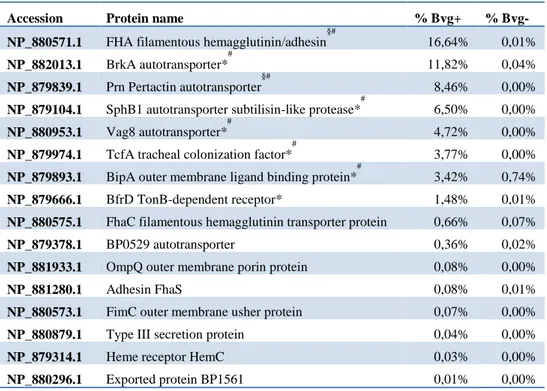
OMV samples from BP536 and BP537 were digested by trypsin after TCA precipitation. Proteomic analysis was performed by LC-MS/MS using a Data-Dependent Acquisition (DDA) approach. A total of 155 and 247 proteins were quantified in the first and second biological replicate respectively, with the second replicate fully including the proteins quantified in the first one. Percentages of each protein in the total samples were averaged to calculate the fold increase of each protein amount in one Bvg phase with the respect to the other (Table 2). Despite some proteins were identified exclusively in either Bvg+ or Bvg- phase, we set a threshold to determine the Bvg phase specificity: 23 proteins showed a 10-fold increase in Bvg+ vs. Bvg- phase and represented ~58% of the total protein amount in Bvg+ OMV while 26 proteins showed a 10-fold increase in Bvg- vs. Bvg+ phase and represented ~14% of the total protein amount in Bvg- OMV (Fig. 4A). The localization of the quantified proteins was predicted according to PSORTb software and refined for lipoprotein annotation using DOLOP software. The analysis predicted ~90% and ~75% of outer membrane proteins, lipoproteins, extracellular and periplasmic proteins for Bvg+ and Bvg- OMV, respectively (Fig. 4B). Around 10% of the identified proteins had an unknown prediction of localization in both OMV samples but only a minority of proteins was predicted to be inner membrane or cytoplasmic, thus confirming that vesicles are generated by outer membrane blebbing and not by bacterial lysis. This analysis confirmed the main differences already observed in the SDS-PAGE gel, identifying the five most abundant proteins in Bvg+ OMV to have a theoretical molecular weight higher than 90 kDa and the most abundant protein in Bvg- OMV to be the outer membrane porin protein BP0840 (~20%).
23
FIG. 4. Comparative proteomic analysis of B. pertussis OMV: protein quantification through LC-MS/MS and prediction of their subcellular localization. (A) Protein quantification of Bvg+ and Bvg-
OMV: proteins with 10-fold amount increase in Bvg+ vs. Bvg- OMV (light blue), proteins with 10-fold amount increase in Bvg- vs. Bvg+ OMV (dark blue), unregulated proteins (grey). (B) Distribution of quantified proteins in the distinct subcellular localizations based on prediction with PSORTb and DOLOP softwares: Outer Membrane (OM), Periplasm (P), Lipoproteins (L) Inner Membrane (IM), Cytoplasm (C), Extracellular (Ex), Unknown (U).
24
Accession Description [Bordetella pertussis Tohama I] % BVG+ %
BVG- Fold increase Bvg+ vs. Bvg- Fold increase Bvg- vs. Bvg+ PSORTb DOLOP
NP_880571.1 filamentous hemagglutinin/adhesin 16,64% 0,01% 1760,96 0,00 Outer Membrane
NP_882013.1 BrkA autotransporter 11,82% 0,04% 313,79 0,00 Outer Membrane
NP_879839.1 pertactin autotransporter 8,46% 0,00% 1771,79 0,00 Outer Membrane
NP_879104.1 autotransporter subtilisin-like protease SphB1 6,50% 0,00% 3284,82 0,00 Outer Membrane
NP_880953.1 Vag8 autotransporter 4,72% 0,00% 1421,45 0,00 Outer Membrane
NP_881876.1 translocation protein TolB 3,86% 8,69% 0,44 2,25 Periplasmic
NP_879974.1 tracheal colonization factor 3,77% 0,00% 3094,18 0,00 Outer Membrane
NP_879893.1 outer membrane ligand binding protein 3,42% 0,74% 4,60 0,22 Outer Membrane
NP_879898.1 serotype 2 fimbrial subunit 3,03% 0,00% 5846,89 0,00 Extracellular
NP_882014.1 molecular chaperone GroEL 2,82% 3,73% 0,75 1,32 Cytoplasmic
NP_879650.1 outer membrane porin protein BP0840 2,75% 20,28% 0,14 7,38 Outer Membrane
NP_879093.1 exported protein 2,24% 2,15% 1,04 0,96 Unknown lipo
NP_882074.1 peptidyl-prolyl cis-trans isomerase 2,19% 2,18% 1,01 0,99 Periplasmic
NP_882329.1 amino acid ABC transporter substrate-binding protein 1,50% 1,25% 1,20 0,83 Periplasmic
NP_879666.1 TonB-dependent receptor BfrD 1,48% 0,01% 171,18 0,01 Outer Membrane
NP_881875.1 peptidoglycan-associated lipoprotein 1,28% 2,76% 0,46 2,15 Outer Membrane lipo
NP_880337.1 iron binding protein 1,18% 0,37% 3,24 0,31 Periplasmic
NP_880224.1 ABC transporter substrate-binding protein 1,17% 0,75% 1,56 0,64 Unknown
NP_881568.1 lipoprotein 1,13% 1,71% 0,66 1,52 Outer Membrane lipo
NP_879744.1 outer membrane protein A 1,06% 1,26% 0,84 1,19 Outer Membrane
NP_881062.1 serine protease 0,90% 2,95% 0,31 3,27 Periplasmic
NP_880436.1 peptidase 0,69% 1,59% 0,43 2,32 Outer Membrane
NP_881028.1 peptide ABC transporter substrate-binding protein 0,68% 0,01% 58,54 0,02 Periplasmic
NP_880575.1 filamentous hemagglutinin transporter protein FhaC 0,66% 0,07% 9,06 0,11 Outer Membrane
NP_879258.1 exported protein 0,61% 1,28% 0,48 2,09 Periplasmic lipo
25
NP_879135.1 exported protein 0,56% 0,68% 0,82 1,23 Periplasmic
NP_880049.1 sn-glycerol-3-phosphate ABC transporter substrate-binding protein UgpB 0,56% 1,82% 0,31 3,27 Periplasmic
NP_881874.1 hypothetical protein BP3341 0,54% 1,43% 0,38 2,64 Unknown
NP_879068.1 lipoprotein 0,45% 0,09% 4,83 0,21 Unknown lipo
NP_881542.1 ABC transporter substrate-binding protein 0,42% 0,33% 1,30 0,77 Unknown
NP_880721.1 glycine/betaine ABC transporter substrate-binding protein 0,40% 0,61% 0,65 1,53 Periplasmic
NP_880844.1 outer membrane protein assembly factor BamB 0,37% 0,90% 0,41 2,43 Outer Membrane
NP_881124.1 zinc protease 0,37% 0,39% 0,94 1,06 Outer Membrane
NP_879378.1 autotransporter BP0529 0,36% 0,02% 14,91 0,07 Outer Membrane
NP_881639.1 adenosylhomocysteinase 0,36% 0,18% 2,02 0,49 Cytoplasmic
NP_881351.1 ABC transporter substrate-binding protein 0,33% 1,64% 0,20 4,97 Periplasmic
NP_881504.1 TonB-dependent receptor 0,30% 2,76% 0,11 9,33 Outer Membrane
NP_881019.1 enolase 0,28% 0,39% 0,71 1,40 Cytoplasmic
NP_880170.1 outer membrane protein 0,27% 0,97% 0,27 3,64 Periplasmic
NP_881493.1 DNA-binding protein 0,26% 0,03% 9,57 0,10 Unknown
NP_882283.1 pertussis toxin subunit 2 0,26% 0,00% #DIV/0! 0,00 Unknown
NP_879610.1 polyribonucleotide nucleotidyltransferase 0,22% 0,31% 0,72 1,39 Cytoplasmic
NP_879922.1 outer membrane protein assembly factor 0,22% 0,59% 0,37 2,69 Outer Membrane lipo
NP_880169.1 outer membrane protein assembly factor 0,22% 0,79% 0,27 3,67 Outer Membrane
NP_880053.1 leu/Ile/val-binding protein 0,21% 0,46% 0,47 2,13 Periplasmic
NP_880738.1 efflux system inner membrane protein 0,21% 1,79% 0,12 8,38 Cytoplasmic
Membrane
NP_881866.1 chaperone SurA 0,21% 0,56% 0,38 2,66 Periplasmic
NP_880063.1 lipoprotein 0,20% 0,61% 0,32 3,10 Unknown lipo
NP_882317.1 hypothetical protein BP3819 0,19% 0,52% 0,38 2,66 Unknown
NP_879005.1 penicillin-binding protein 0,19% 1,93% 0,10 10,40 Cytoplasmic
Membrane
NP_879899.1 NADP-dependent malic enzyme 0,18% 0,03% 6,15 0,16 Cytoplasmic
26
NP_878925.1 elongation factor Tu 0,17% 0,85% 0,20 4,95 Cytoplasmic
NP_882263.1 lipoprotein 0,15% 0,09% 1,74 0,58 Outer Membrane lipo
NP_882282.1 pertussis toxin subunit 1 0,15% 0,00% #DIV/0! 0,00 Extracellular
NP_879312.1 exported protein 0,15% 7,15% 0,02 47,22 Unknown
NP_879963.1 lipoprotein 0,15% 0,07% 1,98 0,50 Outer Membrane lipo
NP_879023.1 glycerol-3-phosphate ABC transporter substrate-binding protein 0,15% 0,00% 145,71 0,01 Periplasmic
NP_882284.1 pertussis toxin subunit 4 0,14% 0,00% #DIV/0! 0,00 Unknown
NP_879606.1 ketol-acid reductoisomerase 0,13% 0,10% 1,33 0,75 Cytoplasmic
NP_882045.1 DNA-binding protein HU-beta 0,13% 0,07% 1,72 0,58 Unknown
NP_881135.1 Outer membrane protein assembly factor BamE 0,13% 0,41% 0,31 3,24 Unknown
NP_880780.1 exported protein 0,12% 0,62% 0,20 4,99 Extracellular lipo
NP_879256.1 exported protein 0,12% 0,73% 0,17 6,03 Cytoplasmic
Membrane
NP_879501.1 exported protein 0,12% 0,38% 0,32 3,11 Periplasmic
NP_882347.1 catalase 0,12% 0,08% 1,45 0,69 Periplasmic
NP_879904.1 2-oxoglutarate dehydrogenase complex subunit dihydrolipoamide
succinyltransferase
0,12% 0,05% 2,31 0,43 Cytoplasmic
NP_882286.1 pertussis toxin subunit 3 0,11% 0,00% #DIV/0! 0,00 Unknown
NP_881063.1 sigma factor regulatory protein 0,10% 0,16% 0,62 1,61 Periplasmic
NP_879065.1 bacterioferritin 0,10% 0,23% 0,42 2,36 Cytoplasmic
NP_880264.1 extracellular solute-binding protein 0,10% 0,05% 1,90 0,53 Periplasmic
NP_880329.1 glutamine synthetase 0,10% 0,03% 3,75 0,27 Cytoplasmic
NP_881814.1 thiol:disulfide interchange protein 0,09% 0,28% 0,33 3,06 Periplasmic
NP_879452.1 carboxy-terminal processing protease 0,09% 0,27% 0,33 3,05 Cytoplasmic
Membrane
NP_880306.1 glutamine ABC transporter substrate-binding protein 0,08% 0,12% 0,71 1,41 Periplasmic
NP_881933.1 outer membrane porin protein OmpQ 0,08% 0,00% #DIV/0! 0,00 Outer Membrane
NP_881966.1 exported protein 0,08% 0,09% 0,85 1,18 Unknown
27
NP_881784.1 peptide ABC transporter substrate-binding protein 0,08% 0,11% 0,72 1,38 Periplasmic
NP_880714.2 DNA-binding protein Bph2 0,08% 0,04% 2,14 0,47 Cytoplasmic
NP_881280.1 adhesin FhaS 0,08% 0,01% 6,30 0,16 Outer Membrane
NP_880732.1 exported protein 0,07% 0,07% 1,08 0,93 Unknown
NP_880573.1 outer membrane usher protein FimC 0,07% 0,00% #DIV/0! 0,00 Outer Membrane
NP_880572.1 chaperone protein FimB/FhaD 0,07% 0,00% 139,94 0,01 Periplasmic
NP_879836.1 D-alanyl-D-alanine carboxypeptidase 0,07% 0,91% 0,08 12,67 Periplasmic
NP_882081.1 exported protein 0,07% 0,02% 3,94 0,25 Periplasmic
NP_880577.1 exported protein 0,07% 0,08% 0,86 1,17 Periplasmic
NP_881416.1 lipoprotein 0,07% 0,44% 0,15 6,78 Cytoplasmic
Membrane
NP_882264.1 exported protein 0,06% 0,06% 1,01 0,99 Unknown
NP_879015.1 thiol:disulfide interchange protein DsbA 0,06% 0,04% 1,77 0,57 Periplasmic
NP_881828.1 ATP synthase subunit alpha 0,06% 0,01% 4,66 0,21 Cytoplasmic
NP_881191.1 lipoprotein 0,06% 0,30% 0,20 4,89 Unknown lipo
NP_881050.1 cytosol aminopeptidase 0,06% 0,04% 1,44 0,70 Cytoplasmic
NP_881830.1 ATP synthase subunit beta 0,06% 0,01% 6,62 0,15 Cytoplasmic
NP_879712.1 peptidase 0,06% 0,04% 1,32 0,76 Unknown
NP_880524.1 glycerol-3-phosphate ABC transporter substrate-binding protein 0,06% 0,05% 1,03 0,97 Periplasmic
NP_879609.1 30S ribosomal protein S15 0,05% 0,07% 0,80 1,25 Cytoplasmic
NP_879842.1 membrane protein 0,05% 0,02% 2,78 0,36 Unknown
NP_880735.1 lipoprotein 0,05% 0,15% 0,36 2,75 Unknown lipo
NP_880592.1 peptidyl-prolyl cis-trans isomerase B 0,05% 0,12% 0,42 2,39 Cytoplasmic
NP_882088.1 exported protein 0,05% 0,01% 9,19 0,11 Unknown
NP_880868.1 autotransporter 0,05% 0,11% 0,46 2,20 Outer Membrane
NP_881247.1 inosine-5'-monophosphate dehydrogenase 0,05% 0,05% 1,01 0,99 Cytoplasmic
NP_880725.1 beta-ketothiolase 0,05% 0,07% 0,65 1,54 Cytoplasmic
28
NP_880879.1 type III secretion protein 0,04% 0,00% 9,48 0,11 Outer Membrane
NP_879855.1 phosphate ABC transporter substrate-binding protein PstS 0,04% 0,16% 0,27 3,68 Periplasmic
NP_880574.1 fimbrial adhesin 0,04% 0,00% #DIV/0! 0,00 Extracellular
NP_880108.1 exported protein 0,04% 0,02% 2,51 0,40 Unknown
NP_881812.1 membrane-bound transglycosylase 0,04% 0,19% 0,20 5,02 Unknown lipo
NP_881126.1 chaperone protein DnaK 0,04% 0,07% 0,52 1,91 Cytoplasmic
NP_879408.1 exported protein 0,04% 0,05% 0,78 1,29 Periplasmic
NP_882325.1 exported protein 0,04% 0,06% 0,66 1,52 Unknown
NP_882066.1 peroxiredoxin 0,04% 0,03% 1,25 0,80 Cytoplasmic
NP_882235.1 exported protein 0,04% 0,07% 0,49 2,03 Outer Membrane
NP_879409.1 exported protein 0,04% 0,08% 0,44 2,28 Unknown
NP_880114.1 amino acid ABC transporter substrate-binding protein 0,03% 1,48% 0,02 44,97 Periplasmic
NP_879067.1 lipoprotein 0,03% 0,06% 0,51 1,97 Unknown lipo
NP_880267.1 amino acid-binding periplasmic protein 0,03% 0,01% 3,20 0,31 Periplasmic
NP_879314.1 heme receptor HemC 0,03% 0,00% 8,17 0,12 Outer Membrane
NP_879807.1 exported protein 0,03% 0,16% 0,18 5,43 Unknown lipo
NP_881587.1 membrane protein 0,03% 0,03% 0,97 1,03 Unknown
NP_881196.1 lipoprotein 0,03% 0,13% 0,21 4,73 Unknown lipo
NP_879636.1 azurin 0,03% 0,08% 0,34 2,95 Periplasmic
NP_880994.1 citrate synthase 0,03% 0,07% 0,39 2,58 Cytoplasmic
NP_880045.1 leucine/isoleucine/valine ABC transporter substrate-binding protein 0,03% 0,02% 1,17 0,86 Periplasmic
NP_882085.1 glycine/betaine ABC transporter substrate-binding protein 0,03% 0,06% 0,47 2,14 Periplasmic
NP_881539.1 N-acetyl-gamma-glutamyl-phosphate reductase 0,03% 0,01% 2,18 0,46 Cytoplasmic
NP_880241.1 amino acid ABC transporter substrate-binding protein 0,03% 0,07% 0,37 2,72 Unknown
NP_881692.1 outer membrane lipoprotein LolB 0,02% 0,09% 0,29 3,48 Unknown lipo
NP_882015.1 co-chaperonin GroES 0,02% 0,12% 0,19 5,19 Cytoplasmic
29
NP_879524.1 lipoprotein 0,02% 0,06% 0,35 2,85 Unknown lipo
NP_879539.1 aspartate--tRNA ligase 0,02% 0,02% 1,30 0,77 Cytoplasmic
NP_881365.1 superoxide dismutase 0,02% 0,03% 0,78 1,28 Periplasmic
NP_882357.1 peptide ABC transporter substrate-binding protein 0,02% 0,01% 4,17 0,24 Periplasmic
NP_881413.1 hypothetical protein BP2815 0,02% 0,01% 2,46 0,41 Unknown
NP_881382.1 lipoprotein 0,02% 0,02% 1,32 0,76 Unknown lipo
NP_879179.1 ABC transporter substrate-binding protein 0,02% 0,04% 0,52 1,92 Periplasmic
NP_880217.1 exported protein 0,02% 0,11% 0,19 5,40 Unknown
NP_882152.1 50S ribosomal protein L17 0,02% 0,04% 0,48 2,08 Cytoplasmic
NP_879154.1 stringent starvation protein A 0,02% 0,01% 1,38 0,72 Cytoplasmic
NP_881865.1 LPS-assembly protein LptD 0,02% 0,04% 0,51 1,98 Outer Membrane
NP_880864.1 cell surface protein 0,02% 0,03% 0,62 1,62 Unknown
NP_879131.1 N-acetylmuramoyl-L-alanine amidase 0,02% 0,02% 0,81 1,23 Cytoplasmic
NP_880586.1 exported protein 0,02% 0,02% 0,72 1,39 Unknown
NP_881648.1 outer membrane protein 0,02% 0,16% 0,11 9,23 Outer Membrane
NP_879309.1 acetylornithine transaminase 0,02% 0,04% 0,40 2,50 Cytoplasmic
NP_879334.1 sulfite oxidase subunit YedY 0,02% 0,00% 18,90 0,05 Unknown
NP_880221.1 aspartate-semialdehyde dehydrogenase 0,02% 0,07% 0,22 4,50 Cytoplasmic
NP_881167.1 succinyl-CoA ligase subunit alpha 0,02% 0,49% 0,03 31,90 Cytoplasmic
NP_880986.1 putrescine ABC transporter substrate-binding protein 0,02% 0,02% 0,62 1,61 Periplasmic
NP_881001.1 malate dehydrogenase 0,02% 0,04% 0,39 2,56 Cytoplasmic
NP_882247.1 cytochrome c oxidase subunit 2 0,02% 0,04% 0,38 2,65 Cytoplasmic
Membrane
NP_880476.1 2 3 4 5-tetrahydropyridine-2 6-carboxylate N-succinyltransferase 0,01% 0,01% 1,27 0,79 Cytoplasmic
NP_881473.1 c'cytochrome 0,01% 0,03% 0,55 1,81 Periplasmic
NP_880458.1 peptidoglycan-binding protein 0,01% 0,02% 0,62 1,62 Cytoplasmic
Membrane
NP_879905.1 2-oxoglutarate dehydrogenase complex subunit dihydrolipoamide
dehydrogenase
30
NP_880776.1 glutathione reductase 0,01% 0,02% 0,65 1,54 Cytoplasmic
NP_881115.1 isocitrate dehydrogenase 0,01% 0,09% 0,14 6,94 Cytoplasmic
NP_882134.1 aldehyde dehydrogenase 0,01% 0,01% 1,66 0,60 Cytoplasmic
NP_879841.1 membrane protein 0,01% 0,02% 0,58 1,74 Unknown
NP_881695.1 cyclase 0,01% 0,04% 0,27 3,74 Cytoplasmic
NP_881693.1 hypothetical protein BP3128 0,01% 0,02% 0,69 1,46 Unknown
NP_881168.1 succinyl-CoA ligase subunit beta 0,01% 0,13% 0,09 11,44 Cytoplasmic
NP_881781.1 osmotically inducible lipoprotein B 0,01% 0,03% 0,39 2,60 Unknown lipo
NP_882271.1 imidazoleglycerol-phosphate dehydratase 0,01% 0,01% 1,29 0,78 Cytoplasmic
NP_881374.1 short-chain dehydrogenase 0,01% 0,04% 0,30 3,32 Cytoplasmic
NP_880304.1 4-hydroxy-tetrahydrodipicolinate synthase 0,01% 0,01% 1,07 0,94 Cytoplasmic
NP_880836.1 adenylosuccinate synthetase 0,01% 0,02% 0,60 1,68 Cytoplasmic
NP_882170.1 single-strand DNA-binding protein 0,01% 0,02% 0,68 1,48 Cytoplasmic
NP_881665.1 molybdate ABC transporter substrate-binding protein ModA 0,01% 0,30% 0,03 31,87 Periplasmic
NP_879417.1 exported protein 0,01% 0,09% 0,10 9,98 Unknown
NP_879686.1 lipoprotein 0,01% 0,02% 0,45 2,25 Unknown lipo
NP_880302.1 serotype 3 fimbrial subunit 0,01% 0,00% 8,55 0,12 Extracellular
NP_879680.1 exported protein 0,01% 0,02% 0,53 1,89 Unknown
NP_880801.1 exported protein 0,01% 0,00% 15,11 0,07 Unknown
NP_882120.1 elongation factor G 0,01% 0,07% 0,12 8,10 Cytoplasmic
NP_880162.1 elongation factor Ts 0,01% 0,03% 0,28 3,60 Cytoplasmic
NP_882233.1 bifunctional UDP-N-acetylglucosamine
pyrophosphorylase/glucosamine-1-phosphate N-acetyltransferase
0,01% 0,01% 0,81 1,23 Cytoplasmic
NP_879756.1 ADP-L-glycero-D-manno-heptose-6-epimerase 0,01% 0,01% 0,88 1,13 Cytoplasmic
NP_879343.1 DNA polymerase III subunit beta 0,01% 0,01% 0,73 1,36 Cytoplasmic
NP_880296.1 exported protein BP1561 0,01% 0,00% 6,29 0,16 Outer Membrane
NP_878933.1 DNA-directed RNA polymerase subunit beta' 0,01% 0,03% 0,27 3,65 Cytoplasmic
31
NP_881651.1 ABC transporter substrate-binding protein 0,01% 0,02% 0,42 2,37 Cytoplasmic
Membrane
NP_879451.1 peptidase 0,01% 0,06% 0,12 8,40 Unknown
NP_879765.1 antioxidant protein 0,01% 0,55% 0,01 78,08 Cytoplasmic
NP_881266.1 exported protein 0,01% 0,02% 0,44 2,29 Unknown
NP_879209.1 exported protein 0,01% 0,01% 0,64 1,56 Periplasmic
NP_880834.1 30S ribosomal protein S21 0,01% 0,01% 0,61 1,63 Cytoplasmic
NP_880684.1 aconitate hydratase 0,01% 0,11% 0,06 16,49 Cytoplasmic
NP_881490.1 phospho-2-dehydro-3-deoxyheptonate aldolase 0,01% 0,03% 0,21 4,69 Cytoplasmic
NP_878936.1 peptidoglycan-binding protein 0,01% 0,02% 0,40 2,53 Unknown
NP_881498.1 nucleotide-binding protein 0,01% 0,01% 0,78 1,28 Cytoplasmic
NP_879794.1 glyceraldehyde-3-phosphate dehydrogenase 0,01% 0,99% 0,01 163,89 Cytoplasmic
NP_879530.1 exported protein 0,01% 0,01% 0,52 1,92 Unknown
NP_880485.1 trigger factor 0,01% 0,01% 0,55 1,83 Cytoplasmic
NP_880438.1 exported protein 0,01% 0,07% 0,08 11,99 Periplasmic
NP_882258.1 outer membrane protein 0,01% 0,01% 0,76 1,31 Outer Membrane
NP_880997.1 succinate dehydrogenase flavoprotein subunit 0,01% 0,02% 0,25 3,94 Cytoplasmic
Membrane
NP_881474.1 adenylosuccinate lyase 0,01% 0,00% 1,88 0,53 Cytoplasmic
NP_879519.1 exported protein 0,01% 0,01% 0,62 1,61 Periplasmic
NP_880710.1 LPS-assembly lipoprotein LptE 0,00% 0,02% 0,22 4,61 Unknown lipo
NP_879616.1 triosephosphate isomerase 0,00% 0,00% 0,97 1,03 Cytoplasmic
NP_882148.1 30S ribosomal protein S13 0,00% 0,01% 0,68 1,47 Cytoplasmic
NP_882131.1 50S ribosomal protein L29 0,00% 0,00% 1,54 0,65 Cytoplasmic
NP_881531.1 serine hydroxymethyltransferase 0,00% 0,04% 0,10 9,79 Cytoplasmic
NP_882109.1 thiazole synthase 0,00% 0,00% 0,96 1,04 Cytoplasmic
NP_880202.1 lipoprotein 0,00% 0,01% 0,23 4,33 Unknown lipo
NP_882053.1 ornithine carbamoyltransferase 0,00% 0,01% 0,32 3,11 Cytoplasmic
32
NP_881497.1 exported protein 0,00% 0,01% 0,30 3,36 Periplasmic
NP_880599.1 aspartate kinase 0,00% 0,03% 0,12 8,20 Cytoplasmic
NP_881402.1 exported protein 0,00% 0,02% 0,20 4,98 Unknown
NP_881354.1 lipoprotein 0,00% 0,05% 0,06 15,65 Unknown lipo
NP_882097.1 hypothetical protein BP3584 0,00% 0,01% 0,34 2,96 Cytoplasmic
NP_880178.1 phosphoenolpyruvate synthase 0,00% 0,02% 0,15 6,82 Cytoplasmic
NP_882020.1 exported protein 0,00% 0,02% 0,16 6,34 Periplasmic
NP_879943.1 muramoyltetrapeptide carboxypeptidase 0,00% 0,01% 0,18 5,48 Unknown
NP_881202.1 50S ribosomal protein L20 0,00% 0,00% 0,54 1,84 Cytoplasmic
NP_881446.1 aromatic amino acid aminotransferase 0,00% 0,02% 0,15 6,81 Cytoplasmic
NP_879766.1 sulfate-binding protein 0,00% 0,06% 0,04 25,87 Periplasmic
NP_879832.1 secreted protein 0,00% 0,00% 0,86 1,17 Cytoplasmic
Membrane
NP_880073.1 bacterioferritin comigratory protein 0,00% 0,00% 0,51 1,96 Unknown
NP_879384.1 carboxymuconolactone decarboxylase 0,00% 0,00% 137,64 0,01 Unknown
NP_880450.1 exported protein 0,00% 0,01% 0,18 5,51 Periplasmic
NP_881385.1 aminotransferase 0,00% 0,01% 0,20 5,12 Cytoplasmic
NP_881159.1 hypothetical protein BP2532 0,00% 0,01% 0,20 5,07 Cytoplasmic
NP_880356.1 capsular polysaccharide export protein 0,00% 0,04% 0,04 24,39 Outer Membrane lipo
NP_880501.1 aromatic amino acid aminotransferase 0,00% 0,02% 0,11 9,33 Cytoplasmic
NP_880234.1 translation initiation factor IF-3 0,00% 0,01% 0,19 5,14 Cytoplasmic
NP_879751.2 30S ribosomal protein S1 0,00% 0,02% 0,09 11,40 Cytoplasmic
NP_879762.1 electron transfer flavoprotein subunit beta 0,00% 0,02% 0,07 13,64 Cytoplasmic
NP_879972.1 chaperone protein ClpB 0,00% 0,02% 0,07 14,45 Cytoplasmic
NP_882128.1 50S ribosomal protein L22 0,00% 0,00% 0,30 3,29 Cytoplasmic
NP_881779.1 cell division topological specificity factor 0,00% 0,00% 0,20 5,07 Cytoplasmic
NP_879190.1 glutamate-1-semialdehyde 2 1-aminomutase 0,00% 0,01% 0,05 19,12 Cytoplasmic
33
NP_882151.1 DNA-directed RNA polymerase subunit alpha 0,00% 0,01% 0,05 20,29 Cytoplasmic
NP_881778.1 septum site-determining protein 0,00% 0,01% 0,02 41,37 Cytoplasmic
NP_882254.1 alcohol dehydrogenase 0,00% 0,01% 0,00 204,68 Cytoplasmic
NP_881994.1 phosphopantetheine adenylyltransferase 0,00% 0,01% 0,00 417,36 Cytoplasmic
NP_880652.1 succinate-semialdehyde dehydrogenase 0,00% 0,02% 0,00 #DIV/0! Cytoplasmic
NP_879763.1 electron transfer flavoprotein subunit alpha 0,00% 0,06% 0,00 #DIV/0! Unknown
NP_881967.1 hypothetical protein BP3441 0,00% 0,01% 0,00 #DIV/0! Unknown
Table 2. Comparative proteomics data: proteins quantified in OMV from BP536 and BP537. With LC-MS-MS analysis, 247 proteins were identified and quantified in two
biological replicates of Bvg+ (BP536) and Bvg- (BP537) OMV. For each protein, accession number and protein description of B. pertussis Tohama I database are listed. In addition, the average (n=2 biological replicates, n=3 technical replicates) of the calculated percentage for each protein in the total sample is given (Average area*MolecularWeight/Sum(Average area*MolecularWeight)). Finally, the fold increase of each protein amount in Bvg+ vs. Bvg- and Bvg- vs. Bvg+ is reported together with the PSORTb and DOLOP prediction of subcellular localization.
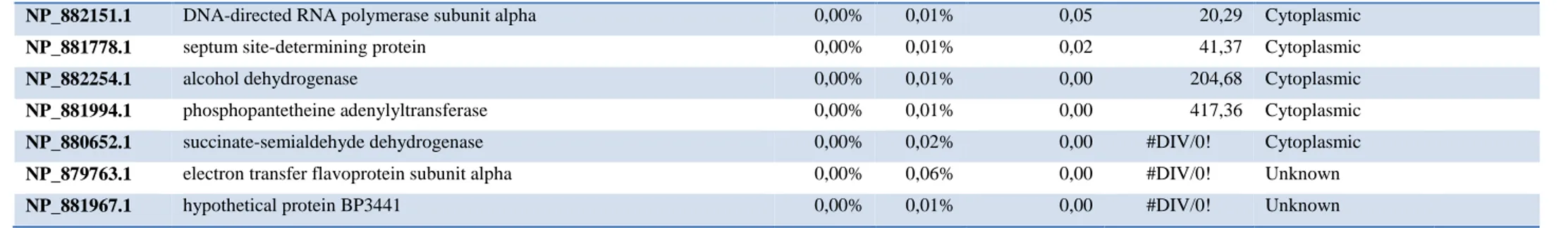
34 Vaccine antigen selection
We applied two main criteria for antigen selection: putative antigens should be either Bvg+ exclusive or showing at least a 2.5 fold increase as compared to Bvg- and they should have a predicted outer membrane localization according to PSORTb. The list of the resulting selected antigens is reported in Table 3. Based on their high abundance in OMV (each higher than 1% of total protein amount) and excluding the proteins already part of the currently available aP formulation, we focused on six promising candidates to be assessed for their adhesive properties and vaccine potential: BrkA, Vag8, TcfA, SphB1, BipA and BfrD. Interestingly, five of the selected antigens had a predicted autotransporter structure.
Table 3. OMV-based antigen selection. Antigens were selected from the total proteins quantified in
OMV based on the following criteria: Bvg regulation, localization prediction and abundance. Percentages refer to the total protein composition of OMV from either Bvg+ or Bvg- phase averaged between the two biological replicates. # known B. pertussis protective antigens *=antigen selected for further analysis, §=antigen currently included in a commercial vaccine.
Accession Protein name % Bvg+ %
Bvg-NP_880571.1 FHA filamentous hemagglutinin/adhesin
§# 16,64% 0,01% NP_882013.1 BrkA autotransporter* # 11,82% 0,04% NP_879839.1 Prn Pertactin autotransporter §# 8,46% 0,00%
NP_879104.1 SphB1 autotransporter subtilisin-like protease*
#
6,50% 0,00%
NP_880953.1 Vag8 autotransporter*
#
4,72% 0,00%
NP_879974.1 TcfA tracheal colonization factor*
#
3,77% 0,00%
NP_879893.1 BipA outer membrane ligand binding protein*
#
3,42% 0,74%
NP_879666.1 BfrD TonB-dependent receptor* 1,48% 0,01%
NP_880575.1 FhaC filamentous hemagglutinin transporter protein 0,66% 0,07%
NP_879378.1 BP0529 autotransporter 0,36% 0,02%
NP_881933.1 OmpQ outer membrane porin protein 0,08% 0,00%
NP_881280.1 Adhesin FhaS 0,08% 0,01%
NP_880573.1 FimC outer membrane usher protein 0,07% 0,00%
NP_880879.1 Type III secretion protein 0,04% 0,00%
NP_879314.1 Heme receptor HemC 0,03% 0,00%
35 Recombinant E. coli strains adhesion and adhesion inhibition assays
In order to evaluate the adhesive properties of each single protein we exploited E. coli as an heterologous background system for surface exposure of the selected antigens. We generated E. coli strains constitutively expressing the selected full length proteins and checked for their ability to bind to A549 respiratory epithelial cells as compared to wild type E. coli. A549 cells were infected with E. coli strains for three hours, then washed to remove unbound bacteria, fixed and stained with an anti-E. coli antibody. We found that BrkA, SphB1, Vag8 and TcfA conferred adhesive ability to E. coli when expressed on the membrane. On the contrary, heterologous expression of BipA and BfrD had no effect on strain adhesiveness (Fig. 5A). We continued the analysis by testing if the E. coli strains singularly expressing the selected antigens were detected with the anti-OMV
serum we had generated. By flow cytometry, anti-OMV serum was able to recognize the specific heterologously expressed BrkA, Vag8 and BipA (Fig. 5B). This result proved not only that BrkA, Vag8 and BipA antibodies had been elicited after mice immunization with B. pertussis OMV but also that these proteins are properly expressed and surface-exposed on E. coli. Finally, to determine whether the inhibition of B. pertussis adhesion promoted by anti-OMV serum correlated with the presence of
specific antibodies against the selected antigens we tested its inhibitory effect on the adhesion of recombinant E. coli strains. We found that anti-OMV serum caused reduction of adhering E. coli at high serum concentration only when BrkA was expressed on the bacterial surface. Unexpectedly, Vag8-expressing E. coli was not inhibited despite the presence of specific anti-Vag8 antibodies in the serum (Fig. 5C).
36
FIG. 5. Functional and immunological characterization of the selected antigens heterologously expressed on E. coli. A. Adhesion of
recombinant E. coli strains on A549 cells. A549 cells were infected with bacteria for 3 hours. Cell-associated bacteria were quantified by fluorescence reading after staining with a rabbit anti-E. coli antibody. Results represent mean ± SD of one representative of three independent experiments each performed in triplicates, ****=P < 0.0001, ns= non-significant. B. FACS analysis on recombinant E. coli strain with anti-OMV serum: recombinant E. coli strains were collected in early exponential phase, stained with mouse anti-OMV antibody and finally fixed with 3.7% formaldehyde. Recognition of surface exposed B. pertussis antigens was checked by flow cytometry using FACS Canto II flow cytometer (BD Biosciences). C. Impact of anti-OMV serum on E. coli adhesion to A549 cells: anti-OMV serum was serially diluted in infection medium and incubated with recombinant E. coli strains for 1 h. A549 cells were then infected with the bacteria/sera mixtures for 3 hours and cell-associated bacteria were quantified by fluorescence reading. Results represent mean ± SD of one representative of three independent experiments each performed in triplicates.
37 Recombinant BrkA protein production and vaccination
To analyze the protective potential of BrkA, recombinant His-tagged protein was expressed, purified and used to immunize mice three times, four weeks apart. Fifteen days after the final vaccination, mice were challenged with aerosolized B. pertussis and seven days later the bacterial load in the lungs was evaluated by CFU counting. This experiment showed that immunization with recombinant BrkA resulted in about 100 fold reduction of bacterial load in the lung as compared to control mice treated with adjuvant only (Fig. 6).
FIG. 6. Protection induced by BrkA in the mouse B. pertussis aerosol challenge model: B. pertussis
infection was followed by performing CFU counts on lungs from groups of 10 mice seven days after aerosol challenge. Protection is reported as compared to mice immunized with the adjuvant only. Results represent mean ± SD; ****=P < 0.0001.
38
2.2 Discussion and conclusions
The increase in pertussis outbreaks and the recent observations with the employment of the baboon model for pertussis infection clearly indicated that a more potent vaccine is needed to prevent not only the disease but also the colonization and therefore transmission of the bacterium. The control of the bacterial burden is governed by multiple mechanisms leading to bacterial killing and interference to the infection process. Among them, the presence of functional antibodies at the epithelial barrier able to target key adhesins is a valuable countermeasure to inhibit the attachment of bacteria and initiation of colonization. In this study, we used outer membrane vesicles to identify potential novel antigens with adhesive properties. Indeed, OMV mainly contain outer membrane proteins and lipoproteins in the membrane bilayer and periplasmic proteins in their lumen. Aiming at preventing B. pertussis colonization of the human respiratory tract, we checked the ability of anti-OMV serum to inhibit B. pertussis adhesion on A549 respiratory epithelial cells. Interestingly, anti-OMV serum proved to be particularly powerful in preventing B. pertussis adhesion to respiratory epithelial cells. Clearly, the whole inhibitory effect on bacterial adhesion is likely determined by the additive effects of different antibodies targeting various antigens displayed by OMV and present on B. pertussis surface. We therefore decided to deepen the analysis at the single protein level, looking for proteins present in OMV that could elicit anti-adhesive antibodies after immunization. The comparative proteomic analysis between OMV from Bvg+ and Bvg- allowed the identification and quantification of over two hundred proteins and showed that ~58% of proteins quantified in Bvg+ OMV are specific for that phase and their amount drops to ~0,1% in Bvg- OMV. This finding is in agreement with the well-established regulation of the virulence genes by the BvgAS two-component system in B. pertussis. Interestingly, among the 23 Bvg+ specific proteins,
39
10 proteins were predicted to be outer membrane proteins and represented ~53% of the total Bvg+ OMV protein amount. Among the Bvg+ specific proteins we found the best known and characterized B. pertussis adhesins such as FHA, 69K and Fimbriae together with Pertussis Toxin. On the contrary, Adenylate Cyclase Toxin and Dermonecrotic Toxin were not identified. On the other hand, the majority of Bvg- specific proteins appeared to be cytoplasmic contaminants, possibly indicating that B. pertussis strain BP537 is more prone to lyse during growth. Finally, among the proteins that did not result to be specific for one or the other phase, the analysis showed a slightly increased amount of lipoproteins, outer membrane and periplasmic proteins in Bvg- OMV vs. Bvg+ OMV. Therefore, it is tempting to hypothesize that most of the proteins in Bvg- OMV are overrepresented to compensate for the lack of the Bvg+ outer membrane proteins and the outer membrane porin protein BP0840 perfectly supported this hypothesis, resulting to be the most abundant protein in Bvg- OMV and representing ~20% of the total protein amount as compared to ~2.5% in Bvg+ OMV. The NTA analysis further supported the hypothesis of a compensative mechanism, showing no differences in size distribution and quantity of proteins per vesicle in both OMV samples. The protein composition of OMV described in the present study differs substantially from what has been shown in a previous report (Raeven, van der Maas et al. 2015). While the high quantity of BrkA and Vag8 is confirmed in both studies, the overall composition in terms of presence and relative abundance is drastically different. This is likely due to the distinct OMV purification methods and B. pertussis strains employed in the two studies. The proteomic analysis presented in this study allowed the identification of an initial list of putative antigens, which were narrowed down to 16 interesting candidates: this first antigen selection resulted in seven known B. pertussis protective antigens including two of the current aP antigens, thus validating our strategy to identify virulence factors. It is intriguing to underline that proteomic analysis of two
40
currently circulating B. pertussis strains under in vitro Bvg+ and Bvg- conditions (de Gouw, de Jonge et al. 2014), combined to in silico prediction for surface- expression, resulted in a panel of top 15 candidates which perfectly mirrors the OMV-based antigen selection described in our study. Then, albeit with variations in the strains and methods employed, the membrane composition of OMV seems to closely reflect the composition of whole bacteria. We decided to focus our attention on the most abundant Bvg+ outer membrane proteins and interestingly five autotransporter proteins were at the top of our list. Autotransporters are possibly the simplest bacterial secretion systems (type V): they consist only of a single polypeptide chain organized in a passenger and translocator domain and they have the ability to autonomously translocate across the outer membrane (Fan, Chauhan et al. 2016). Therefore, the heterologous expression of the autotransporters on the surface of E. coli was selected to characterize their adhesive properties. This approach allowed the demonstration that BrkA, TcfA, SphB1 and Vag8 conferred to E. coli a significant increased ability to adhere to lung epithelial cells. To the best of our knowledge, this is the first time that B. pertussis autotransporters were rationally characterized for their adhesive properties exploiting a heterologous E. coli background. The involvement in adhesion of BrkA was previously demonstrated in in vitro analysis and in mouse infection studies with B. pertussis mutant strains showing
2-fold and 2-log reduction in the ability to adhere to cell lines and to colonize the murine lungs, respectively (Fernandez and Weiss 1994, Elder and Harvill 2004). Also, the loss of TcfA was previously shown to cause 10-fold reduction in the number of bacteria isolated from tracheas after B. pertussis aerosol challenge (Finn and Stevens 1995). Nevertheless, deletion of individual virulence factor genes could have limited effects on the ability of B. pertussis to efficiently infect the respiratory tract of mice, suggesting they may perform redundant functions. This was the case of a Vag8 deletion mutant, which was as efficient as the parental B. pertussis strain in colonizing murine lungs