UNIVERSITÀ DEGLI STUDI DI VERONA
DEPARTMENT OF BIOTHECNOLOGY
GRADUATE SCHOOL OF NATURAL SCIENCE AND ENGINEERING DOCTORAL PROGRAM IN BIOTHECNOLOGY
CYCLE XXXI
DECIPHERING Pseudomonas syringae pv. actinidiae
VIRULENCE AND COMMUNICATION
WITH HOST PLANT
S.S.D. AGR/12 Plant PathologyCoordinator: Prof. Matteo Ballotari
Tutor: Prof. Annalisa Polverari
Co-tutor: Dr. Elodie Vandelle
ABSTRACT
The gram-negative bacterium Pseudomonas syringae pv. actinidiae (Psa) is the causal agent of the bacterial canker of kiwifruits leading to important economic losses in New Zealand, Italy, Korea and Japan the main producers worldwide.
To identify virulence targets and develop new effective targeted control strategies, molecular mechanisms involved in host plant recognition by Psa and related infection processes was currently studied.
To this aim, the Psa CRAFRU 8.43 strain was transformed with constructs carrying the green fluorescent protein (GFP)-encoding reporter gene under the control of the promoter of several Psa genes, selected for their putative role in pathogenicity based on literature. Our results showed that hrpA1 gene promoter, involved in the early steps of bacterial infection, is induced in a minimal medium (mimicking apoplast conditions), with an earlier and at higher levels in presence of Actinidia deliciosa leaf extract, indicating a role downstream of host recognition by bacteria. The characterization of hrpA1-inducing kiwi extract showed that such signal(s) are kiwi-specific and smaller than 10KDa in size.
Moreover, to elucidate the signalling pathway(s) involved in host-mediated hrpA1 induction, a chemical library was screened to identify molecules able to block such activation. Nineteen candidate molecules were obtained, displaying different inhibition levels. According to the role of HrpA1 in plant hypersensitive response (HR) induction, HR in model plants wasquantifiedto obtain a visible phenotype correlated with hrpA1 promoter induction and thus demonstrate the inhibitory effect of selected molecule, dicoumarol, in a plant system.
Moreover, the molecules responsible for Psa quorum sensing activation are still unknown, since Psa possess three LuxR-solos proteins (PsaR1, R2, R3) lacking LuxI enzymes responsible for AHL synthesis. Thus, to identify putative PsaR1 ligands, its recombinant autoinducer-binding domain was produced in E. coli and chemical libraries were screened using a high throughput fluorescence-based thermal shift assay. Four molecules inducing a significant thermal shift were identified as putative PsaR1 ligands; moreover, the presence of the same or other putative PsaR1 ligands in kiwi plants was confirmed by a similar thermal shift effect in presence of kiwi plant extracts.
Quercetin and luteolin, PsaR1 putative ligands, tested on Psa virulence traits (motility and biofilm) showed an increase of swarming motility and reduction biofilm formation.
ACKNOWLEDGEMENTS
I would like to thanks Dr. Marco Scortichini (CREA: Council for Agricultural Research and Analysis of Agricultural Economics) to provide us the Psa strains used in this study and Dr. Vittorio Venturi (IGEB: International Centre for Genetic Engineering and Biotechnology, Trieste) to provide us all Psa GFP-transformed strains.
I kindly thanks Dr. Teresa Colombo tocurate the bioinformatic design of the custom microarray and Dr Nicola Vitulo to curate thestatistical data analysis of the microarray data; and the biochemistry group of the biotechnology department (Univ.of Verona: Prof. Paola Dominici; Dr. Alessandra Asegno and Prof. Massimo Crimi) to provide us recombinant PsaR1 protein. I also want to express my gratitude to my advisors Prof. Annalisa Polverari and Dr. Elodie Vandelle for their kind, strong and patient supervision.
CONTENTS
i
CONTENTS
Chapter 1: Introduction
1. Pathogenicity and virulence factors of plant pathogenic bacteria
1.1. Abilities to colonize and enter a plant: bacterial motility1.2. Abilities to colonize and enter a plant: biofilm 1.3. Abilities to overcome plant resistance
1.4. The type III secretion system (T3SS) 1.5. Quorum sensing in gram-negative bacteria
1.5.1 Non-canonical quorum sensing system: the LuxR “solos” family
1.6. Role of GacS/GacA two component system in the regulation of bacterial signaling network
2. Pseudomonas syringae pv. actinidiae, the causal agent of kiwifruit bacterial
canker
2.1. Psa outbreaks and identification of Psa biovars 2.2. Pathogenically relevant Psa traits
3. REFERENCES
AIMS OF THESIS
Chapter 2: Identification of signaling pathways involved in early
virulence induction in Pseudomonas syringae pv. actinidiae (Psa)
1. ABSTRACT
2. INTRODUCTION
3. MATERIALS AND METHODS
3.1. Culture media3.2. Bacterial strains
3.3. Plant leaf extracts and fractions 3.4. Monitoring gene promoters induction
CONTENTS
ii
3.5. Screening of chemical molecule library 3.5.1 Screening data analysis approach
3.6. Evaluation of Psa-induced plant cell death
3.6.1 Effect of dicoumarol on Pst-AvrB cell death induction
4. RESULTS
4.1.
A high-throughput system (HTS) to monitor promoter induction of Psa virulence gene promoters induction4.1.1 Assessment of the conditions for the monitoring of virulence gene promoter induction
4.1.2 Characterization of the hrpA1 gene promoter inducing factors: specific effect of the kiwi extract
4.1.3 Characterization of the hrpA1 gene promoter inducing factors: kiwi leaf extract fractions
4.1.4 Assessment of gene promoter induction in presence of a kiwifruit leaf extract in PBS
4.1.5 hrpA1 gene promoter induction with fractions of kiwi leaf, extracted in PBS 4.1.6 hrpA1 gene promoter induction with bacterial spent media and surfactant
4.2.
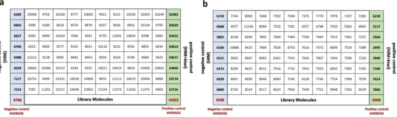
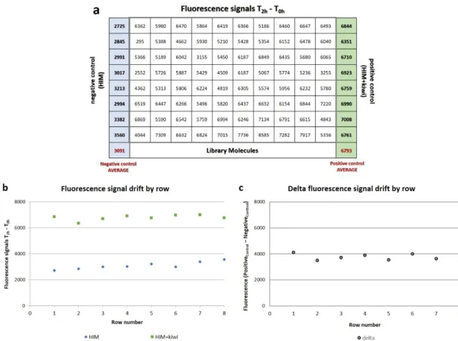
Identification of molecules affecting hrpa1 promoter activity: high-throughput screening of a chemical library4.2.1 Plate acceptance criteria: spatial uniformity assessment
4.2.2 Plate acceptance criteria: quality control (QC) parameters
4.2.3 Statistical approaches in the selection of candidate molecules
4.2.4 Candidate molecules validation in a secondary screening
4.3.
Set up of a method to evaluate the pathogenicity-reducing activity of selected molecules in planta5. DISCUSSION
6. REFERENCES
Chapter 3: Investigating the function of putative environmental
sensors in Pseudomonas syringae pv. actinidiae (Psa) virulence:
LuxR-solos
1. ABSTRACT
2. INTRODUCTION
CONTENTS
iii
3. MATERIALS AND METHODS
3.1. Culture media3.2. Bacterial strains, PsaR1-NT protein and plant extracts 3.3. Bioinformatic analysis of PsaR1 LuxR-solos
3.4. Thermal shift assay
3.5. Gene expression analysis by microarray
3.6. Phenotypic effect of putative PsaR1-NT ligands: motility assay 3.7. Phenotypic effect of putative PsaR1-NT ligands: biofilm assay
4. RESULTS
4.1. Investigating the function of LuxR-solos sensors in Psa virulence-related traits 4.1.1 Bioinformatics characterization of PsaR1 protein
4.1.2 Thermal shift assay as high throughput system to identify PsaR1 putative ligands: optimization of experimental conditions and screening of a molecule library
4.1.3 PsaR1 putative ligands validation and characterization using thermal shift assay
4.2.
Evaluation of genes affected by PsaR14.3. Effects of PsaR1 putative ligands on Psa phenotypes: motility and biofilm formation 4.3.1 Effect of PsaR1 putative ligands on Psa phenotype: preliminary tests on swarming
motility and biofilm formation
5. DISCUSSION
6. REFERENCES
CHAPTER 1: INTRODUCTION
1
Chapter 1: INTRODUCTION
1. Pathogenicity and virulence factors of plant pathogenic
bacteria
Pseudomonas syringae, the most studied plant pathogenic bacterial species, countsapproximately 60 pathovars [1] infecting economically important crops, with continuous new disease outbreaks. A recent example is the kiwifruit canker outbreak in New Zealand and Europe caused by P. syringae pv. actinidiae [2].
Pseudomonas syringae strains show epiphytic and endophytic interconnected phases of growth:
in the former bacteria live on the surface of plant tissues such as leaves, stems, flowers and fruits while in the latter bacteria enter the plant and colonize the intercellular apoplast space [3]. Entering the plant requires bacterial motility, mediated by flagella and type IV pili and also recognition and taxis towards infection sites and nutrients. In this transition from epiphytic to endophytic phases, bacteria get in contact with the plant cell wall and need to overcome host plant resistance, to evoke disease. At this stage, bacterial pathogenicity is largely dependent on the ability to assembly a functional type III secretions system (T3SS), encoded by hrp genes (for hypersensitive response and pathogenicity) and responsible for the secretion of many effectors; these hrp genes are present in most plant pathogenic bacteria including P. syringae pathovars and their expression is induced only in presence of plant tissue [4].
Thus, to evoke disease, bacteria can manipulate the plant cell metabolisms by producing effectors or other pathogenicity factors such as toxins, hormones or lytic enzymes, to the advantage of bacteria. The timing and efficient expression of these virulence factors is regulated by a complex signaling network, which has been only partially elucidated in model species [5].
1.1. Abilities to colonize and enter a plant: bacterial motility
Motility is an important trait in bacteria, to reach more and better nutrients, avoid unfavorable environments and spread onto or within host tissues.
Bacteriacan move by different systems: swarming, swimming and twitching motilities occur by rotating flagella or pili, which are protein structures anchored to the cell membrane (Fig. 1).
CHAPTER 1: INTRODUCTION
2
Figure 1: A scheme of bacterial movement: swarming, swimming and twitching [6].
Moreover, chemotaxis allows bacteria to move and orient according to environmental stimuli [7] as shown by mutations in chemotaxis genes, which impair bacterial spreading [8-9]. Bacteria swarm as a group of cells across host surface [10]; this motility, mediated by flagella, is also assisted by the biosynthesis of surfactantsand cell-cell interactions. Bacteria synthesize and secrete surfactants (short for “surface active agent”), amphipathic molecules onthe swarming front, that reduce superficial tension on the substrate [8]. Direct contact between bacteria and the surface may have a crucial role, but the mechanisms of surface sensing must be elucidated [6]. Because surfactants are effective at high concentrations, their production is regulated by quorum sensing (QS) to ensure a sufficient bacterial population to produce beneficial surfactants [11].
Swimming motility is defined as the movement of individual cells in liquid or low-viscosity media [10] and despite it requires a functional flagellum, unlike swarming motility doesn’t need neither QS systems nor biosurfactants [12].
Mutations on flagella synthesis or function abolish colony spreading, confirming flagella crucial role in swarming and swimming [13].
Twitching motility requires the type IV pili, which produce a movement by repetitive extension and retraction, and is important in exploration and initial attachment on surfaces, as well as in the development of biofilm architecture, thereforedepending on QS system [14-15].
CHAPTER 1: INTRODUCTION
3
1.2. Abilities tocolonize and enter a plant: biofilm
Biofilm are bacterial communities in which cells are immersed in a matrix attached to a surface, concentrating water and nutrients and protecting bacteria from adverse conditions, outside and inside the plant.
The mixture of extracellular factors represents the “biofilm matrix” composed primarily of EPSs, proteins, eDNA and lipids. The components of this matrix support biofilm 3-dimensional structure composed by bridge, channels and pores which play a key role in cell-cell interaction, nutrient utilization and horizontal gene transfer; moreover, the matrix provides a physical barrier against antibiotics or plant defense compounds and protects biofilm cells from environmental stresses such as UV radiation, pH changes, osmotic stress and desiccation [16]. External bacterial components such as flagella, pili and LPSs play an important function in the initial stages of biofilm formation on surfaces, while the phases of growth, maturation and disassembly of biofilms depend on the biosynthesis of EPSs, proteins, eDNA, and lysis products [17].
The importance of LPSs in biofilm formation was demonstrated by various mutations related to LPSs synthesis in Pseudomonas aeruginosa [18], P. syringae [19] and Xanthomonas citri [20] which reduced the ability to form a biofilm.
Bacterial autoaggregation, differs from biofilm in the abundance of extracellular factors, surface components and cell density. It can be visualized macroscopically by clumping of cells in liquid culture, and represents a survival strategy triggered in hostile environments, involving LPSs and outer membrane structures, such as type IV pili, fimbriae, flagella and proteinaceous polymeric appendages. Therefore, bacterial aggregation on a surface precedes the phase of biofilm formation. The transition from bacterial aggregation to mature biofilm can be summarized in the following steps: (i) interactions among bacteria near plant microenvironment (ii) interactions between bacteria and plant surfaces (iii) biofilm formation. These phases, as a whole, are also common to plant growth promoting bacteria and may thus have negative or positive effectson plant fitness depending on the bacterial species involved [21] (Fig. 2).
CHAPTER 1: INTRODUCTION
4
Figure 2: A scheme of bacterial autoaggregation and biofilm development, and their relationship with plant colonization [21].
Bacterial motility (twitching, swimming and swarming) in combination with chemotaxis are all involved in the various stages of biofilm development [22]. Initially, free-swimming phenotype allows bacteria to reach a suitable surface while the surface-swarming phenotype allows bacteria to adhere and to move on the surface [23]. Afterwards, motility is involved also in biofilm dispersal, when bacteria spread to colonize new habitats. Chemotaxis regulates the site of biofilm establishment, as observed in Ralstonia solanacearum, for example, which is attracted by host plant exudates, and colonizes specific positions of the plant surface with optimal nutrient availability [22].
1.3. Abilities to overcome plant resistance
Plant pathogenic bacteria enter through wounds or natural openings (i.e. stomata and hydathodes), proliferate in intercellular spaces (the apoplast) and then deliver effector molecules (virulence factors) into plant cells to enhance their microbial fitness. Plants, on the other side, rely on the innate immunity of each cell, and the systemic signals released from infection sites.
Intricate interactions between plant immune responses and bacterial virulence strategies occur. Plants evolved a first layer of pathogen recognition, relying ontransmembrane pattern recognition receptors (PRRs), that respond to slowly evolving microbial- or pathogen-associated molecular patterns (MAMPS or PAMPs) [24-25] and can activate for example a signaling cascade in the stomatal guard cells to close stomata as part of the PAMP-triggered immunity (PTI). A major consequence of PTI is the inhibition of bacterial growth through
CHAPTER 1: INTRODUCTION
5
production of antimicrobial compounds, restriction of nutrients in the apoplast and the reinforcement of plant cell walls [26].
Moreover, most plant pathogenic bacteria can induce a resistant response in non-host plants often including a hypersensitive response (HR) characterized by a programmed cell death (PCD) at the infection site. As a counter-defence strategy, P. syringae evolved virulence factors such as toxins (i.e. coronatine and syringomycin) and effectors delivered by the type 3 secretion system (T3SS) which can act on plant metabolism or interfere with hormonal signaling, to re-establish for instance stomata opening. In turn, some plant genotypes in a species can evolve new specific receptors (Fig. 3) recognizing such bacterial effectors, to achieve the so-called effector-triggered immunity (ETI). As a consequence, bacterial growth is restricted as above, and plant cells typically undergo an ETI- triggered hypersensitive response.
Figure 3: Schematical representation of the “zigzag model” illustrating the quantitative output of the plant immune system. PTI= PAMP-trigger immunity, ETI= effector-triggeredimmunity, ETS= effector-triggered susceptibility, HR= hypersensitiveresponse [27].
1.4. The type III secretion system (T3SS)
In conclusion, it is now well accepted that the effector proteins injected into hostcells via the type III secretion system (T3SS) are responsible for pathogenicity and disease occurrence in host plants while in resistant genotypes they can be recognized triggering a resistance-associated HR [28].
The key component of T3SS is the Hrp pilus, which functions in the translocation of bacterial effectors inside the plant cell. The major subunit of the TTSS pilus is the HrpA protein [29].
CHAPTER 1: INTRODUCTION
6
P. syringae mutants in hrpA lose the ability to cause disease in host plants and to elicit the
defense-associated HR in non-hosts [4].
Despite the primary sequences of hrpA share a low homology among various pathovars, the secondary structure is remarkably similar, consisting almost exclusively of α-helices, and a β-stand only in the amino terminus, which is dispensable for function [30]. The hypervariability of the primary sequences of the pilus principal subunit may reflect evolutionary adaptation, to avoid the recognition by the host defense system.
While the Hrp pilus is constructed, by adding HrpA subunits at the distal site, also the secretion of effector proteins occurs, presumably because the expression of T3SS and effectors genes is coordinately regulated [31].
Two alternative models were proposed to explain the secretion of effector proteins namely “conduit” and “guiding filament” model. In the former model, proteins exit from the tip of the Hrp pilus, while in the latter the Hrp pilus carries effector proteins with it, requiring a physical interaction between pilus and proteins, so that newly secreted proteins would be localized near the base of the pilus (Fig. 4) [32-33].
Figure 4: Schematical representation of type 3 secretion system (T3SS), and alternative secretion models of effector proteins [32, 34].
In all examined strains of P. syringae, the hrp locus consists of 27 open reading frames (ORFs) located in the chromosome and organized into six operons. Among hrp genes, three classes can be distinguished: (i) the first class encodes core components of T3SS; (ii) the second class encodes regulatory proteins (HrpL, HrpR, HrpS and HrpV); (iii) the third class encodes secreted proteins such as extracellular components of T3SS (e.g. HrpA) [26, 35].
CHAPTER 1: INTRODUCTION
7
In a saprophytic environment, P. syringae does not express hrp genes at a significant level, until it comes in contact with plant apoplast. HrpL, HrpR, HrpS, three positive regulators, are involved in the transcriptional regulation of the T3SS genes. HrpR and HrpS interact directly and in conjunction with the sigma factor σ54 required for the maximal expression of hrpL gene;
HrpL is a sigma factor that directs the transcription of all T3SS genes. On the other hand, HrpV is a negative regulator of T3SS-associated genes [4, 36].
1.5. Quorum sensing in gram-negative bacteria
Many bacterial traits, either linked to pathogenicity or simply associated to environmental fitness are regulated at the population level. Population density changes bacterial behaviors related to motility, biofilm formation, gene expression, virulence activation and communication with other bacteria. The ability to perceive bacterial density is described as quorum sensing (QS) [37].
The QS circuit, also present in most in gram-negative phytopathogenic bacteria, was first identified in the luminescent bacterium Vibrio fischeri [38] and consists of two proteins called LuxI and LuxR. LuxI-like proteins synthesizes specific acyl homoserine lactones (AHL) known as autoinducers, the concentration of which increases with increasing of cell density. LuxR-like proteins bind the cognate AHL, and after reaching a threshold concentration, the LuxR-autoinducer complex binds a specific promoter DNA sequence called “lux box” activating target gene transcription. In V. fischeri, this mechanism regulates luminescence, from which the names of LuxR/LuxI are derived. Moreover, in V. fischeri, the LuxR-autoinducer complex also binds the luxR promoter with a repressive effect, thus acting as a negative feedback regulation of the circuit (Fig. 5a).
The canonical QS networkin Pseudomonas spp. was mainly investigated in Pseudomonas
aeruginosa which contains two LuxI/R-type systems, namely LasI/LasR and RhlI/RhlR. Both
LasI and RhlI are autoinducer synthases that catalyze the formation of N-(3-oxododecanoyl)-homoserine lactone and N-(butyryl)-N-(3-oxododecanoyl)-homoserine lactone, respectively. At high cell density, LasR binds its cognate autoinducer, and the resulting complex binds “lux boxes” in the promoters of several genes encoding virulence factors, such aslasB, lasA and toxA, responsible to produce an elastase, a protease and the exotoxinA, respectively. The LasR-autoinducer complex also activates the expression of rhlR, encoding the second LuxR-like protein of P.
aeruginosa. In turn, the RhlR-autoinducer complex activates a second class of target genes,
CHAPTER 1: INTRODUCTION
8
lecA encoding a cytotoxic lectin and rhlI, involved in the synthesis of RhlR-specific autoinducer
(Fig. 5b) [39-40].
LuxR proteins consist of two domains: the amino-terminal domain, involved in the binding of the autoinducer, and the carboxyl-terminal domain, binding DNA sequences. In absence of the autoinducer, the amino-terminal domain inhibits the DNA binding on the carboxyl-terminal. The carboxyl-terminal domains are also involved in the multimerization of LuxR proteins, required for binding DNA sequences. Regions responsible for DNA binding on “lux box”are conserved helix-turn-helix (HTH), suggesting that the target specificity of these systems derives from selectivity for the cognate autoinducers. In fact, alteration in the acyl side chain of the autoinducer, can inhibit LuxR activity and target genes expression [38].
Figure 5: Schematical representation of quorum sensing (QS) system in gram-negative bacteria: (a) Vibrio fischeri LuxI/LuR QS system [41] and (b) Pseudomonas aeruginosaLasI/LasR and RhlI/RhlR QS system [42].
CHAPTER 1: INTRODUCTION
9
1.5.1 Non-canonical quorum sensing system: the LuxR “solos” family
Bacterial quorum sensing plays a crucial role in ecological fitness by regulating cell-cell communication. Many plant pathogenic bacteria in particular, employ the QS system to regulate their virulence factors; in addition, it was recently found thatcomponents of the QS system can also be involved in the interaction between bacteria and eukaryotic hosts [43]. In fact, AHLs can act as inter-kingdom signals because they can be perceived by plants, which in turn respond regulating plant gene expression [44]. On the other hand, plants can produce low molecular weight compounds interfering with the bacterial QS system, acting as agonists or antagonists of AHLs [45-46].
In the last decade studies revealed a family of proteins in plant-associated bacteria sharing highly similarity with QS LuxRs, but lacking a cognate LuxI protein, for this reason called “solos”. LuxR-solos have been identified in several bacterial species, including the object of this Thesis, P. syringae pv. actinidiae. These proteins cannot bind AHLs, due to the lack of highly conserved aminoacids in the AHL-binding domain, but there is evidence that they could bind plant compounds [47], which makes them interesting candidates in plant-pathogen communication.
Figure 6: Schematical representation of interkingdom signaling between plants and bacteria involving canonical and non-canonical quorum sensing (QS) systems [48].
CHAPTER 1: INTRODUCTION
10
Up to date, LuxR-solos proteins participating in interkingdom signaling by the detection and binding to host signal molecules [47] have been identified in several plant pathogenic bacteria, such as Xanthomonas oryzae pv. oryzae (OryR) [49], X. campestris pv. campestris (XccR) [50],
Pseudomonas fluorescens (PsoR) [51], and P. syringae pv. actinidiae (PsaR2) [52].
A major achievement in this intriguing inter-kingdom communication, mediated by a non-canonical QS system, would be the identification of plant molecules possibly binding
LuxR-solos, among the many low molecular weight secondary metabolites produced by plants [48].
1.6. Role of GacS/GacA two component system in the regulation of bacterial signaling network
In nature, microorganisms are subjected to a myriad of environmental stimuli, such as changes in temperature, osmolarity, pH and nutrient availability. In response, bacteria have developed multiple systems to adaptat to these environmental fluctuations.
The sensor kinase GacS and the response regulator GacA are members of a two-component system found in gram-negative bacteria and also in plant pathogenic pseudomonads [36]. GacS sensor kinase detects environmental signal molecule(s) and undergoes autophosphorylation, and in turns fosforilates the GacA response regulator, activating a signal transduction cascade leading to gene expression and bacterial adaptation to changing conditions [53].
In Pseudomonas syringae pv. tomato DC 3000, GacS/GacA is upstream of the regulatory hierarchy controlling the expression of several bacterial traits. Among the genes affected by this system there are hrpL, hrpR and hrpS -well-known essential components in the induction and assembly of the T3SS - and the ahlI and ahlR, key genes of the QS system. Moreover, GacS/GacA controls upstream events in the expression of several phenotypes related to pathogenicity, such as bacterial motility, elicitation of hypersensitive response (HR) and in
CHAPTER 1: INTRODUCTION
11
2. Pseudomonas syringae pv. actinidiae, the causal agent of
kiwifruit bacterial canker
Pseudomonas syringae pv. actinidiae (Psa) is a gram-negative bacterium, aerobic, rod-shaped,
motile by polar flagella, that can grow epiphytically and endophytically causing the kiwifruit canker. Psa can spread by rain, insects, animals and human activities and penetrates host tissues through wounds or natural openings, such as stomata or lenticels, or through pollen dissemination.
Infections can spread more efficiently in spring under mild, humid weather, with temperatures ranging from 12 to 18 °C. Leaf symptoms include brown–black leaf spots often surrounded by a chlorotic halo and blossom necrosis. From primary infection sites, bacteria can move systemically through the twigs and trunk causing extensive cankers along the trunk and bleeding of a whitish to orange ooze (Fig. 7) [55-56].
Figure7: Life cycle and symptoms of Psa across the seasons. Green arrows represent the epiphytic inoculum; blue arrows represent how Psa move in the plant (dashed arrowindicateshypothetical pathway) and red arrows represent symptoms observed in each season after different type of infections. Green boxes describe the various entry ways of Psa and red boxes describe symptoms [57].
CHAPTER 1: INTRODUCTION
12
Disease control strategies, necessary to minimize the impact of the epidemic and to contain economic losses, include good orchard hygiene practices and chemical treatments with copper-based compounds (in Europe) and streptomycin, in Countries where this is allowed (New Zealand and Asian countries). However, none of the available practices is effective enough to cure the disease, while Psa bacterial strains can evolve to become resistant to copper and antibiotics. Additional available tools consist in elicitors, such as bacterial proteins and polysaccharides (i.e. harpins, chitosan), that activate plant defense and induce resistance. However, the use of elicitors alone is not recommended and the duration of protection provided depends on the elicitor, pathogen and crop [58]. Recently, copper resistance has been identified in New Zealand Psa strains, acquired through horizontal gene transfer (HGT) [59], so in order to reduce this phenomenon and to develop new control strategies, new natural compounds from different sources i.e. essential oils (EOs) are currently studied [60].
2.1. Psa outbreaks and identification of Psa biovars
Psa was first identified in Japan (1989) [61], and subsequently in South Korea and Italy (1994) [62-63], in evolutionarily divergent populations: while the Japanese and Italian strains produced phaseolotoxin, the Korean strains synthesized the phytotoxin coronatine [64].
In Europe, Psaoutbreaks of new highly virulent strains have been reported for the first time in Italy (2008) [65] ad then in Portugal [66], Spain [67], France [68], and Turkey [69], and also in other continents including New Zealand [70] and Chile [71] in 2010.
Psa outbreaks attracted much attention due to the severity and global economic impact of the disease, caused by a recently emerged pathogen. In subsequent years, Psa strains from diverse origins have been investigated using various methods, such as multilocus sequence typing (MLST) analysis of housekeeping genes or effector genes, or whole-genome sequencing of several Psa strains.
From a taxonomic point of view, P. syringae “sensu lato” (P. syringae–related species and pathovars) consists in nine genomospecies, corresponding to species with no distinguished phenotypical features according to Wayne et al. [72]. In particular, genomospecies 1 corresponds to P. syringae “sensu stricto” and includes P. syringae pv. syringae, while P.
syringae pv. tomato belongs to genomospecies 3 [73], and P. syringae pv. actinidiae belongs
to genomospecies 8. Usually P. syringae pv. tomato is the well-recognized bacterial model to study emerging Pseudomonas phytopathogenic bacteria, and this can be applied to Psa since
CHAPTER 1: INTRODUCTION
13
genomospecies 3 and 8, to which both pathovars belong, are closely related. Nevertheless, Berge et al. identified two clades among microorganisms belonging to genomospecies 3 and 8, called phylogroups 1a and 1b, which include P. syringae pv. tomato and P. syringae pv.
actinidiae, respectively [74-75]. Moreover, within the genomospecies 8, Psa is more closely
related to P.s. pv. theae [76]. The Asian origin of Psa and P.s. pv theae as well as the genetic closeness between their hosts. i.e. Actinidiaceae and Theaceae families, could explain their high similarity [77]. Importantly, Psa, P.s. pv theae and P. s. pv. avellanae (genomospecies 8), the latter elevated to the species level and further named P. avellanae [78], are so closely related to have been recently considered belonging to the P. avellanae species [79].
On the basis of genetic diversity, toxin production and other phenotypic features, Psa strains were classified into different biovars [80]: strains originally isolated in Japan (1989) and in Italy (1994), were assigned to biovar 1, which can synthesize phaseolotoxin, from the argKtox gene cluster. Psa strains isolated in South Korea (1994) were classified as biovar 2, and can produce coronatine [81]. The most aggressive Psa biovar 3 responsible for the global outbreak of bacterial canker of kiwifruit doesn’t produce phaseolotoxin nor coronatine but carries a plasmid of about 160 kb, absent in biovar 1 and 2, and encodes four putative clade-specific TTSS effectors (hop-H1, hop-Z5, hopAM1-2 and hopAA1-2) [55, 82, 84].
Strains belonging to biovar 4, detected in New Zealand and Australia, were recently re-classified as a new pathovar actinidifoliorum (Pfm) based on phylogenetic and phenotypic differences causing only leaf spots but no canker symptoms [85]. Biovar 5 and biovar 6, recently identified in Japan, have an extremely limited distribution and may be endemic at present. However, there is no guarantee that they remain so in the future [86-87]. At present,the most severe biovar 3, can be found in most infected orchards worldwide.
Several genome analyses performed so far revealed that epidemics in Europe, New Zealand and Chile of Psa 3 biovar originated from independent introductions [82], probably from a single founder variant from China [87]. There are evidence supporting two phases in the recent evolution of Psa biovar 3. In the first phase DNA mobile elements contributed to differentiation and developing of more adapted clones during the initial spread in China; subsequently highly virulent and adapted biovar 3 strains spread to Europe, Chile and New Zealand, that represent ecological niche lacking competitive selections represented by the highly sensitive A. chinensis [88].
On the basis of recent evidence of short-term genome evolution of Psa biovar 3, modern control strategies should focus on methods that should recombination to reduce the risk of developing
CHAPTER 1: INTRODUCTION
14
variants with enhanced fitness or virulence [88]. In fact, copper-resistant strains were recently reported for Psa biovar 3 in New Zealand, but not yet in European strains [59, 88].
2.2. Pathogenically relevant Psatraits
Similarly, to other pseudomonads, Psa can organize cell communities within biofilms, composed of bacterial cells merged in a dense matrix of exopolysaccharides (EPSs), proteins, and extracellular DNA, in which bacteria are protected against host defense mechanisms, unfavorable environment or chemicalagents. Biofilm formation and dispersal cycles reflect the pathogenic cycle of spreading and survival on plants. In fact, it was reported that Psa biofilm formation is involved in different phases of kiwifruit infection both outside and inside the host plant [89].
Detailed biochemical studies revealed that Psa EPSs are mainly composed of rhamnose, fucose and glucose, in two polysaccharides: a branched α-D-rhamnan with side chains of a terminal α-D-Fucf and an α-D-1, 4-linked glucan, considered novel and Psa -specific polysaccharides [90], because such branched rhamnans were never reported from Pseudomonas species before. As in other plant pathogenic bacteria, Psa biofilm formation is likely regulated by the QS system, although Psa possesses three putative LuxR-solos protein, and the regulation of density-dependent behaviors is poorly understood. As previously mentioned, these LuxR-solos, namely PsaR1, PsaR2 and PsaR3, could be involved in detection of host signals or QS signals from other bacterial species. In particular, a bioinformatics analysis revealed that PsaR2 likely belongs to the subfamily of LuxR-solos found in plant-associated bacteria and proposed to bind uncharacterized plant signal(s) [52]. On the other hand, PsaR1 and PsaR3 are more similar to LuxR-type protein of the canonical QS system, suggesting that these two putative receptors could also detect AHLs from other bacteria. All Psa LuxR-solos are involved in in planta survival, as shown by phenotypic analyses of Psa mutants, while PsaR3 mutant is also impaired in swarming motility and lipase production [52]. However, very few LuxR-solos have been studied in relation to their possible role as sensors foro other types of signals. For example, a recent seminal paper showed that a LuxR-solo from Photorhabdus luminescens responds to an endogenous signal which is not an AHL [91-92]. Therefore, it cannot be excluded that Psa LuxR-solos may participate to a novel QS system involving other types of signals.
CHAPTER 1: REFERENCES
15
6.
REFERENCES
1) Bull CT, De BoerSH, Denny TP, Firrao G, Fischer-Le Saux M, Saddler GS, Scortichini M,Stead DE, Takikawa Y. Comprehensive list of names of plant pathogenic bacteria, 1980-2007.Journal of Plant Pathology.2010; 92 (3), 551-592
2) Butler MI, Stockwell PA, Black MA, Day RC, Lamont IL, Poulter RT. Pseudomonas syringae pv. actinidiae from recent outbreaks of kiwifruit bacterial canker belong to different clones that originated in China.PLoS One. 2013;8(2):e57464.
3) Xin XF, He SY. Pseudomonas syringae pv. tomato DC3000: a model pathogen for probing disease susceptibility and hormone signaling in plants. Annu Rev Phytopathol. 2013;51:473-98
4) Jin Q, Thilmony R, Zwiesler-Vollick J, He SY. Type III protein secretion in Pseudomonas syringae. Microbes Infect. 2003;5(4):301-10.
5) Ichinose Y, Taguchi F, Mukaihara T. Pathogenicity and virulence factors of Pseudomonas syringae. J Gen Plant Pathol.2013; 79: 285.
6) Kearns DB. A field guide to bacterial swarming motility. Nat Rev Microbiol. 2010;8(9):634-44. 7) Wadhams GH, Armitage JP. Making sense of it all: bacterial chemotaxis. Nat Rev Mol Cell Biol.
2004;5(12):1024-37.
8) Kearns DB, Losick R. Swarming motility in undomesticated Bacillus subtilis. Mol Microbiol. 2003;49(3):581-90.
9) Kojima M, Kubo R, Yakushi T, Homma M, Kawagishi I. The bidirectional polar and unidirectional lateral flagellar motors of Vibrio alginolyticus are controlled by a single CheY species. Mol Microbiol. 2007;64(1):57-67
10) Patrick JE, Kearns DB. Swarming motility and the control of master regulators of flagellar biosynthesis. Mol Microbiol. 2012;83(1):14-23
11) Ochsner UA, Reiser J. Autoinducer-mediated regulation of rhamnolipid biosurfactant synthesis in Pseudomonas aeruginosa. Proc Natl Acad Sci U S A. 1995;92(14):6424-8.
12) Ha DG, Kuchma SL, O'Toole GA. Plate-based assay for swimming motility in Pseudomonas aeruginosa. Methods Mol Biol. 2014;1149:59-65.
13) Chevance FF, Hughes KT. Coordinating assembly of a bacterial macromolecular machine. Nat Rev Microbiol. 2008;6(6):455-65.
14) Otton LM, da Silva Campos M, Meneghetti KL, Corção G. Influence of twitching and swarming motilities on biofilm formation in Pseudomonas strains. Arch Microbiol. 2017;199(5):677-682.
15) Burrows LL. Pseudomonas aeruginosa twitching motility: type IV pili in action. Annu Rev Microbiol. 2012;66:493-520.
16) Flemming, HC. Biofilms and environmental protection. Water Sci. Technol. 1993; 27, 1–10. 17) Flemming HC, Wingender J. The biofilm matrix. Nat Rev Microbiol. 2010;8(9):623-33.
18) Lau PC, Lindhout T, Beveridge TJ, Dutcher JR, Lam JS. Differential lipopolysaccharide core capping leads to quantitative and correlated modifications of mechanical and structural properties in Pseudomonas aeruginosa biofilms. J Bacteriol. 2009;191(21):6618-31.
19) Deng WL, Lin YC, Lin RH, Wei CF, Huang YC, Peng HL, Huang HC. Effects of galU mutation on Pseudomonas syringae-plant interactions. Mol Plant Microbe Interact. 2010;23(9):1184-96.
20) Li J, Wang N. The wxacO gene of Xanthomonas citri ssp. citri encodes a protein with a role in lipopolysaccharide biosynthesis, biofilm formation, stress tolerance and virulence. Mol Plant Pathol. 2011;12(4):381-96
21) Bogino PC, Oliva Mde L, Sorroche FG, Giordano W. The role of bacterial biofilms and surface components in plant-bacterial associations. Int J Mol Sci. 2013;14(8):15838-59.
22) Danhorn T, Fuqua C. Biofilm formation by plant-associated bacteria. Annu Rev Microbiol. 2007;61:401-22.
23) Conrad JC. Physics of bacterial near-surface motility using flagella and type IV pili: implications for biofilm formation. Res Microbiol. 2012;163(9-10):619-29
CHAPTER 1: REFERENCES
16
24) Dangl JL, Jones JD. Plant pathogens and integrated defence responses to infection. Nature. 2001; 411(6839):826-33.
25) Jones JD, Dangl JL. The plant immune system. Nature. 2006;444(7117):323-9.
26) Xin XF, Kvitko B, He SY. Pseudomonas syringae: what it takes to be a pathogen.Nat Rev Microbiol. 2018;16(5):316-328.
27) McHale L, Tan X, Koehl P, Michelmore RW. Plant NBS-LRR proteins: adaptable guards. Genome Biol. 2006; 7 (4): 212.
28) Collmer A, Badel JL, Charkowski AO, Deng WL, Fouts DE, Ramos AR, Rehm AH, Anderson DM, Schneewind O, van Dijk K, Alfano JR. Pseudomonas syringae Hrp type III secretion system and effector proteins. Proc Natl Acad Sci U S A. 2000;97(16):8770-7
29) Roine E, Wei W, Yuan J, Nurmiaho-Lassila EL, Kalkkinen N, Romantschuk M, He SY.Hrp pilus: an hrp-dependent bacterial surface appendage produced by Pseudomonas syringae pv. tomato DC3000.Proc Natl Acad Sci U S A. 1997;94(7):3459-64.
30) Koebnik R. The role of bacterial pili in protein and DNA translocation. Trends Microbiol. 2001;9(12):586-90.
31) Xiao Y, Hutcheson SW. A single promoter sequence recognized by a newly identified alternate sigma factor directs expression of pathogenicity and host range determinants in Pseudomonas syringae.J Bacteriol. 1994;176(10):3089-91.
32) He SY, Jin Q. The Hrp pilus: learning from flagella. Curr Opin Microbiol. 2003;6(1):15-9.
33) Lohou D. Lonjon F, Genin S, Vailleau F. Type III chaperones & Co in bacterial plant pathogens: a set of specialized bodyguards mediating effector delivery. Front Plant Sci. 2013; 22;4:435.
34) Koebnik R. The role of bacterial pili in protein and DNA translocation. Trends Microbiol. 2001;9(12):586-90.
35) Alfano JR, Collmer A. The type III (Hrp) secretion pathway of plant pathogenic bacteria: trafficking harpins, Avr proteins, and death. J Bacteriol. 1997;179(18):5655-62
36) Brencic A, Winans SC. Detection of and response to signals involved in host-microbe interactions by plant-associated bacteria.Microbiol Mol Biol Rev. 2005;69(1):155-94.
37) Pfeilmeier S, Caly DL, Malone JG. Bacterial pathogenesis of plants: future challenges from a microbial perspective: Challenges in Bacterial Molecular Plant Pathology.Mol Plant Pathol. 2016;17(8):1298-313. 38) Schaefer AL, Hanzelka BL, Eberhard A, Greenberg EP. Quorum sensing in Vibrio fischeri: probing
autoinducer-LuxR interactions with autoinducer analogs. J Bacteriol. 1996;178(10):2897-901.
39) Pesci EC, Pearson JP, Seed PC, Iglewski BH. Regulation of las and rhl quorum sensing in Pseudomonas aeruginosa. J Bacteriol. 1997;179(10):3127-32.
40) Latifi A, Foglino M, Tanaka K, Williams P, Lazdunski A. A hierarchical quorum-sensing cascade in Pseudomonas aeruginosa links the transcriptional activators LasR and RhIR (VsmR) to expression of the stationary-phase sigma factor RpoS. Mol Microbiol. 1996;21(6):1137-46.
41) Waters CM, Bassler BL. Quorum sensing: cell-to-cell communication in bacteria. Annu Rev Cell Dev Biol. 2005;21:319-46.
42) Henke JM, Bassler BL. Bacterial social engagements. Trends Cell Biol. 2004;14(11):648-56.
43) Schikora A, Schenk ST, Hartmann A. Beneficial effects of bacteria-plant communication based on quorum sensing molecules of the N-acyl homoserine lactone group. Plant Mol Biol. 2016;90(6):605-12. 44) Schikora A, Schenk ST, Stein E, Molitor A, Zuccaro A, Kogel KH. N-acyl-homoserine lactone confers resistance toward biotrophic and hemibiotrophic pathogens via altered activation of AtMPK6. Plant Physiol. 2011;157(3):1407-18.
45) Keshavan ND, Chowdhary PK, Haines DC, González JE. L-Canavanine made by Medicago sativa interferes with quorum sensing in Sinorhizobium meliloti. J Bacteriol. 2005;187(24):8427-36.
46) Bauer WD, Mathesius U. Plant responses to bacterial quorum sensing signals. Curr Opin Plant Biol. 2004; 7(4):429-33.
47) Subramoni S, Venturi V. LuxR-family 'solos': bachelor sensors/regulators of signalling molecules. Microbiology. 2009;155(Pt 5):1377-85.
CHAPTER 1: REFERENCES
17
48) Patel HK, Suárez-Moreno ZR, Degrassi G, Subramoni S, González JF, Venturi V. Bacterial LuxR solos have evolved to respond to different molecules including signals from plants. Front Plant Sci. 2013;12;4:447.
49) Ferluga S, Venturi V. OryR is a LuxR-family protein involved in interkingdom signaling between pathogenic Xanthomonas oryzae pv. oryzae and rice. J Bacteriol. 2009;191(3):890-7.
50) Zhang L, Jia Y, Wang L, Fang R.A proline iminopeptidase gene upregulated in planta by a LuxR homologue is essential for pathogenicity of Xanthomonas campestris pv. campestris. Mol Microbiol. 2007;65(1):121-36.
51) Subramoni S, Gonzalez JF, Johnson A, Péchy-Tarr M, Rochat L, Paulsen I, Loper JE, Keel C, Venturi V. Bacterial subfamily of LuxR regulators that respond to plant compounds. Appl Environ Microbiol. 2011;77(13):4579-88.
52) Patel HK, Ferrante P, Covaceuszach S, Lamba D, Scortichini M, Venturi V. The kiwifruit emerging pathogen Pseudomonas syringae pv. actinidiae does not produce AHLs but possesses three luxR solos. PLoS One. 2014;9(1):e87862.
53) Heeb S, Haas D. Regulatory roles of the GacS/GacA two-component system in plant-associated and other gram-negative bacteria. Mol Plant Microbe Interact. 2001;14(12):1351-63.
54) Chatterjee A, Cui Y, Yang H, Collmer A, Alfano JR, Chatterjee AK. GacA, the response regulator of a two-component system, acts as a master regulator in Pseudomonas syringae pv. tomato DC3000 by controlling regulatory RNA, transcriptional activators, and alternate sigma factors. Mol Plant Microbe Interact. 2003;16(12):1106-17.
55) Scortichini M, Marcelletti S, Ferrante P, Petriccione M, Firrao G. Pseudomonas syringae pv actinidiae: a re-emerging, multi-facted, pandemic pathogen. Mol Plant Pathol. 2012;13(7):631-40.
56) Donati I, Mauri S, Buriani G, Cellini A, Spinelli F. Role of Metcalfa pruinosa as a Vector for Pseudomonas syringae pv. Actinidiae. Plant Pathol J. 2017;33(6):554-560.
57) Vanneste JL. The Scientific, Economic, and Social Impacts of the New Zealand Outbreak of Bacterial Canker of Kiwifruit (Pseudomonas syringae pv. actinidiae). Annu Rev Phytopathol. 2017;4;55:377-399. 58) Cameron A, Sarojini V. Pseudomonas syringae pv. actinidiae: chemical control, resistance mechanisms
and possible alternatives. Plant Pathology 2014; 63, 1–11.
59) Colombi E, Straub C, Künzel S, Templeton MD, McCann HC, Rainey PB. Evolution of copper resistance in the kiwifruit pathogen Pseudomonas syringae pv. actinidiae through acquisition of integrative conjugative elements and plasmids. Environ Microbiol. 2017;19(2):819-832.
60) Vavala E, Passariello C, Pepi F, Colone M, Garzoli S, Ragno R, Pirolli A, Stringaro A, Angiolella L. Antibacterial activity of essential oils mixture against PSA. Nat Prod Res. 2016; 30(4):412-8.
61) Takikawa, Y, Serizawa S, Ichikawa T, Tsuyumu S, Goto M. Pseudomonas syringae pv. actinidiae pv. nov.: The causal bacterium of canker of kiwifruit in Japan. Ann. Phytopath. Soc. Japan. 1989; 55, 437– 444.
62) Koh YJ, Cha BJ, Chung HJ, Lee DH. Outbreak and spread of bacterial canker in kiwifruit. Korean Journal of Plant Pathology. 1994; 10:68-72.
63) Scotichini M. Occurrence of Pseudomonas syringae pv. actinidiae on kiwifruit in Italy. Plant Pathol. 1994; 43:1035-1038.
64) Han HS, Koh YJ, Hur JS, Jung JS. Identification and characterization of coronatine-producing Pseudomonas syringae pv. actinidiae. J. Microbiol. Biotechnol. 2003; 13:110- 118.
65) Ferrante P, Scortichini M. dentification of Pseudomonas syringae pv. actinidiae as Causal Agent of Bacterial Canker of Yellow Kiwifruit (Actinidia chinensis Planchon) in Central Italy. J Phytopathology. 2009; 157(11‐12):768 - 770
66) Balestra GM, Renzi M, Mazzaglia A. First report of bacterial canker of Actinidia deliciosa caused by Pseudomonas syringae pv. actinidiae in Portugal. New Dis. Rep. 2010; 22:10
67) Balestra, GM, Renzi M, Mazzaglia A. First report of Pseudomonas syringae pv. actinidiae on kiwifruit plants in Spain. New Dis. Rep. 2011; 24:10.
CHAPTER 1: REFERENCES
18
68) Vanneste JL, Poliakoff F, Audusseau C, Cornish DA, Pailard S, Rivoal C, Yu J. First report of Pseudomonas syringae pv. actinidiae, the causal agent of bacterial canker of kiwifruit in France. Plant Dis. 2011; 95:1311
69) Bastas K, Karakaya A. First report of bacterial canker of kiwifruit caused by Pseudomonas syringae pv. actinidiae in Turkey. Plant Dis. 2012; 96:452.
70) Everett KR, Taylor RK, Romberg MK, Rees-George J, Fullerton RA, Vanneste JL, Manning MA. First report of Pseudomonas syringae pv. actinidiae causing kiwifruit bacterial canker in New Zealand. Australas. Plant Dis. Notes 2011; 6:67-71.
71) European Plant Protection Organization, 2011. First report of Pseudomonas syringae pv. actinidiae in Chile. EPPO Reporting Service 3:55
72) Wayne LG, Brenner DJ, Colwell RR, Grimont PAD, Kandler O, Krichevsky MI, Moore LH, Moore WEC, Murray RGE, Stackebrandt E, Starr MP, and Truper HG. Report of the ad hoc committee on reconciliation of approaches to bacterial systematics. Int. J. Syst. Bacteriol. 1987; 37:463–64.
73) Gardan L, Shafik H, Belouin S, Broch R, Grimont F, Grimont PA.DNA relatedness among the pathovars of Pseudomonas syringae and description of Pseudomonas tremae sp. nov. and Pseudomonas cannabina sp. nov. (ex Sutic and Dowson 1959). Int J Syst Bacteriol. 1999;49 Pt 2:469-78.
74) Bull CT, Koike ST. Practical benefits of knowing the enemy: modern molecular tools for diagnosing the etiology of bacterial diseases and understanding the taxonomy and diversity of plant-pathogenic bacteria. Annu Rev Phytopathol. 2015;53:157-80.
75) Berge O, Monteil CL, Bartoli C, Chandeysson C, Guilbaud C, Sands DC, Morris CE. A user’s guide to a database of the diversity of Pseudomonas syringae and its application to classifying strains in this phylogenetic complex. 2014; PLOS ONE 9:e105547
76) Scortichini M, Marchesi U, Di Prospero P. Genetic Relatedness among Pseudomonas avellanae, P. Syringae pv. Theae and P.s. pv. Actinidiae, and their identification. European Journal of Plant Pathology. 2002; 108: 269.
77) Marcelletti S, Ferrante P, Petriccione M, Firrao G, Scortichini M. Pseudomonas syringae pv. actinidiae draft genomes comparison reveal strain-specific features involved in adaptation and virulence to actinidia species. PLoS One. 2011; 6(11): e27297.
78) Janse JD, Rossi P, Angelucci L, Scortichini M, Derks JHJ, Akkermans ADL, De Vrijer R, PG Psallidas. Reclassification of Pseudomonas syringae pv. avellanae as Pseudomonas avellanae (spec. nov, the Bacterium Causing Canker of Hazelnut (Corylus avellana L.). Systematic and Applied Microbiology. 1996; 19(4): 589-595.
79) Scortichini M, Marcelletti S, Ferrante P, Firrao G.A Genomic Redefinition of Pseudomonas avellanaespecies. PLoS ONE. 2013; 8(9): e75794.
80) Chapman JR, Taylor RK, Weir BS, Romberg MK, Vanneste JL, Luck J, Alexander BJ. Phylogenetic relationships among global populations of Pseudomonas syringae pv. actinidiae. Phytopathology. 2012;102(11):1034-44.
81) Lee YS, Kim GH, Koh YJ, Zhuang Q, Jung JS. Development of Specific Markers for Identification of Biovars 1 and 2 Strains of Pseudomonas syringae pv. actinidiae. Plant Pathol J. 2016;32(2):162-7. 82) McCann HC, Rikkerink EH, Bertels F, Fiers M, Lu A, Rees-George J, Andersen MT, Gleave AP,
Haubold B, Wohlers MW, Guttman DS, Wang PW, Straub C, Vanneste JL, Rainey PB, Templeton MD. Genomic analysis of the Kiwifruit pathogen Pseudomonas syringae pv. actinidiae provides insight into the origins of an emergent plant disease. PLoS Pathog. 2013;9(7):e1003503
83) Marcelletti S, Ferrante P, Petriccione M, Firrao G, Scortichini M. Pseudomonas syringae pv. actinidiae Draft Genomes Comparison Reveal StrainSpecific Features Involved in Adaptation and Virulence to Actinidia Species. PLoS ONE. 2011; 6(11): e27297.
84) Cunty A, Poliakoff F, Rivoal C, Cesbron S, Fischer‐Le Saux M, Lemaire C, Jacques MA, Manceau C, Vanneste JL. Characterization of Pseudomonas syringae pv. actinidiae (Psa) isolated from France and assignment of strains Psa biovar 4 to a de novo pathovar: Pseudomonas syringae pv. actinidifoliorum pv. nov. Plant Pathol. 2015; 64, 582–596.
CHAPTER 1: REFERENCES
19
85) Sawada H, Miyoshi T, Ide Y. Novel MLSA group (Psa5) of Pseudomonas syringae pv. actinidiae causing bacterial canker of kiwifruit (Actinidia chinensis) in Japan. Jpn. J. Phytopathol. 2014; 80, 171–184. 86) Fujikawa T, Sawada H. Genome analysis of the kiwifruit canker pathogen Pseudomonas syringae pv.
actinidiae biovar 5. Sci Rep. 2016; 19;6:21399.
87) McCann HC, Li L, Liu Y, Li D, Pan H, Zhong C, Rikkerink EHA, Templeton MD, Straub C, Colombi E, Rainey PB, Huang H. Origin and Evolution of the Kiwifruit Canker Pandemic.Genome Biol Evol. 2017;9(4):932-944.
88) Firrao G, Torelli E, Polano C, Ferrante P, Ferrini F, Martini M, Marcelletti S, Scortichini M, Ermacora P. Genomic Structural Variations Affecting Virulence During Clonal Expansion of Pseudomonas syringae pv. actinidiae Biovar 3 in Europe.Front Microbiol. 2018; 5;9:656.
89) Renzi M, Copini P, Taddei AR, Rossetti A, Gallipoli L, Mazzaglia A, Balestra GM. Bacterial canker on kiwifruit in Italy: anatomical changes in the wood and in the primary infection sites. Phytopathology. 2012;102(9):827-40
90) Ghods S, Sims IM, Moradali MF, Rehm BH. Bactericidal Compounds Controlling Growth of the Plant Pathogen Pseudomonas syringae pv. actinidiae, Which Forms Biofilms Composed of a Novel Exopolysaccharide. Appl Environ Microbiol. 2015;81(12):4026-36.
91) Brameyer S, Heermann R. Quorum Sensing and LuxR Solos in Photorhabdus.Curr Top Microbiol Immunol. 2017; 402:103-119.
92) Brachmann AO, Brameyer S, Kresovic D, Hitkova I, Kopp Y, Manske C, Schubert K, Bode HB, Heermann R. Pyrones as bacterial signaling molecules. Nat Chem Biol. 2013; 9(9):573-8.
AIMS OF THESIS
20
AIMS OF THESIS
The aim of this work was to investigate on the plant signal molecules and Psa genes and sensors involved in plant-bacteria communication during the early phases of infection, in order to start a preliminary characterization of downstream Psa signaling pathways.
The first objective was the application of a chemical genetics approach to obtain the gene(s) activation in a mimicking plant apoplast environment and subsequently set up a high-throughput system in order to screen a natural molecule library to detect molecules interfering with signaling pathway between plant signal molecules and bacterial gene activation. Moreover, molecule with gene promoter inhibition effect was tested to obtain its phenotypic effect on Psa.
The second objective was investigating the role of the LuxR-solos PsaR1 as a putative plant molecule sensor. At first molecule libraries were screened by high-throughput thermal shift assay to probe putative PsaR1 ligand(s); subsequently Psa virulence-associated phenotype (i.e. motility and biofilm) were set up to elucidate the effect of PsaR1 and binding molecules.
CHAPTER 2: Abstract
21
Chapter 2
: Identification of signaling pathways involved
in early virulence induction in Pseudomonas
syringae pv. actinidiae (Psa)
1.
ABSTRACT
The gram-negative bacterium Pseudomonas syringae pv. actinidiae (Psa) is the causal agent of the bacterial canker of kiwifruits, which causes important economic losses worldwide.
As with other plant pathogenic bacteria, plant colonization is enabled by the expression of crucial virulence factors. However, the regulatory networks controlling pathogenicity and virulence are not fully understood. The identification of plant components that may trigger pathogenicity and of inhibitory signals can be very helpful in developing new effective control strategies. To this aim, we are currently studying the molecular mechanisms involved in host plant recognition and downstream virulence induction.
Psa CRAFRU 8.43 strain was transformed with constructs carrying the green fluorescent protein (GFP)-encoding reporter gene under the control of the promoters of different Psa genes, selected based on their putative role in pathogenicity, reported inthe literature. Promoter activity induction was then monitored following GFP fluorescence in different conditions. One of those genes, hrpA1, encodes an essential component of the type III secretion system. Our results showed that hrpA1 gene promoter is activated in a minimal medium (mimicking the apoplast conditions) and is induced earlier and more strongly in presence of Actinidia deliciosa leaf extract, as expected based on its role downstream host recognition by bacteria. This system was used to characterize hrpA1-inducing kiwi extracts, showing that such signal(s) are kiwi-specific and their size is smaller than 10KDa.
Moreover, to start dissecting the signaling pathway(s) involved in host-mediated hrpA1 induction, a chemical library was screened to identify molecules that could block such activation. Nineteen candidate molecules were selected, displaying different inhibition levels. Because hypersensitive response and pathogenicity are dependent on a functional hrp cluster, we tested the ability of one candidate molecule, dicoumarol, to reduce HR-associated programmed cell death in tobacco plants, to correlate the inhibition of hrpA1 gene expression with the possible reduction of an hrpA1-dependent phenotype.
CHAPTER 2: Introduction
22
2.
INTRODUCTION
As already mentioned in Chapter 1, among the different biovars of Pseudomonas syringae pv.
actinidiae, biovar 3 is the most severe and widespread, and responsible for the global outbreak
of bacterial canker of kiwifruit, in last decade [1]. However, the basis of biovar 3 aggressiveness has been only recently investigated. Among the few examples, there is a screening of a mutant population for altered lipase secretion in Psa biovar 3, which led to the identification of several genes belonging to different functional categories i.e. dsbA, mcp, pilI and gacS genes [2]. The
role of these genes in pathogen survival and/or growth in the plant was then assessed, revealing a group of new important bacterial virulence factors.
The importance of these genes in virulence of other bacterial species is described: dsbA (thiol-disulfide oxidoreductase), is involved in type 2 secretion (T2SS) and in particular in (thiol-disulfide bond formation on secreted enzymes during their passage through the periplasm [3] and is required for the expression of the type III secretion system, intracellular survival and bacterial motility of P. aeruginosa [4]. Mcp (methyl-accepting chemotaxis proteins) are involved in chemotaxis, as a key element during invasion of plant tissues and regulate, among others, the bacterial motility mediated by type IV pilus, and biofilm formation, in response to chemical signals [5]. The pilI gene is also involved in pilus biosynthesis and type IV pilus-mediated bacterial motility [6]. Lastly, the sensor kinase GacS, in the GacS/GacA two-component system, [7] is an upstream regulator of the signalling cascade controlling the expression of various genes involved in type 3 secretion system (T3SS), elicitation of hypersensitive response (HR), quorum sensing (QS) system, motility and in planta survival [8], in P. syringae pv.
tomato.
HrpA1 protein, belonging to the same protein family of HrpA, is a type III helper protein required for full expression of other Hrp proteins affecting the transcription and/or RNA stability of the hrpRS operon [9].
Currently, there are some examples of the induction of bacterial virulence by plant-derived molecules. For example, in P. syringae, hrpA gene transcription [10], protein accumulation and secretion [11] are strongly induced in the presence of plant-derived compounds of unknown chemical nature. Despite the involvement of plant molecules in plant-microbe communication and bacterial virulence induction has repeatedly been suggested, the nature of plant signals and corresponding bacterial signaling pathways are still not elucidated. In this context, chemical genetics can offer several advantages compared to classical genetic approaches, allowing to test
CHAPTER 2: Introduction
23
a multitude of molecules for their effect on the modulation of gene expression or on specific phenotypic traits [12].
Progress in synthetic and combinational chemistry provides some new available molecule libraries, that can be used to modulate target genes, protein functions and phenotypes and dissect signalling pathways in different organisms [13,14].
In recent years, chemical genetics has been increasingly used in microbiology to assist new drugs discovery and to probe virulence mechanism; for example, compounds blocking specific responsescould uncover the regulation of complex biological responses [15].
On these bases, the aim of this work was to shed light on the plant signal molecules possibly involved in plant-bacteria communication during the early phases of infection and to start a characterization of downstream signaling pathways in Psa. Coherently, the chemical genetics approach was applied focusing on the expression of hrpA1 a gene, with a prominent role in assembly of the T3SS, and on the expression of the above mentioned genes, (dsbA, mcp, pilI,
gacS and hrpA1), shown to be involved in P. syringae virulence.
To this purpose, Psa CRAFRU 8.43 strain was transformed with the plasmid pBBR1MCS-5 (GmR) containing the green fluorescent protein (GFP)-encoding gene under the control of the above-mentioned virulence gene promoters in order to assess their responsiveness in different conditions. Once optimized the experimental conditions to get a reproducible activation of the promoter of these virulence genes in vitro, a chemical library was subsequently screened to identify the molecules able to block such activation.
CHAPTER 2: Materials and Methods
24
3.
MATERIALS AND METHODS
3.1. CULTURE MEDIA
The composition of media used to grow or incubate bacterial strains aredescribed in Table 1 and Table 2. KB (King’s B medium) PEPTONE 20 gr GLYCEROL 10 ml K2HPO4 1.5 gr MgSO4 1.5 gr WATER Up to 1000 ml pH 7.2
Table 1: King’s B medium composition
HIM (hrp-inducing medium)
KH2PO4 5.5 gr K2HPO4 1.5 gr (NH4)SO4 1.0 gr MgCl2 0.34 gr NaCl 0.1 gr GLYCEROL 2 ml WATER Up to 1000 ml pH 5.5
CHAPTER 2: Materials and Methods
25 3.2. BACTERIAL STRAINS
To evaluate induction of gene promoters, Pseudomonas syringae pv. actinidiae (Psa) strain CRAFRU 8.43, belonging to biovar 3 and isolated in Italy in 2008 from Actinidia deliciosa [16], was transformedas described [17] with the plasmid pBBR1MCS-5 (GmR) containing the
green fluorescent protein(GFP)-encoding sequence under the control of different virulence gene promoters, namely hrpA1, dsbA, mcp, pilI and gacS. The same Psa strain transformed with GFP but without any promoter sequence was used as negative control. All transformed strains were produced in collaboration with Dr V. Venturi (International Centre for Genetic Engineering and Biotechnology ICGEB, Trieste).
Psa strains J35 (biovar 1), KN.2 (biovar 2), CRAFRU 8.43 and V13 (biovar 3) were kindly provided by Dr. Marco Scortichini (CREA: Council for Agricultural Research and Analysis of Agricultural Economics) while Pseudomonas syringae pv. tomato carrying the avirulence gene AvrB (Pst-AvrB) was kindly provided by Prof. J. Dangl (University of North Carolina).
3.3. PLANT LEAF EXTRACTS AND FRACTIONS
Leaves and petioles of Actinidia deliciosa cv. Hayward from a lot of twenty plants grown in greenhouse were grinded in a kitchen juice extractor and squeezed completely; the collected gross extract was centrifuged repeatedly (5000 rpm, 4 °C, 10 minute) discarding the pellet until clarification, and the supernatant sterilized through a 20 µm filter before storing at -20 °C. The same procedure was used to obtained tomato and cucumber leaf extracts.
Kiwifruit plant leaf extract was fractionated by centrifugal filter units (Amicon ® Ultra–4, Merck Millipore Ltd., Tullagreen, Carrigtwohill, Co. Cork, IRL.) with molecular size cut-off from 100 to 10 kDa, according the manufacturer’s instructions. The extract fraction retained on the filters and the flow through were resuspended in milliQ water to the original sample volume. Kiwifruit leaf extract in PBS and its fractions, as well as bacterial spent media and surfactants, were kindly provided by Dr. Francesco Spinelli (University of Bologna).
3.4. MONITORING GENE PROMOTERS INDUCTION
All Psa GFP-transformed strains, grown on KB-agar plate, were inoculated in 15 ml of KB liquid medium supplemented with gentamicin (40 µg/ml final concentration) and incubated over-night at 28 °C under shaking (200 rpm).
CHAPTER 2: Materials and Methods
26
The cells, recovered through centrifugation (5000 rpm, room temperature, 15 minute), were resuspended in fresh HIM or KB, supplemented or not with kiwifruit leaf extract (1% final concentration), to reach an optical density of OD600 = 1 or OD600 = 0.1, respectively.
Bacterial cultures of each strain (200 µl) were aliquoted in wells of transparent 96-multiwell plates and GFP fluorescence emission was measured every 15 minutes up to 8 hours, while OD600 was measured at 0, and 2, 4 and 8 hours of incubation at room temperature, with
fluorescence reader settings at λexc = 485nm and λem = 535nm, while optical density was
measured at λOD = 600nm.
Fluorescence values of the control strain (Psa GFP-transformed without promoter) were subtracted to fluorescence values obtained for each gene promoter, at each time point. Similarly, bacterial OD600 at time zero was subtracted to each OD600 measurement along the
experiment.
The same procedure, using a black 96-multiwell plates, was applied also to obtained time-course of all strains incubated in HIM supplemented with kiwifruit leaf extract in PBS and the time course of Psa GFP-transformed strain under the control of hrpA1 gene promoter (hrpA1::GFP) in HIM supplemented with spent media and surfactants.
Each test was carried out in three independent replicates.
As described above Psa carrying the hrpA1::GFP construct and the Psa control strain were prepared in HIM supplemented or not with kiwifruit, cucumber or tomato leaf extracts (1% final concentration) and 200 µl of each bacterial suspension was aliquoted in wells of black 96-multiwell plate. GFP fluorescence emission was measured at 0, 2 and 4 hours using plate reader settings mentioned above.
The GFP fluorescence values of control strain was subtracted to fluorescence values of
hrpA1::GFP obtained with various extracts at each time point. The same procedure was used to
analyze also kiwifruit leaf extract fractions. For each test, three independent experiments were performed.
3.5. SCREENING OF CHEMICAL MOLECULE LIBRARY
The Psa hrpA1::GFP strain suspension was prepared as described above in HIM supplemented with kiwifruit leaf extract (1% final concentration) and library molecules (0.01 mg/ml final concentration) of SCREEN-WELL® library Natural Product Library provided by the "Enzo life sciences" manufacturer. The library consists of 502 natural compounds and are supplied into 96-multiwell plates, dissolved in 100% dimethylsulfoxide (DMSO) in a volume of 100 μl
![Figure 2: A scheme of bacterial autoaggregation and biofilm development, and their relationship with plant colonization [21]](https://thumb-eu.123doks.com/thumbv2/123dokorg/8249735.129501/10.892.221.674.127.402/figure-scheme-bacterial-autoaggregation-biofilm-development-relationship-colonization.webp)
![Figure 6: Schematical representation of interkingdom signaling between plants and bacteria involving canonical and non-canonical quorum sensing (QS) systems [48].](https://thumb-eu.123doks.com/thumbv2/123dokorg/8249735.129501/15.892.180.714.654.1070/schematical-representation-interkingdom-signaling-bacteria-involving-canonical-canonical.webp)