FACOLTA’ DI SCIENZE MATEMATICHE FISICHE E NATURALI
DOTTORATO DI RICERCA IN IMMUNOLOGIA XX CICLO
(2004-2007)
Neisseria gonorrhoeae strains of recent isolation in Italy:
study of surface-exposed antigens and sera
from infected patients
DOTTORANDA: Stefania Starnino
TUTOR: Dr.ssa Paola Stefanelli COORDINATORE: Prof. Paolo Rossi
Index
1. Introduction 4
1.1 Epidemiology 7 1.2 Biology of Neisseria gonorrhoeae 9
1.3 Clinical manifestations of gonorrhoea 12
1.4 Virulence factors 15
1.5 Adherence and invasion 26
1.6 Prevention 28
1.7 Aims 34
2. Materials & Methods 35
2.1 Samples collection 35
2.2 Bacterial strains and phenotypic analyses 36
2.2.1 Serotyping 36
2.3 Molecular analyses 38
2.3.1 DNA preparation, PCR amplification 38
and DNA sequencing 2.4 Genotypic analyses 42
2.4.1 opa-typing 41
2.4.2 Pulsed Field Gel Electrophoresis (PFGE) 41
2.4.3 Neisseria gonorrhoeae- Multi Antigen Sequence 43
Type (NG-MAST) 2.5 Ng-mip and Ng-ompA gene sequence analyses 44
2.6 Western blot analysis 45
2.7 Serum Bactericidal Activity 46
3. Results 47
3.1 Characteristics of the study population 47 3.2 Phenotypic analyses 48 3.3 Molecular analyses 50 3.3.1 Genotypic analyses 51
3.4 Sequence analyses of mip and ompA genes 56
and their gene products 3.5 Western blot analysis 60
3.6 Serum bactericidal activity 61
4. Conclusions 62
5. Acknowledgments 67
6. References 68
1. Introduction
Neisseria gonorrhoeae, the gonococcus, is a Gram negative diplococcus
cause of the sexually transmitted disease named gonorrhoea. Citations to the contagious nature of gonococcal infection dates back to biblical times (Lev. 15:1-15-19) and in ancient Chinese writings (Handsfield M, 1990), making gonorrhoea one of the oldest recorded human disease. Hippocrates referred to acute gonorrhoea as “strangury” obtained from the “pleasures of Venus” in the fourth and the fifth centuries B.C. (Sparling PF, 1999). Neisser described the causative agent of gonorrhoea in 1879, and in 1882 Leistikow and Löffler finally cultivated the gonococcus (Sparling PF, 1999).
Today, gonococcal infection remains a major global health problem with increasing infection rates worldwide (Tapsall JW et al, 2001). The World Health Organization (WHO) estimates more than 60 million new cases of gonorrhoea per year (Tapsall JW, 2005) make it an important public health issue. The highest incidence of disease and sequelae occurs prevalently in developing countries. The above approximations are suggested to be underestimated by almost half due to inadequate reporting measures where the disease is concentrated as well as the prevalence of asymptomatic infections (Tapsall JW et al, 2001). Mainly teen-agers and young adults, aged less than 20 or 25 years, are at risk for infection. High-risk groups for gonorrhoea are also commercial sex workers and homosexual active men; moreover, a previous episode of gonorrhoea or a more general history of sexually transmitted diseases (STDs), is also documented to be a risk factor for gonorrhoea infection (Giuliani M et al, 1998).
N. gonorrhoeae is an exclusive human pathogen highly adapted to mucosa
tissue, and transmitted by sexual contact. Although the urethra and the uterine cervix serve as the initial sites for gonococcal infection in men and women, respectively, infection of the conjunctiva, pharynx and rectal mucosa are also reported (Edwards JL et al, 2004). In addition, susceptibility to chronic complications associated with N. gonorrhoeae infection particularly affect women and, as consequence, their fertility; the newborns, and the risk of acquisition of with Human Immunodeficiency Virus (HIV) in both sexes. In particular, gonococcal infection enhances of five-fold the risk of HIV-1 transmission (Laga M et al, 1993). Gonorrhoea can be effectively treated with antibiotics. However, as with most bacteria under antibiotic pressure, gonococcal antibiotic resistance has recently emerged (CDC annual report, 2003).
In fact, surveillance of antimicrobial resistance in several country showed a progressively increase in resistance over time (Gonococcal Isolate Surveillance Program (USA) [GISP], Gonococcal Resistance to Antimicrobials Programme (Canada and United kingdom) [GRASP] and Australian Gonococcal Surveillance Program [AGSP] ). In particular, the emergence and spread of gonococcal resistance to the quinolones in West Pacific Region, but also in Australia and in Nord Europe (Tapsall JW et al, 2005) has been dramatically increased. Also for penicillin, used to treat gonococcal infection since 1980 (Handsfield H et al, 1999), it was observed antimicrobial resistance mediated not only by penicillinase production but also by chromosomal changes in specific genes. Antibiotic resistance in N.
gonorrhoeae represents an important obstacle to effective treatment and
as the important health suggest the need of an integrated approach in which provision of effective treatment is essential for the individual patient to interrupt transmission chain and to reduce overall burden. Accurate surveillance data on the distribution of determinants of resistance is also essential for national and international treatment guidelines. In 2002, it was established a European network to identify reference centres for the European Gonococcal Susceptibility Programme (EuroGASP). The Istituto Superiore di Sanità (I.S.S.), at the Department of Infectious Parasitic and Immune-mediated Diseases, is the laboratory of experts for Neisseria
gonorrhoeae in Italy.
Gonorrhoea rarely kills directly, yet the morbidity and sequelae and/or complications are substantial, although not readily quantifiable in term of human health costs. Nonetheless, some estimates of the benefits of effective treatment have been gathered. One calculation suggests that, for every 100 women treated successfully for gonorrhoea, 25 cases were of pelvic inflammatory disease, 7 cases of ophtalmia neonatorum, and 6 instances of infertility will be prevented (Tapsall JW, 2005). Oven and Piot (WHO, 1995) have estimated the cumulative impact of treating successfully gonorrhoea in a core group.
1.1 Epidemiology
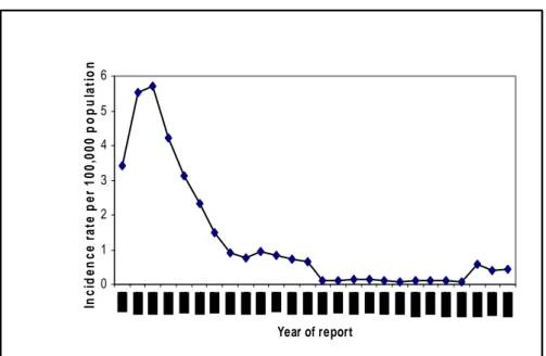
Gonorrhoea declined, from a peak during the Second World War, largely due to the advent of penicillin, and then increased during the sixties and early seventies (Danielsson D et al, 1990). This increase was speculated to be caused by a number of different factors such as increasing treatment failures due to the antibiotic resistance, the introduction of oral contraceptives, the liberation of sexual mores and an increasing travel and tourist industry. Gonorrhoea is a continuing threat to public health worldwide, exspecially in developing countries and some lower socio-economic groups in developed countries. Cases of gonorrhoea in Europe have decreased since the early 1980, with the most dramatic decline in 1985 (Giuliani M et al, 1998; Hughes G et al, 1998). This decline in Europe coincides with the establishment of comprensive educational programmes for changes in sexual behaviour due to the concerns for AIDS and HIV (Panchaud C et al, 2000). The disproportionate impact of AIDS related mortality, on high risk population subgroups may have also contributed to this decrease (Chesson HW et al, 2003). Numbers and rates of acute STIs in any European countries have, however, again been increased since the mid 1990s, the trend for Italy is showed in Figure 1. The incidence of gonorrhoea in the country is estimated from statutory notifications and from more detailed reports received from Sexually Transmitted Diseases (STD) Clinics which voluntarily report the cases to the Istituto Superiore di Sanità since 1991. Data from STD clinics and notifications were available at http://www.simi.iss.it. National incidence for the year 2006 was reported to be 0.43 per 100.000 inhabitants (http://www.data.euro.who.int.cisid).
0 1 2 3 4 5 6 Year of report In ci d en c e r a te p er 1 00, 000 p o p u la ti o n
Figure 1. Incidence of gonococcus infections per 100,000 population in Italy; data from CISID and National surveillance database.
1.2 Biology of Neisseria gonorrhoeae
The bacterial family of Neisseriaceae comprises Gram-negative cocci and rods. The family has four genera: Neisseria, Moraxella, Acinetobacter and
Kingella. The genus Neisseria contains two species primarily pathogenic to
humans: N. gonorrhoeae and N. meningitidis as well as several usually apathogenic commensal species such as N. lactamica, N. cinerea, N.
subflava, N. sicca and N. elongata.
The organisms are aerobic, enhanced by humidity (most of the species). The growth needs carbon dioxide approximately 5%. Neisseriaceae are oxidase and catalase producing (with the exception of N. elongata), do not produces endospores, do not exhibit flagella for motility and have an optimal growth temperature of 35-37 °C (Knapp JS et al, 1984).
The human is the only natural host for N. gonorrhoeae, which survives poorly outside the human body due to its sensitivity to extreme temperatures desiccation, oxidation, toxic substances as fatty acids, as well as to the fastidious nutritional requirements of the organism (Knapp JS et al, 1984). One of the most striking feature of N. gonorrhoeae is the marked degree of variation between isolates. The source of this variation lies not only in spontaneous mutation inside the genes, but also in genetic exchange of DNA with other Neisseria species.
Strains of N. gonorrhoeae are naturally competent for DNA uptake, and can also exchange genetic material by conjugation. The high degree of random recombinant between chromosomal genetic loci, probably mediate by transformation, has to be frequent in nature, and this makes N. gonorrhoeae an example of pathogenesis with a fully sexual, i. e. panmictic or non-clonal, population structure (Hamilton HL et al, 2006). This causes
genotypic and phenotypic variability, which is important for evasion or adaptation to immune response of the host, and for development of antibiotic resistance spread.
N. gonorrhoeae infects epithelial surfaces in the human genital tract,
including urethra, endocervix and fallopian tubes. It also infects conjunctiva, and infection can damage the cornea leading to blindness (Harry TC et al, 2005; MacDonald N et al, 2008).
Rarely, N. gonorrhoeae invades the bloodstream, causing disseminated gonococcal infection. To persist in the male urethra and avoid being washed away by the passage of urine, N. gonorrhoeae has developed effective methods to adhere to epithelial cells, and can also be found intracellular (Apicella M et al, 1996). During gonococcal infection, PMNs (polymorphonuclear cells) migrate into the urethral lumen and attach to the cells and extracellular matrix present in the surrounding environment. During the extravasation from the blood and attachment to surfaces, PMNs are activated and exhibit increased phagocytosis activity. Most gonococci taken up by the PMNs are killed, but up to 2% can survive due to their resistant to oxidative killing (Shafer WM et al, 1989). The interaction of N.
gonorrhoeae with PMNs is an important aspect of gonococcal disease that
remain unclear. Several studies have suggested N. gonorrhoeae survives within PMN (Bouvet JP et al, 1994; Kenney CD et al 2002) and may replicate in PMN phagosomes with time (Prince GA et al, 2005). In fact, prolonged survival of even a low percentage of internalized bacteria might have a crucial role in the outcome of infection. The persistence of gonococci in resident macrophages, which represent a reservoir of infection, could provide a means to overcome host immune response and promote the
infection. N. gonorrhoeae replicating within PMNs, represents an attractive model to explain the chain of persistence of the infection and its possible role in transmission.
Successful colonization by gonococcus can result in a broad spectrum of clinical manifestations of the disease that are, in part, determined by either the specific strain initiating the infection and by the anatomical site infected. An example is the male urethra and the upper female genital tract, in which the gonococci interact with epithelial cells, triggers cytokine release and promotes the influx of PMNs.
1.3 Clinical manifestations of gonorrhoea
N. gonorrhoeae is primarily transmitted by direct human-to-human contact
between the mucosal membranes of the urogenital tract, anal canal, or the oropharinx epithelium of the mucous membranes of the urethra in males, and the endocervix in females (Figure 2). The incubation period is typically 1-7 days, but can vary greatly, between women and men. In males, the acute urethritis associated with dysuria and urethral discharge may in rare cases be complicated later by urethral stricture, prostatitis and epididymitis that may lead to infertility (Bolan G et al, 1999).
However, even untreated, the symptoms of urethritis usually spontaneously decrease within 3-4 weeks. Asymptomatic infection is estimated to occur in approximately 10-20% of infected men, but this may vary depending on the population studied.
In females, the bacteria mostly cause uncomplicated cervicitis and/or urethritis characterized by vaginal discharge, dysuria and sometimes irregular bleeding. The infections are more often asymptomatic in women (approximately 50%), favouring the progress of the infection involving uterus and fallopian tubes. Complications primary include Pelvic Inflammatory Disease (PID), as endometritis, salpingitis, tubo-ovarian abscesses and subsequently ectopic pregnancies and infertility (Figure 2). In addition. anorectal and/or oropharyngeal gonorrhoea, often asymptomatic, are common among homosexual men, but also among heterosexuals. N.
gonorrhoeae may also cause conjunctivitis, mostly in neonates, defined as
“ophtalmia neonatorum” after, exposure to infected secretion of the mother during delivery. Conjunctivitis may, if untreated, result in blindness (Harry
TC et al, 2005; MacDonald N et al, 2008). Disseminated Gonococcal Infection (DGI) may occur in up to 0.5-3% of infected individuals, most often associated with untreated asymptomatic urogenital infection. Moreover, people deficient in late-acting complement factors: C7, C8 and C9 seems to be more likely to develop the DGI. It has been documented 2 DGI cases in Italy. One of the two caused by an imported, multi-drug resistant strain characterized at ISS laboratory and published by Dal Conte et al, 2006 (paper I). Several clinical manifestations of DGI are documented: dermatitis, arthritis, septicaemia, endocarditic and meningitis. The biological basis in HIV transmission, is related to the increased load of HIV in the semen and vaginal fluid in persons with a concurrent gonorrhoea infection (Anzala AJ et al, 2000).
Oropharyngeal Pharyngitis infection
Figure 2. Transmission and outcome of infection with N. gonorrhoeae.
N. gonorrhoeae Anal or genital
infection Local irritation, discharge Asymptomatic (especially women) Systemic spread (1%)
Cervical infection Ascending infection of uterine cavity,
N. gonorrhoeae
(symtomatic or fallopian tubes (pelvic inflammatory disease, infertility, ectopic pregnancy) asymtomatic)
Blindness Surface colonization
N. gonorrhoeae Eye infection
of infant during birth
1.4 Virulence Factors
Several virulence factors have been identified in gonococcus allowing the bacterium to successfully infect human host.
Many of the most important virulence factors include pili, opacity-associated (Opa) outer membrane proteins and lipo-oligosaccharide (LOS). All of them undergo to phase and/or antigenic variation (Snyder LA et al, 2001). Antigenic variation helps the pathogen, first of all, to evade the human antibody response; secondly to more efficiently bind to the mucosa and encounter phagocytes.
Pili
Pili are flexible filamentous, composed of thousands of pilin (pilE) protein subunits, in association with other pilus associated proteins. Pili extend several μm (Nassif X et al, 1999) in all directions from the surfaces of gonococci. Pili have a wide variety of functions which, in Neisseria species, include bacterial aggregation, adhesion, invasion and host cell signalling modulation (Griffiss JM et al, 1999; Merz AJ et al, 1999). Furthermore, pili are though to contribute to gonococcal pathogenesis not only by promoting the initial anchoring to the mucosal surfaces; these surface appendages are also involved at a later stage of the infection during exposure to human monocytes (Knepper B et al, 1997).
Pili play a critical role in the first steps of initial attachment to host cells (Dehio C et al, 2000). The complement regulatory protein, CD46
(membrane cofactor receptor) is a human-specific, transmembrane protein that is expressed by all nucleated cells (Kallstrom H et al, 1997). In human epithelial cell line, (Kallstrom H et al, 2001), CD46 has been demonstrated the receptor for gonococcal pilus. The binding of pili with CD46 is probably mediated by the phase variable adhesin protein PilC located at the tip of the pilus fibre, and/or PilE (major pilus subunit) (Jonsson AB et al, 1994; Rudel T et al, 1995; Nassif X et al, 1999). This interaction results in a rapid cytoplasm calcium flux derived from intracellular calcium stores and, in the host cell cytoskeleton rearrangements. Most strains carry two homologous, but not identical pilC genes, pilC1 and pilC2. A knockout of both these genes results in the lack of expression of pili (Jonsson AB et al, 1995). Pili variation in the amino acid sequence of the major subunit pilE, has been demonstrated to influence the adhesive properties towards various types of epithelial cells (Knepper B et al, 1997).
The pili antigenic variation could explain not only the ability of gonococcus to escape the human immune response but also its ability to adhere to different cells in different sites, such as the cervix and the urethra.
Opa proteins
N. gonorrhoeae outer membrane proteins contains a class of proteins called
opacity proteins (Opa). The expression of these proteins contributes to colony opacity and to intracellular aggregation (Swanson J, 1978). Opa proteins are integral outer membrane proteins synthesized as precursors containing signals for inner membrane transport. A single gonococcal strain can harbour up to 12 opa genes, and Opa protein expression undergoes
phase variation, i.e. expression of each Opa protein can be independently switched to an “on” or “off” state.
Prediction in the secondary structure suggests that mature Opa proteins possess eight membrane-spanning domains arranged as anti-parallel β strands, giving rise to a membrane-embedded β barrel with four extracellular loops (Bhat KS, et al 1992).
Sequence variation between Opa proteins is observed predominantly within the central two loops, that have been termed “hypervariable domain” 1 (HV-1) and 2 (HV-2). Despite variations in amino acid sequence, almost all currently characterized gonococcal Opa proteins can be grouped into two major classes, according to their binding specificity for human surface receptor: OpaHS–type (the term OpaHS denotes heparansulphate-recognising
Opa proteins), and the large group of OpaCEA-type (the term OpaCEA
comprises all Opa proteins recognising carcinoembryonic antigen CEA).
Pathogenic Neisseriae use OpaHS to engage glycosamminoglican side chain
of HSPGs to initiate internalization. In addition, to interact with HSPGs OpaHS is also able to bind extracellular matrix (ECM) proteins such as
fibronectin and vibronectin (Van putten et al, 1998). This represents an additional bacteria-host-cell contact point and allows invasion into the cell with low HSPG expression level. Upon CEACAM binding, OpaCEA–
positive bacteria, can be internalized by professional phagocytes (granulocytes and macrophages) but also epithelial and endothelial cells, Figure 3. CEACAM1 (or CD66a), the broadest tissue expression of all CEACAMs, was also found on B and T cells. Recently, it has been demonstrated that gonococcal stimulation of the ITIM (immune-receptor tyrosine-based inhibition motif)–bearing CEACAM1 expressed on T cells,
arrests the activation and proliferation of these cells in response to cytokines, suggesting that OpaCEA-type gonococci are able to modulate T
cell response via CEACAM1 (Boulton et al, 2002).
The extensive variation and rapid evolution of the opa genes repertoire, as well as sufficient homology between the conserved region of the opa genes, has been exploited to provide a high-resolution typing method for short term transmission of gonorrhoea.. opa-typing is high discriminatory and identical opa-types is believed to be a good indicator for sexual tracing, or part of a short chain of disease (O’Rourke M et al, 1995; Ward H et al, 2000; Palmer HM et al, 2001).
Figure 3. Schematic diagram of CEACAM-dependent interactions of OpaCEA.expressing
Neisseriae with different human cell types (a). CEACAMs expressed on epithelial cells are clustered by tightly adhering OpaCEA-positive bacteria (b). Rearrangements of the actin
cytoskeleton are involved in the subsequent internalization (c). Presence of bacteria derived lipooligosaccharide, or bacteria-induced pro-inflammatory cytokines, stimulate the expression of CEACAM1 on endothelial cells, allowing enhanced adhesion of OpaCEA
-expressing Neisseriae and favouring bloodstream widespread (d). In activated T cells, binding of the OpaCEA-positive gonococci to CEACAM1 inhibits T-cell proliferation and
activation; similarly, interaction with DC (Dendritic cell) inhibits its maturation (e, f). Binding to CEACAMs on PMNs results in opsonin-independent phagocytosis (g) (Gray-Owen SD et al, 2006).
Lipo-oligosaccharide
Lipopolysaccharide (LPS) constitute a family of toxic glycolipids which are integral in the outer membranes of Gram-negative organisms. These molecules are critical for the integrity and functioning of the outer membrane (Gronow S et al, 2001). They are also important surface antigens and are highly immune-stimulatory properties.
LPS molecule generally consists of a highly hydrophobic Lipid A, a covalently attached region, which can be divided into an inner and outer core, and a polymer of a repeating saccharide.
In contrast to the lipopolysaccharide (LPS) that is prevalent among Gram-negative bacteria, the pathogenic Neisseriae have within their outer membranes, LOS molecules, which lack the repeating O-antigen sugar that comprises the polysaccharide side chain of LPS (Giardina PC et al, 1999). LOS produced by N. gonorrhoeae is one of the major antigenic and immunogenic components of the bacterium. Antibodies against LOS have several important functions in gonococcal infection. LOS also contribute to serum resistance, an important feature especially associated with systemic infection; conversely, serum sensitive strains are often associated to local infections.
The oligosaccharide substitutions of LOS exhibit both inter and intrastrain variability (Apicella MA et al, 1987; Demarco de Hormaeche R, et al 1988). A single N. gonorrhoeae strain and even a single gonococcal cell can express several LOS structures at any given time (Apicella MA et al, 1987). Interconversion of LOS oligosaccharides occurs spontaneously, and is dependent upon the presence or absence of available substrates and enzymes
involved in LOS biosynthesis (Burch CL et al, 1997). Some N. gonorrhoeae strains can take an activate form of N-acetylneuraminic (sialic) acid from human blood (CMP-N-acetylneuraminic) and covalently attach it to galactose residues on LOS. The spontaneous conversion of oligosaccharide determinants can change the manner in which the gonococcus associates to host tissue and, hence, can potentially change the course of gonococcal disease. Different LOS structures have been shown to correlate with disease symptomatology: lacto-N-neotretraose LOS is associated with symptomatic gonococcal infection, while lactosyl LOS is associated with asymptomatic gonococcal infection (Schneider H, ). Bacteria with sialylated LOS become serum resistant not only for the presence of the sialic acid, ubiquitous in the host cell, but also because the membrane attack complex (MAC) does not deposit around the altered LOS.
Moreover, LOS oligosaccharide side chain terminate in epitopes that mimic sugar moieties of mammalian glycosphingolipids such as structures on human erythrocyte membrane (Mandrell RE et al, 1988). This form of molecular mimicry not only provides the bacterium a method of immune escape, but also allows the bacterium to use host-derived molecules that, normally associate with the mimicked structure (Harvey H, et al 2001).
Porins
The gonococcal porins are named porin B IA (PorB1A 35KDa) and porin B IB (PorB1B 37KDa) (Gotschlich EC, et al 1987). The native structure of
Neisseria porins is trimeric, with each porin comprising three single
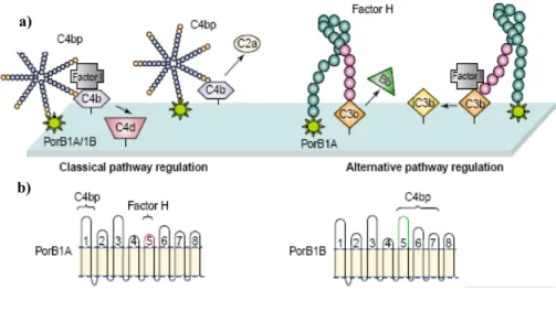
polypeptides. Neisseria porins function as pores, essential for bacterial survival and for ions exchange of ions with the surrounding environment. They form aqueous transmembrane channel and the insertion process leads to the formation of a functional channel at low membrane potential (Edwards JL et al, 2004). A unique feature ascribed to gonococcal porin is its ability to translocate into eukaryotic cell membranes, where it forms a voltage-gated channel that is modulated by host cell ATP and GTP (Rudel TH et al, 1996). ATP and GTP regulate gating of the size and ion selectively of pores, causing a transient change in mammalian membrane potential and interference with host cell signalling. This, has been implicated in pathogenesis due to induction of apoptosis of target cells (Muller, et al 1999). Gonococcal porins play an important role in enabling the bacteria to evade complement-mediated killing. The ability of certain gonococcal strains to down-regulate early complement cascade, prior to the C3 deposition, may be partly responsible for milder symptoms seen in individuals infected with such strains. The complement system has an important role in the innate immune system, mediating direct bactericidal killing and bacterial clearance by enhancing opsonic phagocytosis.
Porins, mediate evasion of complement-dependent bactericidal activities by binding factor H, the main fluid-phase regulatory protein of the alternative pathway of complement, or C4, that inactivate C3b and C4b respectively
(Figure 4, a). This binding interfere with the activation, degranulation and phagocytosis of neutrophils. Porins from N. gonorrhoeae, as well as previously described LOS, have been shown to influence the development of serum resistance. The gonococcal porin PorB1A is critical in modulating stable serum resistance, which is not mediated by LOS sialylation. Binding of PorB1A to factor H occurs via the surface-exposed region of the 5th loop of porin (Figure 4 a and b) (Ram S et al, 1998). In addition, several gonococcal strains bearing PorB1A can also down regulate the classical complement pathway regulatory protein C4b-binding protein, which appear to interact with the amino-terminal loop of porin. Moreover, certain serum resistant PorB1B strains may also contain C4bp-binding site.
Previous studies have identified an association between PorB1A isolates and higher resistance to bactericidal activities by normal human sera (Van putten JP et al, 1998; Bauer FJ et al, 1999). Furthermore, PorB1A isolates have been associated with invasive disease like DGI, and this may be affected by the fact that PorB1A but not PorB1B also promotes Opa-independent invasion of epithelial cells.
Any individual gonococcal strain expresses only one of two different proteins, the PorB1A or the PorB1B. Rarely, strains expressing both of the proteins occur (Gill M et al, 1994) . The antigenic expression of PorB within a strain is stable; however, diversities among strains constitute serovar classification using monoclonal antibodies as suggested by Sandstrom E et al, 1980; Knapp JS et al, 1984.
a)
b)
Figure 4. Picture showing the role of porin in complement regulation on the surface of N. gonorrhoeae. a) Regulation of the classical pathway occurs within C4b-bp binds to porin. Factor H bound to porin regulates in alternative pathway. b) Schematic representation of PorB1A and PorB1B proteins. Loops 1 and 5 are essential C4b-bp and for factor H, respectively (Massari P et al, 2003).
IgA
The pathogenic Neisseria spp. produce extracellular IgA1 proteases, which cleave humoral and secretory IgA1 at the hinge region and it has been implicated as a virulence factor in the pathogenesis of human mucosal infections caused by gonococcus (Kilian M et al, 1996). Cleavage of intact IgA1 (or S-IgA) results in the loss of Fcα-mediated secondary effector functions such as inhibition of adherence (Hajishengallis G et al, 1992) despite the retention of antigen-binding activity by the Fab α fragments (Mansa B et al, 1986). Moreover, it has been hypothesized that Fab α fragments may block the binding to antibodies and thereby inhibits, for istance, complement activation and bacteriolysis (Kilian M et al, 1996). IgA1 protease secreted by N. gonorrhoeae have recently been suggesting to have several effects when the bacterium is present in the sub-epithelium rather than in the lumen or on the mucosal surface. In particular, recent evidences suggest that the lysosome phagosomal protein LAMP-1 is cleaved by IgA1 protease, and that may contribute to intracellular gonococcal survival (Hauck CR et al, 1997).
1.5 Adherence and invasion
The first stage in the infection process is the colonization of a mucosal surface composed of columnar epithelial cells (Figure 5). Some of the surface components of N. gonorrhoeae are involved in attachment and invasion of mammalian cells, as previously described (Griffiss JM et al, 1999). N. gonorrhoeae first attaches to the cell surface by pili, shortly after initial attachment, the bacteria become more tightly attached to the epithelial surface.
The second stage of binding and the phagocytic uptake, appear to be mediated in part by Opa protein. Opa is not sufficient for tight binding and invasion, but loss of Opa protein is associated with loss of the ability to adhere and invade epithelial cells. These proteins also mediate the binding of N. gonorrhoeae each other to form micro colonies by exposed sugars of LOS of another bacterium. Micro colony formation may aid in the initial colonization of the mucosal surface. Opa proteins are also involved in the cytoskeleton rearrangements required for endocytosis after the adhesion (Grassmè H et al, 1996; Grassmè H et al, 1997). In addition porins contribute to the ability of gonococci to survive inside phagocytes. In particular, of inhibition phagosome maturation down regulates immunologically important, opsonin-dependant cell surface receptors (Bjerknes RH et al, 1995). Porins may play a role in preventing the release of reactive oxygen species from PMNs to extracellular milieu which is consistent with the degranulation inhibition attributed to the action of porin (Haines KA et al, 1991).
Figure 5. Colonization of the human host begins at apical surfaces. Pili and Opa are primarily involved in adhesion process. Porins may facilitate the cytoskeleton rearrangements required for actin-mediated entry of gonococcus into target cells. Following internalization, bacteria could either be killed by macrophages, and/or inhibit phagosome maturation, determining host cell damage (www.textbook.bacteriology.net/neisseria.htlm).
1.6 Prevention
The first step of a correct prevention at all STIs is the reduction of contact at risk. Changes in response to the emerging HIV/AIDS pandemic saw dramatic reductions in the incidence of many acute STIs in the late 1980s (Van der Heyden JH et al, 2000; Adler M et al, 2000). However, the progress in HIV/AIDS therapy and the success in the reduction of case fatality rate, decreased in parallel the attention to its prevention.
Because of the important role of the bacterial STDs in HIV transmission, the WHO has concluded strategies to control bacterial STDs (WHO, 1992). It is noteworthy, how potential transmission in STDs, since sexual intercourse provide limited contacts, often display peculiar characteristics. In particular, there are two main aspects to be considered in gonococcal prevention: antimicrobial resistance control and looking for an effective anti-gonococcal vaccine. In fact, as above mentioned in this Thesis, high degree of genotypic variability of N. gonorrhoeae, makes this pathogen prompt to genetic changes with implications also in antimicrobial resistance. In this context, multidrug resistant gonococcal strains, represent a focal point for treatment, reduction of spread, and a possible role to acquire other STI infections. Antimicrobial surveillance is important to monitor the change in drugs susceptibility of the bacterium and to suggest changes in therapy protocol.
An effective vaccine to prevent the disease is an important goal. Human trials using partially lised gonococci, purified pilin or purified porin, failed to confer protection upon natural exposure (Sparling PF et al, 1994). These vaccine formulations, although immunogenic, failed to protect likely due to the intrinsic ability of the gonococcus to undergo high-frequency phase and
antigenic variation of surface structures (Meyer TF et al, 1994). It is clear that patients with uncomplicated gonococcal infection develop antibodies, but they are not reactive against gonococcal antigen from a different strain without any immunological memory (Hedges SR et al, 1999). Thus prompted researchers to look for surface antigens with conserved sequences among strains. Two surface- exposed gonococcal proteins: the macrophages infectivity potentiator Mip) and the outer membrane protein A (Ng-OmpA) are recently studied in their structure and in their role during gonococcal infection. In particular, Ng-Mip is involved in intracellular survival, during macrophages interaction. Ng-OmpA is an outer membrane protein possibly involved in the adhesion to epithelial cells, as well as in intra-macrophages survival.
Both of those molecules show interesting features as hypothetical candidates for therapeutic and/or prophylactic therapy against gonorrhoea.
Macrophage Infectivity Potentiator
Macrophage Infectivity Potentiators (MIPs) are surface-exposed or secreted proteins already described as virulence factors in different pathogens such as
Legionella pneumophila (Lp), Chlamydia trachomatis (Ct), Trypanosoma cruzi (Tc), and Escherichia coli (Ec) (Kohler R et al, 2003). These proteins
exhibit peptidyl-prolyl cis/trans isomerise (PPI-ase) activity, and catalyses the slow cis/trans isomerization of prolyl peptide bonds in oligopeptides involved in the proline mediated folding of proteins (Fisher et al, 1992). MIPs belong to the enzyme family of FK506-binding protein (FKBP) (Fischer G et al, 1992; Fischer G et al, 1984; Schmid F et al, 1993) inhibited by T cells immune-modulatory cyclosporines. In particular, FK506 and rapamycin, which are important immunosuppressant are used to treat transplant recipients (Fanghanel J et al, 2004). The addition of PPI-ase inhibitors to Ct and Lp resulted in reduced infectivity of these organisms suggesting that the inhibition of PPIase activity interferes with the biology of the infection, confirming the role of PPI-ase in bacterial virulence (Lundemose AG et al, 1993; Moro A et al, 1995).
It has been described the identification and characterization of a Ng-MIP in
N. gonorrhoeae, showing high homology to MIPs proteins (Leuzzi R et al,
2005).
Ng-MIP is a basic 29 KDa protein and the presence of a L18XXC21 motif at
the putative site cleavage, suggests that the mature polypeptide could be a lipoprotein anchored to the outer membrane through Cysteine at amino acid position 21. Ng-MIP also contains a putative dimerization domain at the N-terminal, supporting the hypothesis that it could exist as a dimer. The
C-terminal of Ng-MIP has high degree of homology with the PPI-ase domain of Lg-MIP and Tc-MIP, suggesting that it could also showed a PPI-ase activity.
Gonococci mutant strains, generated by deletion of mip gene (Δmip), have the same ability as the wild type, to adhere and to be internalized in macrophages, but have a reduced survival after the infection due to the macrophage killing (Leuzzi R et al, 2005). Therefore, Ng-MIP seems to be important for survival of N. gonorrhoeae inside macrophages, highlighting a possible role in promoting the persistence of gonococcal infection. Ng-MIP is specific for macrophages since it does not influence the invasion and the survival of gonococcus in other cells, such as the epithelial cells. Currently, little is known regarding the role of Ng-MIP interaction with phagocytic cells as well as its PPI-ase activity in N. gonorrhoeae.
The structure of FK506 complex of Ng-Mip provides a starting point for the design of more specific inhibitors lacking the immune-suppressant property of the molecule. Such inhibitors could be of prime interest for developing new therapies against gonococcal infection.
Outer Membrane protein A
Outer Membrane Protein A (OmpA) is an abundant structural protein of the outer membrane of Gram negative bacteria (Sonntag et al, 1978). It assembles in the outer membrane via an N-terminal eight transmembrane amphipatic β-barrel region with the C-terminal region retained in the periplasm (Koebnik R et al, 2000; Pautsch A et al, 1998). Unlike other surface-exposed of the bacterial cell envelope, OmpA is highly conserved among the Enterobacteriaceae and throughout the evolution (Poolman JT, et al 1996). OmpA functions include first of all the maintenance of structural membrane integrity during the invasion into mammalian cells (Weiser JN et al, 1991). OmpA-deficient in E. coli exhibit attenuated virulence invasive capacity and resistance to serum bactericidal activity (Weiser JN, et al 1991). In particular, OmpA contributes to serum resistance by binding to the complement regulatory protein C4bp (Prasadarao NV et al, 2002) leading to a decrease in serum killing, a mechanism used by many pathogens to avoid complement attack.
Interestingly, in contrast to most of the soluble foreign proteins that produce a weak immune response or tolerance, when administered in the absence of an adjuvant, OmpA from different species induces specific humoral (Poolman JT, 1996) and cytotoxic responses (Kim SK et al, 2000) also in the absence of adjuvant. In particular, it has been demonstrated that the recombinant 40 KDa OmpA from Klebsiella pneumoniae, interacts with human immune cells and particularly with antigen presenting cells (APCs) via endocytic receptors on host cell surfaces (Jeannin P et al, 2002). This property contributes to explain its unusual immunogenicity driving interest
for future vaccine strategies as target for antigen delivery and for cytotoxic T-lymphocytes (CTLs) induction.
Serino L. and colleagues, have recently described a gonococcal OmpA-like protein, defined as Ng-OmpA, playing a significant role in the entry and in the intracellular survival in macrophages. In particular, the knockout mutant (ΔOmpA), demonstrated reduced recovery in a mouse model of infection when compared with the wild type strain, suggesting that Ng-OmpA play an important role in in vivo colonization by N. gonorrhoeae. Ng-OmpA is a surface-exposed lipoprotein with a predicted molecular mass of 23 KDa, showing a significant homology with the OmpA expressed by several microrganisms such as Escherichia coli, and Salmonella typhimurium (Serino L et al, 2007). Currently, little is known regarding Ng-OmpA and its exact function and role in the pathogenesis of gonorrhoea. However, studies regarding OmpA features, as possible candidate vaccine, further investigations will be performed.
1.7 Aims
Data regarding the trend and the group at risk for N. gonorrhoeae infection originates mainly from North Europe and United States. Few publications exist from Central and South Europe, including Italy concerning the epidemiology and microbiological characteristics of circulating strains. From April 2003 to December 2007, a study on clinical and microbiological characteristics of the isolates was undertaken.
The aims of the present Thesis were:
- to investigate the phenotypic and genotypic features of isolates. In particular, the antimicrobial susceptibility and the analysis of the molecular changes responsible for antibiotic resistance.
- to analyse the genetic relatedness among gonococcal strains circulating in the study period in Italy.
- to analyse the mip and ompA gene sequences, as well as their gene products, to investigate gene conservation among a selected group of isolates showing different phenotypic and genotypic characteristics associated with different clinical pictures.
- to evaluate the recognition, by human sera collected from infected patients, of purified Ng-Mip and Ng-OmpA proteins.
- to evaluate the role of Ng-Mip and Ng-OmpA in the serum bactericidal activity using N. gonorrhoeae knockout strains for the two genes.
2. Materials & Methods 2.1 Samples collection
N. gonorrhoeae clinical isolates were accompanied by a patient
questionnaire with epidemiologic information about nationality, residence, sexual orientation, previous antibiotic use, number of sexual partners, as well as clinical information (sex, age, diagnosis, HIV infection and immune status compromising). Sera samples, acute and convalescent, when available, were obtained from patients at approximately two-weeks of interval. Those sera were sent to I.S.S., for serological studies and for storage at -80°C.
2.2 Bacterial strains and phenotypic analysis
A total of 729 cases were investigated during the study period. In particular, 583 N. gonorrhoeae isolates were stored in BHI (Brain Hearth Infusion) broth with 20% glycerol at -80°C.
The isolates were cultured as purified colonies on Thayer Martin medium with 2% IsoVitaleX (Oxoid LTD, UK) at 37°C in 5% CO2. Knockout F62 N. gonorrhoeae: Δmip and ΔompA, strains were cultured on Thayer Martin
medium added with 1% IsoVitaleX (Oxoid LTD, UK) and 5 mg/ml of erythromycin (Sigma Aldrich, USA).
2.2.1 Serotyping
Serological group (serotyping) of all isolates was obtained with Phadebact GC serovar test (Boule Diagnostics, Sweden) by a co-agglutination using monoclonal antibodies directed against PorB1A and PorB1B proteins encoded by the mutually exclusive alleles of the porB gene, porB1A and
porB1B, respectively (Knapp JS et al, 1984; and Sandstrom E et al, 1980).
Bacterial suspension in sterile Phosphate Buffered Saline (PBS) was used to react with the monoclonal antibodies. The strains with cross-reactivity (IA/IB) were re-tested to confirm the result.
2.2.2 Antimicrobial susceptibility analysis
The antimicrobial susceptibility test for: ciprofloxacin, penicillin, tetracycline, azithromycin, spectinomycin and ceftriaxone was determined by the E-test (AB Biodisk, Sweden) following the manufacturer’s instructions. Strains were plated onto Thayer Martin (TM) plates supplemented with 2% IsoVitaleX (Oxoid, Ltd UK) cultured over night (o. n.). After 24h of growth, gonococcal cell suspensions, prepared in sterile PBS, up to a density of 0.5 McFarland, were plated on TM agar supplemented with 1% IsoVitaleX at 37°C and 5% CO2. Minimum
Inhibitory Concentration (MIC) breakpoints for each antimicrobial agent are shown in Table 1.
For tetracycline, to confirm the MIC close to breakpoint, the agar dilution method was also included. Penicillinase production was determined using the chromogenic nitrocefin test (Oxoid, Ltd UK).
N. gonorrhoeae reference strain ATCC 49226 was includes in each test, as control.
Table1. Breakpoints as defined by NCCLS (CSLI), 2006.
Antimicrobial agent Susceptible (mg/L) Intermediate (mg/L) Resistant (mg/L) Penicillin ≤0.06 0.12-1 ≥2 Tetracycline ≤0.25 0.5-1 ≥2 Ciprofloxacin ≤0.06 0.12-0.5 ≥1 Spectinomycin ≤32 64 ≥128 Ceftriaxone ≤0.25 - - aAzithromycin - - ≥1
a Azithromycin breakpoint was that from the Centre for Disease Control and Prevention
(2003) Neisseria gonorrhoeae reference strains for Antimicrobial Susceptibility, B88, August 2005.
2.3 Molecular analyses
2.3.1 DNA preparation, PCR amplification and DNA sequencing
Chromosomal DNA used as template was prepared with QIAamp DNA minikit (QIAGEN, Hilden, Germany) according to the manufacturer’s instructions. Plasmid DNA was prepared by boiling a bacterial suspension in PBS after 24 h of growth.
Amplification reactions were carried out as previously described for each target gene and oligonucleotides are listed in Table 2. All purified PCR products (QIAquick PCR purification Kit, QIAGEN) were subjected to automated sequencing. Sequences analyses were performed with Chromas version 6 software, and BLAST website program (http:// www.ncbi.nlm.nih.gov/BLAST) was used to compare the sequences with those available on the website.
Table 2.Primers used for PCR amplification and sequencing to study antimicrobial resistance.
Antimicrobial
agent Targets involved in resistance Primer sequences (5’→3’)
Penicillin Plasmid PPNGa Β-lactamase (Toronto-Rio/ African plasmid) BL1 (forward) 1545TACTCAATCGGTAATTGGCT1564 BL2 (forward) 3606 CACCTATAAATCTCGCAAGC3625 BL3 (reverse) 4564CCATAGTGTTGAGTATTGCGAA4585 BL4 (reverse) 6528TCATTCGTGCGTTCTAGGA6546 Ciprofloxacin Chromosomal QRNGb gyrA gyrA1 (forward) 160CGGCGCGTACTGTACGCGATGCA182 gyrA1X (reverse) 438AATGTCTGCCAGCATTTCATGTGAG A413 parC parC1 (forward) 166ATGCGCGATATGGGTTTGAC185 parC2X reverse 20CGACAACAGCAATTCCGCAA401 Tetracycline Chromosomal CMRNGc rpsJ rpsJ1 (forward) 23TCCGCCTGAAAGCTTATGAT42 rpsJ2 (reverse) 246TTATCGGTCCAATCCACGAT227 Plasmid TRNGd TetMe (American /Dutch type plasmid) UF (forward) 825CTCGAACAAGAGGA842 AR (reverse) 1602GCATTCCACTTCCCAAC1586 DR (reverse) 1267TGCAGCAGAGGGAGG1253
a Penicillinase-producing N. gonorrhoeae b Quinolone-resistant N. gonorrhoeae
c Chromosomally-mediated resistance N. gonorrhoeae d Tetracycline resistant N. gonorrhoeae
e The primer sequences derived from the sequences of TetM pOZ100 (UF, AR) and TetM
pOZ100 (AR) which accession number were L12241 and L12242.
2.4 Genotypic analyses
For genotypic analysis the gonococcal strains were typed by PCR-Restriction Fragment Length Pattern (RFLP) of opa genes, Pulsed-Field Gel Electrophoresis (PFGE) and N. gonorrhoeae Multi-antigen Sequence Typing (NG-MAST).
2.4.1 PCR-RFLP of opa genes
Typing of opa genes was performed on a subset of samples, 128, and based on method previously described by O’ Rourke et al, 1985. The 11 opa genes were amplified with a single pair of primers (opa-up 663GCG ATT ATT TCA
GAA ACA TCC G 687 and opa-down 1227GCT TCG TGG GTT TTG AAG CG 1207) and
digested with 40 U of TaqI (New England, BioLabs) for 2 h. Gonococcal yielding identical restriction fragment patterns were considered to have the same Opa-Type (OT). Isolates differing by even a single fragment were assigned to different OTs. Isolates that gave identical patterns with TaqI, were then digested with 40 U of HpaII (New England, BioLabs) restriction enzyme for 2 h (Van M et al, 1999) in order to confirm the result.
2.4.2 Pulsed-Field Gel Electrophoresis
Bacteria were suspended in sterile PBS to a final concentration of 4 McFarland. From each suspension 200 μl were used for preparation and digestion of bacterial DNA. The suspension after centrifugation were resuspended in lysis buffer (EC+RNA-asi 50μg/ml+Lisozima 1mg/ml) and mixed to an equal volume of 1.2% (wt/vol) low melting preparative-grade
solidified plugs were incubated o. n. at 37° C with 40U of SpeI (New England, Biolabs) (Mavroidi A et al, 2001) and 30 U of NheI (New England, Biolabs). The slides of agarose containing the chromosomal DNA fragments were inserted into the wells of 1% agarose gel (Bio-Rad) and the electrophoresis was performed in CHEF-Mapper II apparatus (Bio-Rad, Hercules, CA). The run comprised the following parameters: 0.5s and 54s of ramping time and a running time of 21h at 6V/cm in a 0.5X TBE buffer (45 mM Tris-borate, 45 mM Tris base, 45 mM boric acid and 1 mM EDTA). The Lambda ladder PFG marker (New England, Biolabs) was used as molecular size standard. The reference N. gonorrhoeae strain FA1090 was included in each run. The gels were stained with 2mg/ml ethidium bromide destained in double distilled water, photographed under UV lights and digitized with Gel Doc 2000 system apparatus (Bio-Rad, CA).
DNA restriction patterns were compared to determine their relatedness. Strains with patterns varying by three or less fragments were considered as the same PFGE type (Tenover FC et al, 1997; Goering RV et al, 1993). The PFGE banding patterns were analyzed with the Diversity Database Fingerprinting Software (version 2, Bio-Rad Laboratory).
2.4.3 N. gonorrhoeae Multi-antigen Sequence Typing
Amplification of por and tbpB genes was performed on a total of 175 gonococci chosen among sensitive and resistant to drugs. PCR parameters were those suggested by Martin I et al, 2004.
Oligonucleotides were those listed in Table 3.
Primers for por gene were designed on conserved regions encoding the pre-loop 2 and pre-pre-loop 8 of the protein, in order to amplify both classes of alleles (por IA and por IB respectively). After being sequenced selected fragments, for por and tbpB gene, respectively were entered into a publicly accessible database on the NG-MAST website: www.ng-mast.net, in order to define the corresponding alleles and the resulting Sequence Type (ST).
Table 3. Primers used for amplification and sequencing of por and tbpB alleles for
NG-MAST typing.
Primers Nucleotide positiona
por forward 350 CAA GAA GAC CTC GGC AA 366
por reverse 1086 CCG ACA ACC ACT TGG T1070
tbpB forward 1098 CGT TGT CGG CAG CGC GAA AAC 1119
tbpBreverse 1686 TTC ATC GGT GCG CTC GCC TTG 1665
a Numbering is based on por and tbpB sequences of N.gonorrhoeae MS11strain (accession
2.5 Ng-mip and Ng-ompA gene sequence analyses
The entire mip and ompA genes were amplified from 20 N. gonorrhoeae clinical isolates. The primers used for PCR reactions are showed in Table 4. Amplifications parameters were: 15 min at 95°C, followed by 35 cycles of which 1 min of denaturation at 95°C, 1 min of annealing at 55°C or 60°C for mip and ompA genes, respectively, 1 min of elongation at 72°C. Purified PCR products (QIAquick PCR purification Kit, QIAGEN), were subjected to automatic sequencing. Nucleotide and amino acid sequences were analyzed with Chromas software version number 6 and with BLAST website (http://www.ncbi.nlm.nih.gov/BLAST). Multiple sequence alignments were performed with DNAMan software version 5.2.10 (Lynnon BioSoft, Canada).
Table 4.Primers used for PCR amplification and sequencing of mip and ompA genes.
Primers Nucleotide positiona
mip1 forward 816AAC ACC ATT TTC AAA ATC AG 798
mip2 reverse 1TTA ATT TAC TTT TTT GAT GTC G 22
ompA forward 1ATGACTTTCTTCAAACCCTC 20
ompA reverse 678 TTA CAT GTG CCG TGC GGC 661
aNumbering is based on mip and ompA gene sequences of N. gonorrhoeae FA1090
2.6 Western blot analysis
Western blot analysis was performed according to standard procedures (Laemmli UK, 1970). Recombinant Ng-OmpA or Ng-MIP (kindly provided from Novartis, Italy), were run on 12% acrylamide at a 100V for 1h. After blotting the nitrocellulose membranes, were incubated for 2 h at room temperature (r. t.) with sera from gonorrhoea infected patients, then washed twice with saline buffer added with 0.05% of Tween 20. Then, were incubated for 1 h at r. t. with antihuman IgG antiserum (Fc-specific)-Alkaline Phosphatase (Sigma Aldrich, USA). Peroxidase substrate (SIGMA FAST BCIP/NBT Sigma Aldrich) was used for the detection reaction. Molecular weight standard (New England, Biolabs) was included in each gel. Polyclonal mouse antiserum, raised against recombinant Ng-Mip and Ng-OmpA proteins, respectively, were used as positive controls.
2.7 Serum bactericidal activity
A total of six sera samples from patients infected with gonorrhoea were used, after heat inactivation (at 56°C for 30 min), in the serum bactericidal activity. These sera samples were separately tested against 6 N. gonorrhoeae strains, as well as, Δmip and ΔompA knockout strains. Gonococcal strains after 24 h of growth were suspended in GC broth supplemented with 1% IsoVitaleX (Oxoid, Ltd UK) up to an optical density of 0.2. The broth culture was monitored until it reached 0.4 OD (mid-log growth phase). Aliquots of 12.5 μl of the cell suspensions (containing about 100 colony forming unit), were added to 25 μl of the heat-inactivated serum sample (serially diluted), and 12.5 μl of baby rabbit serum, as complement source . The final volume of all reaction mixtures was 50 μl. The microplate was then incubated at 37°C in 5% CO2 for 1 h. Ten microliter aliquots of the
reaction mixtures were plated onto GC agar at time 0 and after 60 min. Percentage of survival was expressed as the percentage of colonies on a plate at 60 min compared with those on the plate at 0 min.
3. Results
3.1 Characteristics of the study population
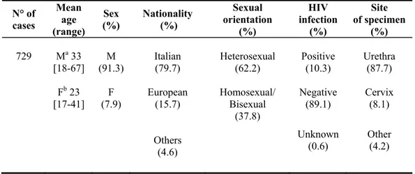
From April 2003 to December 2007 a total of 729 cases of gonorrhoea were recorded in our database. As shown in Table 5, considering the range of different ages among males and females infected, it was noteworthy that most of the infections occurring among males, 18-67 year age range; while for females it was 17-41. More than 60% of infected patients were among heterosexuals. Since the cases were predominantly among male, the male urethra was the most frequent site of isolation with discharge/dysuria the main associates symptoms.
Interestingly, it has been recorded 3 cases of disseminated gonococcal infections.
A mean of 10.3% of patients with gonorrhoea were also HIV-1 positive.
Table 5. Clinical data of patients infected with N. gonorrhoeae strains isolated in Italy
during the study period.
N° of cases Mean age (range) Sex (%) Nationality (%) Sexual orientation (%) HIV infection (%) Site of specimen (%) 729 Ma 33 [18-67] M (91.3) Italian (79.7) Heterosexual (62.2) Positive (10.3) Urethra (87.7) Fb 23
[17-41] (7.9) F European (15.7) Homosexual/ Bisexual (37.8) Negative (89.1) Cervix (8.1) Others (4.6) Unknown (0.6) Other (4.2) a M: Male. b F: Female.
3.2 Phenotypic analyses
Serotyping was performed on a total of 481 isolates. Most of the gonococci isolated were serotype IB (90.1%), the remaining were serotype IA/IB and IA, 7.2% and 2.7%, respectively. Those strains showing a cross-reactive serotype, IA/IB, were re-tested in order to confirm the result.
As showed in Table 6, gonococci resistant to ciprofloxacin, penicillin, and tetracycline were recorded in the study.
Ciprofloxacin resistant strains (QRNG) were 46% of all gonococci analysed. Few strains showed reduced susceptibility to ciprofloxacin (Table 6).
Penicillin resistance was observed in 20% of the analysed strains (Table 6). Most of the isolates (97%) were PPNG (i.e. carrying African type or Toronto/Rio, β-lactamase-producing plasmid). The remaining 3 % were CMRNG, with a negative β-lactamase nitrocefin test.
Tetracycline resistant gonococci were 28.3% of the total. The 44.6% showed a TRNG phenotype (carrying plasmid with TetM determinant, American or Dutch type); the 23% were CMRNG. Noteworthy, 32.4% of tetracycline resistant gonococci showed both the TRNG/CMRNG phenotypes.
Interestingly, more than 40% were strains with reduced susceptibility to penicillin or tetracycline Table 6.
Antimicrobial susceptibility analysis for azithromycin was performed on a total of 172 gonococci out of 481, the 11.6 % of them were resistant (Table 6).
Multidrug resistant strains were also found in this study. Twenty-four percent and 4.4% resulted resistant to 2 or 3 antibiotics, respectively.
Resistance to ciprofloxacin plus tetracycline was the most frequently combination found (17.3%).
Table 6. Percentages of susceptible, intermediate and resistant N. gonorrhoeae strains by
antimicrobial agents. Antimicrobial agent N° of isolates S (%) I (%) R (%) Ciprofloxacin 481 45.7 4 46 Penicillin 481 19.1 49.4 20.4 Tetracycline 481 22.2 42 28.3 Azithromycin 172 - - 11.6 aS: Susceptible bI: Intermediate cR: Resistant
3.3 Molecular analyses
Resistance to ciprofloxacin was conferred by amino acid changes in gyrA and parC genes. In particular, sequence analyses were performed on a total of 94 out of 129 ciprofloxacin resistant strains. The results revealing the following amino acid substitutions: S91→F, D95→G/A/N in gyrA gene,
S87→I/R/N, D86→N/H, E91→K in parC gene. However, the most frequent
pattern mutations was: S91→F, D95→N in gyrA gene and D86→N in parC
gene with a MIC range was 8-32 mg/L.
All but one ciprofloxacin resistant strains, showed at least one amino acid substitution in both of the gene. The exception showed an amino acid substitution only in the parC gene. This was associated with a lower MIC
value for ciprofloxacin, 6 mg/L.
Penicillin resistance was prevalently associated to β-lactamase producing plasmid: Toronto/Rio (61%), and African type plasmid 39%. Chromosomally mediated penicillin resistance was 3%.
Resistance to tetracycline was conferring either by plasmid carrying TetM determinant (TRNG) or by point mutation in rpsJ with the gene amino acid substitution Val57→Met (CMRNG). In particular, among TRNG isolates,
the American type plasmid was the most frequent. The co-existence of both the plasmid and chromosomally mediated resistance (TRNG/CMRNG) was identified in 32.4% of the gonococci analysed.
3.3.1 Genotypic analyses
A representative number (40%) of resistant and susceptible isolates was typed by molecular methods including opa-typing, NG-MAST, and PFGE analysis.
Opa-type profiles are shown in Figure 6. In particular, opa-type 1 was the most frequent (66.4 %) identified among the antimicrobials resistant strains examined. Most of the gonococci resistant to ciprofloxacin, penicillin or to both of these two drugs, showed opa-type 1. However among 7 opa-type profile identified, opa-type 1 to 6 (Figure 6, lanes 1 and 6) were recovered in ciprofloxacin resistant strains while opa-type 1, 2. 3, 5 and 7 were prevalent in penicillin resistant strains.
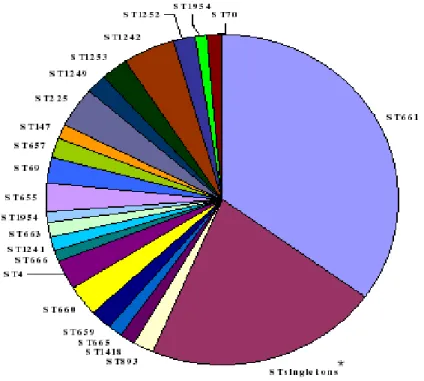
NG-MAST analysis performed on a total of 175 strains. A total of 66 STs were identified, comprising both novel and previously described STs (Figure 7). Among 62 STs observed 39 occurred only once (singletons), most of them associated exclusively with homosexual transmission. The remaining 23 were clusters of two, three and four isolates. Interestingly, it has been identified the presence of a large cluster of isolates showing sequence type ST661, most of which were opa-type 1. The transmission of ST661 was associated with heterosexual behaviour. All the isolates identified as ST661 were serogroup IB and were resistant to penicillin, ciprofloxacin and resistant or had reduced susceptibility to tetracycline. The percentage of ST661 isolates resistant to one, two or three drugs was 3.7%, 83.3% and 13%, respectively. In particular, ST661 clone was circulating in Italy in Turin province. Its circulation in this geographic area peaked from October to December 2003. The isolation rate decreased and the appearance of 3 other STs was seen. These 3 STs: ST657, ST1252, and ST1253, which
were exclusively found in Turin, shared the same tbpB allele with ST661 but have different por allele. However, two of the STs (ST657 and ST1252) each different from ST661 by a non-synonymous point mutation in the por gene fragment and would therefore be considered to be part of the same cluster. ST657 and ST1252 profile analysis were identical and closely related to that of ST661, respectively. Conversely, ST1253 had an unrelated profile to strains ST661, ST657 and ST1252. This is consistent with the relatedness by the NG-MAST result described.
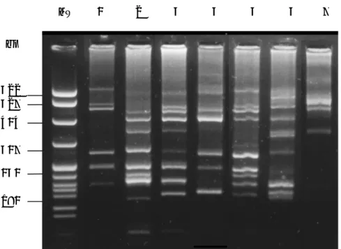
PFGE analysis performed on a total of 76 strains. In particular, those strains representing the same opa-type, showed also the same PFGE pattern profile (Figure 8, lane 1) confirming the presence of a clone.
PFGE analysis showed identical profiles among the strains belonging to ST661 and ST657 (Figure 8, lane 1 and 2) and a closely related profile for strains of ST1252 (Figure 8, lane 3). ST1253 had an unrelated profile to strains of ST661, ST657 and ST1252 (Figure 8, lane 4).
2 6 7 MW 1 3 4 5 bp 622 527 404 307 242 180
Figure 6. Examples of opa-type identified in the study. Lane MW, pBR322 MspI digest
molecular DNA marker (New England , Biolabs). Lanes 1-7, opa-type from 1 to 7, respectively.
Figure 7. Pie chart depicting different N. gonorrhoeae Sequence Types (STs) identified
among the Italian isolates examined. ST661 represents the largest cluster of isolates identified. ST singletons* include the total number of the STs represented by only one isolate; indicates STs that are not previously identified. The remaining STs are clusters of two, three or four isolates.
MW 1 2 3 4
Figure 8. PFGE patterns of SpeI-digested genomic DNA from N. gonorrhoeae isolates
belonging to STs: ST661, ST657, ST1252 and ST1253 (lane 1-4). MW: Molecular weight marker (New England, Biolabs).
3.4 Sequence analyses of mip and ompA genes and their gene products To investigate mip and ompA genes conservation PCR amplification and sequences analyses were performed on selected strains representing the STs most frequently found in the country. In particular, gonococci were chosen to represent different clinical and microbiological characteristics (Table 7). Comparison of mip gene sequences of 20 N. gonorrhoeae clinical isolates, and F62 strain, revealed a high degree of gene conservation. However, deduced amino acid sequences revealed the presence of 2 amino acid substitutions, S59→G and E160→K, compared with N. gonorrhoeae FA1090
(Figure 9). Most of the isolates showed the presence of both these substitutions. Six strains showed only the presence of. S59→G localized in
the N-terminal region of the gene (Figure 8). E160→K substitution was
localized in the C-terminal region, involved in the catalytic activity of the protein (PPI-ase activity). In particular, the latter did not alter the biological activity of the protein. An hypothetical Ng-Mip protein structure, obtained with software SwissPdb Viewer (http://www.expasy.org/spdbe/), is shown in Figure 10.
OmpA gene sequences as well as their deduced amino acid sequences were
completely identical either among the clinical isolates tested, the F62, and the FA1090 strain.
Table 7. Microbiological features of strains selected for ompA and mip gene sequence analysis a TC: Tetracicline. b PG: Penicillin. c CI: Ciprofloxacin. Year of isolation Serotype MIC TCa (mg/L) MIC PGb (mg/L) MIC CIc (mg/L) Sequence Type by NG-MAST 2003 IB 1 32 6 ST 661 2003 IB 0.5 32 6 ST 661 2003 IB 1 32 1 ST 661 2003 IB 2 32 32 ST 661 2003 IB 1 32 8 ST 661 2003 IB 1 32 6 ST 661 2004 IA/IB 24 32 4 ST 147 2004 IB 16 32 8 ST 1253 2005 IA/IB 0.25 0.032 0.08 ST 1242 2005 IB 0.38 1.5 32 ST 225 2007 IA/IB 2 0.5 32 ST 954 2007 IB 2 32 32 ST 225 2007 IB 16 0.094 0.003 ST 1954 2007 IB 2 0.5 32 ST 935 2007 IB 2 0.38 12 ST 663 2007 IB 2 0.38 32 ST 935 2007 IB 0.25 0.016 0.004 ST 660 2007 IB 1 0.38 32 ST 69 2007 IB 2 1 32 ST 1241 2007 IB 1 0.125 0.004 ST 4
G617-mip -MNTIFKISA LTLSAALALS ACGKKEAAPA SASEPAAASA AQGDTSSIGG
59
G627-mip -MNTIFKISA LTLSAALALS ACGKKEAAPA SASEPAAASA AQGDTSSIGG FA1090-mip -MNTIFKISA LTLSAALALS ACGKKEAAPA SASEPAAASA AQGDTSSIGS F62-mip -MNTIFKISA LTLSAALALS ACGKKEAAPA SASEPAAASA AQGDTSSIGG G617-mip TMQQASYAMG VDIGRSLKQM KEQGAEIDLK VFTDAMQAVY DGKEIKMTEE G627-mip TMQQASYAMG VDIGRSLKQM KEQGAEIDLK VFTDAMQAVY DGKEIKMTEE FA1090-mip TMQQASYAMG VDIGRSLKQM KEQGAEIDLK VFTDAMQAVY DGKEIKMTEE F62-mip TMQQASYAMG VDIGRSLKQM KEQGAEIDLK VFTDAMQAVY DGKEIKMTEE G617-mip QAQEVMMKFL QEQQAKAVEK HKADAKANKE KGEAFLKENA AKDGVKTTAS G627-mip QAQEVMMKFL QEQQAKAVEK HKADAKANKE KGEAFLKENA AKDGVKTTAS FA1090-mip QAQEVMMKFL QEQQAKAVEK HKADAKANKE KGEAFLKENA AKDGVKTTAS F62-mip QAQEVMMKFL QEQQAKAVEK HKADAKANKE KGEAFLKENA AKDGVKTTAS G617-mip GLQYKITKQG EGKQPTKDDI VTVEYEGRLI DGTVFDSSKA NGGPATFPLS
160
G627-mip GLQYKITKQG KGKQPTKDDI VTVEYEGRLI DGTVFDSSKA NGGPATFPLS FA1090-mip GLQYKITKQG EGKQPTKDDI VTVEYEGRLI DGTVFDSSKA NGGPATFPLS F62-mip GLQYKITKQG KGKQPTKDDI VTVEYEGRLI DGTVFDSSKA NGGPATFPLS G617-mip QVIPGWTEGV RLLKEGGEAT FYIPSNLAYR EQGAGEKIGP NATLVFDVKL G627-mip QVIPGWTEGV RLLKEGGEAT FYIPSNLAYR EQGAGEKIGP NATLVFDVKL FA1090-mip QVIPGWTEGV RLLKEGGEAT FYIPSNLAYR EQGAGEKIGP NATLVFDVKL F62-mip QVIPGWTEGV RLLKEGGEAT FYIPSNLAYR EQGAGEKIGP NATLVFDVKL G617-mip VKIGAPENAP AKQRY
G627-mip VKIGAPENAP AKQRY FA1090-mip VKIGAPENAP AKQ-- F62-mip VKIGAPENAP AKQRY
Figure 9. Alignment of Ng-Mip amino acid sequences. G617 and G627: clinical isolates. Sequence of FA1090 N. gonorrhoeae strain was derived from the NCBI database available at accession number AE004969.
Amino-acid substitutions in Ng-Mip protein and their positions are indicated in bold. Ng-Mip dimerization domain, localized at N-terminal region, are underlined; PPI-ase domains, localized at C-terminal region, are highlighted in yellow.
Figure 10. Ng-Mip structure with the secondary structure elements α-helices and
β-strands. The side chains of residues interacting between the two monomers (violet and orange), are depicted as ball-and-stick models (SwissPdb, Viewer http:// www.expasy.org/spdbe/).