University of Cagliari
GRADUATE SCHOOL
Biology, Biochemistry of Human and Environment
Bioenergetics of Human Movement
Year XXV°
Leg vein pressure pulser (LVPP): a mechatronic device for spinal cord
injured patient standing in for the ineffectiveness of paralyzed leg
muscles to pump blood from leg veins towards heart
Scientific fields
BIO/09 PHYSIOLOGY
M-EDF/02 METHODS AND DIDACTIS OF SPORTS ACTIVITIES
Submitted by:
dr. Luca Angius
Post graduate coordinator
prof. Emanuele Sanna
Supervisor
prof. Alberto Concu
Index
List of figures and tables 3
List of abbreviations 4
Introduction 5
Chapter 1 – Cardiovascular adjustments during exercise in healthy subjects 6
Chapter 2 – Biomechanics of walking and his hemodynamic effect 9
Chapter 3 – Spinal cord injury, causes and pathophysiological aspects 12
Chapter 4 – Cardiovascular adjustments during exercise in spinal cord injured subjects 15
Chapter 5 – Rehabilitation devices in spinal cord injured population, state of the art 18
Chapter 6 – Study 1 (DIMECA). An inflatable pneumatic system for blood pressure recovery 21
Chapter 7 - Study 2 (POLITO). An inflatable pneumatic system for blood pressure recovery 38
Chapter 8 – Discussion 51
List of figures and tables
Fig.1. Illustration of central peripheral mechanisms regulating cardiovascular 7
Fig. 2. Effects of walking on blood vessel 10
Fig. 3. Distribution of spinal segments in the human body 12
Fig. 4. The LVPP – DIMECA 21
Fig. 5. The pneumatic circuit 23
Fig. 6. The device under working conditions 24
Fig. 7. Timing diagram of the electro-valves’ on-off law imposed by the PLC 26
Fig. 8. Impedance cardiography 28
Fig. 9. Air pressure vs. time just upstream each cuff bladder 30
Fig. 10. Stroke Volume (SV) values for two tested subjects 33
Fig. 11. Diastolic filling rate (SV/DT) in the same conditions 34
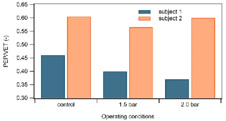
Fig. 12. PEP/VET ratio in different operating conditions 35
Fig. 13. Sleeves tires down the leg and one sleeve under the foot 38
Fig. 14. Solenoid valves and programmable logic controller 39
Fig. 15. Subject in the study wearing the automatic 42
Fig. 16. Time courses of cuff pressure in both legs 43
Fig. 17. Operator measuring cardiac volumes 46
Fig 18. Group response during the various phases of the study 49
List of abbreviations
ASIA American Spinal Injury Association
CO Cardiac output
DIMECA Department of Mechanical Engineering, Chemistry and Materials, University of
Cagliari
EDV End diastolic volume
HR Heart rate
LDL Low-density lipoprotein
LVPP Leg vein pressure pulser
MAP Mean arterial pressure
PEP Pre-ejection period
PEP/VET Pre-ejection period / Ventricular ejection time
PLC Programmable logic controller
POLITO Department of Mechanical and Aerospace of the Polytechnic of Turin
SCI Spinal cord injury
SD Standard deviation
SPLAB Laboratory of Sports Physiology
SV Stroke volume
SV/DT Stroke volume/Diastolic time
INTRODUCTION
There are a number of pathological conditions of the venous segment of the cardiovascular system leading to a venous insufficiency or a difficult return of blood from the extremities to the heart. Venous insufficiency results in a not complete filling of the heart during the diastolic phase, from which a reduced cardiac output (CO). This condition leads to a mismatch between oxygen demand and availability of the organs. Impairment of CO is often associated with a reduction quality of life.
Venous insufficiency of the lower limbs is more compromised respect to upper limbs, due to the antigravity direction of the blood under conditions of ambulation or orthostatic. In the health subject, this potential condition of venous insufficiency is effectively counteracted by the contraction of the muscles of the legs, especially the calf (triceps surae). Obviously, this compensation effect is particularly effective during walking which the rhythmic contraction of the calf compresses the large veins thus producing an increase in blood pressure. However, even in the absence of ambulation, muscle tone of the legs determines a sufficient pressure action on the antigravity venous vessels, thus avoiding situations of venous insufficiency.
The sensitive but efficient antigravity control system on the venous flow return to the heart is reduced, or is even missing in pathological conditions that compromise the contractile ability of the leg muscles such as, for example, in the case of spinal cord injury which follows a condition of paraplegia. Therefore, in these patients there is a serious need to recover the venous insufficiency and restore a normal diastolic function.
CHAPTER 1
CARDIOVASCULAR ADJUSTEMETS DURING EXERCISE IN HEALTHY SUBJECTS
Ventilatory and hemodynamic control of the circulatory apparatus is vital to guarantee enough oxygen supply to the organ. In particular, during physical activity cardiovascular and ventilator systems, increase their activity to satisfy the energy demand of the working muscle. At rest, the oxygen consumption of the body in normal subject is 250 ml∙min-1 with a cardiac output of 5 l∙min-1. During strenuous
aerobic efforts, endurance athletes can reach values of oxygen consumption of 6 l∙min-1 and the cardiac output value above 35 l∙min-1 (Lewis et al., 1983; Mitchell et
al., 1983).
Accordingly, the cardiovascular apparatus operates some important adjustments to enhance blood flow delivery, such as increase of heart rate (HR), stroke volume (SV) and cardiac output (CO).
Elevation of cardiac output represents a potential stressful mechanism for the heart if not balanced by other mechanisms. As result, reduction of peripheral vascular resistance decline by a release of end-products of muscle metabolisms thus maintaining mean arterial pressure stable or slightly increased respect to rest.
Mechanical and neural mechanisms both act in favor of cardiac filling during exercise. Rhythmic contraction of working muscles and respiratory activity move an important quantity blood from peripheral areas to the heart thus enhancing cardiac preload (i.e. muscle pump) and facilitating the Frank-Starling mechanism (Laughlin, 1987; Crisafulli et al, 2003b).
Regarding the neural regulation of the cardiovascular apparatus, both central and peripheral structures cooperate to regulate the cardiovascular response. “Central
command” and the “exercise pressor reflex” are involved in this complex regulation.
Central command is described as a feed-forward mechanism where descending signals from higher brain centers cause a parallel activation of motor and cardiovascular areas (located in the medulla) (Krogh et al., 1913; Goodwin et al. 1972). Thus, an increase in sympathetic activity leads to an increase of heart rate, stroke volume and mean arterial pressure response during voluntary contraction of the muscle. Sympathetic tone is also linked to the level of muscle activation (Strange et al. 1993, Thornton et al. 2002).
The exercise pressor reflex is peripheral mechanism evoked by the stimulation of free nervous fibers (i.e., group III and IV) in the muscle (McCloskey et al., 1972; Kaufman et al. 1984). While type III fibers are more sensitive to mechanical variations (i.e. mechano-receptors), type IV fibers mainly respond to the metabolic alteration (i.e. metabo-receptors), (Kaufman et al. 1984).
Therefore, when oxygen supply is not enough to satisfy the demand, accumulation of metabolites in the muscle stimulate the group III and IV, which subsequently activate cardiovascular control areas by a feedback reflex, i.e. the metabo-reflex. As consequence, an increase in sympathetic tone elevates arterial blood pressure by increasing cardiac output or systemic vascular resistance or even both (Bastos et al. 2000; Crisafulli et al 2003).
Recent works demonstrate that the activation of this system is dependent of muscle intensity (Crisafulli et al. 2006, 2008), thus, confirming that this system acts to corrects a possible mismatch between oxygen supply and demand. Therefore, the
activation of central command and exercise pressor reflex participate in the increase of cardiac output and cardiac contractility.
The increase of the sympathetic tone is continuously balanced by the
“arterial baro-reflex” that modulates muscle vasodilation (i.e. reduction of
peripheral vascular resistance) and heart chronotropism (i.e. reduction of heart rate) to contrast any excessive variations of blood pressure (Fadel et al. 2001; Sheriff 2006).
Recently many studies revealed that central command resets the level of activation of the arterial baro-reflex during exercise (Raven et al, 2002; Gallagher 2006).
As result, during exercise the sympathetic activity prevails respect to the parasympathetic in order to stimulate cardiovascular areas activity (Fig.1)
Fig.1. Illustraion of central peripheral mechianims regulating cardiovascluar response.
CHAPTER 2
BIOMECHANICS OF WALKING AND HIS HEMODYNAMIC EFFECT
To understand the mechanisms that underlie the leg vein pressure pulser (LVPP), i.e. the device which is the main target of this research, it may be useful to describe in detail the venous system of the lower limbs.
This consists of:
- deep and superficial veins, plantar (sole Lejars); - veins of the lower limbs deep and shallow.
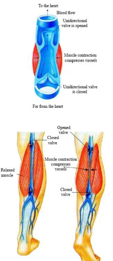
The foot, the ankle and calf muscles constitute a real pump “device” which gives a great help during the ascent phase of the deoxygenated blood to the heart. During walking, the bodyweight is downloaded to the forefoot and venous sole of Lejars: then follows a compression action to ensure a first movement of blood from the peripheral veins of the leg. During the plantar flexion-extension, the movement of the ankle and the resulting contraction of the calf muscles, gives further speed blood flow by exerting mechanical pressure on the veins, allowing a better rising to the cardiac pump. However, this operation must be supported by the presence of some valves placed on the veins and venules allowing a movement of the blood in only one direction.
In the complex biomechanics of walking several physiological actions are divided in two types of forces:
- aspirate forces, due to the action respiratory and muscle release; - driving forces, due to the residual pressure in the peripheral blood circulation, the so called “vis a tergo”, the arterial pulse adjacent to the veins,
the thrust and the plantar muscle contraction exerted by the muscles of the calf.
These forces act to varying degrees depending on the posture and are certainly limited or absent in patients with spinal cord injury.
In principle, in an upright position property, the pressure inside of the superficial veins of the lower limbs assumes a maximum value of about 90 mmHg: venous reflux disease is almost completely absent and therefore it may have conditions of venous dilation. Considering the supine position, the pressure decreases gradually, to equal the diameter of the veins of the lower limbs, as the angle of inclination of the limbs themselves with respect to the heart.
During walking the muscles of the calf implement the actions of rhythmic compression on the veins of these arts with the consequent activation of the one-way valves (Fig. 2). The return of blood to the heart is therefore favored in the supine position with legs raised or during the motion of the subject. The device developed is intended to act at the level of the lower limbs by imposing the actions of rhythmic compression combined with the forced plantar flexion-extension.
CHAPTER 3
SPINAL CORD INJURY, CAUSES AND PATHOPHYSIOLOGICAL ASPECTS
Spinal cord injury causes
The spinal cord is composed of a bundle of nerves that allows communication between the brain and the rest of the human body and vice-versa. When there is a partial or complete spinal cord lesion an interruption of information between the brain and the region of the body located below the lesion itself occurs.
Statistical investigations show that spinal cord injures interest’s predominantly young male adults. In USA the incidence is approximately 55 new cases per year/1 million inhabitants, of whom 35/1 one million survive. 80 % of the total cases comprise male patients while 80% of them are less than 40 years old.
In Italy the total population of paraplegic subjects during 2000, is about 60-70.000 people with an incidence of about 25 new cases per year/1 million inhabitants.
Spinal cord injury (SCI) can be classified into two main categories: - traumatic (75% of the cases);
- non-traumatic (25% of the cases);
In most cases, the traumatic events that lead to a SCI are caused by car crash, home and workplace accidents and sports injuries. Non-traumatic lesions are often caused by vascular disease or tumor which leads to the onset of infections and cysts.
In 1982 the American Spinal Injury Association (ASIA) defined the basic terminology for SCI, distinguishing between quadriplegia, paraplegia, neurological level, level of preservation of sensation, motor level, and skeletal or spinal injury incomplete, complete lesion. According to this, the neurological level of a spinal cord injury is identified by the caudal segment of the spinal cord, which preserves unchanged the capacity sensory and motor from both sides of the body.
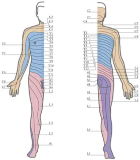
It is useful to explain the meaning of the following terms: myelomere and dermatome. Myelomere refers to each of the segments, in which the spinal cord can be divided with reference to the subdivision of the corresponding vertebrae. The dermatome identifies the portion of the surface of the skin innervated by a single sensory root of a spinal nerve; to each spinal segment corresponds then a dermatome.
The distribution of the segments is represented by bands transverse to the longitudinal trunk and limbs following the diagram in Fig. 3.
Pathophysiological aspects of spinal cord injury
Patients suffering from spinal injury can present numerous complications depending on the degree of injury and neurological level. In general, the most typical effects are:
- pulmonary complications;
- cardiovascular complications; - deep vein thrombosis;
- gastro-intestinal complications.
In particular patients with lesion of the bone have a decreased muscle mass, a reduction in venous return and lung function as well as an alteration of the homeostatic mechanisms of control. All this contributes to a change in the normal cardiovascular kinetics.
Clearly, muscle function is more reduced in subjects with the highest level of lesion with consequent reduction of physical activity, onset of obesity and increased risk of cardiovascular disease.
The programs for the rehabilitation of patients with SCI are highly personalized. These programs also have different influences: primarily it is the individual factors (age, weight, height, joint limitation) and environmental factors (family support, lifestyle, economic and employment conditions). The patient's rehabilitation program includes a series of measures aimed at prevention of pressure sores, respiratory complications.
CHAPTER 4
CARDIOVASCULAR ADJUSTMENTS DURING EXERCISE IN SPINAL CORD INJURED SUBJECTS
During physical tasks, optimal oxygen delivery to the muscle is a fundamental requirement of the cardiovascular system; in fact a deficiency of the last is detrimental for muscle performance.
Oxygen supply depends by cardiac output that is mainly influenced by cardiac preload (i.e. venous return) and cardiac contractility. Increase of both of these two variables participates in the increase of stroke volume (Laughlin, 1987; Crisafulli et al, 2003b). Central command, muscle pump and the exercise pressor reflex are the systems that participate in the increase of stroke volume, heart rate and as consequences cardiac output (Fadel, 2001).
In particular, individuals with chronic spinal cord injury (SCI) experience various kind of circulatory dysregulation, depending on the level of lesion in the spinal cord. According to this, cardiac output is strongly impaired in SCI individuals respect to able bodied subjects (Hopman et al. 1993a, 1993b,) due to a reduction of cardiac filling (i.e. reduced venous return).
This deficiency is caused by the inability to distribute blood during exercise below the level of lesion of the spinal cord (Hopman et al., 1993, 1998, Thijssen DH et al., 2009), thus causing an accumulation of blood in lower limbs and at splenic level (i.e. blood pooling phenomenon).
Two main factors have been associated with blood pooling: the first is the incapacity to recruit the muscle pump during exercise and the second is the lack of
sympathetic control below the spinal cord lesion. In particular, this last aspect has been recently elucidated by Crisafulli et al. (2009) where activation of the muscle metabo-reflex was reduced. In this work SCI individuals showed an impaired ventricular filling rate (VFR) in comparison with what was observed in able-bodied subjects during the metabo-reflex response. This was responsible for the reduced SV and CO of SCI persons compared with able-bodied individuals.
Because of these, regulation of cardiac output at peripheral level is blunted leading to a central limitation and may contribute to the increase of risk of cardiovascular disease in SCI patients.
Cardiovascular disease is often present in subject with SCI; in particular epidemiological studies reported these as major cause of death (DeVivo et al. 1993, 2002; Bauman et al., 1999). Left ventricular diastolic dysfunction caused by impairment of cardiac preload, often experience a cardiac remodeling in this population, leading to a decrease of ventricular mass and performance (Kessler et al., 1986; Nash et al., 1991).
In particular the cardiac structure is influenced by cardiac preload and systemic pressure, hence a reduction of these two lead to a decline and alteration of pump capacity of the heart associated with a decline of cardiovascular function (Cooper et al., 1992). In addition several studies reported atrophy and impairment of vascular reactivity tone in lower limbs because are extremely inactive.
However, regular training has the capacity to reverse in part negative effects of paraplegia. Most of the people who perform regular physical exercise, report some benefits such as: increase of VO2max (Cowell et al., 1986; Hoffman, 1986; Hooker et
cardiovascular function (Davis et al., 1987) and reduction of LDL concentration (Warburton et al., 2001, 2006).
CHAPTER 5
REHABILITATION DEVICES IN SPINAL CORD INJURIED POPULATION, STATE OF THE ART
In order to reduce impairment of venous return, numerous attempts have been developed to replace the muscle pump of lower limbs. Most of devices include anti-G suits (Hopman et al., 1992), anti-shock trousers, neuromuscular stimulation (Davis et al., 1990; Glaser et al., 1994) and pneumatic devices.
These seem particularly suited for rehabilitation due to their portability, low cost, high force-to-weight ratio and tailoring features. Reduction of the hemodynamic response can be observed in paraplegic patients; therefore a means to replace muscle action on leg in order to restore venous return when tonus is lost would be a challenging target. Restoring blood circulation efficiency in motion impaired patients can improve their general conditions.
Although the basic idea of stimulating upper body circulation by means of some leg and abdomen compression device is not new (Hills et al., 1972). Unfortunately commercial devices don’t always take into the due account the mechanism of physiological action, and are somehow not well focused on reproducing its effects. Some studies suggest that for individuals with spinal cord injury the use of devices that increase venous return to the heart could augment exercise capacity by preventing the redistribution of blood to the lower extremities (Bazzi et al., 1996; Pitetti et al., 1994).
Devices should be comfortable and safe, needing the design of soft and continuum robots lacking of rigid structures that could cause damage or pain
meanwhile resulting easy to control, design and build, which they are actually not. Their mechanical behavior and modeling features depend on the task they are designed for (Robinson and Davis, 1999; Cowan and Walker 2008; Gorissen et. al., 2011). Balloon actuators, which seem a natural choice for our requirements, are still very different in structure and working principle: some have been designed to handle sensors or end effectors, as trunk robots (Hannan and Walker, 2003, Chang and Menon 2012) with mainly bending features in one dimension, or bi-dimensional bending plates (Daerden et al., 2001); some can walk or crawl by axially stretching and outstretching (Manuello Bertetto and Ruggiu, 2004); others are meant for following curved paths, e.g. inspecting pipes for different applications, either medical or industrial (Manuello Berretto A. and M. Ruggiu, 2001). But while flexible robots are often equipped with rigid grippers or end effectors, only a few applications of continuum soft manipulators with swelling axial- symmetric characteristics (no beam-like bending behavior) are available.
Due to these considerations it is reasonable that external devices could improve diastolic function in this special population. This thesis tries to offer a new contribution in this field by investigating a novel approach to the problem. In this work we investigated the response of the cardiovascular system during the application of new robotic-pneumatic devices able to combine passive mobilization with a progressive increase of dynamic action to lower limbs.
The work is divided in two studies where our group from the Laboratory of Sports Physiology of the Medical Sciences Department, University of Cagliari (SPLAB), collaborated with the Department of Mechanical Engineering, Chemistry and Materials of the University of Cagliari (DIMECA) and the Department of Mechanical and Aerospace of the Polytechnic of Turin (POLITO).
While the two groups of engineers developed the two devices, our group was responsible to evaluate and validate the prototypes from a cardiovascular point of view.
This was possible thanks to our wide knowledge in the field, especially in the monitoring of the cardiovascular apparatus beat-to-beat by means of impedance cardiography (Concu A. et al. 1993; Crisafulli A. et al, 2003b, 2008).
Despite this is not the gold standard for hemodynamic assessment (Fick and the dye-dilution methods) this technique described afterwards, is able to monitor hemodynamic response non-invasively that is an important aspect if we consider some application in special population.
Furthermore, is able to detect accurately changes of systolic and diastolic events of the heart contrary to invasive techniques.
CHAPTER 6
STUDY 1 (DIMECA)
AN INFLATABLE PNEUMATIC SYSTEM FOR BLOOD PRESSURE RECOVERY
In this first study three toroidal balloons actuators each consisting of an inflatable bladder bounded by an inextensible fabric coat acting as and constrain has been chosen for its geometrical, mechanical, pneumatic and shape properties (Fig. 4).
In Fig. 4 is represented the LVPP robotic device prepared by the INGCA group (LVPP-Dimeca). The device is applied on a limb of a healthy subject and consists of 3 inflatable sleeves, in order to exert a pressure in a centripetal direction.
The sleeve (1) acts on the venous sole of the foot in order to recover the pressure coming from the ground during walking; the sleeve (2) is positioned so as to implement, during the stages of inflating and emptying, a bending plantale followed by a dorsiflexion of the ankle, thereby producing a rotation of the ankle in the sagittal plane so as occurs during the journey; the sleeve (3) produces radial compression of the calf simulating the pumping action on the veins of the leg exerted by the lateral and medial gastrocnemius muscles and soleus muscles during the elevation of the heel while walking.
The device, presented in previous works (Manuello and Meili 2011, Manuello and Meili 2012) has now been upgraded in order to perform a two-leg actuation and its controlling system revised and tuned.
Impedance cardiography technique has been successfully used (Concu et al., 1993) to get heart efficiency indexes and was adopted in this study to measure such parameters in working conditions. After giving a review of physiological and fundamentals underlying the idea, the system pneumatic scheme will be illustrated along with its timing diagram. Some encouraging results will finally be presented and discussed.
Technical aspects of device
The system was built connecting three sphygmomanometer cuffs for each leg into an air supplied circuit: one had to massage the patient’s foot sole, one his ankle and one his calf. A branch of the two symmetrical twin circuits is shown in Fig. 5.
Downstream the air supply (a) an air service unit (b) allows to fine tune the supplying pressure along with filtering inlet air. Six electro operated pneumatic flow control solenoid 3/2 valves (c) are piloted by a PLC (d) so as to intermittently inflate the cuffs’ bladders (f). Upstream each bladder an in-line unidirectional flow control regulator (e) helps smoothening the inflow pressure change allowing free outflow. In both the outflow branches collecting way 1 of each valve an in-line Venturi ejector (g) helps deflation, inflated by a flow regulator working as a check valve not to allow for outflow along the checked line.
Experimental procedure
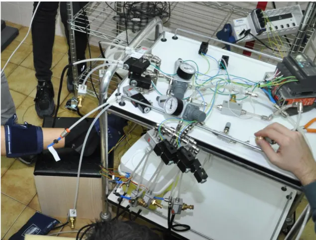
A group of eleven informed healthy people (between 20 and 30 years) underwent two cycles of four min respectively of intermittent leg massages (Fig. 6). Feeding air pressure upstream the system was tuned using the air service unit (b), and set on 1.5 bars at first, then raised to 2.0 bars. None had any history of cardiac or respiratory disease or was taking any medication at the time of the study. Each volunteer gave written consent to take part in this study after they had been given detailed information on the procedures and risks. Subjects were asked to refrain from caffeine, alcohol and physical activity for 12 h, and eating for 3 h, prior to the visit.
All experiments were carried out in a temperature-controlled room (room temperature set at 22º C, relative humidity between 40-50%). The study was performed according to the Declaration of Helsinki and was approved by the local ethics committee.
Pressure levels were chosen in preliminary tests so as not to result uncomfortable for the tested subject. Circuit pressure upstream each bladder was measured on one leg by means of three 0-15 psi Honeywell 24 PC Series piezo-resistive differential pressure gauges with 15 mV/psi sensitivity, one for each cuff, and acquired by means of a NI cDAQ-9172 CompactDAQ chassis, equipped with a NI9219 4-Channel, 24-Bit, analog input module. It is to be noted that operating pressure sensibly drops inside the bladders, reaching values about one order of magnitude smaller then supplying values. Activation was performed with the foot and calf cuffs for 3 min at about 0.3 Hz frequency (3.5 s period) and the on-off timing diagram imposed by the programmable logic controller (PLC) to the solenoids is shown in Fig. 7.
Hemodynamic data collection
Hemodynamic parameters were measured by means of impedance cardiography (NCCOM 3, BoMed, Irvine, CA), which allows continuous beat-to-beat cardio dynamic measure during exercise. The device was connected to the subject by arranging eight spot electrodes: two pairs were thoracic and cervical injecting electrodes, whereas two other pairs were sensing electrodes placed above the cervical and below the thoracic pairs (Fig. 8).
By means of a digital chart recorder (ADInstruments, PowerLab 8sp, Castle Hill, Australia), we stored NCCOM 3-derived analog traces of electrocardiogram, thorax impedance (Z0), and Z0 first derivative (dZ/dt).
Afterward, stored impedance traces were cleaned from signals affected by movement and respiratory artifacts and analyzed taking particular care to calculate hemodynamic variables only from traces not affected by impedance artifacts.
SV was assessed by using the Sramek-Bernstein equation:
where VEPT was the volume of electrical participating tissue and was derived using a normogram from sex, height, and weight of the subject; Z0 was the thorax
impedance measured at the end of cardiac diastole; dZ/dtmax was the maximal Z0 first
derivative during cardiac systole; and VET was the left ventricular ejection time, measured as the interval between the beginning and the minimum of the deflection in dZ/dt trace during systole.
Fig. 8. Impedance cardiography heart efficiency measurements. By means of disposable electrodes a constant intensity electrical current circulates into the
thorax. Changes in the bioelectrical impedance (Zt) are detected. They are proportional to left ventricle systolic flow.
Heart rate (HR) was calculated as the reciprocal of the electrocardiogram R-R interval, and CO was obtained by multiplying SV and HR. Also measured was the pre-ejection period (PEP), identified as the time interval between the electrocardiogram Q wave and the beginning of the dZ/dt deflection during systole. By calculating the PEP-to-VET (PEP/VET) ratio, we obtained an index inversely related to myocardial contractility.
(a)
(b)
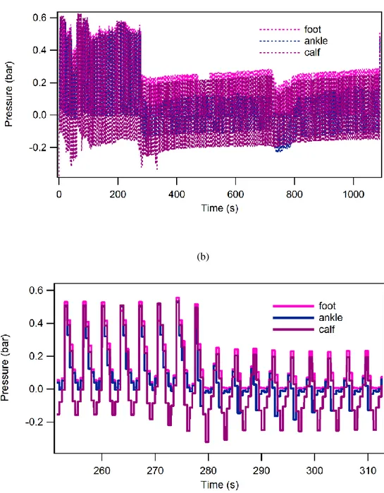
Fig. 9. Air pressure vs. time just upstream each cuff bladder: (a) global trend ad (b) detail.
Results
A sample of acquired circuit pressure values is shown in Fig. 9. The shown trend highlights some circuit features. First, the ejectors’ efficiency, which during deflation brings about a negative relative pressure mostly visible in the calf bladder curve, is stressed with increasing operating pressure, finally overcoming the inflating action and resulting in lesser an average effective value;
Second, the three bladders pressure peak values appear to be in phase despite the solenoid valve on-off law shown in Fig. 5 gives out-of-phase commands to them. This can be explained with the really slow circuit dynamics;
Third, a much less steep slope of pressure rate has been achieved thanks to the unidirectional flow regulators; it’s worth noting that their check valve prevents air from flowing back in inlet pipe.
In Fig. 10-12 the two typical extreme behaviors (i.e. subject 1 and subject 2) for hemodynamic adjustments observed within the tested group are shown and compared. In Fig. 10 (subject 1) SV values before testing (control) and during the device performance are displayed. Actuation was exerted at 1.5 bar and 2.0 bar upstream the system, and respective values are shown in turn: the 1.5 bar actuation causes an 8% increment while at 2 bar the SV has a further increase to 30% the control value.
In Fig. 11 (subject 1) it is clearly seen how these SV increments, caused by the rhythmic foot-calf compression, are dependent on the progressive, pressure-dependent increase in diastolic filling rate SV/DT: this hemodynamic variable, describing VP, went up to 3% at 1.5 bars and reached 19% at 2.0 bars. Fig. 12 shows the PEP/VET ratio going for subject 1 to -13% its control value at 1.5 bars and getting a further 19% reduction at 2.0 bar supplying pressure.
Fig. 10 above for subject 2 behavior, the graph shows that supplying the system at just 1.5 bar induced in the subject 2 a 10% SV increment. A reverse trend was conversely observed when supplying at 2.0 bars: SV got a negative drop i.e. -3% of the control value.
The reason for this behavior can be explained by means of Fig. 11 where SV/DT value decreases by 12% at 2.0 bar actuation, but for just 1.5 bar it increases of 2% of control value, according to SV trend as above except for the reverse slope.
Considering that intra-ventricular pressure rate depends upon pre-systole stretching degree of myocardic fibers (according to the Frank-Starling) we can also explain the PEP/VET 7% reduction (Fig. 12) as myocardic fiber length reaches its relative maximum with MC increasing as well, whereas for less stretched fibers (i.e. 2.0 bar) PEP/VET gets back to -2%, about the control value.
Fig. 10. Stroke Volume (SV) values for two tested subjects at different supplying pressure.
Is possible that in these subjects, due to a higher compliance of the venous limb segment, an increase of compression of only 0.5 bar (in step 1.5 to 2.0 bar), caused under the mechanical intervention, an important increase in distention of the venous and a greater patency of collateral vessels, producing a kind of venous blood pooling, from which the paradoxical reduction of SV/DT.
CHAPTER 7
STUDY 2 (POLITO)
AN INFLATABLE PNEUMATIC SYSTEM FOR BLOOD PRESSURE RECOVERY
Study population
A group of 19 male healthy subjects aged between 20 and 35 years were recruited, none had any history of cardiac or respiratory disease or was taking any medication at the time of the study. Each volunteer gave written consent to take part in this study after they had been given detailed information on the procedures and risks. Subjects were asked to refrain from caffeine, alcohol and physical activity for 12 h, and eating for 3 h, prior to the visit.
All experiments were carried out in a temperature-controlled room (room temperature set at 22º C, relative humidity between 40-50%). The study was performed according to the Declaration of Helsinki and was approved by the local ethics committee.
Device description
The device was composed by two mechanical actuators made by flexible sleeves to be applied on each leg around foot and leg (Fig. 13). Each sleeve was composed by five inflatable cuffs placed in succession from ankle to knee acting against the triceps surae, while the one placed on the foot mainly acted against the feet sole. The sleeves were inflated with compressed air by means of solenoid valves controlled by a programmable logic controller (Fig. 14).
Fig. 14. (A) solenoid valves; (B) programmable logic controller.
A B
According to this, was possible to impose against the lower limb, different kind of protocols in terms of pressure and spatial-time distribution. All of the protocols involved different sequences of activation-deactivation following a peristaltic compression, having a rostral-caudate trend, with compression of 0.5 bars. The logical scheme was to alternate the activation of the two sleeves to simulate the muscle patter during walking.
Experimental design
Before monitoring the cardiovascular response during the activation of the device, each subject was monitored at rest for 3 min to collect baseline values.
Then, the device was placed to the subject to collect the data (Fig. 15 A). Once the preparation was completed, all volunteers underwent three different protocols, respectively called A, B and C (Fig. 16).
Protocol A. The time of activation was the same for each cuff; the delay between
each was 3 s. The cycle lasted for 18 s. No delay was given between each sleeve. The entire cycle including both sleeves was 36 s kept constant at 0.5 bars?
Protocol B. The time of activation was the same for each cuff; the delay between
each was 1.5 s. The cycle lasted for 10.5 s. No delay was given between each sleeve. The entire cycle including both sleeves was 21 s kept constant at 0.5 bars?
Protocol C. The time of activation for each cuff was: 6 s (cuff 1), 5 s (cuff 2), 4 s
(cuff 3), 4 s (cuff 4), 4 s (cuff 5) and 4 s (cuff 6) with a delay between each of 1 s. The cycle lasted for 10.5 s with a delay of 3 s between the two sleeves. The entire cycle including both sleeves was 27 s with an inflating pressure between 0.3 and 0.5 bar.
On each thigh we placed an elastic sleeve to reduce the venous distention caused by the increase of blood flow during the mechanical actuation (Fig. 12 B). All the four phases (i.e. baseline and three protocols) lasted 3 min each.
Fig. 15. One of the subject in the study wearing the automatic device (A) and elastic sleeve (B). Echocardiographer (C) and impedance cardiometer (D) placed on the
neck and chest can be seen during data collection.
A B
C D
Hemodynamic data
Hemodynamic variables were monitored and recorded beat-to-beat by means of impedance cardiography (NCCOM 3, BoMed Inc., Irvine, CA) which allows a continuous noninvasive hemodynamic assessment throughout all the phases of the protocol.
The device was connected to the subject by eight electrodes: two pairs were thoracic and cervical injecting electrodes, whereas two other pairs were sensing electrodes placed above the cervical and below the thoracic pairs (Fig. 12 D). The signal was recorded by a digital chart recorder (AD Instruments, PowerLab 8sp, Castle Hill, Australia), we stored NCCOM 3-derived analog traces of electrocardiogram, thorax impedance (Z0), and Z0 first derivative (dZ/dt). SV was
assessed by using the Sramek-Bernstein equation:
where VEPT was the volume of electrical participating tissue obtained by a normogram from sex, height, and weight of the subject; Z0 was the thorax impedance
measured at the end of cardiac diastole; dZ/dtmax was the maximal Z0 first derivative
during cardiac systole; and VET was the left ventricular ejection time, measured as the interval between the beginning and the minimum of the deflection in dZ/dt trace during systole. Heart rate (HR) was calculated by R-R interval, and CO was the product between SV and HR. Pre-ejection period was estimated as the time interval between the electrocardiogram Q wave and the beginning of the dZ/dt deflection
during systole. The ratio between PEP/VET was used as index of myocardial cardiac contractility.
During the post analysis all artifacts (i.e. movement and respiratory signals) were removed from the recordings and then manually analyzed by the same operator to obtain estimation of hemodynamic data.
A four chamber view was used to monitor variations of the cardiac chambers during heart cycle (Fig. 14). All measurements were performed by means of bi-dimensional echocardiography (Mindray M5, Mindray DS USA, Inc.) at the end of all phases by the same operator (Fig. 12 C). During the post analysis all images recorded were analysed by the same operator to evaluate end diastolic volume (EDV).
Data and statistical analysis
Data are presented as mean ± standard deviation (SD). The normality assumption was checked using the Kolmogorov–Smirnov test.
Hemodynamic data were averaged for three min during all four phases. One-way ANOVA for repeated measures followed by Bonferroni’s post hoc test were employed to compare the mean group values of SV, HR, CO, SV/TD, PEP/VET and EDV during rest, A, B and C phases. Significance was set at p<0.05 in all cases. Statistics were carried out using commercially available software (GraphPad Prism, GraphPad Software, San Diego, CA, USA).
Results
All subjects completed the study protocol. Fig. 18 (or table 1) shows mean group ± SD response of SV (panel A), HR (panel B), CO (panel C), PEP/VET (panel D), EDV (panel E) and SV/TD (panel F) during all the phases of the study.
SV (panel A) increased significantly during A respect to rest condition (71.81 ± 12.56 vs. 75.88 ± 13.32) while C was higher compared to rest and B (72.42 ± 13.77 vs. 72.39 ± 11.33). HR (panel B) didn’t exhibit any statistical difference during all the phases. CO (panel C) rose significantly only during C respect to rest condition (7.34 ± 1.84 vs. 6.75 ± 2.17). PEP/VET (panel D) decreased significantly during A, B and C respect to rest condition (0.66 ± 0.10, 0.67 ± 0.07, 0.63 ± 0.08 vs. 0.71 ± 0.09) whereas C was significantly lower compared to B (0.63 ± 0.08 vs. 0.67 ± 0.07). EDV (panel E) reported a significant increase only during phase C compare to rest condition (119.00 ± 26.41 vs. 105.00 ± 22.15). SV/DT (panel F) didn’t exhibit any statistical changes during all the phases.
Table and figures
Table 1. Variables during rest and protocol A, B and C.
rest Protocol A Protocol B Protocol C
HR (bpm) 75.88 ± 13.32 71.81 ± 12.56 72.39 ± 11.33 72.42 ± 13.77 SV (ml) 89.27 ± 26.62 99.12 ± 28.13* 93.84 ± 26.29 102.80 ± 27.48*# CO (l·min-1) 6.75 ± 2.17 7.03 ± 2.14 6.95 ± 2.16 7.34 ± 1.84* PEP/VET 0.71 ± 0.09 0.66 ± 0.10* 0.67 ± 0.07* 0.63 ± 0.08*# SV/DT 232.50 ± 86.11 234.00 ± 82.72 233.50 ± 86.15 247.4 ±80.26 EDV (ml) 105.00 ± 22.15 115.80 ± 20.12 114.20 ± 21.80 119.00 ± 26.41* Values are means ± SD during rest and A, B and C protocol of heart rate (HR), stroke volume (SV), cardiac output (CO), pre-ejection period/left ejection time ratio (PEP/VET), stroke volume/diastolic time ratio (SV/TD) and end diastolic volume (EDV). * p<0.05 respect to rest. # p<0.05 respect to B.
SV Rest A B C 0 40 80 120 160 * * # ml HR Rest A B C 0 20 40 60 80 100 bpm CO Rest A B C 0 4 8 12 * l m in -1 PEP/VET Rest A B C 0.0 0.2 0.4 0.6 0.8 1.0 * * *# EDV Rest A B C 0 40 80 120 160 * ml SV/DT Rest A B C 0 100 200 300 400
A
B
D
F
C
E
Fig. 18. Group response during the various phases of the study. Values are presented as mean ± SD (n=19). * p<0.05 respect to rest. # p<0.05 respect to B.
CHAPTER 8
DISCUSSION
Results show that the application of robotic mechanical actuators pneumatic, as our LVPP, is able to generate cycles of compression and decompression on venous structures of the lower limbs similar to what normally happens in the skeletal muscle when subjects are walking.
Especially in the second study where we simulated the mechanics of walking by applying the cuffs in both limbs and activating alternatively in ascendant mode, we were able to further increase CO and EDV.
This finding is very important, because contrary to what developed before in previous studies, we were able not only to increase cardiovascular response but we simulated quite closely the biomechanical pattern of muscle recruitment.
In fact, our prototype mainly differed because implied not only a passive constriction of lower extremities but was able to move the lower extremities thus partially restore muscle pump. Another interesting aspect is that, the operator can change the intensity and the kind of activation (intensity and time of occlusion), thus probably further improving the positive effect.
It has to be taking in account that the experimental groups recruited in the two experiments are healthy subjects with functionally innervated muscles voluntary released. Results however are important and promote a transfer to SCI individuals in order to limit the failure of venous return.
Probably by applying these prototypes we might improve the physical capacity of the patients and hence also their quality of life.
Furthermore, the results of the assessments made by SPLAB robotic actuators, prepared respectively by DIMECA and POLITO year highlighted the possibility to differentiate the therapeutic in two ways: 1) implementation external pressure on the veins of the legs, type explosive, similar to the fast and powerful contraction of the elevator muscles of the calcaneus (the triceps: the surae) while jumping or running: see the prototype prepared by INGCA, 2) implementation pressure more gradual and prolonged the same muscles, comparable to that of the road at low speed and parallel postural adjustments: see the prototype prepared by POLITO.
In conclusion our preliminary finding suggest that is possible to compensate partially the lack of venous return in SCI population thus increasing the quality of life, reducing the risk of cardiovascular disease and then extend their life expectancy.
Study limitations
In our devices LVPP, suitable for producing a pressing action on the lower limbs with the objective of compensating the venous insufficiency, the type of energy transferred inside of the extensible socks by a compressor, is partially dispersed to deform the socks themselves, leading to a considerable reduction of the pressure still available. Moreover, only a part of the residual pressure arrives at the leg perpendicular direction (that is effective), to the area of skin affected because a good part of it assumes other directions resulting in part or totally ineffective in the action pressure on the limb. So, is not possible to know exactly to know which the actual pressure that compress is. This represents a limit of therapeutic effectiveness and safety that is scarcely reliable are devices on the market. In fact, if on the one hand a certain value of externally applied pressure may be totally ineffective due to
excessive fall of energy along the path of compression, on the other hand it might even be excessive and hence harmful because of tissue lesions that may cause.
Future Developments
The research group headed by SPLAB is finalizing a system of monitoring and control of actuators LVPP in closed loop with a two-level hierarchy Master & Slave control strategy.
The highest level, Master, of closed loop control will be able to define the profile of actions imposed by the actuators, through the pressure of the fluid, as a function of the magnitudes of clinical reference (i.e. non-invasive hemodynamic variables). In correspondence with the clinical conditions will set the profiles of the actuators. The main purpose of a dual level of control, Master & Slave, hierarchically organized is to allow a periodic clinical setting system in the hospital and then to be used at home. This strategy enables the daily use of a lightweight periodically according to the clinical variables. In addition, this strategy allows a complete customization of the device in terms of control and needs of each patient.
In order to achieve the Master-slave control system, in an innovative way sensors will be used to contact pressure at the interface actuators-skin, as an integral part of the control system. Such pressure sensors tactile with appropriate dynamic characteristics will be able to perform a dynamic measurement of forces/pressures between the two surfaces in contact (i.e. the outer surface of the sock and the skin).
The geometrical distribution of pressure-time on the skin of the limb in question can be modulated by the action of the flexible actuators for the purpose of restoring the diastolic function of the heart.
The control strategies that will be used will neural type as it is believed that they are particularly suitable to the scope in which the mechatronic system is built to operate. The tactile sensors generate a signal which is suitably conditioned by a transducer to make it readable by electronic devices to microcontroller. The interfacing of the latter to other devices of implementation allows to generate, for example with the use of neural networks, the behaviors that follow specific laws to be adopted on a case by case basis. Note the pressure exerted by the device on the skin of the limb, it may generate more pressure, flow rate and frequency that will act on the control valves of the actuators, at the level of Slave controller.
The considerations on hierarchical control and action strategies above show geometric-time show that the contact pressure sensors play a vital role in the effectiveness of the system in order to achieve the objective of an effective recovery circulatory pursued with the type of mechatronic devices those that will be developed in the project.
REFERENCES
1. B. Chang, A. Chew, N. Naghshineh and C. Menon (2012). A spatial bending fluidic actuator: fabrication and quasi-static characteristics, Smart Mat and
Struct. Vol. 21, pp. 1-7;
2. Bastos GB,Williamson JW, Harrelson T & Nˆobrega ACL (2000). Left ventricular volumes and hemodynamic responses to postexercise ischemia in healthy humans. Med Sci Sports Exerc. 32, 1114–1118;
3. Bauman WA, Adkins RH, Spungen AM, Herbert R, Schechter C, Smith D, Kemp BJ, Gambino R, Maloney P, Waters RL (1999). Is immobilization associated with an abnormal lipoprotein profile? Observations from a diverse cohort. Spinal Cord. Jul;37(7):485-93.
4. Bauman WA, Spungen AM, Adkins RH, Kemp BJ (1999). Metabolic and endocrine changes in persons aging with spinal cord injury. Assist Technol. 11: 88-96;
5. Bazzi-Grossin, C., P. Bonnin, O. Bailliart, H. Bazzi, W. A. Kedra, and J.P. Martineaud (1996). Maximal exercise in spinal cord injured subjects: effects of an antigravity suit. Sci Sports. Vol. 11, pp.173-179;
6. Concu A. and C. Marcello (1993). Stroke volume response to progressive exercise in athletes engaged in different types of training. Eur J Appl Physiol. Vol. 66, pp:11-17;
7. Cooper IV G, Tomanek RJ. (1982). Load regulation of the structure, composition, and function of mammalian myocardium. Circ Res.50:788-798. 8. Cowan, L.S. and A. Walker (2008). “Soft” Continuum Robots: the Interaction of Continuous and Discrete Elements, Artificial Life, Vol. 11, pp. 126, 133.;
9. Cowell LL, Squires WG, Raven PB (1986). Benefits of aerobic exercise for the paraplegic: a brief review. Med Sci Sports Exerc; 18: 501-8;
10. Crisafulli A, Milia R., Lobina A., Caddeo M., Tocco F., Concu A. (2008). Haemodynamic effect of metaboreflex activation in men after running above and below the velocity of the anaerobic threshold. Exp Physiol. 93.4 pp 447– 457;
11. Crisafulli A, Orrù V, Melis F, Tocco F, Concu A (2003b). Hemodynamics during active and passive recovery from a single bout of supramaximal exercise. Eur. J. Appl. Physiol. 89: 209-216;
12. Crisafulli A, R. Milia, S. Vitelli, M. Caddeo, F. Tocco, F. Melis and A. Concu (2009). Hemodynamic responses to metaboreflex activation: insights from spinal cord-injured humans, Eur J Appl Physiol. Vol. 106, pp. 25-33; 13. Crisafulli A, Salis E, Pittau G, Lorrai L, Tocco F, Melis F, Pagliaro P &
Concu A (2006b). Modulation of cardiac contractility by muscle metaboreflex following efforts of different intensities in humans. Am J
Physiol Heart Circ Physio. 291, H3035–H3042;
14. Crisafulli A, Scott AC, Wensel R, Davos CH, Francis DP, Pagliaro P, Coats AJS, Concu A, and Piepoli MF. Muscle metaboreflex-induced increases in stroke volume. Med Sci Sports Exerc. 35: 221–228, 2003;
15. Crisafulli, A., A.C. Scott, R. Wensel, C.H Davos, D.P. Francio, P. Pagliaro, A.J.S. Coats, A. Concu and M.F. Piepoli. (2003). Muscle Metaboreflex-induced Increases in Stroke Volume, Med Sci Sport Exe. Vol. 35, pp. 221-228.
16. Daerden, F., D. Lefeber, B. Verrelst and R. Van Ham (2001). Plated pneumatic artificial muscles: actuators for automation and robotics. IEEE -
ASME Int. Conf. On Advanced Intelligent Mechatronics, Como (Italy) July
8-12;
17. Davis GM, Shephard RJ, Leenen FH. (1987). Cardiac effects of short-term arm crank training in paraplegics: echocardiographic evidence. Eur J Appl
Physiol.; 56: 90-6;
18. Davis, G. M., Servedio, F. J., Glaser, R. M., Gupta, S. C. & Suryaprasad, A. G. (1990). Cardiovascular responses to arm cranking and FNS-induced leg exercise in paraplegics. J Appl Physiol. 69: 671–677,
19. DeVivo MJ, Black KJ, Stover SL (1993). Causes of death during the first 12 years after spinal cord injury. Arch Phys Med Rehabil. Mar;74(3):248-54; 20. DeVivo MJ, Go BK, Jackson AB (2002). Overview of the national spinal
cord injury statistical center database. J Spinal Cord Med; 25: 335-8;
21. Fadel PJ, Ogoh S, Watenapaugh DE, Wasmund W, Olivencia-Yurvati A, Smith ML, Raven PB (2001). Carotid baroreflex regulation of sympathetic nerve activity during dynamic exercise in humans. Am J Physiol Heart Circ
Physiol. 280: H1383-H1390;
22. Gallagher KM, Fadel PJ, Smith SA, Morten Strømstad M, Kojiro Ide I, Secher NH. and Raven PB. Neural Control of the Circulation During Exercise - The interaction of central command and the exercise pressor reflex in mediating baroreflex resetting during exercise in humans. Exp Physiol. 91.1 pp 79–87 79;
23. Glaser, R. M. (1994). Functional neuromuscular stimulation: exercise conditioning of spinal cord injured patients. Int J Sport Med. 15: 142–148; 24. Goodwin GM, McCloskey DI & Mitchell JH (1972). Cardiovascular and
exercise at constant muscle tension. J Physiol. 226, 173–190;
25. Gorissen, B., M. De Volder, A. De Greef and D. Reynaerts (2011). Theoretical and experimental analysis of pneumatic balloon microactuators.
Sens. Actuators A. Vol. 168, pp.58-65.
26. Hannan, M. W. and I. D. Walker (2003). Kinematics and the implementation of an elephant’s trunk manipulator and other continuum style robots, J of
Robotic Syst. Vol. 20, No. 2, pp. 45-63;
27. Hoffman MD (1986). Cardiorespiratory fitness and training in quadriplegics and paraplegics. Sports Med. 3: 312-30.
28. Hooker SP, Wells CL (1989). Effects of low- and moderate-intensity training in spinal cord-injured persons. Med Sci Sports Exerc; 21: 18-22;
29. Hopman MT, Monroe M, Dueck C, Phillips WT, Skinner JS (1998). Blood redistribution and circulatory responses to submaximal arm exercise in persons with spinal cord injury. Scand J Rehabil Med. 30:167–74;
30. Hopman MT, Pistorius M, Kamerbeek IC, Binkhorst RA (1993). Cardiac output in paraplegic subjects at high exercise intensities. Eur J Appl Physiol
Occup Physiol.;66:531–5;
31. Hopman MT, Verheijen PH, Binkhorst RA (1993). Volume changes in the legs of paraplegic subjects during arm exercise. J Appl Physiol.;75:2079–83; 32. Hopman MT, Verheijen PH, Binkhorst RA (1993). Volume changes in the
legs of paraplegic subjects during arm exercise. J Appl Physiol. 75:2079–83; 33. Hopman, M. T. E., Oeseburg, B. & Binkhorst, R. A. (1992). The effect of an
anti-G suit on cardiovascular responses to exercise in persons with paraplegia. Med Sci Sports Exerc. 24: 984–990,
Effects of static and rhythmic twitch contractions on the discharge of group III and IV muscle afferents. Cardiovasc Res. 18, 663–668;
35. Kessler KM, Pina I, Green B, Burnett B, Laighold M, Bilsker M, Palomo AR, Myerburg RJ. SL (1986). Cardiovascular findings in quadriplegic and paraplegic patients and in normal subjects. Am J Cardiol. Sep 1;58(6):525-30;
36. Krogh A & Lindhard J (1913). The regulation of respiration and circulation during the initial stages of muscular work. J Physiol. 47, 112–136;
37. Laughlin MH (1987). Skeletal muscle blood flow capacity: role of muscle pump in exercise hyperemia. Am. J. Physiol. 253: H993-H1004;
38. Lewis SF, Taylor RM, Graham RM, Pettinger WA, Schutte JE, Blomqvist CG (1983). Cardiovascular responses to exercise as functions of absolute and relative work load. J. Appl. Physiol. 54: 1314-1323;
39. Manuello Berretto A. and M. Ruggiu (2001). In-pipe inch-worm pneumatic flexible robot, 2001 IEEE-ASME International Conference on Advanced
Intelligent Mechatronics Proceedings, 6-12 July Como, Italy, pp. 1226-1231.
40. Manuello Bertetto A., S. Meili, A. Concu and A. Crisafulli (2011). Flexible Pneumatic Actuation For Blood Pressure Recovery, Proc. Of Musme, The
International Symposium On Multibody Systems And Mechatronics, Valencia, Spain. 25-28 October, pp. 359- 370.
41. Manuello Bertetto A., S. Meili, A. Concu and A. Crisafulli (2012). An inflatable pneumatic device for blood pressure recovery. Mech bas des of
struct and mach. Volume 40, Number 4, 1 October 2012 , pp. 506-518(13);
42. Manuello Bertetto, A., M. Ruggiu (2004). A Novel Fluidic Bellows Manipulator. J of Rob and Mech. Vol.16, No.6, pp. 604-612;
43. McCloskey DI & Mitchell JH (1972). Reflex cardiovascular and respiratory response originating in exercising muscle. J Physiol. 224, 173–186;
44. Mitchell JH, Kaufman MP, Iwamoto GA (1983). The exercise pressor reflex: its cardiovascular effects, afferent mechanisms, and central pathways. Ann.
Rev. Physiol. 45: 229-242;
45. Nash MS, Bilsker S, Marcillo AE, Isaac SM, Botelho LA, Klose KJ, Green BA, Rountree MT, Shea JD (1991). Reversal of adaptive left ventricular atrophy following electrically-stimulated exercise training in human tetraplegics. Paraplegia. Nov;29(9):590-9.
46. Pitetti, K. H., P. J. Barrett, K.D. Campbell and D.E. Malzahn (1994). The effect of lower body positive pressure on the exercise capacity of individuals with spinal cord injury. Med Sci Sports Exerc, Vol. 26, pp. 463-468.
47. Raven PB., Fadel PJ and Scott A. Smith SA (2002). The influence of central command on baroreflex resetting during exercise. Exerc Sport Sci Rev. Jan;30(1):39-44;
48. Robinson, G. and J.B.C. Davies (1999). Continuum robots - a state of the art,
Proceedings 1999 IEEE International Conference on Robotics and Automation, Detroit (Michigan). May, pp. 2849, 2854;
49. Sheriff DD (2006). Baroreflex resetting during exercise: mechanisms and meaning. Am J Physiol Heart Circ Physiol. 290: H1406-H1407;
50. Strange S, Secher NH, Pawelczyk JA, Karpakka J, Christensen NJ, Mitchell JH, Saltin B (1993). Neural control of cardiovascular responses and of ventilation during dynamic exercise in man. J. Physiol. (Lond.) 470: 693-704; 51. Thijssen DH, Steendijk S, Hopman MT (2009). Blood redistribution during exercise in subjects with spinal cord injury and controls. Med Sci Sports
Exerc. Jun;41(6):1249-54;
52. Thornton JM, Aziz T, Sclungam D, Paterson DJ (2002). Electrical stimulation of the midbrain increases heart rate and arterial blood pressure in awake humans. J. Physiol. 539: 615-621;
53. Warburton DE, Gledhill N, Quinney A. (2001). The effects of changes in musculoskeletal fitness on health. Can J Appl Physiol.; 26(2):161–216; 54. Warburton DE, Nicol CW, Bredin SS. (2006). Health benefits of physical