[page 4] [Hematology Reports 2017; 9:6859]
Hemophagocytic
lymphohistiocytosis, an unclear
nosologic entity:
case report of an adult
man with rising of amylase
and lipase and spinal
cord infiltration
Moris Sangineto, Antonio Perrone, Pasquale Agosti, Viera Boccuti, Anna Campobasso, Carlo Sabbà Department of Interdisciplinary Medicine, University of Bari “Aldo Moro”, Bari, Italy
Abstract
Here we present the case of a 57-years old patient affected by hemophagocytic lymphohistiocytosis (HLH), a rare disease characterized by an uncontrolled immune activation, resulting in clinical and bio-chemical manifestations of extreme inflam-mation. In a previous hospitalization, the patient showed fever, hepato-splenomegaly, pancytopenia, hyperferrtitinemia, lym-phadenopathy and cholestasis. No diagnosis was done, however, he totally recovered after splenectomy. Eight months later, he relapsed, showing also hypofibrinogene-mia, hypertriglyceridehypofibrinogene-mia, hemophagocytic signs in bone marrow, cholestatic jaundice, high LDH and high PT-INR. Interestingly, he presented increased levels of amylase and lipase in absence of radiologic signs of pancreatitis. He was treated with Dexamethasone and Cyclosporine accord-ing to HLH-2004 guidelines. The clinical and biochemical manifestations disap-peared in a few weeks, but he was newly hospitalized for lower limbs hypotonia caused by a hemophagocytic lesion of the cauda equina and lumbar cord. The death occurred in a few days, despite the immuno-suppressive treatment.
Introduction
The histiocytic cells or macrophages are innate immune cells characterized by a strong phagocytic activity, deriving from maturation and tissue migration of the cir-culating monocytes of bone marrow monoblastic origin. The proliferative disor-ders involving histiocytes or histiocyte-like cells are named histiocytic syndromes. Different classifications have tried to delin-eate these different and complex entities. The Komp-Perry classification in 1991,1
considered the histiocytosis of Langherans cells in the first class, the hemophagocytic syndromes in the second one and the malig-nant histiocytosis including the histiocytic lymphoma in the third one. Similar was the more recent Favara classification (1997)2
that distinguished the histiocytic disorders into histiocytosis of dendritic cells, histio-cytosis of macrophages and malignant ones, according to the specific immunohisto-chemical features. A further clarification is needed for the true histiocytic lymphoma that appeared also in Rappaport classifica-tion of lymphomas in 1966,3but according
to the WHO classification (2008),4the last
currently accepted, it is no longer recog-nized as an isolated entity and it is included in B diffuse large cell or anaplastic T lym-phomas. In the context of the histiocytic disorders involving the monocyte-macrophage line cell, a rare and often lethal disease is the hemophagocytic lymphohisti-ocytosis (HLH), characterized by a cytokine explosion secondary to an excessive but ineffective immune response with a conse-quent abnormal activation of CD8+ T lym-phocytes, resulting into INFɣ release, monocyte-macrophages activation and infiltration in tissues. The result is multior-gan failure. It is distinguished by a primitive form, typical of childhood (familiar) and a secondary form related to infections, autoimmune diseases or neoplasms (lym-phomas and solid tumors). However, in a significant percentage of HLH in adulthood no cause is identified (idiopathic form). In 2004, clinical and laboratory criteria neces-sary for HLH diagnosis and a recommended therapeutic protocol have been proposed5.
Here we describe the case of an adult patient with the diagnostic criteria for HLH characterized by a very aggressive and neo-plastic-like behavior.
Case Report
A 57 years old man was hospitalized in February 2015 in our Internal Medicine Division due to fever and pancytopenia. He was in a healthy condition until one year before (February 2014) when he was hospi-talized for unknown origin fever (FUO). On that occasion the total-body computerized axial tomography (CAT) showed a medi-astinum lymphadenopathy, a hepatomegaly with normal bile ducts and a splenomegaly (maximum diameter 18 cm). The bone mar-row histology detected an aspecific mild tri-linear hyperplasia, signs of dyserythro-poiesis, slight excess of lymphoid cells with cytotoxic phenotype; the axillary lymph node biopsy showed a fatty involution. Because of worsening pancytopenia and
cholestasis appearance he underwent splenectomy and liver biopsy. Histological exams showed a splenic tissue with a T cytotoxic lymphocyte proliferation without immunohistochemical and morphological malignancy signs, while the hepatic parenchyma was characterized by plasma-cells and eosinophils, areas of spotty necro-sis and Mallory bodies. After splenectomy and a therapy based on large spectrum antibiotics and immunoglobulins, in a few weeks the patient made a total recovery, when the fever disappeared and the blood examinations normalized. Eight months later (February 2015), at admission to our Division he presented a blood pressure of 100/50 mmHg, a heart frequency of 82 per minute, body temperature 38.7°C, mucocu-taneous pallor, a diffused reduction of vesicular murmur and the liver was palpa-ble 3 cm from costal margin. Blood tests were as follow: hemoglobin (Hb) 9.6 g/dL, red blood cells (RBC) 3.25×10⁶/µL, white
blood cells (WBC) 2.11×10³/µL with 2.8%
of neutrophils and 87% of lymphocytes, platelets (PLT) 22×103/µL, ferritin 17813
ng/mL (n.v. 8-252 ng/mL), albumin 2.3 g/dL, LDH 541 U/L (n.v. 84-246 U/L), total bilirubin (T-bil) 2 mg/dL (direct 1.43 mg/dL), AST 108 U/L (n.v. 15-37 U/L), ALT 130 U/L (n.v. 12-78 U/L), gammaglu-tamyltranspeptidase (GGT) 109 U/L (n.v. 5-55 U/L), alkaline phosphatase (ALP) 204 U/L (n.v. 50-136 U/L), PT-INR 1.15, aPTT
Hematology Reports 2017; volume 9:6859
Correspondence: Moris Sangineto, Department of Interdisciplinary Medicine, University of Bari “Aldo Moro”, Piazza Giulio Cesare 11, 70124 Bari, Italy. Tel.: +39.346.0137005.
E-mail: [email protected] Key words: Hemophagocytic Lymphohistiocytosis, spinal cord, amylase and lipase, splenectomy.
Contributions: the authors contributed equally. Conflict of interest: the authors declare no potential conflict interest.
Received for publication: 21 September 2016. Revision received: 27 November 2016. Accepted for publication: 5 December 2016. This work is licensed under a Creative Commons Attribution-NonCommercial 4.0 International License (CC BY-NC 4.0). ©Copyright M. Sangineto et al., 2017 Licensee PAGEPress, Italy
Hematology Reports 2017; 9:6859 doi:10.4081/hr.2017.6859
Non
commercial
use
only
1.49 Ratio, fibrinogen 115 mg/dL (n.v. 200-400 mg/dL), d-dimers 3.99 ug/mL (n.v. <0.5 ug/mL), reactive C-protein 55.4 mg/L (n.v. <2.9 mg/L), amylase 148 U/L (n.v. 8-53 U/L), lipase 2201 U/L (n.v. 73-393 U/L), creatinine 1.07 mg/dL, estimated glomeru-lar filtration rate (eGFR) 77 mL/min, triglycerides 530 mg/dL. Every serological and cultural test for virus, bacteria and par-asites were negative. A lymphadenomegaly at hepatic hilum and in peripancreatic zone was the lonely CAT alteration. Therefore, a bone marrow biopsy was performed, while the patient appeared icteric (T-bil 18 mg/dL, mostly direct), with a continuous feverover 38.5°C, and he presented a further reduction of leucocytes to 1.2×10³/µL, a risingferritin
to 28545 ng/mL, a worsening coagulation parameters, and persistently high enzymes of cholestasis and pancreatitis. The histo-logical analysis described an interstitial infiltration of histiocytes CD 68 ++ with some hemophagocytosis signs.
Consequently, the diagnosis of the HLH was made and the patient started therapy according to guidelines HLH-2004 with Etoposide exclusion because of bone mar-row hypocellularity, and in addition to large spectrum antiobotics and immunoglobulins at 400 mg/kg/die for 5 days. In the follow-ing weeks the fever disappeared and the patient improved from a clinical and hema-tochemical point of view. In 8 weeks the patient had totally recovered and he contin-ued with maintenance therapy. After about 6
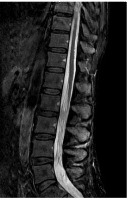
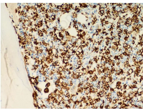
months he was submitted to our Unit with paresthesias, hypotonia and muscle atrophy of the lower limbs, predominantly the left side, which evolved to paresis. On admis-sion, blood tests showed only hyperfer-ritinemia (5189 ng/mL). The CAT, com-pared to the previous one, presented a numerical and volumetric increase in the lateral cervical lymphadenopathy, with evi-dence of lymph nodes also in hepatic hilum, peripancreatic and left supraclavicular regions. PET-CAT showed a diffuse hyper-accumulation of the radiopharmaceutical in the liver, laterocervical and retro-clavicular left region, hepatic hilum, and bone mar-row. Therefore, the patient underwent exci-sional biopsy of lateral cervical left lymph node, that showed a structure subverted by widespread proliferation of histiocytic ele-ments CD68+, comprising focal aspects of hemophagocytosis and many T-lympho-cytes (Figure 1). The brain magnetic-reso-nance-imaging (MRI) was negative for injury, while spine MRI showed pathologi-cal tissue at the level of the cauda equina and leptomeninge covering the spinal cord cone, mainly in the vertebral body L3, and intervertebral space L2-L3 (Figure 2). These findings were interpreted as histio-cytic infiltration of the lumbar subarachnoid space. The bone marrow biopsy described a hypercellular bone marrow with a zonal increase, in the interstitial region, of the proportion of histiocytes, positive for CD68 (PGM1), some of large size with aspects of
erythrophagocytosis (Figure 3). During the hospital stay, despite treatment, the patient presented a recurrence of all laboratory abnormalities of previous hospitalization and a rapid clinical deterioration as well as high fever, jaundice, stupor and death.
Discussion and Conclusions
The HLH in adults is more frequently a secondary form, while the primary form is typical of childhood linked to genetic muta-tions. Infective, autoimmune and neoplastic causes were excluded in this case, indeed we could only suppose this form was an idiopathic hemophagocytic lymphohistio-cytosis, since the unavailability of genetic examinations. Currently, the diagnosis cri-teria are defined in HLH-2004 guidelines as 5 positives out of 8 possibilities: i) fever; ii) splenomegaly; iii) cytopenia affecting ≥2 of 3 lineages in the peripheral blood: Hemoglobin (<90 g/L), Platelets (<100×109/L), Neutrophils (<1.0×10⁹/L); iv
hypertriglyceridemia and/or hypofibrino-genemia (triglycerides ≥265 mg/dL; fib-rinogen ≤1.5 g/L); v) hemophagocytosis in bone marrow, spleen or lymph nodes; vi) low or absent NK-cell activity, vii) ferritin ≥500 microgram/L; viii) soluble CD25 ≥2400 U/mL.5Despite the unavailability of
our hospital laboratory to measure sCD25 and NK-cell activity, our patient showed 6
Case Report
[Hematology Reports 2017; 9:6859] [page 5] Figure 1. Lymph node (hematoxylin and eosin): a dense infiltrate of histiocytes including
small reactive lymphocytes.
Figure 2. Magnetic resonance imaging of spinal cord (T2 image): pathological tis-sue infiltrating cauda equine, mainly in the vertebral body of L3, and interverte-bral space between L2-L3.
Non
commercial
use
positive criteria (fever, splenomegaly, cytopenia, hypertriglyceridemia and hypofrinogenemia, hemophagocytosis signs). Furthermore, the patient showed other clinical and biochemical manifesta-tions that are very common in HLH of adults such as cholestasis, high PT INR and high LDH, although they are still not included in guidelines.6Therefore, it could
be useful considering new diagnose criteria, also in addition to those proposed by Delphi study and H score.7,8 Only a few
hemo-phagocytosis signs were detectable in patient’s bone marrow, in spite of the wide and aggressive clinical manifestation. Accordingly, we agree that the bone mar-row aspect should not be considered as a gold standard, because the typical hemo-phagocytosis signs predominantly appear in the more advanced disease stages, and they are absent in about 40% of cases.9,10
Moreover, this case presented two clinical peculiarities. The first one is the presence of raised amylase and lipase with demonstrat-ed absence of radiological pancreatitis signs and bile ducts dilation and/or obstruction by Magnetic resonance cholangiopancreatog-raphy (MRCP) and Endoscopic retrograde cholangiopancreatography (ERCP). This pancreatic alteration persisted until the beginning of immunosuppressive therapy. The HLH is a systemic syndrome thus every tissue and organ may theoretically be involved by histiocyte infiltration. Therefore, we think that patient’s raised pancreatic enzymes could be the
conse-quence of mild pancreatic ducts inflamma-tion with a mechanism similar to the more typical hepatic involvement. HLH cases with pancreatitis are very rare and only one adult case is described with radiologic signs of pancreatitis though correlated to the pres-ence of ulcerative colitis.11,12 The second
clinical characteristic was the finding of a large histiocyte infiltration in the spinal cord, which determined left lower limb paresis with a progressive muscular atro-phy. The HLH usually involves central nervous structures as the periventricular substance, thalamus and basal ganglia,13
leading to neurological symptoms such as conscience alterations, meningism, focal neurological deficits and epilepsy. Rare cases of HLH are described with spinal cord involvement and probably no case is described with spinal cord involvement and brain savings.14,15Therefore, diagnosing the
HLH in adults may be arduous since the large clinical manifestations variety and the restricted number of diagnosis criteria; fur-thermore deciding an appropriate therapy is very difficult, considering the absence of guidelines for adults. There are only case series of adult patients treated with various regimens mostly based on cyclosporine, dexamethasone, etoposide and more recent-ly on stem cell transplantation, reporting a poor response and a high mortality.16-19
Although no diagnosis was done during patient’s first manifestation, the patient totally recovered after splenectomy and without any immunosuppressive treatment.
In fact, the splenectomy has been suggested as an alternative treatment in idiopathic HLH of adults not responsive to standard therapy.20 The patient showed a relapse
after splenectomy and so he underwent a treatment based on the HLH-2004 protocol, a prospective study of the Histiocyte Society conducted on pediatric patients with no history of immunosoppression or malignancy.5 Etoposide infusion was
excluded for bone marrow hypocellularity, while cyclosporine and dexamethasone were administered with an 8-week induc-tion and following maintenance therapy. However, the patient relapsed after 6 months, the high dexamethasone dosage was useless and the death occurred in a few days. This case report demonstrates that HLH in adults can appear in an unusual way, thus a diagnosis criteria revision is needed to facilitate disease recognition and anticipate the treatment. In addition, further studies are necessary to understand patho-genesis and to choose the optimal treatment in adults. As it has been described in the introductive section, currently HLH is not considered a malignant histiocytosis. However, HLH prognosis is very poor with a high rate of relapse and, although in this syndrome histiocytic cells have no cellular atypia or other malignant aspects, their behaviour is aggressive. Therefore, we agree to treat HLH aggressive forms with lymphoma-type chemotherapies.
References
1. Komp DM, Perry MC. The histiocytic syndromes. Introduction. Semin Oncol 1991;18:1-2.
2. Favara BE, Feller AC, Pauli M, et al. Contemporary classification of histio-cytic disorders. The WHO Committee On Histiocytic/Reticulum Cell Proliferations. Reclassification Working Group of the Histiocyte Society. Med Pediatr Oncol 1997;29: 157-66.
3. Rappaport H. Tumors of the hemato-poietic system. Washington DC: U.S. Department of Defense, Armed Forces Institute of Pathology, 1966.
4. Jaffe ES. The 2008 WHO classification of lymphomas: implications for clinical practice and translational research. Hematology Am Soc Hematol Educ Program 2009;523-531.
5. Henter JI, Horne A, Arico M, et al. HLH-2004: Diagnostic and therapeutic guidelines for hemophagocytic lympho-histiocytosis. Pediatr Blood Cancer
Case Report
Figure 3. Bone marrow biopsy: CD68 immunostain shows a diffuse histocyte infiltrate in hypercellular marrow.
[page 6] [Hematology Reports 2017; 9:6859]
Non
commercial
use
2007;48:124-31.
6. Schram AM, Berliner N. How I treat hemophagocytic lymphohistiocytosis in the adult patient. Blood 2015;125:2908-14.
7. Debaugnies F, Mahadeb B, Ferster A, et al. Performances of the H-Score for diagnosis of hemophagocytic lympho-histiocytosis in adult and pediatric pati-ents. Am J Clin Pathol 2016;145:862-70.
8. Hejblum G, Lambotte O, Galicier L, et al. A web-based delphi study for elici-ting helpful criteria in the positive diag-nosis of hemophagocytic syndrome in adult patients. PLoS One 2014;9:e94024.
9. Ho C, Yao X, Tian L, et al. Marrow assessment for hemophagocytic lymp-hohistiocytosis demonstrates poor cor-relation with disease probability. Am J Clin Pathol 2014;141:62-71.
10. Machaczka M, Klimkowska M. Bone marrow assessment in the diagnosis of acquired hemophagocytic lymphohi-stiocytosis in adults. Am J Clin Pathol 2015;143:308-9.
11. Cwiklinska M, Czogala M, Balwierz W, et al. [Hemophagocytic syndrome in children with different underlying con-ditions]. Przegl Lek 2010;67:430-5. [Article in Polish]
12. Kanaji S, Okuma K, Tokumitsu Y, et al. Hemophagocytic syndrome associated with fulminant ulcerative colitis and presumed acute pancreatitis. Am J Gastroenterol 1998;93:1956-9. 13. Rego I, Severino M, Micalizzi C, et al.
Neuroradiologic findings and follow-up with magnetic resonance imaging of the genetic forms of haemophagocytic lymphohistiocytosis with CNS involve-ment. Pediatr Blood Cancer 2012;58: 810-4.
14. Fujii T, Shimada K, Tanaka S, et al. [An autopsy case of B lymphoblastic leuke-mia/lymphoma with hemophagocytic syndrome infiltrating in the central ner-vous systems]. Rinsho Byori 2013;61:679-684. [Article in Japanese] 15. Gokce M, Balta G, Unal S, et al. Spinal
cord involvement in a child with famili-al hemophagocytic lymphohistiocyto-sis. J Pediatr Neurosci 2012;7:194-6.
16. Otrock ZK, Eby CS. Clinical characteri-stics, prognostic factors, and outcomes of adult patients with hemophagocytic lymphohistiocytosis. Am J Hematol 2015;90:220-4.
17 Arca M, Fardet L, Galicier L, et al. Prognostic factors of early death in a cohort of 162 adult haemophagocytic syndrome: impact of triggering disease and early treatment with etoposide. Br J Haematol 2015;168:63-8.
18. Li J, Wang Q, Zheng W, et al. Hemophagocytic lymphohistiocytosis: clinical analysis of 103 adult patients. Medicine (Baltimore) 2014;93:100-5. 19. Parikh SA, Kapoor P, Letendre L, et al.
Prognostic factors and outcomes of adults with hemophagocytic lymphohi-stiocytosis. Mayo Clin Proc 2014;89: 484-92.
20. Jing-Shi W, Yi-Ni W, Lin W, Zhao W. Splenectomy as a treatment for adults with relapsed hemophagocytic lympho-histiocytosis of unknown cause. Ann Hematol 2015;94:753-60.
Case Report
[Hematology Reports 2017; 9:6859] [page 7]