with Massive Oxidation of Plasma Albumin
Luca Musante,*
†Giovanni Candiano,* Andrea Petretto,
‡Maurizio Bruschi,*
†Nazzareno Dimasi,
§Gianluca Caridi,* Barbara Pavone,
储¶** Piero Del Boccio,
储¶**
Monica Galliano,
††Andrea Urbani,
储¶** Francesco Scolari,
‡‡Flavio Vincenti,
§§and
Gian Marco Ghiggeri*
*Laboratory on Pathophysiology of Uremia,
‡Mass Spectrometry Core Facility,
§Laboratory of Molecular Medicine, G.
Gaslini Children Hospital, and
†Renal Child Foundation, Genoa,
储Department of Biomedical Science, Universita` degli
Studi di Chieti e Pescara, Chieti,
¶Centro Studi sull’Invecchiamento, Fondazione Universita` “G. D’Annunzio,” Chieti,
**IRCCS-Fondazione Santa Lucia, Rome,
††Department of Biochemistry, University of Pavia, Pavia, and
‡‡Department
of Nephrology, University of Brescia, Brescia, Italy; and
§§Transplant Service, University of California, San Francisco,
San Francisco, California
The basic mechanism for idiopathic FSGS still is obscure. Indirect evidence in humans and generation of FSGS by oxidants in experimental models suggest a role of free radicals. In vitro studies demonstrate a main role of plasma albumin as antioxidant, its modification representing a chemical marker of oxidative stress. With the use of complementary liquid chromatography electron spray ionization tandem mass spectrometry (LC-ESI-MS/MS) and biochemical methods, plasma albumin was characterized in 34 patients with FSGS; 18 had received a renal transplant, and 17 had IgM mesangial deposition. Patients with FSGS that was in remission or without recurrence after transplantation had normal plasma albumin, and the same occurred in patients with primary and secondary nephrites and with chronic renal failure. In contrast, patients with active FSGS or with posttransplantation recurrence had oxidized plasma albumin. This finding was based on the character-ization of albumin Cys 34 with an mass-to-charge ratio of 511.71 in triple charge that was consistent with the formation of a cysteic acid carrying a sulfonic group (alb-SO3ⴚ). The exact mass of albumin was increased accordingly (ⴙ48 Da) for incorporation of three oxygen radicals. Direct titration of the free sulfhydryl group 34 of plasma albumin and electrophoretic titration curves confirmed loss of free sulfhydryl group and formation of a fast-moving isoform in all cases with disease activity. This is the first demonstration of in vivo plasma albumin oxidation that was obtained with an adequate structural approach. Albumin oxidation seems to be specific for FSGS, suggesting some pathogenetic implications. Free radical involvement in FSGS may lead to specific therapeutic interventions.
J Am Soc Nephrol 18: 799 – 810, 2007. doi: 10.1681/ASN.2006090965
F
SGS is a degenerative disease of the kidney that is characterized by focal accumulation of extracellular ma-trix in glomeruli. It is a heterogeneous condition that includes different variants (1– 4), possible genetic background (5), and variable response to drugs that modify long-term out-come (6). The basic lesion ranges from minimal glomerular involvement to segmental-global glomerulosclerosis that prob-ably represents an evolutionary stage in patients who are un-responsive to drugs (1,4). On clinical grounds, FSGS usually has an abrupt onset with heavy proteinuria, hypoalbuminemia, and frequent progression to renal failure. Notwithstanding some recent advances on the characterization of genes that areresponsible for congenital FSGS (7–11), little is known of the pathogenesis of the idiopathic form. The disease typically psents clinical recurrences after infectious episodes and a re-markable percentage of 30 to 50% of patients who have FSGS and receive renal allograft have posttransplantation recurrence (12–15). Abnormal T cell responses, have been suggested to be involved in the pathogenesis of the idiopathic form and in posttransplantation recurrences but remain poorly defined and lack specificity (16). On the basis of the rapid posttransplanta-tion recurrence, an implicaposttransplanta-tion of circulating plasma factors that persist over time and modify glomerular permeability to proteins long has been suspected (13,14). Unfortunately, the identification and the characterization of the circulating factors have been elusive (17,18), and, more recently, other pathoge-netic mechanisms have been proposed (19).
In an attempt to elucidate the pathogenesis of FSGS, we embarked on a proteomics approach to study plasma proteins in these patients. Our previous studies in patients with FSGS (20) demonstrated massive oxidation of plasma albumin, re-sulting in modification of the structure of the protein. We
Received September 6, 2006. Accepted December 18, 2006.
Published online ahead of print. Publication date available at www.jasn.org. L.M. and G.C. contributed equally to this work.
Address correspondence to:Dr. Gian Marco Ghiggeri, Laboratory on Pathophysiol-ogy of Uremia, G. Gaslini Children Hospital, Largo G. Gaslini, 5. 16148 Genova, Italy. Phone:⫹39-010-380742; Fax: ⫹39-010-395214; E-mail: labnefro@ospedale-gaslini.ge.it
proposed that massive oxidation plays a role in the pathogen-esis of the proteinuria that is associated with FSGS. In this study, we devised a mass spectrometry technique for analysis of native plasma albumin that is more reproducible and abol-ished the need for chemical manipulation of the protein. The structural analysis of albumin then was extended to a wide cohort of patients with nephrotic syndrome and to patients with posttransplantation recurrence of FSGS. Children who had other forms of primary and secondary glomerulonephritis and/or chronic renal failure (end-stage renal failure [ESRF]) and were undergoing various treatments also were studied to define specificity of our structural approach. New method-ologic assays also were developed to titrate free sulfhydryl group (SH) of Cys 34 and to determine the occurrence of neutral to acid transition (N-A) in large-scale screening studies.
Materials and Methods
Chemicals
Acrylamide and ampholine were from Amersham Bioscience (Upp-sala, Sweden); N,N⬘-methylenebisacrylamide, SDS, N,N,N⬘,N⬘-tetram-ethylethylenediamine, and electrophoresis calibration standard pro-teins were from Bio-Rad Laboratories (Hercules, CA). All other chemicals, of analytical and electrophoretic grade, were purchased from BDH (Poole, UK). Solutions were prepared fresh using Milli-Q (Millipore, Milan, Italy).
Patients
FSGS/IgM. Thirty-four patients with FSGS, 18 of whom received a renal transplant and 17 of whom had IgM mesangial proliferative glomerulonephritis, were enrolled in the study (Tables 1 and 2). Twelve patients of the FSGS group were presenting nephrotic syndrome at the time of the enrollment; four had been in stable remission in the previ-ous 2 yr. Eleven patients of the group with mesangial IgM deposits were presenting florid proteinuria and nephrotic syndrome at the time of the study, and the remaining six were in stable remission. The general criteria for enrollment were (1) availability of a clear histology diagnosis on the basis of accepted criteria and (2) absence of familiarity and/or relevant mutations of slit diaphragm genes (NPHS2, CD2AP, and ACTN4). Nephrotic syndrome was defined by the presence of florid proteinuria⬎40 mg/h per m2. Renal biopsies were processed by
standard procedures for light microscopy, immunofluorescence stud-ies, and electron microscopy. Clinical and pathologic features (e.g., gender, age at onset of proteinuria, treatment, evolution toward renal failure, renal transplant) are reported in Tables 1 and 2. Patients with FSGS and IgM and with active proteinuria were receiving treatment with steroids alone (at variable dosages) or in association with cyclo-sporin and with angiotensin-converting enzyme inhibitors (ACEi). Ste-roids were given in a starting dosage of prednisolone 2 mg/kg fol-lowed by a gradual taper (21). Cyclosporin was administered at 5 mg/kg starting dosage, followed by dosage adjustment to maintain cyclosporin serum levels between 50 and 100 ng/ml (6). Six patients of the stable remission groups with either FSGS or IgM were still receiving cyclosporin; ACEi was used in one. Eighteen children with FSGS had developed ESRF and had received a cadaver renal transplant. Immu-nosuppression therapy in these patients included tacrolimus or cyclo-sporin, mycophenolate mofetil, and steroids. Fourteen patients of this cohort had posttransplantation recurrence of proteinuria that was treated with plasmapheresis (1.5-ml plasma exchange with albumin as the unique replacement protein) in all but one patient and was associ-ated with cyclophosphamide 2 mg/kg for 2 mo in one patient.
ESRF. Nine patients who had chronic renal failure and were un-dergoing various treatments were enrolled. One was a child (patient 1), two were adolescents (patients 2 and 9), and six were young adults who developed ESRD for various reasons (Table 3). The therapeutic ap-proach was hemodialysis in six cases, and two patients were main-tained on a conservative treatment.
Membranous Glomerulonephritis. Eight patients had received a diagnosis of membranous nephropathy and were all in the proteinuric phase of the disease (Table 4). All but one were adults and were presenting a variable degree of proteinuria from 1 to 8 g/d. Patients with membranous glomerulonephritis (MGN) were receiving steroids as single drug (1 to 2 mg/kg) or in combination with immune suppres-sors (22); in some cases, ACEi (ramipril 5 to 8 mg/m2) had been
associated.
Membrane Proliferative Glomerulonephritis. Four patients had a histology diagnosis of membrane proliferative glomerulonephritis (MPGN) that presented with proteinuria, hematuria, and normal renal function without arterial hypertension (Table 4). All were receiving prednisolone (1 mg/kg) for at least 2 mo.
IgA. Five patients had a recent diagnosis of IgA in the absence of relevant proteinuria. Only one was treated with steroids; in three cases, it was used in a scheme with ACEi (Table 4).
Normal Control Subjects. The control group consisted of 18 healthy adult control subjects.
In all cases plasma was obtained in the morning after an overnight fast. In patients with posttransplantation recurrence, plasma was ob-tained at the onset of proteinuria and during the follow-up. Morning fresh urine was collected in sterile condition without additive and frozen at⫺80°C within 1 h. Plasma was frozen within 1 h and was maintained at⫺80°C under vacuum. Appropriate informed consent was obtained from all patients in the study.
Purification of Albumin from Healthy Donors and Patients
Albumin was purified from plasma of healthy donors and patients with FSGS by preparative continuous monodimensional PAGE electro-phoresis (total acrylamide concentration⫽ 4 to 12%) in native condi-tions with 2-mm gel spacers. All purification steps were performed in a native condition to prevent structural modifications according to Margolis and Kenrick (23). One milliliter of serum was applied to gel, and electrophoresis was run in Tris-borate-EDTA (80/90/2.5 mM) for 12 h with 16 mA at 12°C. Albumin was desorbed from acrylamide by gentle pestle and was maintained in PBS at 4°C for 24 h with two changes of the solution.
Liquid Chromatography Electron Spray Ionization Tandem
Mass Spectrometry (LC-ESI-MS/MS) for Tryptic Digest
Characterization
Albumin after purification was delipidated first in a methanol:ac-etone:tributyl phosphate (1:12:1) with gentle agitation at room temper-ature overnight and then was digested by trypsin. Trypsin was added at an enzyme substrate ratio of 1:30 (wt/wt) in a solution of 100 mM ammonium bicarbonate and 1 mM CaCl2 (pH 8.5). After overnight
incubation at 37°C, the reaction was stopped with formic acid to pH 2. All mass spectrometric measurements were performed using an LTQ linear trap mass spectrometer (Thermo Electron, San Jose, CA) coupled to an HPLC Surveyor (Thermo Electron) and equipped with a Jupiter C18 column 250⫻ 1 mm (Phenomenex, Torrance, CA). Peptides were eluted from the column using an acetonitrile gradient, 5% B for 6 min followed by 5 to 90% B within 109 min (eluent A: 0.1% formic acid in water; eluent B: 0.1% formic acid in acetonitrile) at a flow rate of 50 l/min. The column effluent was directed into the electrospray source.
The spray voltage was 5.0 kV. The capillary of ion trap was kept at 200°C, and the voltage was kept at 2.85 V. Spectra were acquired in automated MS/MS mode: Each full MS scan (in the range 400 to 1800 mass-to-charge ratio [m/z]) was followed by five MS/MS of the most abundant ions; mass that had been analyzed more than two times this way was automatically taken up into an exclusion list for 30 s. Com-puter analysis of peptide MS/MS spectra was performed using Bio-works software, version 3.2 (Thermo Electron) and searched against an ALB protein database. Peptide MS/MS assignments were filtered ac-cording to the following criteria: cross-correlation (Xcorr)ⱖ 1.9 for the
singly charged ions, Xcorrⱖ 2.2 for doubly charged ions, and Xcorrⱖ 3.7
for triply charged ions; peptide probabilityⱕ0.001; change in correla-tion value (⌬Cn) ⱖ0.1; and percentage of ions ⱖ30%. For all protein, two missed cleavages was allowed.
ESI-MS for Exact Mass
Albumin-containing solutions were injected manually (5l) into an on-line flow, using a CapLC system (Micromass, Waters, Milford, MA) coupled with a nano-ESI-Q-TOF (quadrupole–time-of-flight) instru-ment (Micromass, Waters). The sample was eluted at 1l/min on a C4 precolumn LC-Packings, 300m inner diameter ⫻ 20 mm. Elution was achieved isocratically by H2O/ACN 50/50 both with 0.1% TFA and
directed into a mass spectrometer equipped with a nano-Lock-Spray source. A 2500- and 50-V tension was applied to the PicoTip capillary (PicoTip Emitter, tip 10⫾ 1m; New Objective, Woburn, MA) and cone voltage, respectively, and the positive ion mode for ion scan experiment was used to monitor the 700- to 2200-m/z range. Data analysis was performed using Masslynx version 4.0 (Micromass/Wa-ters). The data collected were examined for multiply charged protein
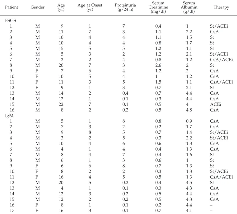
Table 1. Clinical and biochemical parameters in 33 children with idiopathic nephrotic syndrome subdivided
according to renal pathology
aPatient Gender Age(yr) Age at Onset(yr) Proteinuria(g/24 h) CreatinineSerum (mg/dl) Serum Albumin (g/dl) Therapy
FSGS
1
M
9
1
7
0.4
1
St/ACEi
2
M
11
7
3
1.1
2.2
CsA
3
M
10
4
4
1.1
1.5
St
4
M
10
4
4
0.8
1.7
St
5
M
15
5
5
1.2
1.1
St
6
M
5
3
2
1.2
2.1
St/ACEi
7
M
2
2
4
0.8
1.2
CsA/ACEi
8
M
20
7
3
2.6
2
St
9
F
7
6
4
1.2
2
CsA
10
F
10
5
4
1
1.2
CsA
11
F
11
3
5
1.5
1.1
CsA/ACEi
12
F
9
1
3
0.7
2.1
St
13
M
14
2
0.4
0.7
4.4
CsA
14
M
12
1
0.1
0.3
4.4
CsA
15
M
22
7
0.1
0.5
4
ACEi
16
M
8
2
0.2
0.5
4.8
CsA
IgM
1
M
5
1
8
0.8
0.9
CsA
2
M
7
3
2
0.2
1.7
CsA
3
M
9
8
5
0.7
1.4
St/ACEi
4
M
3
2
5
0.3
2.2
St/ACEi
5
M
10
4
6
0.6
1.3
CsA
6
M
4
1
4
0.4
1.3
CsA
7
M
8
4
7
0.4
1.6
St
8
M
6
1
3
0.6
1
St
9
F
6
6
8
0.7
1.3
St
10
F
8
2
2
0.3
1.3
St/ACEi
11
F
16
4
5
0.5
1.3
CsA/ACEi
12
M
20
5
0.2
0.4
4.5
St
13
M
4
1
0.1
0.3
4.3
CsA
14
M
12
3
0.2
0.5
4.4
CsA
15
M
12
2
0.2
0.5
4.3
CsA
16
F
8
1
0.1
0.2
4.4
–
17
F
16
3
0.1
0.7
4.1
–
aTen patients were evaluated during stable remission of nephrotic syndrome. induced by drugs; in these, plasma had been collected after at least 6 mo of normal urine. ACEi, angiotensin-converting enzyme inhibitor; CsA, cyclosporine; St, steroids.
spectra, which then were integrated to provide a single combined spectrum for the protein injected. A maximum entropy deconvolution algorithm (MaxEnt1) was used to deconvolute multiply charged spec-tra and produce molecular mass specspec-tra.
Electrophoretic Titration Curves
For electrophoretic titration curves, we used total plasma in antico-agulant citrate dextrose. Titration was performed according to Bruschi et al. (24). Both methods for electrophoresis runs and calculations have been described previously (24,25).
“In Gel” Determination of Free SH Accessibility
Free SH group titration in plasma albumin was done with the male-imide-PEO2-Biotin
(biotinyl-3-maleimidopropionamidyl-3,6-dioxact-anediamine) assay (Pierce, Rockford, IL) according to the manufactur-er’s instructions. After reaction with maleimide- PEO2-Biotin, plasma
proteins first were separated in monodimensional polyacrylamide gels performed according to Laemmli (26) without mercapto-ethanol. After electrophoresis (total acrylamide concentration⫽ 5 to 16%; degree of cross-link⫽ 2.67%) reactivity of SH groups with maleimide was al-lowed at pH 6.5 for 2 h at 37°C. Streptavidin that was conjugated with
Table 2. Clinical parameters in 18 children who had idiopathic FSGS and developed ESRF and received a renal
graft.
Patient
No. Gender Onset(yr) ESRF(yr) Transplantation(yr) RecurrenceDays for Therapy Outcome
1
M
4.2
6
7
1
PL
Stable proteinuria
2
F
4.3
7
8
7
–
Stable proteinuria
3
M
10.5
14
15
1
PL
Remission
4
F
4.2
12
13
9
PL
Remission
5
M
9.6
11
13
15
PL
Remission
6
M
12.3
30
34
15
PL
Stable proteinuria
7
F
13.7
18
19
3
PL
Frequent recurrence
8
M
0.6
4
5
22
PL
Remission
9
M
2.2
4
6
1
PL
Remission
10
M
14
14
16
30
PL/Cy
ESRF
11
M
11
11
15
600
PL
Normal function
12
F
36
45
50
30
PL
Stable proteinuria
13
F
28
32
33
1
PL
ESRF
14
F
5
28
32
4
PL
Nephrectomy
No recurrence
1
F
5.2
8
10
–
–
–
2
F
3.0
6
9
–
–
–
3
F
6.3
7
8
–
–
–
4
M
5.2
16
17
–
–
–
aFourteen patients of this cohort presented recurrence of proteinuria and were treated with a combination of
plasmapheresis and cyclophosphamide. The other four patients had stable and prolonged good outcome. Cy, cyclophosphamide; ESRF, end-stage renal failure; PL, plasmapheresis.
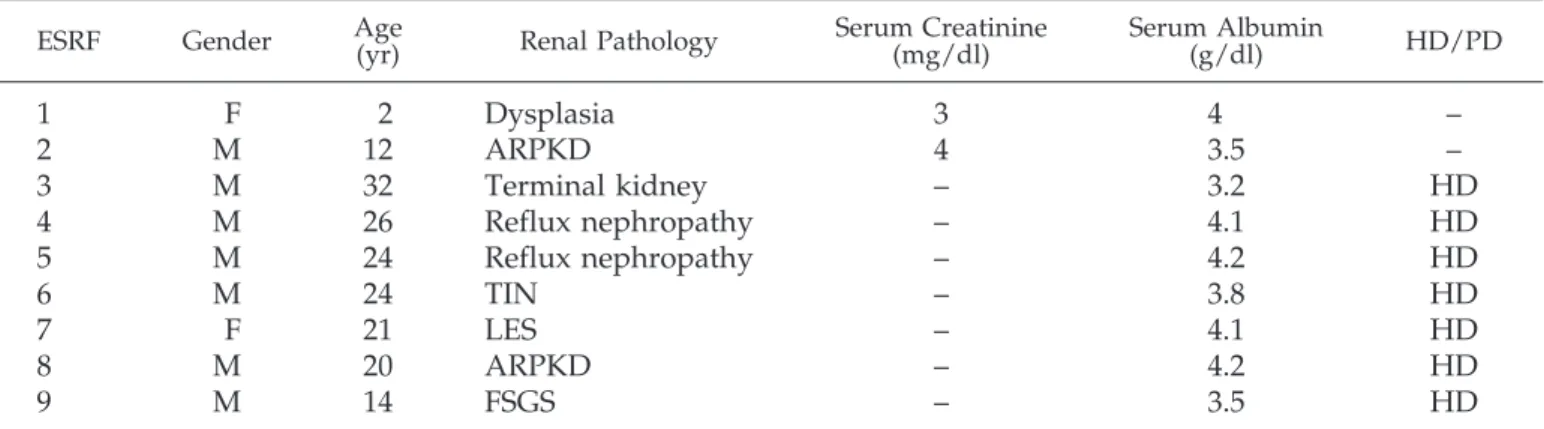
Table 3. Clinical and biochemical parameters in nine patients with chronic renal failure from various renal
pathologies
aESRF Gender Age(yr) Renal Pathology Serum Creatinine(mg/dl) Serum Albumin(g/dl) HD/PD
1
F
2
Dysplasia
3
4
–
2
M
12
ARPKD
4
3.5
–
3
M
32
Terminal kidney
–
3.2
HD
4
M
26
Reflux nephropathy
–
4.1
HD
5
M
24
Reflux nephropathy
–
4.2
HD
6
M
24
TIN
–
3.8
HD
7
F
21
LES
–
4.1
HD
8
M
20
ARPKD
–
4.2
HD
9
M
14
FSGS
–
3.5
HD
aSeven patients had developed ESRF and were undergoing hemodialysis (HD). Two children presented severe renal lesions
but were still on a conservative treatment. ARPKD, autosomal recessive polycystic kidney disease; LES, lupus erythmatosus systemic; PD, peritoneal dialysis; TIN, tubulointerstitial nephropathy.
horseradish peroxidase was used to determine biotin incorporation with 2-(4⬘-hydroxyazobenzene)-benzoic acid using the EZ Biotin Quan-titation Kit (Pierce) at 500 nm with correction for the amount of albumin as determined by Coomassie R-250. A calibration curve that consisted of four dilutions of the same serum with known concentration of albumin was used as standard. Specificity of the maleimide dye for the free SH group of Cys 34 was demonstrated by preventing the binding with methyl-methanethiosulfonate that specifically binds this group at pH 5.
Albumin Degradation In Vitro
A total of 100g of purified albumin was incubated at 37°C for 16 h with 0.1g/ml (wt/vol) of bovine trypsin in a medium that contained 50 mM Tris-HCl (pH 7.8) and 5 mm CaCl2. The digestion was stopped
by addition of Laemmli reducing sample buffer (10 mM Tris-HCl [pH 8.8], 1% [wt/vol] SDS, 1 mM EDTA, 20% [vol/vol] glycerol, and 5% [vol/vol]-mercaptoethanol) (26). After 1 h, the samples were loaded on 8 to 18% (wt/vol) gradient SDS-PAGE and run at 45 mA at 12°C. Gel was fixed with 40% (vol/vol) ethanol and 10% (vol/vol) acetic acid and stained by silver nitrate.
Albumin Gene DNA Sequence
Genomic DNA was extracted from patient blood. The 14 coding exons of human albumin gene and their intro-exon junctions were PCR-amplified with specific primer pairs as already described by Watkins et al. (27). Amplicons were purified by Exo-SAP-IT (Amersham Bioscience, Milan, Italy) and directly subjected to direct sequencing (ABI 3100; Applera, Milan, Italy).
Glutathione Assay
Reduced glutathione (GSH) levels were determined in plasma and in red blood cells after trichloroacetic precipitation (28% wt/vol). GSH was determined by dithio-bis (2-nitrobenzoic acid) at 412 nm with correction with a blank reagent and using free cysteine as the standard (28).
Statistical Analyses
Simple statistical tests based on one-way ANOVA and2test were
used to verify differences in free SH titration and N-A transition among different cohorts of patients. Data are mean⫾ SEM.
Results
Chemical and Structural Characterization of Plasma
Albumin in FSGS
The structural approach to plasma albumin in FSGS was done originally in seven patients, using LC-ESI-MS/MS analy-sis after reduction and alkylation of purified albumin (20). This approach is not readily applicable for extensive analysis, be-cause the sulfonic group at Cys 34 that is generated upon oxidation is a stable end product in the native protein but may undergo further rearrangements during alkylation (personal observation by G.C.). Therefore, we devised a new proteomic approach to plasma albumin that was based on characterization by LC-ESI-MS/MS of trypsin digestion products of the native protein that are more stable then the alkylated products and gave highly reproducible results. Proteomic analysis of plasma albumin now was extended to 12 patients with FSGS, five with the idiopathic form and seven with posttransplantation recur-rence of the disease. In the first step, albumin was purified in nondenaturing conditions using preparative electrophoresis in native gels giving a mean recovery yield⬎90%. Fine structure analysis by LC-ESI-MS/MS and determination of the exact mass by ESI-MS protein analysis then was repeated in all patients.
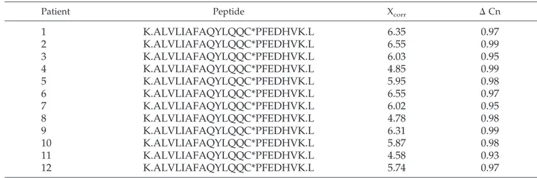
LC-ESI-MS/MS. The spectrum of albumin after digestion with trypsin indicated the presence of a parent fragment m/z 827.96 (K.ALVLIAFAQYLQQCSO3PFEDHVK.L) in the triply
charged state. The m/z 827.96 compound was studied by MS/MS showing the presence of an m/z 511.71 ion in triple charge that was consistent with a sequence in which the Cys 34 brings three additional oxygen residues, indicating oxidation of the SH to cysteic acid (⫹48; Figure 1). Control albumin was characterized by the absence of the m/z 827.96 ion in triple charge. In Table 5 are reported the sequences with relative Xcorr
and⌬Cn of the MS/MS characterization of the m/z 827.96 ion in the cohort of patients with primary nephrotic syndrome and post-transplant recurrence. Xcorr and ⌬Cn were in all cases
highly significant, confirming the presence of a cysteic group in position 34.
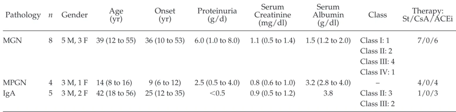
Table 4. Clinical and biochemical parameters in patients with different primary renal pathologies involving the
glomerulus
aPathology n Gender Age(yr) Onset(yr) Proteinuria(g/d) CreatinineSerum (mg/dl) Serum Albumin (g/dl) Class Therapy: St/CsA/ACEi MGN 8 5 M, 3 F 39 (12 to 55) 36 (10 to 53) 6.0 (1.0 to 8.0) 1.1 (0.5 to 1.4) 1.5 (1.2 to 2.0) Class I: 1 Class II: 2 Class III: 4 Class IV: 1 7/0/6 MPGN 4 3 M, 1 F 14 (8 to 16) 9 (6 to 12) 2.5 (0.5 to 4.0) 0.8 (0.6 to 1.0) 3.2 (2.8 to 4.0) – 4/0/4 IgA 5 3 M, 2 F 42 (18 to 56) 25 (12 to 35) ⬍0.5 0.9 (0.5 to 1.2) 3.8 Class II: 3
Class III: 2
1/0/3
a
Overall, 17 patients were enrolled. They were selected among the most frequent causes of glomerular diseases that cause nephrotic syndrome (MGN), intermediate proteinuria (membrane proliferative glomerulonephritis关MPGN兴) and hematuria (IgA).
ESI-MS. Analysis of the exact mass of plasma albumin was used to confirm the result of the structural analysis in a large cohort of patients. In all patients with FSGS, the bulk of plasma albumin presented an exact mass of 66.555 kD, whereas this isoform was only minimally detected in normal plasma in which the major band presents a mass of 66.507 kD (data not shown).
Biomarkers of Albumin Oxidation in Patients with FSGS/IgM
On the basis of sulfonic transformation, the free SH of Cys 34 of albumin should not be titrated any more and could become a specific marker of oxidation. We devised a method for direct “in gel” determination of free Cys 34 in plasma that could be
used in screening analysis. In this technique, the free Cys of albumin was targeted by maleimide-PEO2biotin, and the
reac-tion then was revealed by streptavidin. This assay was ex-tended, with the analysis of N-A transition by electrophoretic titration curves (24), to the whole cohort of patients. Overall, 23 patients with primary FSGS/IgM and florid proteinuria and 14 who were presenting posttransplantation recurrence of protein-uria were studied. Ten patients with FSGS/IgM in stable re-mission and four who received a renal transplant but were free from recurrence represented the negative control group for disease activity.
Cys 34 Titration. The results are given in Figure 2, the former reporting an example of in gel titration in a selection of patients from each separate clinical subset as above. It is evi-dent the lack of albumin band in the PEO2-maleimide gel in
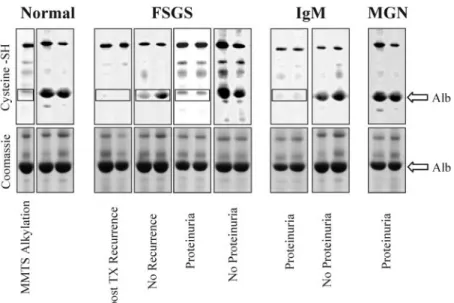
patients with proteinuria (see boxes in Figure 2). Figure 3 reports extensive results that confirm a marked decrease of Cys 34 titration in all patients with FSGS and IgM in both pre- and posttransplantation phases compared with normal people and with MGN with some differences within categories. In partic-ular, patients with active FSGS and IgM had markedly lower levels than in the remission phase (0.43⫾ 0.12 and 0.230 ⫾ 04
versus 1.94⫾ 0.42 U; P ⬍ 0.003 and P ⬍ 0.001, respectively);
patients with posttransplantation recurrence showed only mildly lower levels than in patients with no recurrence (0.27⫾ 0.04 versus 0.71⫾ 0.19). Normal people and other nephrotic patients with MGN had instead the same concentration (normal control subjects 2.75 ⫾ 0.33; MGN 2.48 ⫾ 0.67). For other categories of renal diseases, see below. Overall, in patients with active FSGS/IgM, Cys 34 seems massively oxidized, whereas during the remission phase, a normal albumin pool is reconsti-tuted.
N-A Transition. The rationale and the technique to define N-A transition have been described extensively (24). Briefly, changes in electrical charge were determined by an
electro-Figure 1. Mass spectrometry analyses of plasma albumin in
FSGS. Example of LC-ESI-MS/MS spectrum of the Cys 34 tryptic fragments of albumin from a patient with FSGS. Albu-min that was purified from the patient showed, after digestion with trypsin, the parent fragments mass-to-charge ratio (m/z) 511.7 in triply charged state, indicating the formation of a cysteic residue in place of the sulfhydryl group in position 34 of albumin. Analysis of control albumin did not reveal the forma-tion of the m/z 511.7 fragment because of lack of ionizaforma-tion of nonoxidized free sulfhydryl groups (SH).
Table 5. Sequence of the m/z 827.96 ion deriving from trypsin fragmentation of albumin that was purified from
patients with FSGS
aPatient Peptide Xcorr ⌬ Cn
1
K.ALVLIAFAQYLQQC*PFEDHVK.L
6.35
0.97
2
K.ALVLIAFAQYLQQC*PFEDHVK.L
6.55
0.99
3
K.ALVLIAFAQYLQQC*PFEDHVK.L
6.03
0.95
4
K.ALVLIAFAQYLQQC*PFEDHVK.L
4.85
0.99
5
K.ALVLIAFAQYLQQC*PFEDHVK.L
5.95
0.98
6
K.ALVLIAFAQYLQQC*PFEDHVK.L
6.55
0.97
7
K.ALVLIAFAQYLQQC*PFEDHVK.L
6.02
0.95
8
K.ALVLIAFAQYLQQC*PFEDHVK.L
4.78
0.98
9
K.ALVLIAFAQYLQQC*PFEDHVK.L
6.31
0.99
10
K.ALVLIAFAQYLQQC*PFEDHVK.L
5.87
0.98
11
K.ALVLIAFAQYLQQC*PFEDHVK.L
4.58
0.93
12
K.ALVLIAFAQYLQQC*PFEDHVK.L
5.74
0.97
aIn the case of normal albumin, no ions with this m/z could be detected in triple charge. MS/MS in triple charge of this
ion demonstrated the presence of a cysteic acid residue with m/z 511.7. Xcorrand⌬Cn were in all cases highly significant and
probative for the chemical characterization above. C*, cysteic acid; Cn, correlation value; MS/MS, tandem mass spectrometry; m/z, mass-to-charge ratio; Xcorr, cross-correlation.
phoretic technique that measures changes in isoelectric point at any given pH from 4 to 9. This technique also gives an estimate of the amount of the protein with a charge change that is inferred from the formation of isoform with different electro-phoretic behavior. In all of the cases studied, the variation in electrical charge involved 100% of the protein, and Figure 4A shows an example of this change. Overall, the screening for N-A transition confirmed the data on SH titration with some
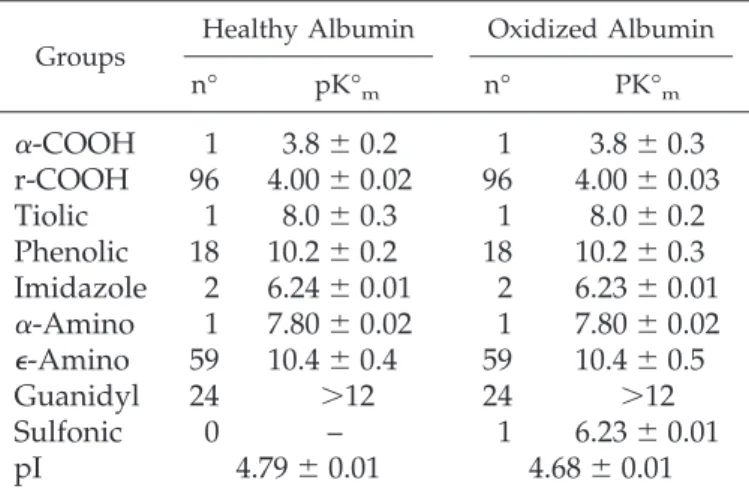
difference for patients without posttransplantation recurrence. In fact, N-A transition was common to all patients with active FSGS and IgM and to those with posttransplantation recur-rence, whereas, in the absence of proteinuria, in patients with-out posttransplantation, recurrence and in patients with MGN, a normal electrophoretic behavior was reconstituted. The per-centage of patients with N-A transition in the various groups is illustrated in Figure 4C and clearly shows a difference among categories. Table 6 reports the mean⫾ SD of ionization param-eters reproducing experimental curves of 10 normal control subjects and patients with proteinuria, according to the Linder-stro¨m-Lang equation. Considering the parameters reported in Table 6, this difference can be attributed to the presence of a sulfonic group in position 34 of the sequence that modifies our original assumption reported previously (24).
Titration of Albumin Cys 34 in Patients with ESRF and
with Other Glomerular Diseases
A few other groups of patients with different renal diseases were enrolled and used for demonstrating the specificity for FSGS of sulfonation of albumin Cys 34. The following groups of patients were enrolled (Tables 3 and 4): (1) Nine patients with chronic renal failure, two of whom were still on a conservative regimen; (2) four children with primary MPGN; and (3) five patients with IgA. In all cases, a normal titration of Cys 34 by PEO2-maleimide was observed. The results presented in Figure
5 show a typical pattern of albumin staining with the dye. It was found that maleimide staining, in particular, is specific for the free SH group of Cys 34 because was abolished by
prein-Figure 2. Titration of the Cys 34 free SH groups of albumin in FSGS, IgM, and membranous glomerulonephritis (MGN). Titration
of albumin free SH 34 by a direct “in gel” technique that uses PEO2-maleimide-biotin (top) as a specific label. According to this
method, plasma proteins first are labeled with PEO2-maleimide-biotin and then are separated by monodimensional
electrophore-sis in nondenaturing conditions. After separation, the reaction is developed with streptavidin. Albumin from healthy people showed PEO2-maleimide staining corresponding to albumin (arrows), whereas albumin of patients with FSGS had no staining,
indicating absence of a free SH that is unique in albumin at position 34 of the sequence (boxes). To prove specificity of the maleimide staining for the free SH, one sample had been treated previously with methyl-methanethiosulfonate (MMTS) that specifically blocked the free SH and prevented the maleimide binding.
Figure 3. Estimation of Cys 34-free SH group of albumin in the
whole cohort of patients with FSGS, IgM, and MGN. Results are expressed as arbitrary unit of biotin per milligram of albumin. A calibration curve with several dilutions of the same plasma was prepared to obtain a confidence limit for linearity of the assay.
cubation of the protein with methyl-methanethiosulfonate at pH 5.
Albumin Gene DNA Sequence
The 14 coding exons of human albumin gene and their intro-exon junctions were sequenced in all patients who were en-rolled in the study. In all cases, normal sequence was found, excluding a genetic basis for altered oxidation.
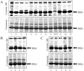
Susceptibility to Digestion by Trypsin
Because the antioxidation effect of albumin has been consid-ered for a long time a suicidal reaction that modifies suscepti-bility to digestion by common proteases, albumin was purified from plasma of four patients with idiopathic FSGS and four with posttransplantation recurrence and submitted to mild di-gestion with trypsin (see the Materials and Methods section). The results reported in Figure 6 demonstrate the formation of fragments in the majority of affected cases, confirming the original assumption (lanes f through n). Normal albumin was not digested following the same conditions (lanes a through e).
GSH in Plasma and in Erythrocytes
GSH levels were determined in plasma and in red blood cells of proteinuric patients with FSGS and in control subjects. Lev-els were slightly decreased in patients with active nephrotic syndrome compared with control subjects in both plasma (8.1⫾ 0.2 versus 8.4 ⫾mol/L) and in red blood cells (6.8 ⫾ 0.8
versus 7.5⫾ 0.8 nmol/mg hemoglobin), but the difference did
not reached statistical significance because of the low number of cases.
Figure 4. Electrophoretic titration curves for neutral to acid N-A
transition. (A) Example of electrophoretic titration curve of a plasma sample from a patient with FSGS and for comparison of normal plasma stained with Coomassie R-250. The two proteins migrate as a single and homogeneous band throughout the pH range: Between 4 and 4.5 and between 7 and 9, they overlap, whereas in the pH range between 4.5 and 7, oxidized albumin migrates with a more acid charge. (B) Theoretical electro-phoretic curve of albumin in which a cysteic group replaces a free sulfhydryl residue. According to the Linderstro¨m-Lang theory, this acid charge shift fits with the introduction of a sulfonic acid group in the molecule. Determination of albumin charge along a stable pH gradient was done according the procedure described by Bruschi et al. (24). (C) Percentage of patients with N-A transition in patients with nephrotic syn-drome and different clinical activity. Presence and/or absence
of fast albumin in the pH range 4.5 and 7 was evaluated in all patients that was defined in positive cases as N-A transition. Bars indicate the percentage of patients with N-A transitions in different study cohorts.
Table 6. Ionization parameters of healthy and oxidized
albumin calculated according to Linderström-Lang
equation
aGroups Healthy Albumin Oxidized Albumin n° pK°m n° PK°m
␣-COOH
1
3.8
⫾ 0.2
1
3.8
⫾ 0.3
r-COOH
96
4.00
⫾ 0.02
96
4.00
⫾ 0.03
Tiolic
1
8.0
⫾ 0.3
1
8.0
⫾ 0.2
Phenolic
18
10.2
⫾ 0.2
18
10.2
⫾ 0.3
Imidazole
2
6.24
⫾ 0.01
2
6.23
⫾ 0.01
␣-Amino
1
7.80
⫾ 0.02
1
7.80
⫾ 0.02
⑀-Amino
59
10.4
⫾ 0.4
59
10.4
⫾ 0.5
Guanidyl
24
⬎12
24
⬎12
Sulfonic
0
–
1
6.23
⫾ 0.01
pI
4.79
⫾ 0.01
4.68
⫾ 0.01
an°, number of ionizable amino acid; pK°m, pK° medium
Discussion
The underlying mechanism of antioxidant response in living systems has been investigated widely for a long time. Several aspects related to antioxidant response in human plasma re-main unresolved because, despite comparable or even in-creased exposure to oxidants, plasma levels of major intracel-lular antioxidant molecules such as GSH and related enzymes are much lower than in the intracellular compartment (0.006
versus 0.75 mM/L). Studies that used in vitro models of
oxida-tion convincingly indicated that plasma albumin is involved in plasma antioxidant activity by means of the unique free sulf-hydryl groups of Cys 34 that is transformed into a sulfonic group by oxidation. In vitro models also suggest that the inter-mediate of the reaction is a sulfenic (SOH-alb) that in vivo may rapidly react with free GSH to form nonmercapto albumin or form dimers of the protein (29). Overall, this reaction should involve 20 to 25% of albumin that is the amount of nonmer-capto plus dimeric albumin in plasma. Even if in vitro studies furnish a solid chemical background to a participation of albu-min into the antioxidant response in plasma, evidence that albumin oxidation takes place in vivo has never been reported, with the exception of scattered reports of partial albumin oxi-dation in ESRF (30) and diabetes that had been obtained with indirect techniques (31,32). Studies in patients with ESRF are of particular interest because they show the increase of nonmer-captoalbumin that should represent the product of reaction of free plasma thiols such as GSH and/or homocysteine with the sulfenic derivative of albumin. The quota of sulfenic albumin in patients with ESRF is estimated to represent less than the 5% of
total albumin. In this study, we demonstrate for the first time that plasma albumin in patients with active FSGS undergoes massive and stable oxidation with chemical modification of the unique free SH of Cys 34 to a sulfonic group (SO3⫺) that is an
end product of the reaction. This change involves some relevant alterations of the protein with formation of an adduct with ⫹48-Da molecular weight, changes of the net charge as a result of additional negative residues, and loss of free SH titration. Therefore, in vivo plasma oxidation produces a more stable derivative than the one described in in vitro models, and, most important, SO3⫺-alb does not dimerize but undergoes
proteol-ysis. This later event may be considered a suicidal effect that results in albumin removal from plasma. Accordingly, many albumin fragments can be detected in urine of patients with FSGS. As a whole, these data demonstrate that oxidation of plasma albumin occurs in vivo, and this is the first demonstra-tion of what seems to be a crucial physiologic process. Overall, the process of oxidation of albumin in FSGS seems substantially different from oxidation that is reported in patients with ESRF because, as already discussed, it mainly involves only the for-mation of an instable intermediate in this condition. According to the literature on the topic (31,32), the amount of sulfonic end product in patients with ESRF is approximately 5% of total albumin and is under the limit of sensitivity of the maleimide assay. Lack of observation of this peculiar structural modifica-tion on the basis of a direct spectroscopic approach in other cohorts of patients with primary and secondary glomerulone-phritis with and without nephrotic syndrome and in young adults with ESRF suggests, therefore, a good degree of speci-ficity for FSGS.
This key observation leads to two considerations. The first is that albumin, as a result of the high plasma levels, is the major antioxidant substance in plasma and is higher than other anti-oxidants. In other words, the molar level of the free SH of Cys
Figure 5. Titration of the Cys 34 free SH group of albumin by the
PEO2-maleimide technique in patients with end-stage renal
failure (A) and in other patients with type 1 membranoprolif-erative glomerulonephritis (B) or with IgA (C). The two young-est patients of the cohort with renal failure were maintained on a conservative treatment, whereas seven were treated with hemodialysis (see Table 3). Also in this case, specificity of maleimide for the free SH of Cys 34 was proved by inhibition of the binding with MMTS that is specific for this group (see sample 10 [MMTS] in A).
Figure 6. Susceptibility to fragmentation of albumin that was
purified from patients and control subjects. Albumin after purifi-cation was incubated with trypsin as indicated in the Materials and Methods section. Lanes a through e indicate albumin that was purified from normal control subjects; lanes f through i indicate albumin that was purified from patients with active FSGS; lanes j through m indicate albumin that was purified from patients with posttransplantation recurrence of proteinuria.
34 of albumin is higher than free GSH by a factor of 100 (0.8
versus 0.008 mM). Second, massive oxidation of plasma
albu-min implicates new pathogenic mechanisms in FSGS related to oxidation. In this view, data deriving from the determination of biomarkers of albumin oxidation (SH titration and N-A transi-tion) suggest some clinical correlations: (1) In pretransplanta-tion patients, oxidapretransplanta-tion is associated with proteinuria, whereas in patients with stable remission, there is a clear trend to normalization; (2) the effect of the nephrotic syndrome per se can be ruled out on the basis of data on patients with MGN; and (3) patients with posttransplantation recurrence also present the same signs of albumin oxidation that are attenuated in absence of recurrence. Lack of complete normalization of free SH in patients in stable relapse and in those who do not have a recurrence could suggest presence of oxidants at lower levels, but we cannot readily exclude that albumin in patients with FSGS/IgM have a stable reduction in antioxidation potential. This is a central point because an altered response of albumin to oxidation could represent a potential trigger of renal toxicity. However, albumin gene sequence in patients with FSGS did not reveal any mutants or sequence variants, ruling out the possi-bility of a primary defect involving albumin structure. The finding of massive oxidation of albumin in patients with active FSGS also suggests that free radicals are produced in excess in these patients. Several literature data on both human and ex-perimental FSGS strongly support the implication of free rad-icals. In fact, practically all animal models of nongenetic FSGS are based on free radical generation. They include puromycin and Adriamycin nephrosis in rats and Mvp 17⫺/⫺mice (33– 37). Renal infusion of H2O2also induces proteinuria in rats and
upregulates the expression of factors such as C/EBP homology protein (GADD 153) that also are upregulated in human FSGS and rat puromycin nephrosis (38). The Adriamycin model is of particular interest because the quinone structure of the mole-cule allows a direct participation in redox reaction (39,40) and may act directly as a free radical.
Few data on free radical generation during nephrotic syn-drome are available in humans. However, increased oxidation for inherited defects that are associated with coenzyme Q de-ficiency as in the case of CoQ2 mutations produces renal lesions that resemble FSGS (41). Increased peroxidation of membrane lipids and consumption of intraerythrocyte GSH in children with FSGS also support an implication of free radicals (28,42); however, these changes are only indirect and reflect peroxida-tion by lipophilic substances that probably are not involved here. In this context, a central point is the type of oxidant involved. Several studies that investigated the structural effect of various oxidants on plasma proteins in vitro (43,44) clearly indicated that oxidation of thiol groups is specific of N-chlora-mine derivatives of ␣-amino acids. In contrast, HOCl reacts preferentially with methionine residues and with ascorbate, and more lipophilic-chloramines are able to cross cell mem-branes and oxidize intracellular components such as GSH and hemoglobin. Because N-chloramines derive from the reaction of HOCl that is produced by phagocytes and free amino acids, we hypothesized that phagocytes are the source of oxidants in FSGS. Stability and reactivity of N-chloramines differ
depend-ing on their structure, but-chloramines are more stable than other similar compounds and probably propagate damage from the source of HOCl. Their half-life, however, is in the order of a few minutes, and we have no chance to demonstrate high levels of N-chloramines in plasma of patients. Analysis of sulfonation of the free Cys 34 SH may serve as a good way to reveal their effect, and we propose a wider utilization for clinical and experimental studies.
The observation of massive oxidation of albumin in FSGS and the possibility to have readily available laboratory assays may lead to preemptive therapy before the development of massive proteinuria. Our finding also lends support to the current use of plasmapheresis for recurrence of FSGS. The success of plasmapheresis in inducing remission of proteinuria in patients with recurrent FSGS may be due to exchange of albumin (replacement of oxidized with normal albumin) rather than the assumed removal of a circulating permeability factor.
Conclusion
These data demonstrate massive oxidation of plasma albu-min in patients with FSGS that induces stable sulfonation of Cys 34. Oxidation of albumin is associated with disease activity and posttransplantation recurrence of proteinuria. The analyt-ical methods that we have described allow rapid determination of albumin oxidation and may lead to prompt therapeutic intervention.
Acknowledgments
This work was conducted with the financial support of the Italian Ministry of Health and of a grant from the Renal Child Foundation. We also acknowledge Fondazione Mara Wilma e Bianca Querci for the financial support of the project “Nuove evoluzioni sulla multifattori-alita` della sindrome nefrosica.”
Data were discussed critically with Prof. R. Gusmano, and we ac-knowledge her role. The manuscript was revised by A. Capurro.
Disclosures
None.
References
1. Korbet SM: Primary focal segmental glomerulosclerosis.
J Am Soc Nephrol 9: 1333–1340, 1998
2. Border WA: Distinguishing minimal-change disease from mesangial disorders. Kidney Int 34: 419 – 434, 1988 3. Cohen AH, Border WA, Glassock RJ: Nephrotic syndrome
with glomerular mesangial IgM deposits. Lab Invest 38: 610 – 619, 1978
4. McAdams AJ, Valentini RP, Welch TR: The nonspecificity of focal segmental glomerulosclerosis. The defining char-acteristics of primary focal glomerulosclerosis, mesangial proliferation, and minimal change. Medicine (Baltimore) 76: 42–52, 1997
5. Caridi G, Perfumo F, Ghiggeri GM: NPHS2 (Podocin) mu-tations in nephrotic syndrome. Clinical spectrum and fine mechanisms. Pediatr Res 57: 54R– 61R, 2005
6. Ghiggeri GM, Catarsi P, Scolari F, Caridi G, Bertelli R, Carrea A, Sanna-Cherchi S, Emma F, Allegri L, Cancarini G, Rizzoni GF, Perfumo F: Cyclosporine in patients with
steroid-resistant nephrotic syndrome: An open-label, non-randomized, retrospective study. Clin Ther 26: 1411–1418, 2004
7. Tryggvason K, Wartiovaara J: Molecular basis of glomer-ular permselectivity. Curr Opin Nephrol Hypertens 10: 543– 549, 2001
8. Boute N, Gribouval O, Roselli S, Benessy F, Lee H, Fuchs-huber A, Dahan K, Gubler MC, Niaudet P, Antignac C: NPHS2, encoding the glomerular protein podocin, is mu-tated in autosomal recessive steroid-resistant nephrotic syndrome. Nat Genet 24: 349 –354, 2000
9. Shih NY, Li J, Karpitskii V, Nguyen A, Dustin ML, Kana-gawa O, Miner JH, Shaw AS: Congenital nephrotic syn-drome in mice lacking CD2-associated protein. Science 286: 312–315, 1999
10. Caridi G, Bertelli R, Carrea A, Di Duca M, Catarsi P, Artero M, Carraro M, Zennaro C, Candiano G, Musante L, Seri M, Ginevri F, Perfumo F, Ghiggeri GM: Prevalence, genetics, and clinical features of patients carrying podocin muta-tions in steroid-resistant nonfamilial focal segmental glo-merulosclerosis. J Am Soc Nephrol 12: 2742–2746, 2001 11. Caridi G, Bertelli R, Di Duca M, Dagnino M, Emma F,
Onetti Muda A, Scolari F, Miglietti N, Mazzucco G, Murer L, Carrea A, Massella L, Rizzoni G, Perfumo F, Ghiggeri GM: Broadening the spectrum of diseases related to podo-cin mutations. J Am Soc Nephrol 14: 1278 –1286, 2003 12. Artero M, Biava C, Amend W, Tomlanovich S, Vincenti F:
Recurrent focal glomerulosclerosis: Natural history and response to therapy. Am J Med 92: 375–383, 1992
13. Savin VJ, Sharma R, Sharma M, McCarthy ET, Swan SK, Ellis E, Lovell H, Warady B, Gunwar S, Chonko AM, Artero M, Vincenti F: Circulating factor associated with increased glomerular permeability to albumin in recurrent focal segmental glomerulosclerosis. N Engl J Med 334: 878 – 883, 1996
14. Dantal J, Bigot E, Bogers W, Testa A, Kriaa F, Jacques Y, Hurault de Ligny B, Niaudet P, Charpentier B, Soulillou JP: Effect of plasma protein adsorption on protein excretion in kidney-transplant recipients with recurrent nephrotic syn-drome. N Engl J Med 330: 7–14, 1994
15. D’Amico G: Natural history of idiopathic IgA nephropathy and factors predictive of disease outcome. Semin Nephrol 24: 179 –196, 2004
16. Koyama A, Fujisaki M, Kobayashi M, Igarashi M, Narita M: A glomerular permeability factor produced by human T cell hybridomas. Kidney Int 40: 453– 460, 1991
17. Ghiggeri GM, Artero M, Carraro M, Candiano G, Musante L, Bruschi M, Zennaro C, Ginevri F, Caridi G, Faccini L, Perfumo F, Gusmano R: Glomerular albumin permeability as an in vitro model for characterizing the mechanism of focal glomerulosclerosis and predicting post-transplant re-currence. Pediatr Transplant 8: 339 –343, 2004
18. Musante L, Candiano G, Bruschi M, Zennaro C, Carraro M, Artero M, Giuffrida MG, Conti A, Santucci A, Ghiggeri GM: Characterization of plasma factors that alter the per-meability to albumin within isolated glomeruli. Proteomics 2: 197–205, 2002
19. Reiser J, Oh J, Shirato I, Asanuma K, Hug A, Mundel TM, Honey K, Ishidoh K, Kominami E, Kreidberg JA, Tomino Y, Mundel P: Podocyte migration during nephrotic syn-drome requires a coordinated interplay between cathepsin L and a3 integrin. J Biol Chem 279: 34827–34832, 2004
20. Musante L, Bruschi M, Candiano G, Petretto A, Dimasi N, Del Boccio P, Urbani A, Rialdi G, Ghiggeri GM: Character-ization of oxidation end product of plasma albumin ‘in vivo.’ Biochem Biophys Res Commun 349: 668 – 673, 2006 21. ISKDC: Primary nephrotic syndrome in children: Clinical
significance of histopathologic variants of minimal change and of diffuse mesangial hypercellularity. A report of the International Study of Kidney Disease in Children. Kidney
Int 20: 765–771, 1981
22. Ponticelli C, Zucchelli P, Imbasciati E, Cagnoli L, Pozzi C, Passerini P, Grassi C, Limido D, Pasquali S, Volpini T, et al.: Controlled trial of methylprednisolone and chlorambucil in idiopathic membranous nephropathy. N Engl J Med 310: 946 –950, 1984
23. Margolis J, Kenrick KG: Polyacrylamide gel electrophore-sis in a continuous molecular sieve gradient. Anal Biochem 25: 347–362, 1968
24. Bruschi M, Musante L, Candiano G, Santucci L, Zennaro C, Carraro M, Del Boccio P, Gusmano R, Perfumo F, Urbani A, Ghiggeri GM: Transitions of serum albumin in patients with glomerulosclerosis ‘in vivo’ characterization by elec-trophoretic titration curves. Electrophoresis 27: 2960 –2969, 2006
25. Attanasio F, Bruschi M, Candiano G, Galletto R, Musante L, Schulein M, Rialdi G: Analytical titration curves of gly-cosyl hydrolase Cel45 by combined isoelectric focusing-electrophoresis. Electrophoresis 20: 1403–1411, 1999 26. Laemmli UK: Cleavage of structural proteins during the
assembly of the head of bacteriophage T4. Nature 227: 680 – 685, 1970
27. Watkins S, Madison J, Galliano M, Minchiotti L, Putnam FW: Analbuminemia: Three cases resulting from different point mutations in the albumin gene. Proc Natl Acad Sci
U S A 91: 9417–9421, 1994
28. Ginevri F, Ghiggeri GM, Candiano G, Oleggini R, Bertelli R, Piccardo MT, Perfumo F, Gusmano R: Peroxidative damage of the erythrocyte membrane in children with nephrotic syndrome. Pediatr Nephrol 3: 25–32, 1989 29. Meucci E, Mordente A, Martorana GE: Metal-catalyzed
oxidation of human serum albumin: Conformational and functional changes. Implications in protein aging. J Biol
Chem 266: 4692– 4699, 1991
30. Himmelfarb J, McMonagle E: Albumin is the major plasma protein target of oxidant stress in uremia. Kidney Int 60: 358 –363, 2001
31. Suzuki E, Yasuda K, Takeda N, Sakata S, Era S, Kuwata K, Sogami M, Miura K: Increased oxidized form of human serum albumin in patients with diabetes mellitus. Diabetes
Res Clin Pract 18: 153–158, 1992
32. Soejima A, Matsuzawa N, Hayashi T, Kimura R, Ootsuka T, Fukuoka K, Yamada A, Nagasawa T, Era S: Alteration of redox state of human serum albumin before and after hemodialysis. Blood Purif 22: 525–529, 2004
33. Ghiggeri GM, Cercignani G, Ginevri F, Bertelli R, Zetta L, Greco F, Candiano G, Trivelli A, Gusmano R: Puromycin aminonucleoside metabolism by glomeruli and glomerular epithelial cells in vitro. Kidney Int 40: 35– 42, 1991
34. Ginevri F, Gusmano R, Oleggini R, Acerbo S, Bertelli R, Perfumo F, Cercignani G, Allegrini S, D’Allegri F, Ghiggeri G: Renal purine efflux and xanthine oxidase activity dur-ing experimental nephrosis in rats: Difference between
puromycin aminonucleoside and adriamycin nephrosis.
Clin Sci (Lond) 78: 283–293, 1990
35. Thakur V, Walker PD, Shah SV: Evidence suggesting a role for hydroxyl radical in puromycin aminonucleoside-induced proteinuria. Kidney Int 34: 494–499, 1988
36. O’Bryan T, Weiher H, Rennke HG, Kren S, Hostetter TH: Course of renal injury in the Mpv17-deficient transgenic mouse. J Am Soc Nephrol 11: 1067–1074, 2000
37. Binder CJ, Weiher H, Exner M, Kerjaschki D: Glomerular overproduction of oxygen radicals in Mpv17 gene-inac-tivated mice causes podocyte foot process flattening and proteinuria: A model of steroid-resistant nephrosis sen-sitive to radical scavenger therapy. Am J Pathol 154: 1067–1075, 1999
38. Bek MF, Bayer M, Muller B, Greiber S, Lang D, Schwab A, August C, Springer E, Rohrbach R, Huber TB, Benzing T, Pavenstadt H: Expression and function of C/EBP homol-ogy protein (GADD153) in podocytes. Am J Pathol 168: 20 –32, 2006
39. Doroshow JH, Locker GY, Ifrim I, Myers CE: Prevention of
doxorubicin cardiac toxicity in the mouse by N-acetylcys-teine. J Clin Invest 68: 1053–1064, 1981
40. Bertelli R, Ginevri F, Gusmano R, Ghiggeri GM: Cytotoxic effect of adriamycin and agarose-coupled adriamycin on glomerular epithelial cells: Role of free radicals. In Vitro
Cell Dev Biol 27A: 799 – 804, 1991
41. Quinzii C, Naini A, Salviati L, Trevisson E, Navas P, Di-mauro S, Hirano M: A mutation in para-hydroxybenzoate-polyprenyl transferase (COQ2) causes primary coenzyme Q10 deficiency. Am J Hum Genet 78: 345–349, 2006 42. Kinra S, Rath B, Kabi BC: Indirect quantification of lipid
peroxidation in steroid responsive nephrotic syndrome.
Arch Intern Med 82: 76 –78, 2000
43. Carr AC, Hawkins CL, Thomas SR, Stocker R, Frei B: Relative reactivities of N-chloramines and hypochlorous acid with hu-man plasma constituents. Free Radic Biol Med 30: 526–536, 2001 44. Carballal S, Radi R, Kirk MC, Barnes S, Freeman BA, Alvarez B: Sulfenic acid formation in human serum albu-min by hydrogen peroxide and peroxynitrite. Biochemistry 42: 9906 –9914, 2003