TECHNICAL NOTE
The OTSC clip for endoscopic organ closure in NOTES: Device and
technique
MARC O. SCHURR
1,2, ALBERTO AREZZO
3, CHI-NGHIA HO
2, GUNNAR ANHOECK
2,
GERHARD BUESS
4& NICOLA DI LORENZO
51
Steinbeis University Berlin, IHCI-Institute, Tuebingen, Germany,2Ovesco Endoscopy GmbH, Tuebingen, Germany, 3
University of Turin, Department of Surgery, Turin, Italy,4University of Tuebingen, Section for Minimally Invasive Surgery, Tuebingen, Germany, and5Tor Vergata University, Department of Surgery, Rome, Italy
Abstract
The closure of the gastrotomy in Natural Orifice Transluminal Endoscopic Surgery (NOTES) is a prerequisite for transgastric endoscopic procedures in the abdominal cavity. Different techniques have been proposed and are under experimental or early clinical investigation. We describe the technique of using an over-the-scope-clip system (OTSC), made of super-elastic Nitinol and a specially designed tissue-approximating double jaw endoscopic grasper for gastric closure. The OTSC is a clipping system mounted at the tip of the endoscope and is used for the treatment of gastrointestinal bleeding or gastrointestinal organ perforations. An enlarged version of the OTSC is now under investigation for NOTES. The closure procedure consists of two steps. First the margins of the perforation are approximated by means of an endoscopic grasper that has two mobile and one fixed jaw, thus providing two independent tissue grasping areas. Each half of this twin grasper is used to grasp one side of the perforation wound margins. Then the margins are approximated and pulled towards the OTSC cap at the tip of the scope. Then the clip is released and the access hole is closed by compression. The OTSC clip can be applied for organ closure in NOTES in experimental studies. The technique allows closing the access site from inside the gastric cavity without leaving material on the peritoneal surface of the organ.
Key words: OTSC clip, closure, NOTES
Introduction
Natural Orifice Transluminal Endoscopic Surgery (NOTES) is currently of major research interest and may offer significant clinical potential for endoscopic procedures in the future, although many issues are still unresolved (1).
A number of transluminal techniques to approach the abdominal cavity are under investigation by researchers and clinicians. Access via natural body orifices can be gained through transgastric, transco-lonic or, in females, transvaginal routes. The transluminal access to the abdominal cavity through the organ wall can be performed with different methods, such as flexible endoscopic high frequency needle electrodes from inside or, in the case of transgastric procedures, by means of percutaneous
puncture of the gastric cavity and balloon dilation of the puncture hole.
After completing a NOTES procedure, the closure of the puncture hole is a key requirement in order to avoid continued leakage of fluids into the abdominal cavity with bacterial contamination and the risk of peritonitis.
Different closure techniques have been proposed and are under scientific evaluation in experimental models or under early clinical evaluation. These include the use of conventional endoscopic clips or endoloops applied through the working channel of the endoscope (2,3). Other techniques involve more complicated devices, such as linear endoscopic staplers (4), tissue placating devices (5,6) or septal occluders, originally used for the treatment of cardiac septal defects (7).
Correspondence: M. O. Schurr, Steinbeis University Berlin, IHCI – Institute, Dorfackerstr. 26, D-72074 Tuebingen, Germany. Fax:+49-7071-763574. E-mail: [email protected]
ISSN 1364-5706 print/ISSN 1365-2931 online # 2008 Informa UK Ltd DOI: 10.1080/13645700802275114
We herewith describe a further alternative for endoscopic organ closure in NOTES, using the OTSC clip (Over-The-Scope-Clip), a novel type of clip attached at the tip of the endoscope (Ovesco Endoscopy GmbH, Tuebingen, Germany). The OTSC clip as a CE-marked device is used clinically for various endoscopic procedures, such as the treatment of gastrointestinal bleeding and iatrogenic lesions of the digestive tract, e.g. colonic perforations after endoscopic interventions. Now an enlarged version of the OTSC clip is being evaluated for the use in NOTES.
Material and methods OTSC closure system for NOTES
The OTSC system and the application techniques have been tested for technical feasibility in ex vivo bench-top experiments and in acute animal experi-ments in the porcine model (n53, domestic pig, approx. 50 kg).
The OTSC clipping system was originally devel-oped to compress tissue for dealing with hemorrhage and for closing gastrointestinal organ wall defects from the lumen without surgical repair. The OTSC is available in versions that fit to common gastro-scope and colonogastro-scope diameters.
The OTSC system for NOTES consists of an enlarged OTSC clip and ancillary instrumentation to facilitate the approximation of the wound margins of transluminal access holes.
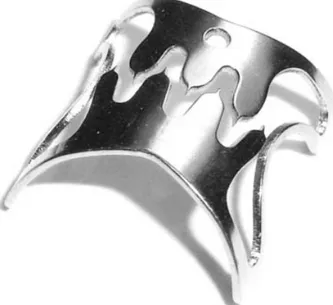
The OTSC (Figure 1) is made of Nitinol and can approximate wound margins like a surgical clamp. It is preloaded in bent shape on its application device
(Figure 2), a disposable cap mounted on the tip of the endoscope. It can be released by means of a thread that runs through the working channel of the endoscope and pushes the clip down. Once released from its application device the OTSC returns to its original closed shape due to the superelastic proper-ties of the material, without the need for external forces. It then exerts a force of approx 8–9 N, thus securely compressing the target tissue, but not inducing a tissue necrosis. The diameter of the clip is 12 mm, which allows closing perforations of up to 15 mm with one clip. In case the lesion is larger, two clips can be placed next to each other. For organ closure usually the traumatic version of the OTSC is used, which has pointed teeth for enhanced tissue grip. The atraumatic version is used for blunt compression of tissue, e.g. for hemostasis.
For closing gastric or colonic access holes different techniques can be used to approximate the wound margins before the OTSC is released. These can include larger endoscopic graspers or suction of the tissue into the applicator cap. A novel twin grasper (Figure 3) can be used to facilitate the approxima-tion of the tissue in NOTES (Ovesco Endoscopy GmbH, Tuebingen, Germany). This grasper has one fixed middle branch and two independently
Figure 1. OTSC, Over-The-Scope-Clip.
Figure 2. OTSC application cap.
Figure 3. Twin grasper for the approximation of perforation holes in NOTES.
mobile lateral branches, subdividing the grasper into two functional parts. The twin grasper can be used through any standard 2.8 mm working channel.
OTSC closure technique for NOTES
After completing the intra-abdominal part of the NOTES procedure, the endoscope is withdrawn from the organ lumen, e.g. the gastric cavity in case of a transgastric procedure. The OTSC system is mounted to the tip of the endoscope and re-inserted. The access lesion is then visualized. Re-insertion of the endoscope and finding the transluminal access lesion in the organ wall is usually not difficult, due to the size of the lesion. However, a guidewire can be left inside the hole in order to facilitate targeting the lesion again quickly after re-insertion of the device.
The actual closure procedure consists of two steps:
N
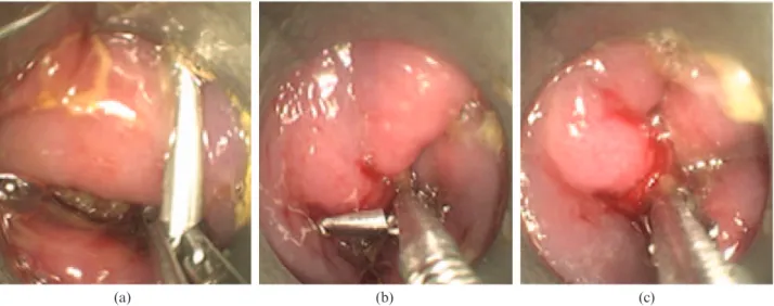
Approximation of the wound margins: The twin grasper is inserted and the first lateral wound margin is grasped (Figure 4a). Then the device is slightly pulled back into the OTSC applicator cap and the second jaw of the grasper is opened while the device is positioned over the second lateral wound margin (Figure 4 b). The second branch is also closed. This maneuver approximates the wound margins (Figure 4 c). Depending on the shape and location of the lesion repeat trials may be needed until the margins have been well approximated. Under more tangential approaches to the organ wall this can be more difficult than under more perpendicular approaches. Before application of the clip, the secure grip of the gastric wall is visualized with the endoscope.N
Application of the OTSC clip: The twin grasper is pulled back into the OTSC applicator cap to establish a close contact between the cap and the target tissue. Then the OTSC clip is released through rotating a hand wheel pulling the thread in the working channel of the endoscope tight. The clip is visually inspected and tightness of the closure is verified by means of air inflation and the ability to maintain organ distension.Figure 5 shows the intraluminal, Figure 6 shows the extraluminal aspect of the clipping site after the closure of a gastrotomy in the domestic pig.
The closed lesions were visually inspected for closure tightness and were tested by air inflation. Air-tight closure was verified before withdrawing the endoscope. In earlier studies on colonic tissue, OTSC has demonstrated its ability to provide a pressure-tight wall closure, resistant to pressures of 20 mm Hg (9). Animal experimental research conducted with OTSC was approved by the responsible governmen-tal agency, as according to German legal provisions.
Discussion
The general feasibility of full-thickness hollow organ closure with folding clips has already been demon-strated for closing the gallbladder after laparoscopic cholecystotomy by Mentges and Buess1991 (8).
The OTSC is a new device than can be applied for the closure of transluminal GI organ access to the abdominal cavity. The tissue compression capabil-ities provide for a tight closure of the target lesion. This has been studied for colonic perforations in the porcine animal model, including investigation of the tissue healing process over two weeks, with good
(a) (b) (c)
success (9). This was the reason to study a technique for the application of OTSC also in the field of NOTES access closure. The OTSC clip has been successfully used clinically in different areas of conventional flexible endoscopy (10). Healing of the gastric wall after closure has not been examined in our experiments. Clinical experience, collected with OTSC for gastric closure on a case level outside of NOTES applications, however, has shown pro-mising results (11).
The technique described here allowed closing gastric and colonic access perforations in the porcine
animal model. The porcine model of appropriate size (w50 kg) corresponds well to the human with regard to anatomy and organ wall thickness of the GI tract.
Figure 6 demonstrates that the full organ wall has been captured and involved into the tissue captured by OTSC. Therefore OTSC can provide a full thickness closure of the gastric wall after NOTES in the experimental model.
The handling of OTSC and the ancillary equip-ment for organ closure, e.g. the twin grasper, is simple and corresponds to conventional flexible endoscopic techniques, such as clipping or band ligating. At the same time the big tissue capture and high compression force set OTSC apart from conventional clips passed through the working channel or endoloops.
The current data base on OTSC and on other techniques proposed for organ closure after NOTES does not yet allow determining clear advantages or disadvantages of the different options.
Case numbers reported so far are still too low to fully judge the performance of the techniques proposed in terms of closure result, ease of use and reliability. Further experimental and clinical research into closure procedures for NOTES will be needed. In our opinion OTSC, being a simple and effective device, might be well positioned for further investigation.
Conflict of interests according to the
international Committee of Medical Journal Editors
Marc O. Schurr and Gerhard Buess are among the founders of Ovesco Endoscopy and hold shares in the company. Chi-Nghia Ho and Gunnar Anho¨ck are employees in the R&D department of Ovesco Endoscopy. The other authors have no disclosures to make. Material for experiments has been provided free of charge by Ovesco Endoscopy.
References
1. Buess G, Cuschieri A. Raising our heads above the parapet: ES not NOTES. Surg Endosc. 2007;21:835–7.
2. Pai RD, Fong DG, Bundga ME, Odze RD, et al. Transcolonic endoscopic cholecystectomy: a NOTES survival study in a porcine model. Gastrointest Endosc. 2006;64:428–34. 3. Fong DG, Pai RD, Thompson CC. Transcolonic endoscopic
abdominal exploration: a NOTES survival study in a porcine model. Gastrointest Endosc. 2007;65:312–8.
4. Magno P, Giday SA, Dray X, Chung SS, et al. A new stapler-based full-thickness transgastric access closure: results from an animal pilot trial. Endoscopy. 2007;39:876–80.
5. McGee MF, Marks JM, Jin J, Williams C, et al. Complete En-doscopic Closure of Gastric Defects Using a Full-Thickness Figure 6. Laparoscopic view of the clipping site after intraluminal
closure of a gastrotomy. Note the impression at the gastric surface, indicating the involvement of the muscular wall into the tissue capture by the clip.
Tissue Plicating Device. J Gastrointest Surg. 2007 Oct 24 [Epub ahead of print].
6. Mc Gee MF, Marks JM, Onders RP, Chak A, et al. Complete Endoscopic Closure of Gastrotomy After Natural Orifice Translumenal Endoscopic Surgery Using the NDO Plicator. Surg Endosc. 2007 Sep 3 [Epub ahead of print].
7. Perretta S, Sereno S, Forgione A, Dallemagne B, et al. A new method to close the gastrotomy by using a cardiac septal occluder: long-term survival study in a porcine model. Gastrointest Endosc. 2007;66:809–13.
8. Mentges B, Buess G, Melzer A, Gutt C, et al. Experimental laparoscopic cholecystotomy. Surg Endosc. 1991;5:51–6.
9. Schurr MO, Hartmann C, Kirschniak A, Ho C-N, et al. Experimental study on a new method for colonoscopic closure of large bowel perforations with the OTSC clip. Biomed Tech (Berl). 2008;53:45–51.
10. Kirschniak A, Kratt T, Stuker D, Braun A, et al. A new endoscopic over-the-scope clip system for treatment of lesions and bleeding in the GI tract: first clinical experiences. Gastrointest Endosc. 2007;66:162–7.
11. Kirschniak A, Traub F, Kueper MA, Stuker D, et al. Endoscopic treatment of gastric perforation caused by acute necrotizing pancreatitis using over2the2scope clips: a case report. Endoscopy. 2007;39:1100–2.