Università di Pisa
Divisione di Radiologia Diagnostica ed
Interventistica
Direttore Prof. C. Bartolozzi
Tesi di Specializzazione - Anno Accademico 2010-2011
Can Diffusion weighted MR imaging add diagnostic
information in the evaluation of uterine tumor?
Relatore: Chiar. mo Prof. Carlo Bartolozzi
INDEX
Abstract
3
Introduction 4
Materials and Methods 8
Results 10
Discussion 11
Tables 14
Pictures 17
Conclusion 22
References 23
2ABSTRACT
Purpose: to evaluate the role of DW imaging in the detection of cervical and endometrial cancers and correlate the histopathological grading and ADC values.
Materials and Methods:
53 women with positive biopsy for cervical and endometrial cancer enrolled in the study underwent MR examination; based on biopsy 34/53 patients had cervical cancer and 19/53 patients had endometrial cancer.
MR study protocol included DWI sequence obtained with a single-shot echo-planar sequence (EPI) in the axial or sagittal plane, with b value of 500s/mm2.
MR protocol also included fast spin-echo T2w.i. sequences acquired in the sagittal and axial planes. Lesions’ signal intensity and ADC values were evaluated on DWI images. Lesions’ signal intensities on T2w.i and maximum diameter were also collected.
Student’s t test was applied for statistical analysis of collected data; a two tailed p-value of less than 0.05 was considered as significant.
Results:
On DWI, only 30/53 tumors were appreciable , in particular 29/30 lesions (96.7%) were
cervical cancers; 1/19 was an endometrial cancer; 18/19 endometrial cancers and 5/34
cervical cancers were not detectable on DWI images.
No relationship was found between cervical cancers’ size and lesions’ signal intensity on
DWI nor lesions’ signal intensity on T2w.i. (p>.05).
Mean ADC values of G1 graded cervical cancers resulted to be significantly higher than ADC values of G2 and G3 graded neoplasms (p<.05).
Conclusion: DWI can help in detecting the presence of cervical tumor, while it does not result useful in assessing the presence of endometrial neoplasms. 3
ADC values of well differentiated cervical cancers are higher than relative values of more de-differentiated neoplasms
INTRODUCTION
Diffusion-weighted imaging (DWI) is a functional imaging technique, applied in Magnetic Resonance imaging, whose contrast derives from the random motion of water molecules within explored tissues.
Since image contrast is derived from inherent differences in the restriction of the movement of water molecules, no exogenous contrast medium administration is required, so that DWI sequences can now be included in routine patient assessment.
DWI uses the measurement of Brownian movements of water molecules by applying symmetric pairs of equally weighted diffusion sensitizing gradients about the 180°refocusing pulse of a spin-echo T2-weighted sequence. Static water molecules develop additional phase incoherencies from the application of the first diffusion gradient, but these incoherencies are eliminated by the application of the second gradient, resulting in no additional net loss of signal (aside from normal T2 decay) [1]. However, mobile water is not completely rephased by the second gradient owing to movement to a different microenvironment during the application of the first gradient, so that a subsequent reduction in signal intensity is observed. DWI sensitivity to water motion is dependent on three main parameters: the gradient amplitude, the duration, and the time interval(b-value)
between paired diffusion gradients [2]. Tissues containing water that is moving the most freely (eg, within blood vessels, ducts, or the bladder) will demonstrate greater signal losses after the application of the smallest diffusion gradients (<100 sec/mm2). Signal losses caused by water motion in the extracellular space of tumors occur at higher b values because water motion is modified by interactions with hydrophobic cell membranes 4
and macromolecules (increased extracellular space tortuosity) [3]. In solid tumors of high cellularity, there are additional significant reductions in extracellular space, resulting in further restrictions to free water movement.
When DWI is used in gynecologic applications, cervical cancers have been shown to have significantly lower apparent diffusion coefficient (ADC) values than normal cervical tissue [4]. Similar findings have been noted in endometrial cancers, with a tendency toward lower ADC values in higher-grade lesions.
ADC value
The ADC value (measured in mm2/sec) is usually calculated by the slope of the line of the natural logarithm of signal intensity (y axis) versus b values (x axis) [5].
DWI imaging: applications in female pelvic neoplasms Cervical cancer
Cervical cancer is the second most common female cancer in the world and it occurs predominantly in women between the age of 30 and 44 years [6]. The American Cancer Society provides the following list of risk factors for cervical cancer: Human Papillomavirus (HPV) infection, smoking, HIV infection, chlamydia infection, dietary factors, hormonal contraception, multiple pregnancies, exposure to the hormonal drug diethylstilbestrol (DES) and a family history of cervical cancer [7].
Cervical cancer: Histopathology
The World Health Organization (WHO) recognizes two main histological types of invasive cervical cancer.
Squamous carcinoma (which constitute about 85% of all cases)
Several other types of carcinoma (eg adenosquamous carcinoma, adenoid cystic carcinoma, metastatic carcinoma) make up the remaining 3-5% of all cases.
Squamous carcinomas are further typed according to whether they are keratinizing or not keratinizing carcinomas. Keratinizing carcinomas may be well differentiated or moderately differentiated and are composed of large tumour cells. Non-keratinizing carcinomas (poorly differentiated carcinomas) may be of large cell or small cell type [8].
Most cervical squamous cell carcinomas grow at the squamocolumnar junction (SCJ). In young women, the SCJ is located outside the external uterine os, and the tumor tends to grow outward (exophytic growth pattern). In contrast, in elderly patients, the SCJ is located within the cervical canal. In these patients, cervical cancer tends to grow inward along the cervical canal (endophytic growth pattern) [9].
Adenocarcinomas are less commonly found and although each type is histologically distinct, it is not uncommon to find two or more histological forms of adenocarcinoma in a single tumour .The frequent coexistence of glandular and squamous carcinoma suggest that they may have a common origin in the reserve cells of the cervix as well as a common etiology. The most frequent type of adenocarcinoma to be found in the cervix is the
endocervical type of mucinous adenocarcinoma. Three grades of endocervical carcinoma are recognized -well differentiated, moderately differentiated and poorly differentiated - depending on the similarity of the tumour cell to the glandular epithelial lining the
endocervix. In fact, in endocervical adenocarcinoma, endocervical type, different subtypes can be found (endometrioid type, clear cell type, papillary serous type, intestinal type, mixed type) [10].
Cervical cancer: MR features
At baselineT2 w.i MR examination, cervical cancers appear as slightly hyperintense masses, while their predominantly signal intensity on baseline T1 w.i is hypointensity. On vascular dynamic study, cervical cancers can show no enhancement or slighly marginal enhancement on arterial phase.
Endometrial cancer
Endometrial cancer is the most common malignancy of the genital tract and overall the endometrium is the fourth most frequent cancer site. Risk factor for endometrial cancer include obesity, diabetes, oestrogen therapy, polycystic ovarian syndrome and
westernization of lifestyle.[11]. Approximately 75% of cases occur in postmenopausal women, with the median age at diagnosis being 70 years.
Endometrial cancer: Histopathology
Endometrial tumors can be have different histology: endometrial carcinoma (with its histopathologic subtypes: villoglandular or papillary, secretory, ciliated cell,
adenocarcinoma with squamous differentiation), adenocarcinoma (serous, mucinous, clear cell), squamous cell carcinoma; undifferentiated carcinoma; mixed carcinoma, metastatic carcinoma and carcinosarcoma.
Adenocarcinomas account for 90% of endometrial neoplasms, whereas uterine sarcomas are relatively rare and account for only 2%–6%; the remaining histologic types include adenocarcinoma, with squamous cell differentiation and adenosquamous carcinoma [12].
Endometrial cancer: MR features
At MR examination, endometrial cancer is iso-hypointense in respect to the
surrounding normal endometrium on baseline T1w.i -weighted images, while it does most commonly show heterogeneous, iso up to hyperintensity on baseline T2wi.
Neoplasms are usually mildly hyperintense on baseline T2w.i. in respect to myometrium.
Endometrial cancer can have different pattern of appearances on baseline T2 w.i.:
hypointense pseudonodular lesions, diffuse endometrial thickening, hypointense
vegetations, or infiltrative endometrial plaques with heterogeneous signal intensity.
Endometrial carcinomas are divided into two types: one shows as a well-demarcated
exophytic mass (Type I), while the other shows as an invasive, endophytic growth toward
the myometrium (Type II). The former subtype is considered to be malignant with favorable
prognosis [13].
MATERIALS AND METHOD
Patients
The present retrospective study received institutional ethics committee approval, and all patients provided informed written consent.
Study group consisted of 53 women (mean age: 54.5 years; range 30–83) years with biopsy positive for cervical and endometrial cancer and enrolled from March 2008 to January 2012.
All the patients performed MR examination after biopsy . Mean time between biopsies and MR examination resulted to be 2 weeks.
MRI study
All MRI examinations were performed with a 1.5 T unit (Excite,Twin Speed, GE Healthcare) with an 8 channel phased-array coil.
MRI protocol included fast spin-echo T2wi.i sequences acquired in the sagittal and axial planes [repetition time/echo time (TR/TE), 4000 ms/85 ms; echo-train length 17; matrix size 320 # 224; band width 31.25 Hz/pixel; field of view 36 cm; NEX: 2; slice thickness: 6 mm; gap: 1 mm); when needed a sagittal or axial FSE T2w.i sequence with fat saturation was performed.
DWI sequence was acquired using single-shot echo-planar imaging (EPI) and array spatial sensitivity encoding technique (ASSET; TR/TE 4000 ms/58.5 ms; matrix size 128x128-
224; field of view 26-34 cm; nex 4; section thickness 6 mm; gap 1 mm; R factor 2) in the
axial or sagittal plane with b values of 0 and 500 s/ mm2. The DWI examination time was 1-2 min.
Statistical analysis
The Student’s t-distribution was used for the two arm comparison and p value acquisition. Receiver operating characteristic analysis and relationship between cervical cancers’ size and lesions’ signal intensity on DWI nor lesions’ signal intensity on T2w.i. or between cervical cancers’ size and lesions’ signal intensity on T2w.i were calculated. Finally we searched relationship between mean ADC values and histological grading. A p-value of less than 0.05 was considered as significant.
RESULTS
At histology, 53 patients had a bioptical diagnosis of a uterine cancer. 34 patients had a
cervical cancer and 19 an endometrial cancer.
Out of 34 cervical cancers, 19 were non keratinizing squamous cell carcinomas, 10
keratinizing squamous cell carcinomas, 4 endocervix adenocarcinomas and 1 cancer with mucinous aspects.
Out of 19 endometrial cancers, 12 patients were endometrioid adenocarcinoma, 1
neuroendocrine tumor, 2 were adenocarcinoma with squamous cell differentiation, 1 was a endometrioid serous adenocarcinomas; 1 patient with endometrioid mucinous
adenocarcinoma;1 case of endometrial metastasis of urotelial cancer and 1 patient with a carcinosarcoma.
Well differentiated cervical cancer G1 had increased ADC value from 1,30 to 1,40 e-9; in mildly differentiated cervical cancers (G2) ADC value could range from 0,82 to 1,24 e -9. 10
In poorly differentiated tumors (G3) ADC value could range from 0,43 to 1,24 e-9. (Table. 2).
On DWI, only 30/53 tumors were appreciable, in particular 29/30 lesions (96.7%) were
cervical cancers; 1/19 was an endometrial cancer; 18/19 endometrial cancers and 5/34
cervical cancers were not detectable on DWI images.
No relationship was found between cervical cancers’ size and lesions’ signal intensity on
DWI nor lesions’ signal intensity on T2w.i. (p>.0.5).(Table. 3- 4).
Mean ADC values of G1 graded cervical cancers resulted to be significantly higher than ADC values of G2 and G3 graded neoplasms (p<.0.5). (Table. 5)
DISCUSSION
Cervical cancer
With increasing tumor cellularity and architectural distortion, there is a reduction in extracellular space, which also becomes increasingly tortuous. These changes are reflected by a reduced ADC value. High-grade adenocarcinomas typically have high
cellular density and so would be expected to have lower ADC values. A trend toward lower ADC values in higher-grade endometrial cancers was noted by Tamai et al [14].
Our study confirms that high-grade adenocarcinomas typically have high cellular density and lower ADC values.
Well differentiated cervical cancer G1 had increased ADC value from 1,30 to 1,40 e-9; in mildly differentiated cervical cancers (G2) ADC value could range from 0,82 to 1,24 e -9. In poorly differentiated tumors (G3) ADC value could range from 0,43 to 1,24 e-9.
A limit of DWI in discovering cervical cancer was correlate to exclusive localization in the uterine fornix (anterior or posterior) or to uterine atrophic structure. In the most of case cervical cancers exhibited a ovalar morphology. Cervical cancers appeared hyperintense 11
on T2 w.i in 25 patients; isointense in 6 patients and hypointense in 1 patient. ADC value at b 500 correlated with histological grading and therefore with prognosis. In fact ADC value from 0,43 to 1,24 e-9 were correlate to a bad prognosis instead a ADC value from 1,30 to 1,40 e-9 was correlate to a good prognosis.
Endometrial cancer.
The use of a high b-value makes images more sensitive to water diffusion; it thus
increases contrast enhancement between normal and cancerous tissue, but this is offset
by decreased signal intensity and anatomic detail in the adjacent structures [15]. DWI can demonstrate uterine endometrial cancer and ADC measurement can provide useful
information for differentiating malignant from benign uterine endometrial cavity lesions.
Higher-grade endometrial cancers exhibit a tendency towards decreased values compared
to those of lower-grade lesions. In our study, using a b value (500), DWI was not useful to
reveal endometrial cancer (DWI failed in detection in 18 patients (94,73%). DWI allowed to
reveal size of lesion in 1 patient. Generally endometrial tumor appeared as a slightly
hypointense lesion on DWI in contrast with marked hyperintensity of normal endometrium,
without revealing margin and tumoral size or as hypointense line in thickened
endometrium. Rarely, as in one case of our study, endometrial tumor appeared as
hyperintense lesion on DWI.
Endometrial cancers is better revealed on T2 w.i where tumor can exhibit a focal
hypontensity in endometrial cavity or a decreased hyperintensity in a thickened
endometrium.
Meanwhile, we confirm as noted by Sachi Kaneda [17] that endometrial cancer appears more elongated than cervical cancer and tends to form a longitudinal shape while cervical cancer tends to appear round or ovoid because in growth endometrial cancer is
sandwiched by the strong uterine muscular wall. Therefore endometrial cancer is forced to migrate in a cranio-caudal direction rather than a lateral direction, thus resulting in an elongated configuration.
Some limitations of this study need to be taken into consideration. First, this was a retrospective study, and the readers knew the diagnosis of endometrial and cervical carcinoma when they reviewed the images, which may have influenced the readers. Second, the study was also limited by the relatively small number of patients included. Third, small cancers’ (<1cm) were exclused from study.
TABLES
Table1:
MR protocol- T2 w.i Technical parameters.
Parameters T2w.i
TR/TE
4000 ms/85 ms
Echo-train length
17
Matrix size
320 # 224
Band width
31.25 Hz/pixel
Field of view
36cm
gap
1mm
Table 2: Histological grading and ADC value calculated in cervical
cancer
Histological
grading
ADC values
(e-9 mm2/s)
G1
1.30-1.40
G2
0.82-1.24
G3
0.45-1.24
14
Table 3:
no relationship between
cervical cancers’ size and lesions’
signal intensity on DWI
Signal
Intensity DW
Number
Mean of
Area
Std Dev
P value
Hyperintense 27
1833,41
1506,16
0,13
Hypointense
5
306,00
200,95
0,03
Isointense
2
2138,00
1558,46
0,77
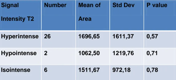
Table 4:
no relationship between
cervical cancers’ size and lesions’
signal intensity on T2 w.i.
Signal
Intensity T2
Number
Mean of
Area
Std Dev
P value
Hyperintense 26
1696,65
1611,37
0,57
Hypointense
2
1062,50
1219,76
0,71
Isointense
6
1511,67
972,18
0,78
15
Table 5:
significantly
relationship between
histological grading and
mean ADC value in G1 cervical cancer (p<.0.5).
Histological
grading
Number
Mean
ADC value
Std Dev
P value
G1
5
1,32
0,07
0,02
G2
14
0,78
0,46
0,04
G3
15
0,85
0,44
0,67
16
PICTURES
Figure. 1: T2w.i, DWI and DWI with color map of G1 cervical
cancer.
Figure.2
:
T2w.i, DWI and DWI with color map of G2 cervical
cancer.
Figure. 3: T2w.i, DWI and DWI with color map of G3 cervical
cancer.
Picture 4: T2 w.i. and DWI in endometrial cancer.
Picture 5:
T2 w.i. and DWI in endometrial cancer.
CONCLUSIONS
DW image can help to detect the presence of cervical tumor; DWI image by means of ADC values, can be helpful to discriminate poorly differentiated from well differentiated cervical tumors. Based on our results, higher ADC values (from 1,30 to 1,40e-9) were noted in G1 tumors while lower values (0,43-1,24 e-9) were observed in G3 tumors. In the cases of
cervical cancer with G2 the ADC were values were variable and therefore controversial
0,82 -1,47 e-9.
Before we analyzed which possible limits Diffusion could have in detection cervical cancer and after this retrospective study. We noticed that DWI failed when cancer was placed only in fornix or if cervical tumor arise from an atrophic structure.
We also analyze which factor could influence the ADC values as: the keratinizing and mixoid or solid components. Keratinizing let reduce ADC value because in deed this element is present in the well differentiated tumors. Mixoid or solid components, instead, let increase ADC value.
In endometrial tumors we considered that DWI is not useful in detection. We arrived to diagnosis and staging only with T2 w.i and T1 w.i after injection of contrast media.
REFERENCES
1:Fujii S, Matsuse E, Kanasaki Y, et al. Detection of peritoneal dissemination in
gynaecological malignancy: evaluation by diffusion-weighted MR imaging. Eur Radiol 2008;18:18–23.
2: Thierry A.G. M. Huisman.”Diffusion-weighted imaging:basic concepts and application in cerebral stroke and head trauma”. European Radiology. Volume 13, Numero 10, 2283-2297, DDI:10.1007/s00330-003-1843-6
3: Thierry A.G. M. Huisman.”Diffusion-weighted imaging:basic concepts and application in cerebral stroke and head trauma”. European Radiology. Volume 13, Numero 10, 2283-2297, DDI:10.1007/s00330-003-1843-6.
4: : Whittaker C, Coady A, Culver L “Diffusion-weighted MR Imaging of Female Pelvic Tumors: A Pictorial Review. Radiographics 2009; 29:759-778.
5: Whittaker C, Coady A, Culver L.”Diffusion-weighted MR Imaging of Female Pelvic “. Tumors: A Pictorial Review,.Radiographics 2009; 29:759-778.
6:R H Reznek, A Sahdev. ”MR imaging in cervical cancer: seeing is believing”. The British Journal of Radiology, 78 (2005), S73–S85
7: Nubia Mu˜noz, Xavier Castellsagu´e, Amy Berrington de Gonz´alez, Lutz
Gissmann. ”Chapter 1: HPV in the etiology of human cancer”.Vaccine 24S3 (2006) S3/1– S3/10
8: “Histologic classification of epithelial tumors”. Holland-Frei Cancer Medicine. 6th edition. Kufe DW, Pollock RE, Weichselbaum RR, et al., editors. Hamilton (ON): BC Decker; 2003. 9:Yoshikazu Okamoto, Yumiko O.Tanaka, Masato Nishida, Hajime Tsunoda, Hiroyuki Yoshikawa,Yuji Itai.”MR Imaging of the Uterine Cervix: Imaging-Pathologic Correlation”.
RSNA, 2003.
10: Imadome K, Iwakawa M, Nakawatari M, Fujita H, Ohno T, Nakamura E, Ohkubo Y, Tamarki T, Kiyohara H, Imai T. “Subtypes of cervical adenosquamous carcinomas classified by EpCAM expression related to radiosensitivity”. Cancer Biol Ther.2010 Nov;10(10):1019-26.Epub 2010 Nov 15.
11: Brian MacMahon. “Risk factors for endometrial cancer” .Gynecologic
Oncology.Volumes 2;issues 2-3 August 1974.page 122-129.
12: “FIGO Staging System for Endometrial Cancer: Added Benefits of MR Imaging”. Peter
Beddy, Ailbhe C.O’Neill; Adam K. Yamamoto, Helen C. Addley, Caroline Reinhold, Evis Sala. Radiographics 2012.
13:Jaime Prat.” Prognostic parameters of endometrial carcinoma”.Human Pathology.Volume 35, Issue 6, Pages 649-662, June 2004.
14:Tamai K, Koyama T, Saga T, et al. Diffusion weighted MR imaging of uterine endometrial cancer. J Magn Reson Imaging 2007;26:682–687.
15: Antonin Levy, Aı¨cha Medjhoul , Caroline Caramella, Elise Zareski,
Oscar Berges, Cyrus Chargari, Be´re´nice Boulet, Franc¸ois Bidault,
Clarisse Dromain and Corinne Balleyguier. “Interest of Diffusion-Weighted Echo-Planar MRImaging and Apparent Diffusion CoefficientMapping in Gynecological Malignancies: A
16: Tamai K, Koyama T, Saga T, et al. Diffusion-weighted MR imaging of uterine endometrial cancer. J Magn Reson Imaging 2007;26:682–687.
17:
Yu-Ching Lin, Gigin Lin, Yu-Ruei Chen, Tzu-Chen Yen,
Chun-Chieh Wang, Koon-Kwan Ng.“Role of Magnetic Resonance Imaging and Apparent DiffusionCoefficient at 3T in Distinguishing between Adenocarcinoma of
the Uterine Cervix and Endometrium”.Chang Gung Med J Vol. 34 No. 1 January-February 2011