UNIVERSITY OF MOLISE
Department of Agricultural, Environmental and Food Sciences
INTERNATIONAL Ph.D. in
“WELFARE, BIOTECHNOLOGY AND QUALITY OF ANIMAL
PRODUCTION”
(
XXVI CYCLE)
Related disciplinary scientific section: 07/G1 (Scienze e Tecnologie Animali)
General Coordinator: Prof. Giuseppe Maiorano
Doctorate Thesis Title:
Cryopreservation of rabbit semen: effectiveness of
different permeable and non-permeable cryoprotectants
on post-thaw sperm quality and
reproductive performances
Supervisor:
Ph.D. Candidate:
Prof. Nicolaia Iaffaldano
Dr. Michele Di Iorio
143442
ACKNOWLEDGEMENTS
The realization of this thesis was only possible due to the several people's
collaboration, to which I desire to express my gratefulness.
I would like to express my deepest gratitude to my supervisor, Prof. Nicolaia
Iaffaldano for her guidance, patience, motivation, caring, and providing me with an
excellent atmosphere for doing research.
I would like to thank in a special way Dr. Maria Pina Rosato, for her help,
encouragement, teaching and advice.
I would like to thank also my Slovak advisor Prof. Peter Chrenek for giving me the
possibility to work in his laboratory, at the Animal Production Research Centre
(APRC Nitra).
I would like to thank the General Coordinator Prof. Giuseppe Maiorano for his
accurate organization of the Ph.D. course.
My grateful appreciation also for Mr. Innocenzo Gentile, who made possible my
research activity in his farm.
Many thanks to the president (Dr. Sergio Pompa) and Director (Dr. Mario
Giovannoli) of Italian National Rabbit Breeders (ANCI-AIA) for allowing me to
use the Central Breeding Farm. I also thank Drs. Paola Principe and Michele
Schiavitto for the technical assistance with semen collection and artificial
insemination.
I would like to thank also Prof. Luca Romagnoli for help with the statistical
analysis of data, and Ana Burton and Amber Burchell for the English revision.
Moreover, I would like also to thank all my PhD colleagues.
I also thank all my friends, who supported me during these years.
Finally, but not the least, I would like to thank my family, for their love, constant
support and encouragement.
I
CONTENTS
Abstract (English) ……… V Riassunto (Italiano) ………. IX
PART 1. INTRODUCTION ………. 1
Chapter 1. Rabbit: History, taxonomy and meat production ………... 1
1.1 History, taxonomy and domestication of the rabbit ……… 1
1.2 Rabbit meat production in the World ……….. 2
1.3 Rabbit meat production in Italy……… 3
1.4 Rabbit meat features……… 4
Chapter 2. Rabbit reproduction ………... 7
2.1 The male ……….. 7
2.1.1 Anatomy of the male rabbit reproductive system ……… 7
2.1.2 Rabbit Spermatozoa ……… 10
2.1.3 Spermatogenesis and sperm maturation ………. 13
2.1.4 Reproductive activity of the buck ……… 15
2.2 The female ………... 15
2.2.1 Anatomy of the female rabbit reproductive system ………. 15
2.2.2 Estrus and ovulation ……… 17
2.2.3 Fertilization and pregnancy ……… 19
2.2.4 Parturition ………... 20
2.2.5 Lactation ……….. 20
Chapter 3. Artificial insemination in rabbit ………... 22
3.1 Rabbit semen collection ……….. 23
3.2 Assessment of semen quality ……….. 24
3.2.1 Macroscopic parameters ………. 24
3.2.2 Microscopic parameters ……….. 25
3.2.2.1 Sperm concentration ………. 25
3.2.2.2 Sperm motility ………... 25
3.2.2.3 Sperm viability ……….. 28
3.2.2.4 Sperm osmotic resistance ……….. 29
3.2.2.5 Acrosome integrity ……… 30
II
3.2.2.7 Apoptosis ………... 32
3.3 Semen dilution and artificial insemination in does ... 33
Chapter 4. Storage of rabbit semen ... 34
4.1 Hypothermic-liquid storage ... 34
4.2 Sperm cryopreservation ... 37
4.3 Cryopreservation of rabbit semen ... 38
4.3.1 Extenders ... 39
4.3.2 Cryoprotectants ... 39
4.3.3 Freezing protocols ... 43
4.3.4 Thawing ... 45
4.3.5 Devices to preserve rabbit sperm ... 45
PART 2. RESEARCH WORKS ... 48
Chapter 5. Research n°1 ... 48
The cryoprotectant used, its concentration and the equilibration time are critical for the successful cryopreservation of rabbit sperm: dimethylacetamide versus dimethylsulfoxide………... 48
5.1 Aim ... 48
5.2 Materials and methods ……… 49
5.2.1 Chemicals ... 49
5.2.2 Animals ... 49
5.2.3 Semen collection ……….. 50
5.2.4 Study 1. Effects of CPA concentrations and equilibration times on post-thaw semen quality ... 51
5.2.4.1 Semen processing ... 51
5.2.4.2 Sperm quality ... 52
5.2.5 Study 2. Comparing the in vivo performance of the DMA and DMSO protocols ………. 58
5.2.6 Statistical analysis ... 58
5.3 Results ... 59
5.3.1 Effects of CPA concentrations and equilibration times on post-thaw semen quality ……….. 59
5.3.2 Comparing the in vivo performance of the DMA and DMSO protocols ………... 60
III
Chapter 6. Research n°2 ... 68
Cryopreservation of rabbit semen using non-permeable cryoprotectants: effectiveness of different concentrations of low-density lipoproteins (LDL) from egg yolk versus egg yolk or sucrose ……… 68
6.1 Aim ... 68
6.2 Materials and methods ……… 69
6.2.1 Chemicals ... 69
6.2.2 Animals ... 69
6.2.3 Semen collection ……….. 70
6.2.4 Experiment 1. Effects of different LDL concentrations, egg yolk and sucrose on postthaw semen quality ………... 71
6.2.4.1 LDL extraction ... 71
6.2.4.2 Extender preparation ... 72
6.2.4.3 Semen processing ... 72
6.2.4.4 Sperm quality ... 72
6.2.5 Experiment 2. Comparing the in vivo reproductive performance of rabbit does inseminated with semen cryopreserved using the most effective CPAs identified in Experiment 1 ……….. 72
6.2.6 Statistical analysis ... 74
6.3 Results ... 74
6.3.1 Effects of different LDL concentrations, egg yolk and sucrose on post-thaw semen quality ... 74
6.3.2 Comparing the in vivo reproductive performance of rabbit does inseminated with 10% LDL versus sucrose ……… 79
6.4 Discussion ... 80
Chapter 7. Research n°3 ... 83
The cryoprotective effect of Ficoll on the rabbit spermatozoa quality ……… 83
7.1 Aim ... 83
7.2 Materials and methods ... 83
7.2.1 Animals ... 83
7.2.2 Semen collection ... 84
7.2.3 Freezing of the semen ... 85
7.2.4 Sperm quality ... 85
IV
7.2.4.2 Analysis of plasma membrane integrity ... 86
7.2.4.3 Ultrastructure analysis ... 88 7.2.5 Fertility analysis ... 89 7.2.6 Statistical analysis ... 90 7.3 Results ... 90 7.3.1 Semen analysis ... 90 7.3.2 Fertility analysis ... 93 7.4 Discussion ... 93 PART 3. Chapter 8. CONCLUSION ... 97 REFERENCES ... 99 List of figures ... 124 List of tables ... 127 List of abbreviations ... 128 List of publications ... 130
V
ABSTRACT
Rabbit breeding for meat production is based mainly on artificial insemination (AI) programs. In order to obtain many of potential advantages of AI, improvement of the storage of rabbit semen is necessary. Therefore, meat rabbit farming would greatly benefit if semen could be stored after collection and subsequently used for AI without affecting fertility. The rabbit sperm can be stored by refrigeration (in liquid or solid extenders) or freezing. The use of frozen semen would provide more practical advantages for the commercial rabbit industry, as cryopreserved semen could potentially maintain the functionality of the sperm cells for months or even years by facilitating the samples transport in time and space.
Cryopreservation has been widely used in the cattle industry, less used in other livestock species, such as pigs, sheep, poultry and rabbit. Therefore, a large number of protocols aimed to cryopreserve the rabbit semen have been developed by many researchers. Each of these protocols has involved in the study of some aspect that can affect the success of the rabbit sperm cryopreservation, such as the composition of the freezing medium, nature of cryoprotectant (CPA) and its concentration, freezing conditions and cooling and warming temperatures. The choice of the CPA is certainly one of the most important aspect for an effective freezing protocol for rabbit semen. The CPAs are chemical compounds included in freezing extenders to reduce the physical and chemical stresses resulting from the cooling, freezing, and thawing of sperm cells. The main biophysical factor that can cause cell destruction during the cryopreservation process is the formation of intracellular ice crystals that can be avoided by increasing the cellular dehydration process, by means of a CPA in the freezing solution. Hence, the addition of the permeable CPAs is a necessary step in the freezing protocol of rabbit sperm, because they minimize cellular injury during cryopreservation. Once CPA has been added to a sperm suspension, a period of time is required for the CPA to permeate the cells. This is called the equilibration time, which varies according to the nature and concentration of the CPA. So the efficacy of the CPAs depends on their type, their concentrations and the exposure time of sperm cells before freezing (equilibration time). Nevertheless, the paradox is that the permeable CPAs themselves could have a toxic effect on sperm (membrane destabilization, protein and enzymes denaturation) related directly to the concentration used and the time of cell exposure. Therefore, it is
VI
necessary the addition of a non-permeable cryoprotectant to offset the cryodamage caused by permeable CPA. Different combination of permeable and non-permeable CPAs have been tested for the cryopreservation of rabbit semen, but none of them has given optimal results to consider one of these as the suitable CPA for freezing rabbit semen. Therefore the process of rabbit sperm cryopreservation still suffers for a lack of standardized freezing protocols. In this thesis were conducted three different researches, that have as common denominator to find an effective freezing protocol for rabbit semen, by studying of the effects of different permeable and non-permeable CPAs. The first study was designed to identify a suitable freezing protocol for rabbit semen by comparing the effects of different CPA concentrations and equilibration times of dimethylacetamide (DMA) and dimethylsulfoxide (DMSO) on the semen post-thaw quality. After establishing the best protocols for each CPA, their efficacy was compared by examining the in vivo fertilizing capacity of the semen samples. The animals used for this study, were 32 rabbit bucks and 342 does. Semen was collected using an artificial vagina and the ejaculates were pooled (4 ejaculates/pool). Pooled semen samples were diluted to a ratio 1:1 (v:v) with a freezing medium composed of Tris-citrate-glucose (TCG) containing 8, 12, or 16% DMA or DMSO (all combined with 2% sucrose as a non-permeating CPA) to give final concentrations of 1% sucrose and 4%, 6% or 8% DMA or DMSO. The diluted semen was loaded in 0.25 mL plastic straws and equilibrated for 5, 15 or 45 min before freezing in liquid nitrogen vapor (5 cm above the liquid nitrogen surface). The variables assessed after thawing were sperm motility, viability, osmotic-resistance, and acrosome and DNA integrity. Marked effects on these variables were shown by the CPA concentration and equilibration time, with best results obtained using DMA 6% or DMSO 8% and equilibration times of 45 minutes. These freezing protocols were selected to compare the two CPAs in an insemination trial. Three groups of 114 rabbit does (28 nulliparous and 86 multiparous in each group), were inseminated with fresh semen or with semen frozen using the optimized DMA or DMSO protocols. Conception rates and numbers of total born were similar respectively for the DMSO-frozen (79.8% and 7.7±0.3 young per kindling) and fresh semen (81.6% and 8.6±0.3) yet higher (P≤0.05) than the rates returned using the DMA-frozen semen (47.4% and 6.7±0.4). Moreover, the numbers of rabbits born alive when DMSO was used in the freezing protocol, despite being lower than those recorded using fresh semen, were higher than when DMA was used as the CPA (P < 0.05). The physiological status of the does (nulliparous or multiparous) had no influence on the fertility and
VII
prolificacy results. Our findings indicate that the cryosurvival of rabbit sperm frozen using DMSO or DMA as the CPA is highly influenced by the concentration of CPA used and the time the semen is exposed to the agent before freezing. According to our in
vivo fertility and prolificacy data, DMSO emerged as more effective than DMA for the
cryopreservation of rabbit sperm.
The second study was designed to identify the most effective non-permeable CPA for the cryopreservation of rabbit semen by comparing the effects of different concentrations of low-density lipoproteins (LDL) on post-thaw sperm quality with those of whole egg yolk or sucrose. The performance of the non-permeable CPAs identified as the most effective was assessed in vivo by determining fertility and prolificacy rates. In this study 32 rabbit bucks and 90 does were used. Pooled semen samples were diluted to a ratio of 1:1 (v:v) in freezing extender (TCG and 16% DMSO as permeable CPA) containing as non-permeable CPAs 6, 8, 10 or 15% LDL from egg yolk, 0.1 M sucrose, or 15% egg yolk. The semen was loaded in 0.25 mL straws and frozen in liquid nitrogen vapor. After thawing, we determined sperm motility, viability, osmotic resistance, and acrosome and DNA integrity. Our results clearly revealed a significant effect of LDL concentration on semen quality. Also, at an optimal concentration of 10%, motility and acrosome integrity were improved over the values recorded for egg yolk (P<0.05). Based on the in vitro data, 3 groups of does (n = 30 each) were inseminated with fresh semen or semen frozen using sucrose or 10% LDL. Sucrose led to a significantly higher conception rate (86.7%) than LDL (66.7%) and reproductive performance was similar to that observed for fresh semen. Our findings indicate the markedly better performance of sucrose in vivo as a non-permeable CPA for the cryopreservation of rabbit semen.
The aim of the third study was to evaluate the effect of addition of Ficoll 70 into the cryopreservation medium containing sucrose and DMSO on rabbit spermatozoa characteristics following freezing/thawing. This large molecular weight polymer elevates the viscosity of medium and, therefore, could better protect spermatozoa during the freezing process. Only ejaculates of good initial motility (> 80%) were used in these experiments. Heterospermic pools were diluted in a freezing medium composed of commercial diluent, 16% DMSO and 2% sucrose (control) or in the same medium enriched with 4% of Ficoll 70 (Ficoll) and frozen in liquid nitrogen vapours for 10 min before being plunged into liquid nitrogen.
VIII
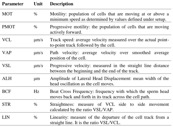
The quality of fresh and frozen/thawed spermatozoa samples was evaluated in vitro using Computer Assisted Semen Analysis (CASA) system, fluorescent probes (peanut agglutinin (PNA)-Alexa Fluor®; annexin V-Fluos) and by electron microscopy. Better cryoprotective effect was observed when the Ficoll 70 was added, compared to the semen cryopreserved with sucrose and DMSO only. The higher values (P˂0.05) of motile and progressively moving spermatozoa immediately after thawing and at 30 min following incubation at 37°C were obtained in the Ficoll group. Moreover, the higher number (P˂0.05) of acrosome intact sperm was found in the Ficoll compared to control group. Furthermore, no significant differences in kindling rates and number of pups born between frozen/thawed and fresh semen group were found. In conclusion, this study showed that the addition of the Ficoll 70 might improve several characteristic of rabbit spermatozoa measured in vitro following freezing/thawing. These researches were important because we have been able to find effective cryopreservation protocols, that showed similar reproductive performances compared to use of fresh semen.
These findings provide direction for future studies designed to address the possibility of using doses of frozen semen for the artificial insemination of rabbits in commercial farms. Moreover, the use of frozen semen will be attractive to the establishment of a gene bank from national or endangered rabbit breeds as gene resources.
IX
RIASSUNTO
Attualmente, i sistemi di allevamento intensivi per la produzione di carne di coniglio si basano principalmente su programmi di inseminazione artificiale (IA). Pertanto, l’allevamento intensivo del coniglio da carne potrebbe usufruire di molti vantaggi se il seme potesse essere conservato dopo la raccolta e successivamente utilizzato per l’IA senza compromettere la fertilità. Due sono le tecnologie disponibili per la conservazione del seme di coniglio: la refrigerazione (in diluenti liquidi o solidi) e il congelamento. L'uso di seme congelato fornirebbe vantaggi più pratici per l'allevamento del coniglio da carne, in quanto, esso potrebbe potenzialmente mantenere la funzionalità delle cellule spermatiche per mesi o anni, facilitando il trasporto dei campioni nel tempo e nello spazio. La crioconservazione del seme è stata ampiamente utilizzata nel settore bovino, meno in altre specie animali, come i suini, ovini, polli e conigli. Pertanto, un gran numero di protocolli di congelamento per il seme di coniglio sono stati sviluppati da molti ricercatori. Diversi sono i fattori che possono influenzare il successo del congelamento del seme, quali la composizione del diluente di congelamento, la natura del crioprotettore (CPA) e la sua concentrazione, le condizioni di congelamento, e le temperature di raffreddamento e scongelamento. La scelta del CPA è sicuramente uno degli aspetti più importanti per ottenere un efficace protocollo di congelamento per il seme di coniglio. I CPA sono molecole, che vengono aggiunte nel diluente di congelamento, per ridurre gli stress fisici e chimici derivanti dal raffreddamento, congelamento e scongelamento degli spermatozoi. Infatti, il principale fattore biofisico che può causare la distruzione cellulare, durante il processo di crioconservazione, è la formazione dei cristalli di ghiaccio intracellulari. La loro formazione può essere evitata aumentando il processo di disidratazione cellulare, mediante l’aggiunta di un CPA nella soluzione di congelamento. Il CPA richiede un periodo di tempo necessario affinché esso penetri nello spermatozoo. Questo tempo è definito tempo di equilibratura, che varia in funzione della natura e della concentrazione del CPA. Quindi l'efficacia di un CPA dipende dalla natura chimica, dalla concentrazione e dal tempo ottimale con cui le cellule spermatiche si espongono ad esso prima del congelamento. Tuttavia, il paradosso è che i CPA permeabili potrebbero avere un effetto tossico sugli spermatozoi (causando la destabilizzazione della membrana e/o la denaturazione delle proteine e degli enzimi). Quest’effetto tossico dipende dalla concentrazione del CPA utilizzata e il
X
tempo di esposizione delle cellule spermatiche con esso. Pertanto, per mitigare questi effetti tossici, è necessaria l'aggiunta di un CPA non-permeabile. Per la crioconservazione del seme di coniglio sebbene, siano stati studiati diversi CPA permeabili e non-permeabili, nessuno di essi ha riportato risultati soddisfacenti. Pertanto il congelamento del seme di coniglio è ancora carente di uno specifico ed efficace protocollo. In questa tesi sono state condotte tre ricerche, che hanno avuto come comune denominatore quello di trovare un protocollo di congelamento efficace per il seme di coniglio, studiando gli effetti di diversi CPA permeabili e non-permeabili. Il primo studio ha avuto come scopo quello di identificare un protocollo di congelamento adatto per il seme di coniglio confrontando gli effetti di diverse concentrazioni di due CPA permeabili (dimetilacetammide (DMA) e dimetilsolfossido (DMSO)) e diversi tempi di equilibratura sulla qualità post-scongelamento del seme. Dopo aver stabilito i migliori protocolli per ogni CPA, la loro efficacia è stata confrontata in vivo, esaminando la capacità fertilizzante del seme congelato con quello fresco. In questa ricerca sono stati utilizzati 32 maschi e 342 femmine. Il seme è stato raccolto mediante una vagina artificiale e costituiti pool di seme (4 eiaculati/pool). I pool sono stati diluiti nel rapporto 1:1 (v:v), con un diluente di congelamento composto da TCG (Tris-acido citrico-glucosio) contenente l’8, il 12 o il 16% di DMA o DMSO, rispettivamente (tutti contenenti il 2% di saccarosio come CPA non-permeabile), ottenendo una concentrazione finale del 4, 6 o 8% di DMA o DMSO, e l’1% di saccarosio. Il seme diluito è stato aspirato in straws da 0.25 mL. Le straws sono state equilibrate a 5°C per 5, 15 o 45 minuti prima di essere congelate sui vapori di azoto (a 5 cm dal livello di azoto). Le variabili valutate dopo lo scongelamento sono state: motilità, vitalità, resistenza osmotica, integrità acrosomiale e integrità del DNA. La concentrazione del CPA e il tempo di equilibratura hanno influenzato le variabili considerate. I migliori risultati sulla qualità post-scongelamento sono stati ottenuti utilizzando il 6% di DMA o l’8% di DMSO e un tempo di equilibratura di 45 minuti. Questi protocolli di congelamento sono stati selezionati per comparare i due CPA in una prova di inseminazione artificiale. Tre gruppi da 114 coniglie (28 nullipare e 86 pluripare in ciascun gruppo) sono state inseminate con seme fresco e seme congelato utilizzando i due migliori protocolli. Il tasso di concepimento e il numero di coniglietti nati sono stati simili tra il seme congelato con il DMSO (79.8% e 7.7±0.3) e il seme fresco (81.6% e 8.6±0.3) e sono stati significativamente più alti rispetto al seme congelato utilizzando la DMA (47.4% e 6.7±0.4). Inoltre, il numero di coniglietti nati
XI
vivi ottenuti da seme congelato con DMSO, pur essendo inferiore rispetto a quello registrato con il seme fresco, è stato più alto rispetto a quello ottenuto utilizzando la DMA (P<0.05). Lo stato fisiologico delle coniglie (nullipare o pluripare) non ha avuto alcuna influenza sui risultati di fertilità e di prolificità. I nostri risultati indicano che la sopravvivenza degli spermatozoi congelati di coniglio, con DMSO o DMA rispettivamente, è stata fortemente influenzata dalla concentrazione del CPA utilizzato e dal tempo di equilibratura. Secondo i nostri dati di fertilità e prolificità in vivo, emerge che il DMSO è più efficace rispetto alla DMA per la crioconservazione degli spermatozoi di coniglio.
Il secondo studio ha avuto come obiettivo quello di identificare il CPA non-permeabile più efficace per il congelamento del seme di coniglio, comparando gli effetti di diverse concentrazioni di lipoproteine a bassa densità (LDL), del tuorlo d’uovo intero e del saccarosio sulla qualità post-scongelamento del seme. I CPA non-permeabili che sono risultati più efficaci nel preservare la qualità post-scongelamento in vitro sono stati successivamente valutati in vivo, determinando le performance riproduttive (tasso di concepimento, fertilità, numero di coniglietti nati e numero di coniglietti nati vivi) e comparandole con il seme fresco.
In questo studio sono stati utilizzati 32 maschi e 90 femmine. I pool di seme sono stati diluiti con un rapporto di 1:1 (v:v) usando un diluente di congelamento composto da TCG, contenente il 16% di DMSO come CPA permeabile e il 6, 8, 10 o 15% di LDL estratte dal tuorlo d’uovo, 0.1 M di saccarosio o il 15% di tuorlo d’uovo come CPA non-permeabile. Il seme diluito è stato aspirato in straws da 0.25 mL e congelato sui vapori d’azoto. Dopo lo scongelamento sono state valutate le seguenti variabili: motilità, vitalità, resistenza-osmotica, integrità dell’acrosoma e del DNA. I risultati ottenuti in vitro hanno rivelato un effetto significativo della concentrazione di LDL sulla qualità post-scongelamento del seme. La concentrazione ottimale è stata quella del 10%, mostrando valori di motilità e integrità dell’acrosoma migliori rispetto a quelli del tuorlo d’uovo (P<0.05). Sulla base dei risultati ottenuti in vitro il 10% di LDL e il saccarosio sono stati scelti come i migliori CPA non-permeabili, e utilizzati per una prova in vivo. Tre gruppi da 30 coniglie sono stati inseminati con seme fresco o con seme congelato utilizzando il saccarosio e il 10% di LDL, rispettivamente. Il tasso di concepimento ottenuto con il seme congelato con il saccarosio (86.7%) è stato significativamente più alto (P<0.05) rispetto a quello del seme congelato con le LDL (66.7%). Le performance riproduttive ottenute con il saccarosio sono state simili a
XII
quelle ottenute con il seme fresco. I nostri risultati indicano che, marcatamente in vivo, il saccarosio è il miglior CPA non-permeabile per il congelamento del seme di coniglio. Lo scopo del terzo studio è stato quello di valutare l’effetto dell’aggiunta del Ficoll 70, nel diluente di congelamento che conteneva saccarosio e DMSO, sulla qualità del seme di coniglio post-scongelamento. Il Ficoll, polimero di elevato peso molecolare, aumenta la viscosità del mezzo e per questo potrebbe proteggere meglio gli spermatozoi durante il processo di congelamento. Per questa sperimentazione sono stati utilizzati 4 maschi e 86 femmine. Sono stati utilizzati solo quegli eiaculati che hanno mostrato una buona motilità iniziale (>80%). I pool di seme sono stati diluiti con i seguenti medium di congelamento: 1) diluente commerciale (DMRS; Minitube, Germany) contenente il 16% di DMSO e il 2% di saccarosio (controllo), 2) Minitube contenente il 16% di DMSO, il 2% di saccarosio e il 4% di Ficoll 70 (Ficoll). Il seme diluito è stato aspirato in straws da 0.25 mL e congelato sui vapori di azoto liquido. La qualità del seme fresco e scongelato è stata valutata in vitro usando il Computer Assisted Semen Analysis system (CASA), sonde fluorescenti (peanut agglutinin (PNA)-Alexa Fluor®; annexin V-FLOUS) e mediante microscopia elettronica. Un migliore effetto crioprotettivo è stato osservato dopo l’aggiunta del Ficoll 70 rispetto al seme congelato con il solo saccarosio e DMSO. I maggiori valori (P<0.05) di motilità totale e progressiva degli spermatozoi subito dopo lo scongelamento e 30 minuti di incubazione a 37°C sono ottenuti nel seme congelato con il Ficoll. Inoltre, il numero maggiore (P<0.05) di spermatozoi con acrosoma integro sono stati ritrovati nel gruppo trattato con il Ficoll rispetto al controllo. Nessuna differenza significativa è stata osservata per il tasso di fertilità e numero di nati tra il seme congelato e il seme fresco. Questo studio ha dimostrato che l’aggiunta del Ficoll 70 potrebbe migliorare la qualità del seme di coniglio post-scongelamento. Queste ricerche sono state importanti, in quanto siamo stati in grado di trovare protocolli di crioconservazione efficaci, che hanno mostrato performance riproduttive simili a quelle del seme fresco.
Queste scoperte forniscono un contributo fondamentale per l’utilizzo di dosi di seme congelato da destinare all’IA negli allevamenti intensivi del coniglio da carne. Inoltre, l’utilizzo del seme congelato potrebbe essere di grande importanza per la creazione di una banca genetica per razze di coniglio nazionali o in via d’estinzione.
1
PART 1. INTRODUCTION
Chapter 1
RABBIT: History, taxonomy and meat production
1.1 History, taxonomy and domestication of the rabbit
Rabbits belong to the order of Lagomorpha, which is divided into two families:
Leporidae (hares and rabbit) and Ochotonidae (pikas). Leporidae has 11 genera, they
range from the highly successful hares and rabbits of the Lepus, Oryctolagus, and
Sylvilagus genera to several endangered genera and species. The Bunolagus genus,
which is one species, the riverine rabbit, it is restricted to Karoo floodplain vegetation. Other rare and endangered lagomorphs include the Sumatran hare (Nesolagus netscheri) in Indonesia, the Amami rabbit (Pentalagus furnessi) in Japan, and the volcano rabbit (Romerolagus diazi) in Mexico. The two main genera of rabbits are the true rabbits (Oryctolagus) and the cottontail rabbits (Sylvilagus). Sylvilagus includes a number of North American cottontails, such as the Eastern, Desert, Brush, Marsh, and Swamp cottontail rabbits. Oryctolagus includes the wild European rabbit and its domesticated descendants, which include all the breeds of domestic rabbits (Table 1.1), (McNitt et al., 2013).
Table 1.1. Scientific classification of rabbits Kingdom: Animalia Phylum: Chordata Subphylum: Vertebrata Class: Mammalia Order: Lagomorpha Family: Leporidae
Genera: Oryctolagus (European wild rabbit and all domestic species) Lepus (hare)
Sylvilagus (swamp rabbit, cottontail)
Fossil records suggest that Lagomorpha evolved in Asia at least 40 million years ago, during the Eocene period. There are currently more than 60 recognised breeds of
2
domestic rabbit in Europe and America, all of them descended from the European rabbit (Oryctolagus cuniculus), the only species of rabbit to have been widely domesticated. It is a seperate species from other native rabbits such as the North American jackrabbits and cottontail rabbits and all species of hares (McNitt et al., 2013).
The European wild rabbit evolved around 4,000 years ago on the Iberian Peninsula. When the Romans arrived in Spain around 200 BC, they began to farm the native rabbits for their meat and fur. The spread of the Roman empire, along with increasing trade between countries, helped to introduce the European rabbit into many parts of Europe and Asia.
With their rapid reproduction rate, and the increase cultivation of land provided ideal habitat, rabbits soon established large populations in the wild. The European rabbit continued to be introduced to new countries as they were explored, or colonised by European adventurers and pioneers. Wild rabbits thrived in many new locations, and populations grew rapidly in countries with suitable habitat and few natural predators. Wild rabbits are said to have been first domesticated in the 5th Century by the monks of the Champagne Region in France. Monks were almost certainly the first to keep rabbits in cages as a readily available food source, and the first to experiment with selective breeding for traits such as weight or fur colour. Rabbits were introduced to Britain during the 12th Century, and during the Middle Ages, the breeding and farming of rabbits for meat and fur became widespread throughout Europe. British colonial expansion was responsible for a new wave of diffusion of the rabbit to many islands and continents (Dalle Zotte, 2014).
1.2 Rabbit meat production in the World
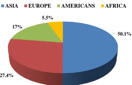
Nowadays the rabbits are bred systematically on a vast scale, with global rabbit meat production reaching 1,739 million metric tonnes in 2013. Such production is, in decreasing order, concentrated in Asia (50.1%), Europe (27.4%), Americas (17%), and Africa (5.5%; Figure 1.1; FAOSTAT, 2013). China is the major rabbit meat producer (723,975 tonnes/year), followed by Venezuela, Italy, Spain, Egypt, and France (Table 1.2; FAOSTAT, 2013). Italy represents the 55% of the rabbit meat production in Europe, followed by Spain with the 13%, France with the 11%, Germany with the 8%, the productions of other countries then, contribute all together at 12%.
3
Figure 1.1. Distribution of rabbit meat production in the World
Table 1.2. Rabbit meat production in the world (FAOSTAT, 2013) Country Production (tonnes)
China 723,975 Venezuela 275,000 Italy 262,332 Korea 149,500 Spain 63,577 Egypt 54,499 France 51,839 Germany 35,225
Rest of the World 128,822
Total 1,739,780
1.3 Rabbit meat production in Italy
At the beginning of the 70’s the Italian rabbit-rearing was characterized by a big number of small and middle dimension farms, only a few of them with a professional character, that produced about 940 thousands quintals of rabbit meat, every year. The increasing consumption drived this field that has fast developed to satisfy the increasing
50.1%
27.4% 17%
5.5%
4
demand of rabbit meat that in the 1990 arrived to be of 4 kg/person/year. Nowadays, the consumption per person is around 4.5 kg/year. The number of breedings is valued by Avitalia to be more than 30,000 units, but this number includes also the “micro” breedings whose production is addressed to the domestic consumption. The number of intensive breedings is extremly inferior and it is estimated to be 8,000 units (from 100 to 400 does) among which about 1,700 are professional breedings (>400 does).
The rabbit breeding with 262,332 tons of products in the 2013 that correspond to 175,000,000 heads per year (FAOSTAT, 2013), represents the fourth zootechnical field, after the bovines, the swines and the bird-rearing, with the 9% of the saleable gross production.
The production of rabbit used for meat is characterized by significative differences between North, Centre and South of the peninsula. This is due partly to a climatic condition; while in the North the rabbit produced has a weight at the slaughter of 2.6-2.8 kg with peaks of 3 kg in Piemonte, in the Centre of Italy the weight decreases to 2.4-2.5 kg while in the South rabbits are slaughtered at around 2 kg of weight (Avitalia, 2010). But the most significative element that distinguishes the South from the North, consists in the lack of integration of the rabbit-rearing chain with the feed-factories that are absent, the few slaughter-houses (the most of them with a farm character) and the almost absence of big breedings with an industrial feature, typical of the North of Italy, in which is possible to solve every kind of climatic problem due to the advanced technology and to obtain heavier animals.
1.4 Rabbit meat features
Rabbit meat has several advantages that support the increase of its use for human consumption. Rabbit meat offers excellent nutritive and dietetic properties (Dalle Zotte, 2002, 2004; Hernàndez and Gondret, 2006; Dalle Zotte and Szendrő, 2011). Its proximate composition demonstrates its protein rich (about 22% when considering the loin ˗ m. Longissimus dorsi ˗ and hindleg meat) (Table 1.3). In addition to high protein content, rabbit meat also contains high levels of essential amino-acid (EAA). In fact, in comparison with other meats, rabbit meat is the richest in lysine (2.12 g/100 g), threonine (2.01 g/100 g), leucine (1.73 g/100 g), valine (1.19 g/100 g), isoleucine (1.15 g/100 g), and phenylalanine (1.04 g/100 g; Dalle Zotte, 2004). The lean meat portion (water and protein contents) is relatively constant (73.0 ± 2.3 g water and 21.5 ± 1.4 g protein/100 g meat) with a decreasing trend from the mid part (loin) to the hind part and
5
then to the fore part of the carcass. Mineral content ranged around to 1.2–1.3 g/100 g meat. The leanest cut of meat in the rabbit carcass is the loin, with an average lipid content of 1.8 g/100 g meat, whereas the fattest portion is the fore leg, with an average lipid content of 8.8 g/100 g (Table 1.3). The most quantitatively important cut is the hindleg, and its lipid content is quite low (on average 3.4 g/100 g) compared to the other meats. Lipid content depends highly on the portion considered, and on feeding (Dalle Zotte, 2002). Based on fatty acid (FA) composition, rabbit meat is highly suited to human consumption. In rabbit meat, unsaturated fatty acids (UFA) represents approximately 60% of the total FA, and the quantity of polyunsaturated fatty acids (PUFA) that accounts for 27 to 33% of total FA is greater than that of other meats, including poultry.
Moreover, rabbit meat contains significant proportions of long-chain PUFA (C20–22), which are formed from the linoleic and α-linolenic acids. Other important products include arachidonic acid (20:4 n-6), eicosapentaenoic acid (EPA, 20:5 n-3) and docosahexaenoic acid (DHA, 22:6 n-3).
In regards to cholesterol content, rabbit meat contains the lowest levels (47.0 and 61.2 mg/100 g), for loin ˗ m. Longissimus dorsi ˗ and hindlegs, respectively (Dalle Zotte and Szendrő, 2011) (Table 1.3) of all the popular meats. Differences in cholesterol content between muscle types have been reported in other species and seem to be related to the differences in fiber type (Chizzolini et al., 1999). To a certain extent, feeding can also influence rabbit meat cholesterol content. Considering the potential human health implications of cholesterol intake, this aspect is relevant and all feeding strategies must be directed to achieving the lowest cholesterol content.
Rabbit meat confers moderately high energy values (from 603 kJ/100 g in the loin to 899 kJ/100 g in the fore legs , Table 1.3). Like other white meats, rabbit meat contains low levels of iron (1.3 and 1.1 mg/100 g for hindleg and loin, respectively; Parigi Bini et al., 1992) and zinc (0.55 mg/100 g in the whole carcass; Lombardi-Boccia et al., 2005; and 1.1 mg/100 g in the hindleg, Hermida et al., 2006).
The organoleptic properties of rabbit meat are: tenderness, juiciness and flavour, like those of other species. Rabbit meat does not have a very strong flavour. It is comparable to chicken. Tenderness varies with muscle age and depends on changes in the proportion and type of conjunctive tissue supporting the muscle fibres. The younger the rabbit is slaughtered, the more tender the meat will be. On the other hand, flavour tends to develop with age (Lebas et al., 1997).
6
Although little research has been done on this, it is known that flavour improves with the quantity of internal fat in the muscle. In the same way, juiciness depends largely on the fat content of the carcass. The fattier the carcass the lower its water content, but the better it retains what juice it does have. Slaughter conditions, especially the onset of rigor mortis, can modify the tenderness and juiciness of rabbit carcasses.
Selection for growth rate combined with confined rearing favour the anaerobic metabolism of rabbit muscle tissue. Animals raised in rational rabbitries therefore have a higher portion of white muscle fibre, which gives the meat a lighter colour (Lebas et al., 1997).
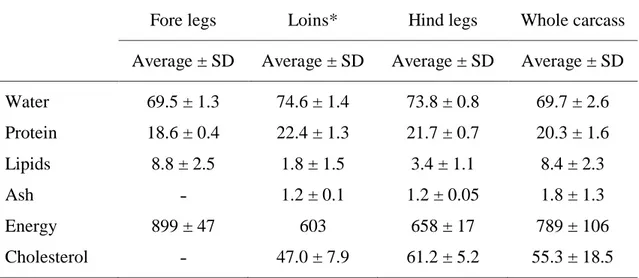
Table 1.3. Proximate composition (g/100 g), cholesterol content (mg/100 g) and energy
value (kJ/100 g) of cuts of rabbit meat
Fore legs Loins* Hind legs Whole carcass Average ± SD Average ± SD Average ± SD Average ± SD
Water 69.5 ± 1.3 74.6 ± 1.4 73.8 ± 0.8 69.7 ± 2.6 Protein 18.6 ± 0.4 22.4 ± 1.3 21.7 ± 0.7 20.3 ± 1.6 Lipids 8.8 ± 2.5 1.8 ± 1.5 3.4 ± 1.1 8.4 ± 2.3 Ash ˗ 1.2 ± 0.1 1.2 ± 0.05 1.8 ± 1.3 Energy 899 ± 47 603 658 ± 17 789 ± 106 Cholesterol ˗ 47.0 ± 7.9 61.2 ± 5.2 55.3 ± 18.5
*Lions, M. L. thoracis et lumborum Source: Hernández and Dalle Zotte, 2010
7
Chapter 2
Rabbit reproduction
2.1 The male
2.1.1 Anatomy of the male rabbit reproductive system
The reproductive organs of the rabbit male include testes, accessory sex glands, ducts and external genitalia (Figure 2.1). The testes are the primary organs of reproduction of the male; they produce spermatozoa (sperm) and hormones (androgens), which affect reproductive function and behavior. The paired testes are ovoid structures measuring about 30-35 × 10-15 millimeters (mm) and weighing approximately 1.5-2 grams (g) (Cerolini et al., 2008).
The testicles are essentially sacs of coiled tubules within which the sperm are formed. This process, known as spermatogenesis, involves changes from a rather normal looking spheroidal cell into the highly specialized spermatozoon, which has transmission of genetic information to the ovum as its only function. Sperm development begins in the walls of the tubules, and as the spermatozoa develop, they move toward the center, or lumen, of the tubule (McNitt et al., 2013).
In embryonic stage, testes lie in the abdominal cavity near the kidneys. They descend into scrotal sacs before birth through inguinal canals. Testes are kept in their original position by spermatic cords. A spermatic cord consists of an artery, a vein and a nerve which are embedded in connective tissue. Testis is covered by a connective tissue coat, known as tunica albuginea. Outside this tunica there is tunica vaginalis which is formed by two layers. Parietal layer lines the scrotum and visceral layer lines the tunica
albuginea. Around the testis, between the two layers of tunica vaginalis, is a narrow
coelomic cavity filled with coelomic fluid. It allows sliding movements of testis. Tunica
albuginea projects inwards as septa and divides the testis into many lobules. These
lobules contain long, convoluted seminiferous tubules, they are lined by the germinal epithelium. Germinal epithelial cells produce sperms by process of spermatogenesis. Among the germinal epithelial cells, there are Sertoli (sustentacular) cells, which nourish the developing spermatozoa. Once the sperm reach the lumen of the tubule, they are transported through the tubule by fluid pressure. This transport takes the sperm to the top of the testis and out into the epididymis (McNitt et al., 2013).
8
Connective tissue among seminiferous tubules contain Leydig (interstitial) cells. They secrete testosterone (male sex hormone) which controls secondary sexual characters. The Leyding cells are under the control of hormones from the anterior pituitary, which is located at the base of the brain. These controlling hormones, regulate the levels of the androgens in the blood, which, in turn, control spermatogenesis and sexual activity of the buck (McNitt et al., 2013). The seminiferous tubules of a testis open into a network called rete testis, it opens into many fine ducts, called vasa efferentia, these vasa open into the epididymis.
The ducts through which the sperm move after leaving the testes proper include the epididymides, deferent ducts, and urethra. The epididymides lie close to the top of the testes and function as a place for maturation of the spermatozoa. Spermatozoa that have not undergone a period of maturation in the epididymides are incapable of fertilizing eggs. The epididymides also serve as a place for storage of spermatozoa; fertile spermatozoa have been recovered from epididymides after eight weeks of storage. The normal time required for movement of sperm through the epididymides is 8 to 10 days. The epididymis is a long, narrow and highly convoluted tubule, it is present along the inner surface of the testis and is divided into three distinct parts:
1) caput epididymis: the anterior part of epididymis, is connected to testis through vasa efferentia. Caput epididymis is also connected to the dorsal abdominal wall by a spermatic cord;
2) corpus epididymis: the middle part of the epididymis, connecting caput epididymis and cauda epididymis;
3) cauda epididymis: the posterior part of the epididymis. The cauda epididymis is joined to scrotal sac by a short, thick, elastic cord known as gubernaculum. The deferent ducts convey the spermatozoa from the epididymis to the urethra and also function to some extent in sperm storage. The accessory sex glands normally add their secretions to the semen at or near the junction of the deferent ducts and the urethra. The urethra is the common passage for both semen and urine. It carries semen from the junction with the deferent ducts to the end of the penis, from which the semen is ejaculated into the female vagina. The bladder empties into the urethra just beyond the point of junction of the urethra and deferent ducts. The external genitalia of the male include the penis, the scrotum and the prepuce. The penis (copulatory organ) is an erectile organ that is used for insertion of the ejaculate into the female tract. It is
9
normally flaccid and rests in the prepuce. It becomes rigid from constriction of the penile veins at the time of breeding. Since arterial blood continues to flow into the organ, it becomes turgid and can thus penetrate the vulva and vagina of the doe. The erect penis is held forward along the abdomen. There is a sensitive tip on the penis, known as the glans penis. Stimulation of the glans penis by the vagina of the female (or by a properly prepared artificial vagina) results in ejaculation. This is due to a reflex contraction of the duct system that forces out spermatozoa stored in the deferent ducts and the last third of the epididymis. Fluids from the accessory glands are also released into the deferent ducts and the urethra at the time of ejaculation. After subsidence of erection, the penis is pulled back into the prepuce by muscular contraction. The penis is covered by a loose sheath of skin, the skin that hangs over the tip of the penis is known as prepuce. The tip of the penis covered by prepuce is called glans penis. The penis is composed of three longitudinal columns, namely, two columns of corpora cavernosa (upper) and one column of corpus spongiosum (lower) made by spongy tissue. The scrotum consists of two relatively hairless sacs that contain the testicles. These function to protect the testes and to provide an area with a lower temperature than that of the body cavity, because spermatogenesis cannot occur at normal body temperature. The testes of the rabbit can move freely in and out of the abdomen and so are not always found in the scrotal sacs (McNitt et al., 2013).
Associated with the male reproductive system of rabbit, there are the accessory sex glands, they include the prostate gland, bulbo-urethral glands (Cowper’s glands), perineal glands and rectal glands. Their secretion are responsible for the production of seminal plasma. The functions of these secretions include adding fluid volume to the ejaculate to facilitate movement of semen through the male and female reproductive tracts, providing nutrients and buffers for the spermatozoa, providing a gelatinous plug to seal the female tract, and providing substances that stimulate contractions of the vagina and uterus of the female to enhance movement of spermatozoa through the tract. Around the base of uterus masculinus, there is prostate gland, it opens into the urethra through many ducts, it secretes an alkaline fluid which activates the spermatozoa and also contributes to the main bulk of semen. Moreover, prostate gland secretes citric acid that enters in the kreb’s cycle to produce ATP. Posterior to the prostate gland, there is a pair of Cowper’s glands, their secretions neutralize the urinary residue and vaginal acidity. Behind the Cowper’s glands, there are two perineal glands, they open into hairless perineal depressions, one on either side of the anus. Their secretions give a
10
characteristics smell to rabbit. Two rectal glands are situated at the sides of the rectum. Their function is not yet known.
Figure 2.1. The reproductive system of the male rabbit. (McNitt et al., 2013)
2.1.2 Rabbit Spermatozoa
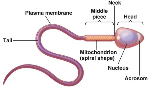
Each spermatozoon has a head that carries the genetic information and a tail that provides propulsion by its whip-like movements. The rabbit spermatozoa is about 50-60 µm long, it has a wide, flattened head and a long, thin and cylindrical segment composed by a neck, an intermediate part and a flagellum or protoplasmic tail (Figure 2.2, 2.3). The whole sperm cell is enveloped by the plasma membrane, whose function is to contain organelles and intracellular components. Thanks to its semi-permeability, it maintains the chemical gradient of ions and of the other soluble components. Moreover, specific membrane proteins facilitate the transport of fructose and glucose from the extracellular environment to the inside of the cell, thus providing it with the energetic substrates it requires for the cell metabolism.
The head includes two structures of fundamental importance: the acrosome (Figure 2.4) and the nucleus.The acrosome is a structure derived from the Golgi apparatus and it is delimited by a trilaminar membrane: it covers two thirds of the front part of the sperm head. Above the acrosome, another structure covers the remaining front part of the head: it is the head cap, composed by sulphur- and fibrous proteins which confer it great resistance. Its biological role, in effect, is to protect the acrosomal content: this includes enzymes with a high proteolytic and glycolytic power, which allow the spermatozoon to
11
penetrate through the zona pellucida during the fecundation of the egg cell. The most important enzyme is acrosin, followed by hyaluronidase, collagenase, acid phosphatase, phospholipase A, arylsulfatase. Inside the head, in the central part, it is possible to find the nucleus, whose function is to transport and transmit the chromosomes, i.e. the genetic information. The nucleus of a ripe spermatozoon is very dense, as it is composed by tightly packed chromatin. The segment adjacent to the head is called neck: this region contains centrioles, which give the impulse for the meiotic cell division. This is also the starting point of the axoneme, a thigh bundle of axial contractile fibres, which continues in the following parts as well.
The segment after the neck, in rabbits, is about 8.8 µm long. It is called intermediate piece and it contains mitochondria, organized in a layer surrounding the axoneme. This part, therefore, constitutes the main energy reservoir for the cell. The long tail segment (40 µm) following the intermediate piece is called main piece: it is not surrounded by any coat of mitochondria but it contains arginine, leucine and other essential amino acids.This segment, too, is composed by bundles of parallel contractile fibres, which end in thin fibrils constituting the final segment. These filaments are projected in the liquid medium like a helix, thus determining the helical movement of the spermatozoa. Moreover, they are able to change their descent energy into ascent energy, thereby advancing against the gravity force following the liquid currents of the female genital apparatus.
12
Figure 2.3. Picture of rabbit spermatozoa obtained by scanning electron microscopy
(magnification × 2000). Provided by Microscopy Interdepartmental Center, University of Molise
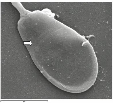
Figure 2.4. Picture of rabbit spermatozoa head obtained by scanning electron
microscopy (magnification × 10000). The arrow highlights the “equatorial zone”, which is the end of acrosomal cap. Provided by Microscopy Interdepartmental Center, University of Molise
13 2.1.3 Spermatogenesis and sperm maturation
Spermatogenesis is a process of division and differentiation of cells from diploid germinal cells termed spermatogonia, to haploid, mature spermatozoa in seminiferous tubules of the testis. The duration of spermatogenesis in rabbit is 53 days (Swierstra and Foote, 1965). The 53-day period may be divided into three phases, including: spermatocytogenesis, where stem cell spermatogonia divide by mitosis to produce other stem cells in order to continue the lineage throughout the adult life of the male, they also divide cyclically to produce committed spermatogonia and primary spermatocytes; meiosis, the period of replication (primary spermatocytes) and then reduction of genetic material into haploid spermatids; and spermiogenesis which is a Sertoli cell aided differentiation of spermatids (Figure 2.5).
Spermiation is the release of spermatids as spermatozoa into seminiferous tubule lumen. Transit of sperm through the efferent ductules and the epididymis is associated with significant maturation changes such as obtaining the capacity of progressive motility; final condensation of the nucleus and further modification of the form of the acrosome; alteration of plasma membrane surface. The most important functional changes in sperm occur in the efferent ductules, caput and corpus of epididymis. Sperm taken from the distal part of corpus and cauda epididymis already have the potential to fertilize. Approximately 8 days are required for transportation of spermatozoa through the excurrent duct system (efferent duct, epididymis and vase deferents). Therefore, a new population of spermatozoa is ejaculated every 50–54 days (Swierstra and Foote, 1965). The cauda epididymis has properties that allow the sperm to be stored for several weeks. Seminal fluids secreted by accessory sex glands added during ejaculation serve as a vehicle, stimulate the metabolism of sperm and provide the energy requirements for passage through the uterus (Gadella et al., 2001).
Most of the spermatozoa are stored in the epididymal tail, but also a lower number of spermatozoa are present in the testis and in the other efferent ducts (Table 2.1).
The spermatozoa in the female reproductive tract, particularly in the isthmus oviduct undergoes certain change called ‘‘capacitation’’, which takes about 5-6 hours in rabbit spermatozoa (Cerolini et al., 2008).
The mechanism of capacitation involves many biochemical changes. These include the removal of adsorbed components from the sperm surface, the reorganization of membrane proteins, a change in membrane lipid composition, increased permeability to
14
certain ions such as Ca2+, a change in internal pH, and an increase in plasma membrane fluidity and in metabolism (Parrish et al., 1993). There is also an increased hyperactivation that is believed to result in the redistribution of membrane components during capacitation (Parrish et al., 1993). In addition, several studies show that there is a decrease in the membrane cholesterol phospholipid ratio during capacitation. All of these changes allow the spermatozoa to undergo the acrosome reaction (AR) following interaction with the zona pellucida, the extracellular matrix of egg cells (Miller et al., 1990). The rabbit acrosome reaction, which involves membrane breakdown and release of enzymes for penetration of egg envelopes, is induced by a specific stimulus in follicular fluid at the site of fertilization.
15
Table 2.1. Number of spermatozoa in different reproductive system portions Reproductive system portion Sperm concentration (× 106)
testis 130180/g
caput and corpus of epididymis 150
cauda of epididymis 12001500
deferent duct 50
2.1.4 Reproductive activity of the buck
Bucks reach sexual maturity at the age between five and eight months, depending on breed and level of nutrition. In a study of New Zealand White bucks, daily sperm output was found to increase from 20 weeks to a mature level at about 31 weeks (McNitt et al., 2013).
The normal ejaculate volume for mature bucks ranged from 0.39 to 1.19 mL, with an average of about 0.6 mL. Sperm per ejaculate is highly variable among bucks as well as among successive ejaculates from the same buck and varies from 146 to 738 million spermatozoa per mL (Bencheikh, 1995; Brun et al., 2006; Castellini et al., 2006b; García-Tomás et al., 2006a,b; Safaa et al., 2008; Lavara et al., 2010). The number of spermatozoa for each ejaculate depends on the breed of the buck, recent use, and level of stimulation.
2.2 The female
2.2.1 Anatomy of the female rabbit reproductive system
The organs of female reproduction system include: the ovaries, oviducts, uterus, cervices, vagina, and external genitalia (Figure 2.6). The ovaries, the primary organs of reproduction, produce eggs, or ova, and hormones (primarily estrogens and progestins). They lie within the abdominal cavity, with one on each side, near the kidneys. The ovaries are ovoid structures with an extent of about 15 × 10 mm and a weight between 300 and 500 mg (Cerolini et al., 2008), depending on the activity of the ovarian components. The middle portion, or medulla, of each ovary, is composed of connective tissue containing nerves and blood vessels. The outer layer, or cortex, contains the ova in various stages of development, as well as other types of tissue, including blood
16
vessels, nerves, and muscle fibers. At the time of a doe’s birth, thousands of undeveloped ova are contained in the germinal epithelium layer of the cortex. From the time of puberty until death or the end of the reproductive life of the female, groups of these undeveloped ova undergo development and are shed (ovulated), or they degenerate. The oviduct is the site of fertilization, functions in a maturation process of spermatozoa known as capacitation, and is the location where early embryonic development occurs. Oviducts are thin tubes, characterized by willowy trend, of about 10-16 cm in length (Cerolini et al., 2008).
The upper end of the oviduct is spread into the ostium tubae, which partially surrounds the ovary. On the edges of the ostium tubae are numerous small projections known as the fimbria. These nearly cover the ovary. Beating of the fimbria causes waves of movement of fluid toward the opening of the oviduct and, at the time of ovulation, sweeps the ova into the oviduct (McNitt et al., 2013).
The uterus is the organ in which the embryo and foetus normally develop and grow. It also provides muscular force for expulsion of the fetuses at birth. The uterus of the rabbit is formed of two distinct horns, which are not connected to form entire body. Each horn of the uterus connects into an individual cervical canal, which opens into the common vagina. The cervices function as muscular plugs to keep the uterine horns closed except at the time of mating and parturition (birth, or kindling) (McNitt et al., 2013).
The vagina is the site of sperm deposition at mating and acts as a channel for the young at parturition. It is long between 6-8 cm and presents longitudinal mucosal folds (Cerolini et al., 2008).
The external genitalia of the doe include the urogenital sinus, which is continuous with the vagina and is the chamber into which the urethra empties urine. The external lips of the urogenital sinus form the vulva, which can be used as an indicator of sexual receptivity of the doe. A doe with a moist red or pink vulva is much more likely to accept service than a doe with a pale, dry vulva. The clitoris is within the urogenital sinus, with the sensitive portion, the glans clitoris, projecting into the urogenital opening. Because the urethra opens into vaginal sinus posterior toward the place where sperm are deposited, urination by the doe following breeding does not necessarily interfere with fertilization.
17
Figure 2.6. Reproductive system of the female rabbit. (McNitt et al., 2013)
2.2.2 Estrus and ovulation
Does become receptive to bucks at the age of about 3.5 months and become able to conceive at the age of 4 or 4.5 months. These ages depends on the breed of rabbit, the smaller breeds generally reaching puberty earlier than the larger breeds. The nutrition level will also affect the age of onset of reproductive function. It is generally not advisable to breed a doe during the first month that it is able to reproduce, because it is still growing and the attainment of mature size may be prevented or delayed if the doe is also expected to produce and feed a litter at this time. However, undue delay in breeding may result in breeding difficulties if a doe becomes too fat. Also, delayed breeding may make a doe more susceptible to hairballs (McNitt et al., 2013).
Rabbit do not have a definite estrous cycle as found in many other animals. At the time of puberty, follicle-stimulating hormone (FSH) from the anterior pituitary gland located at the base of the brain begins inducing growth of follicles, with corresponding development of ova within them. The ova begin their development with a single layer of follicle cells surrounding them. The number of follicular cells gradually increases until numerous layers are present. As a follicle develops further, a fluid-filled cavity forms, with the ovum located in the center upon a hillock of cells. By this time, the follicle has
18
enlarged to such an extent that it bulges from the surface of the ovary. At ovulation the outer layer of the follicle ruptures, and the ovum is expelled along with the fluid (McNitt et al., 2013).
The follicles produce estrogens (the main female sex hormones), that make female receptive for the male (Patton, 1994). Follicular development generally occurs in waves, with 5 to 10 follicles on each ovary at the same stage of development at any one time. Follicles start continuously development, so follicles at several stages of development are always present (Patton, 1994). When follicles reach mature size, they produce estrogens for about 12 to 14 days. After this period, if ovulation has not occurred, these follicles degenerate, with a corresponding reduction in blood concentration of estrogen level and receptivity. After about 4 days a new follicles will begin producing estrogen and the doe becomes receptive again. The doe therefore, has a cycle of 16-18 days, during which she is receptive for about 12-14 days followed by 2-4 days when the doe refuses to mate (Patton, 1994). The receptivity of the does is extremely variable because of individual differences, sexual stimulation and environmental factors, such as nutrition, light and temperature (Cheeke et al., 1987).
Ovulation in the doe occurs only after induction by an external stimulus, such as mating, and occurs 10-13 h after coitus (Patton, 1994). This includes stimulation of the anterior vagina by the penis as well as pressure on the hindquarters from mounting by the buck. Intense sexual excitement or mounting of the doe by other rabbits may also induce ovulation. This may result in a condition known as pseudopregnancy. An ovulatory stimulus results in the release of luteinizing hormone (LH) from the anterior pituitary. This hormone causes rupture of a number of mature follicles on one or both ovaries approximately 10 hours after the stimulus occurs. The number of ova shed from each ovary is a factor that determines the litter size. Other factors include the number of shed ova that are fertilized by the sperm and the number of fertilized eggs that follow the entire intrauterine development process (McNitt et al., 2013). When the ovum has been shed from the ovary, LH stimulates changes in the follicular cells that rapidly develop into a corpus luteum (yellow body), which produces hormones known as progestins. These are necessary throughout pregnancy for development of the embryo. The primary action of the progestins is to stop muscular contractions of the uterus and to stimulate production of nutrients for the embryo. The corpus luteum begins actively secreting within three days after ovulation and continues throughout pregnancy. The hormone output increases until about the fifteenth day of pregnancy and remains at a
19
high level until the last week, when the hormone level begins to fall. The progestins control uterine function, especially inhibition of muscular activity so that the embryo can remain in the uterus and be nourished throughout pregnancy. Moreover, the progestins inhibit sexual receptivity in pregnant doe, although follicles continue to develop and produce estrogens throughout pregnancy. It has been shown that estrogens are necessary for the progestins secretion, occurring in corpus luteum. Since follicles are present at the end of pregnancy, the doe is sexually receptive and able of ovulation immediately after parturition (McNitt et al., 2013).
2.2.3 Fertilization and pregnancy
At the moment of mating the buck deposits several million sperm in the doe’s vagina. The spermatozoa move by contractions of the female tract and by swimming to the middle portion of the oviduct, where fertilization occurs. The first sperm are found in the oviduct within 20 to 30 minutes of mating, although the majority are not found there for several hours. Of the millions deposited into the vagina, only a few thousand reach the site of fertilization (McNitt et al., 2013).
When the eggs are released from the follicle, they are swept into the oviduct and move to the middle third of the oviduct. Fertilization refers to the entry of a sperm cell into the ova and fusion between male and female genetic material (syngamy). Cellular division and development of the embryo begin almost immediately after syngamy. The developing embryo remains in the oviduct until the 8- or 16-cell stage is reached. This takes 48-52 hours, after which the embryo migrates to the uterus, where it floats in the uterine fluid and is nourished by it (Cerolini et al., 2008). During this phase, nutrients enter the embryo by diffusion through the cell membranes. After seven days the embryo becomes too large to be properly nourished solely by diffusion, so it is attached to the wall of the uterus, and the placenta (afterbirth) begins to form. This process is known as implantation. The placenta supplies protection for the embryo and a close connection between the embryonic and maternal circulatory systems. There is no direct connection between these two systems, although the two blood supplies pass very close to each other in their respective vessels. In this way, oxygen and nutrients can diffuse through the vessel walls from the doe to the young, and the wastes from the young can diffuse out to the circulatory system of the doe. Transport of nutrients and oxygen within the embryo is carried out through embryonic circulatory system. Pregnancy in rabbits lasts an average of 30 to 32 days but may be as short as 29 days or as long as 35 days.