Effects of antioxidants, r
educed glutathione and melatonin,
on in vitr
o boar
sperm capacitation and acr
osome exocytosis.
MAR TINA ROCCO Ph.D ThesisThesis Doctoral
INTERNATIONAL Ph.D. in
“WELFARE, BIOTECHNOLOGY AND QUALITY OF ANIMAL PRODUCTION”
2015/2016
Effects of antioxidants, reduced glutathione and melatonin,
on in vitro boar sperm capacitation and acrosome exocytosis.
Hyperactivation Capacitation Acrosome reaction Sperm/oocyte fusion Lipid peroxidation Changes in intracellular calcium storage
Cell Membrane
PROGESTERONE
Ca2+ Ca2+
Author: Martina Rocco
General coordinator: Prof. Giuseppe Maiorano Supervisor: Prof. Angelo Manchisi
Foreign supervisor: Prof. Juan Enric Rodriguez- Gil
IN COLLABORATION WITH:
UNIVERSITY OF MOLISE
Department of Agricultural, Environmental and Food Sciences
INTERNATIONAL Ph.D. in
“WELFARE, BIOTECHNOLOGY AND QUALITY OF ANIMAL PRODUCTION” (XXVIII CYCLE)
Related disciplinary scientific section: 07/G1 (Scienze e Tecnologie Animali)
General Coordinator: Prof. Giuseppe Maiorano
Doctorate Thesis Title:
Effects of antioxidants, reduced glutathione and melatonin, on in vitro boar
sperm capacitation and acrosome exocytosis.
Supervisor: Ph.D. Candidate:
Prof. Angelo Manchisi Dr. Martina Rocco
Foreign Supervisor: 133284
Prof. Juan Enric Rodriguez- Gil
Ringraziamenti, Agraïments, Agradecimientos
A lavoro terminato, desidero ricordare tutti coloro che mi hanno aiutato nella stesura con suggerimenti, critiche ed osservazioni: a loro va la mia gratitudine, anche se a me spetta la responsabilità per ogni errore contenuto in questo lavoro.
Innanzitutto ringrazio il Coordinatore di questo Dottorato il Professor Giuseppe Maiorano, ed il mio tutor Angelo Manchisi per avermi dato la possibilità di iniziarlo e di portarlo a termine, e soprattutto per essersi prodigati per l’accrescimento delle mie basi scientifiche e per l’ampliamento delle stesse attraverso le missioni effettuate all’estero, soprattutto per il lungo periodo concessomi presso la “Universitat Autonoma de Barcelona” che è stato un ottimo contributo per la mia carriera come dottoranda e come futura lavoratrice.
Vull donar el meu agraïment al professor Joan Enric Rodriguez-Gil co-supervisor, i al professor Marc Yeste, segon co-supervisor: sense el seu recolzament i ajuda aquesta tesi no existiria.
Un ringraziamento particolare va ai colleghi ed agli amici che mi hanno incoraggiato o che hanno speso parte del proprio tempo per leggere e discutere con me le bozze del lavoro, e per essere presenti durante le fasi più critiche di questo dottorato, in particolare (in ordine alfabetico) Michele Di Iorio, Siria Tavaniello e Francesco Vizzarri.
També voldria agrair a tot l’equip dels laboratoris de Barcelona i Girona.
En primer lloc a tot el departament de medicina i cirurgia animal que m’han deixat fer servir el laboratori, aparells, i tot allò que necesitava (en ordre alfabètic): Teresa M. Mogas
Amorós, Teresa Rigau Mas, Jordi Miró Roig, M. Jesùs Palomo Peiro, Maria Montserrat Rivera del Álamo.
Y a quien ha sido siempre un valioso apoyo en el laboratorio ya sea a nivel de habilidades o a nivel anímico en particular: Alex Peña y Estela Garcia. Gracias por la ayuda que me habéis ofrecido y por haberme hecho sentir como en casa, y sobre todo por haberme hecho sonreir durante los días mas pesados; y para acabar quisiera agradecer todo el tiempo que nos hemos dedicado los unos a los otros a: Nuria Arcarons, Barbara Azevedo Pereira (y el citometre que para nosotras se ha convertido en una persona), Rafael Betarelli, Sebastian Bonilla Correal,
Manel Bosch, Manuela Costa Garcia, Lluis Ferrè Dolcet, Gaia La Fortezza, Henar Marin Estruch, Federico Noto, Roser Moratò, Anna Placci, Meritxell Vendrell Flotats.
Vorrei infine ringraziare le persone a me più care, quelle che fanno parte della mia famiglia (in ordine d’età), mio padre Roberto, mia madre Silvana, mia sorella Serena, mio cognato
Michele, nonchè i miei amici di sempre Gianfranco Bianco e Lorenza Lauria sempre pronti
ad ascoltarmi e supportarmi, ed anche se ad oggi non c’è più, mio nonno Ennio che sempre mi ha stimolato a studiare; i a la meva sogra la Pepi que és part de la meva família catalana.
I despres un agraïment molt particular va per la meva parella, l’Antoni, que sense ell no hauria pogut somriure tant de gust fins i tot escrivint la tesi. I per acabar a la Jipi, la gata de casa, que ha contribuït amb les seves potes a escriure-la (o desescriure-la).
7
i
CONTENTS
Abstract (English) I
Riassunto (Italiano) III
Resumen (Castellano) V
Resum (Català) VII
PART 1. Introduction
1. Introduction 1
1. Pig reproduction: male genital tract 1
2. Boar sperm morphology 7
3. Sperm journey through female genital tract: processes and molecules
involved 12
4. Chronological events of sperm capacitation 15
5. Acrosome reaction 20
PART 2. Sperm Analisis
6. Sperm analisis 24 6.1. Assessment of macroscopic characteristics of the ejaculate 25
ii
6.3. Sperm motility 26
6.4. Sperm morphology 29
6.5. Sperm agglutination 33
6.6. Osmotic resistance of sperm 34 6.7. Sperm plasma membrane integrity (sperm viability) 35
6.8. Acrosome integrity 37
6.9. Determination of mitochondrial membrane potential (Δψm) 40 6.10. Oxidative stress evaluation by flow cytometry: superoxide /oxide levels
in sperm cells 41
6.11. Changes of plasma membrane fluidity compatible with capacitation 42 6.12. Intracellular calcium levels as capacitation markers 43 6.13. Detection of tyrosine-phosphorylated proteins by western blot analysis 44 6.14. Detection of phosphotyrosine residues location in boar sperm
through immunocytochemistry 47
6.15. Evaluation of free-cysteine residues in sperm nucleoproteins 48 6.16. Evaluation of DNA fragmentation (SCD test adapted for boar sperm) 50
iii
PART 3. - Research works
Main aim of this thesis 52
7. Glutathione 53
7.1. General function, roles and localization 53 7.2. How glutathione interacts in spermatozoa’s mechanisms 55
Research work: Effects of reduced glutathione on in vitro boar sperm
capacitation and acrosome exocytosis 57
8. Melatonin 123
8.1. General function, roles and localization 123 8.2. How melatonin can modifies sperm dynamics 128
Research work: Melatonin impedes the loosing of head disulfide bonds and induces agglutination during the achievement of
boar sperm in vitro capacitation and in vitro acrosome exocytosis 130 PART 4. Conclusions
9. Conclusions 207
10. Apendix 210
10.1. List of abbreviations 213
iv
10.3. List of images 255
10.4. Glutathione image reference 257 10.5. Melatonin images reference 257
I
Abstract
The assisted reproductive technologies allow investigators to discern how mammalian sperm achieve capacitation through a sequence of biochemical modifications that lead to the establishment of fully fertilizing ability. In this way, in vitro studies on boar spermatozoa incubated in a specifically designed in vitro capacitation medium (CM) for 4h show the whole of structural and functional changes that boar sperm suffers during the achievement of the capacitation status. These changes comprise modifications in parameters like sperm motility, membrane lipid disorders, mitochondrial membrane potential, intracellular calcium levels, tyrosine phosphorylation levels of protein and their new arrangement, and increasing of intracellular levels of free cysteine radicals. The addition of antioxidants such as reduced glutathione or melatonin to the medium prevented or modified the majority of capacitation-like changes. The aim of this thesis was to study the effects of two known antioxidant agents, reduced glutathione (GSH) and melatonin, on the achievement of boar sperm in vitro capacitation (IVC) and subsequent in vitro, progesterone-induced acrosome exocytosis (IVAE). For this purpose, in the first study the CM was supplemented with three different concentrations of GSH. Results shown that GSH-sensitive mechanisms had important effects on the achievement of a feasible boar sperm IVC and subsequent, progesterone-induced IVAE. This is concluded by the fact that several important markers of the achievement of boar sperm IVC like motion parameters, changes of sperm mebrane fluidity and the overal tyrosine phosphorylation status are altered by GSH. In the second study, melatonin was added to the CM in three separate concentrations. Supplementation of melatonin resulted in a noticeable negative effect on motility, which was concomitant with a very intense increase in the percentage of agglutination between cells in a dose–dependent manner. Meanwhile, melatonin seems to have a positive effect on superoxids and peroxids levels, and also seems to stabilize the levels of free-cysteine radicals depending on its concentration in the extender. In
II
conclusion, our researchs would suggest that boar sperm capacitation is linked to an increase of free cysteine residues and intracellular reactive oxygen species levels. Furthermore, these increases seem to be instrumental in the launching of subsequent IVAE. Thus, the launching of a mild oxidative stress status might be one of the factors related with the establishment of a feasible IVC and subsequent IVAE. In this way, antioxidants like GSH and melatonin seem to have an effect on ionic and redox mechanisms, altering some biochemical and fisiologic process, and compensing the damageous effects provoked by an excessive exposition of the cells to ROS compounds.
III
Riassunto
Le tecniche di riproduzione in vitro, permettono di investigare riguardo i meccanismi biochimici connessi con la capacitazione spermatica, e la successiva fecondazione. A tal proposito, studi in vitro su spermatozoi di suino incubati per 4 ore in uno specifico medium capacitante (CM), rendono possibile effettuare un’analisi approfondita dei cambiamenti strutturali e funzionali che si verificano a livello spermatico durante il raggiungimento dello status capacitato. Tra i parametri interessati dai suddetti cambiamenti ritroviamo: la motilità spermatica, disordini della membrana lipidica, cambiamenti nel potenziale di membrana mitocondriale, variazioni nei livelli di calcio intracellulari, differenti livelli di fosforilazione delle tirosine proteiche e una nuova ridistribuzione delle stesse, ed infine un incremento dei livelli intracellulari dei radicali liberi di cisteina. L’aggiunta al medium di antiossidanti come, il glutatione ridotto (GSH) o la melatonina sembra intervengano nella prevenzione e/o modificazione della maggior parte dei cambiamenti biochimici associati alla capacitazione. Scopo della tesi è stato di studiare gli effetti di questi agenti antiossidanti, sul raggiungimento della capacitazione in vitro (IVC) degli spermatozoi di suino e sulla successiva esocitosi a livello acrosomiale indotta dall’aggiunta di progesterone in vitro (IVAE). Nel primo studio il medium capacitante è stato integrato con GSH a tre differenti concentrazioni. I risultati ottenuti in vitro hanno mostrato la presenza di meccanismi GSH-sensibili che sembrano interferiscano in maniera importante nel raggiungimento di uno status capacitato, e della successiva IVAE indotta dal progestorene. Infatti la maggioria dei parametri usati come marcatori principali della capacitazione spermatica suina quali, motilità spermatica, cambi nella fluidità delle membrane spermatiche e lo stato di fosforilazione delle tirosine vengono alterati dalla presenza nel medium di GSH.
IV
Il secondo studio invece, è improntato sull’aggiunta di melatonina al medium capacitante, in tre distinte concentrazioni. L’aggiunta di melatonina ha fatto registare un notevole calo nei valori di motilità spermatica, concomitanti con un netto incremento nella percentuale di agglutinazione tra gli spermatozoi, e un’intensità di questi effetti variabile in maniera dose-specifica. Viceversa l’aggiunta di melatonina al medium ha abbassato la concentrazione di perossidi e superossidi presenti a livello intracellulare, nonchè stabilizzato i livelli di radicali liberi delle cisteine sempre con una modalità d’azione dose-effetto. Entrambe, le ricerche hanno dimostrato che la capacitazione produce un incremento dei radicali liberi di cisteina e dei livelli intracellulari di radicali liberi relativi allo stress ossidativo. Entro determinati livelli questi incrementi sembrano tra l’altro rivelarsi fondamentali nell’innesco del successivo IVAE. La presenza di un leggero stress ossidativo viene considerato indispensabile al fine di innescare la capacitazione spermatica e la successiva esocitosi acrosomiale. Inoltre, antiossidanti come il GSH e la melatonina sembra vengano implicati nei meccanismi redox e ionici a livello cellulare, con l’alterazione di alcuni processi fisiologici e biochimici, e che compensino gli effetti dannosi risultanti da un’eccessiva esposizione delle cellule ad alte concentrazioni di radicali liberi quali, superossidi e perossidi intracellulari (ROS: O2
-●
V
Resumen
Las tecnologías de reproducción asistida permiten a los investigadores identificar cómo el esperma de los mamíferos consigue la capacitación a través de una secuencia de modificaciones bioquímicas que permiten a la célula conseguir la habilidad de fertilizar. De esta manera, los estudios in vitro hechos sobre espermatozoides de cerdo incubados en un medio de capacitación específico (CM) durante 4h muestran el conjunto de cambios funcionales y estructurales que el esperma de cerdo sufre durante la consecución del estado de capacitación. Estos cambios comportan modificaciones en parámetros como: motilidad de los espermatozoides, trastornos de los lípidos de la membrana, potencial de la membrana mitocondrial, niveles de calcio intracelulares, niveles de fosforilación de las tirosinas en la proteína y su nueva reorganización, y aumento de los niveles intracelulares de los radicales libres de cisteína. El hecho de agregar antioxidantes como la glutathión reducida o melatonina al medio parece prevenir y/o modificar la mayoría de cambios conectados con la capacitación. El objetivo de esta tesis era estudiar los efectos de dos agentes antioxidantes, la glutathión reducida (GSH) y la melatonina, en el logro de la capacitación in vitro del esperma de cerdo (IVC) y la subsiguiente exocitosis acrosomal in vitro (IVAE), inducida a través de la progesterona. Para este propósito, en el primer estudio, el CM se suplementó con tres concrentraciones distintas de GSH. Los resultados muestran que los mecanismos sensibiles al GSH tuvieron efectos importantes en la consecución del estado de capacitación en el esperma de cerdo, y en la siguiente IVAE inducida por la progesterona. A esta conclusión se llega por el hecho que marcadores importantes de la capacitación en el esperma de cerdo como los parámetros de movimiento, los cambios de fluidez en la membrana del esperma y el estado en general de la fosforilación de la tirosina se ven alterados por el GSH. En el segundo estudio, al CM se añadió la melatonina en tres concentraciones distintas. La suplementación de melatonina dio como resultado un destacable efecto negativo en la motilidad que estuvo
VI
asociada a un intenso aumento en el porcentaje de aglutinación entre células de una manera dosis-dependiente. Mientras tanto, la melatonina parece tener un efecto positivo en los niveles de superóxidos y peróxidos, y también parece estabilizar los niveles de radicales libres de cisteína dependiendo de su concentración en el medio. En conclusión, nuestras investigaciones sugerirían que la capacitación del esperma de cerdo va unida al aumento de los residuos libres de cisteína y de los niveles intracelulares de especies reactivas de oxígeno. Por otra parte, estos aumentos parecen ser el instrumento para el lanzamiento de la sucesiva IVAE. Así, la presencia de un ligero estado oxidativo podría ser uno de los factores relacionados con la activación de la capacitación y de la siguiente exocitosis acrosomal. De esta manera, los antioxidantes como GSH y la melatonina parecen tener efectos en los mecanismos iónicos y redox; alterando algunos procesos bioquímicos y fisiológicos y compensando los efectos dañinos provocados por una excesiva exposición de las células a los radicales libres del oxígeno (peróxidos y superoxidos: O2-● y H2O2).
VII
Resum
Les tecnologies de reproducció assistida permeten als investigadors identificar com l’esperma dels mamífers aconsegueix la capacitació mitjançant una seqüència de modificacions bioquímiques que permeten a la cèl·lula assolir l’habilitat de fertilitzar. D’aquesta manera, els estudis in vitro fets sobre espermatozoides de porc incubats en un medi de capacitació específic (CM) durant 4h mostren el conjunt de canvis funcionals i estructurals que l’esperma de porc pateix durant la consecució de l’estat de capacitació. Aquests canvis comporten modificacions en paràmetres com: motilitat dels espermatozoides, trastorns dels lípids de la membrana, potencial de la membrana mitocondrial, nivells de calci intracel·lulars, nivells de fosforilació de la tirosina a la proteïna i la seva nova reorganització, i augment dels nivells intracel·lulars dels radicals lliures de cisteïna. El fet d’agregar antioxidants com la glutatió reduïda o la melatonina al medi sembla prevenir i/o modificar la majoria de canvis connectats amb la capacitació. L’objectiu d’aquesta tesi era estudiar els efectes de dos agents antioxidants, la glutatió reduïda (GSH) i la melatonina, a l’assoliment de la capacitació in vitro de l’esperma de porc (IVC) i la subsegüent exocitosi acrosomal in vitro (IVAE), induïda a través de la progesterona. Per aquest propòsit, en el primer estudi, el CM es va suplementar amb tres concentracions diferents de GSH. Els resultats mostren que els mecanismes sensibles al GSH van tenir efectes importants en la consecució de l’estat de capacitació a l’esperma de porc i a la següent IVAE induïda per la progesterona. A aquesta conclusió s’hi arriba pel fet que marcadors importants de la capacitació a l’esperma de porc com els paràmetres de moviment, els canvis de fluïdesa a la membrana de l’esperma i l’estat en general de la fosforilació de la tirosina es veuen alterats pel GSH. En el segon estudi, al CM s’hi va afegir la melatonina en tres concentracions diferents. La sumplementació de melatonina va donar com a resultat un destacable efecte negatiu en la motilitat que va estar associada a un intens augment en el
VIII
porcentatge de aglutinació entre cèl·lules d’una manera dosi-dependent. Mentrestant, la melatonina sembla tenir un efecte positiu en els nivells de superòxids i peròxids, i també sembla estabilitzar els nivells de radicals lliures de cisteïna depenent de la seva concentració al medi. En conclusió, les nostres investigacion suggeririen que la capacitació de l’esperma de porc va unida a l’augment dels residus lliures de cisteïna i dels nivells intracel·lulars d’espècies reactives d’oxígen. Per altra banda, aquests augments semblen ser l’instrument pel llançament de la successiva IVAE. Així, la presència d’un lleuger estat oxidatiu podria ser un dels factors relacionats amb l’activació de la capacitació i de la següent exocitosi acrosomal. D’aquesta manera, els antioxidants com el GSH i la melatonina semblen tenir efectes en els mecanismes iònics i redox; alterant alguns processos bioquímics i fisiològics i compensant els efectes danyosos provocats per una excessiva exposició de les cèl·lules als radicals lliures de l’oxigen (peròxids i superòxids: O2-● i H2O2).
- 1 -
Introduction
1. Pig reproduction: male genital tract
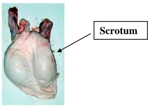
The main principal organs of the boar genital system are scrotum, testicles, epididymis, accessory glands and penis.
Fig. 1: Pietrain pig - General representation of genital apparatus of boar (Singleton, 1997). Scrotum
Fig. 1.2: Boar genital apparatus – scrotum (Dwane Davis picture).
- 2 -
The scrotum is a sac of skin characterized for its thin thickness that is present in the male reproductive system of mammals. The scrotum is divided into two compartments and each one contains one of the two testes. It is continuous with the skin of the lower abdomen and is located directly behind the penis and in front of the anus. The scrotal wall is a thin layer of skin lined with smooth muscle tissue (dartos fascia). The skin contains more pigment than that of surrounding areas and has many sebaceous (oil-producing) glands and sweat glands, as well as some hair. The two compartments of the scrotum are distinguished externally by a middle ridge called the raphe. Internally, the raphe connects to a muscular partition, the septum, which serves to divide the scrotum into its two areas (https://global.britannica.com/science/scrotum).
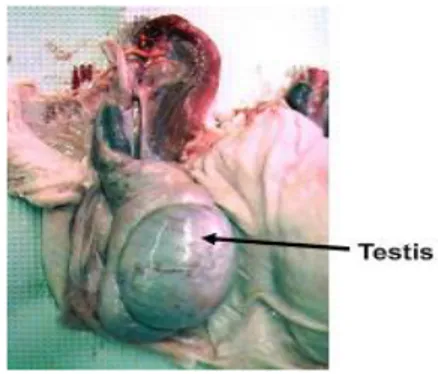
Testicle
Fig. 1.3: Boar genital apparatus - testicle (Dwane Davis picture).
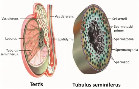
Testicles have two main functions. The first function is the formation of the male germinal cell, the sperm. The second function is acting as an endocrine gland by secreting into the whole body hormones that are important for the regulation of the reproductive function, like androgens. The testicle is composed by a series of thin tubules, called seminiferous tubules (ST). These tubules originate and terminate in the rete testis located in the central portion of the testis.
- 3 -
Fig. 1.4: A horizontal section through the scrotum showing the internal organization of the
testes.
The surface of the testis is covered by a serous membrane resulting from the peritoneum (vaginal tunic) and by a fibrous capsule (tunica albuginea) which contains smooth muscle cells. When these cells contract, there is an increase in pressure in ST which facilitates the transfer of sperm, still immobile at this stage, towards the rete testis and subsequently epididymis. Finally sperm acquire motility after their transit through the epididymis (Sjaastad et al., 2013).
- 4 -
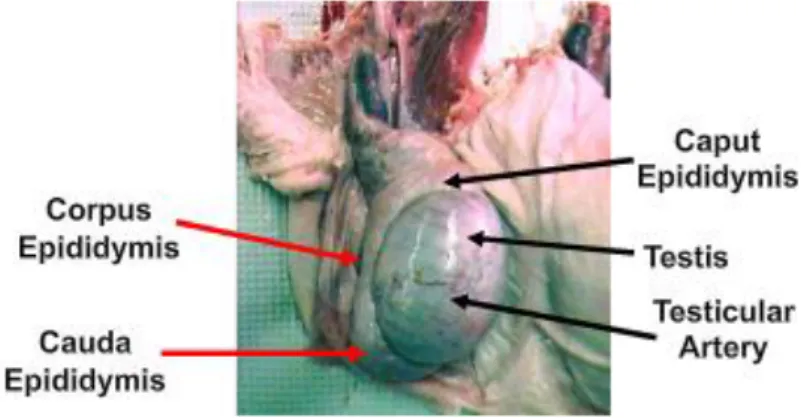
Epididymis
Fig. 1.5: Boar genital apparatus – testis with highlighted epididymis position (Dwane Davis
picture).
Epididymes are two crescent-shaped organs that are adjacent to the testis in manner that each testis-epididymis formation can be considered as a unified functional system. Epididymis can be divided in three gross morphological regions, namely head (caput epididymis), body (corpus
epididymis) and tail (cauda epididymis). The main structure of epìdidymis is a long and unique
convoluted tubule connected to the testis via the efferent ducts and finished by joining the vas
deferens in its caudal part (Guyonnet et al., 2009). Both vas deferens are muscular tubes that
undergo peristaltic contractions during ejaculation, propelling the spermatozoa from epididymis to the urethra (Frandson et al., 2009).
- 5 -
Accesory glands
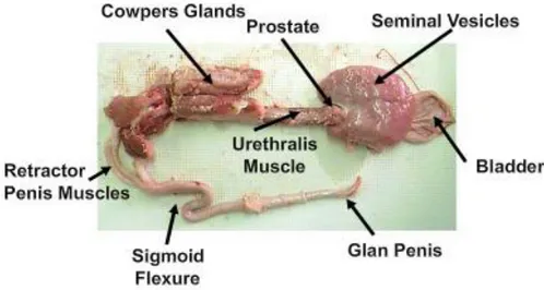
Fig. 1.6: Boar reproductive tract (Dwane Davis picture).
Boar has three main accessory glands, namely the bulbourethral glands, also named Cowper’s glands, the seminal vesicles and the prostate (Badia, 2003). All of them are exocrine glands that release their secretion into the lumen of the urethra by means an androgen-dependent secretory activity (Bonet et al., 2013).The main role of these secretions are to form the ejaculate together with the sperm-rich fraction originated in testes. Thus, the final volume of the boar ejaculate is composed by a 55%-75% of secretions from prostate and urethra, 10%– 25% of secretions from the Cowper’s glands and 15%–20% of secretions from seminal vesicles. Only 2%–5% of the ejaculate volume comes from the caudal region of the epididymis, containing it sperm and epididymal/testicular fluid (Dyce et al., 1999; Badia, 2003; Badia et al., 2005, 2006).
- 6 -
Fig. 1.7: Boar genital apparatus – seminal vesicles; prostate body; bulbourethral glands;
bladder (Dwane Davis picture).
Penis
Fig. 1.8: Boar genital apparatus – penis (Dwane Davis picture).
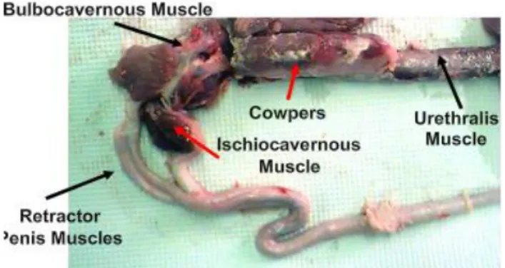
The penis is the male organ of copulation. Under an anatomical point of view, the boar penis is fibro-elastic, with cavernous bodies of reduced size. This implies that erection was mainly due to the relaxation of the retractor penis muscle located at the sigmoid flexure. The boar penis
- 7 -
measures 45 cm-50 cm, with a diameter of 1.5-2 cm even during erection. The gland has a typical corkscrew-shaped shape, which aids to fix the whole peneal structure fits into the corkscrew-shaped cervix canal of the sow (Hollandbeck et al., 1964).
2. Boar Sperm morphology
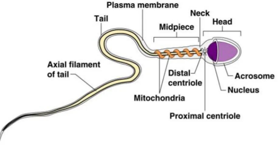
The mammalian mature spermatozoon is a uniflagellar and motile sperm cell. Spermatozoa are the cellular component of ejaculates, being their primary function to fertilize an oocyte and thus, create a new organism composed half by the sperm-gene and half by the gene load from the oocyte.
The mature boar spermatozoon is an elongated cell of about 43–45 μm in full length (Briz, 1994; Holt et al., 2010). The cell is composed by two major distinguishable regions, the head and the tail, separated by a short linking segment called the connecting piece or neck.
Fig. 2: Structure of mature sperm cell
- 8 -
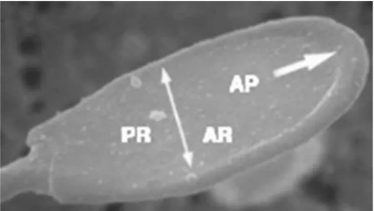
The boar head sperm is a bilaterally flat structure of about 7µm long designated to interact with the oocyte. The two major components of the sperm head are the nucleus and the acrosome. The nucleus constitutes the major part of the sperm head. The nuclear structure is electrodense due to its very rigid nucleo-proteinic composition. Surrounding the antero-lateral area of the nucleus is the sac-like acrosome, a membrane-bound vesicle that forms a cap that covers approximately 80 % of its length. The acrosome has an inner membrane that overlays the nuclear envelope. This inner membrane is merged at the posterior margins of the acrosome with an outer membrane that is lying directly beneath the plasmalemma. Enclosed inside both acrosomal membranes there is a narrow space, the acrosomal matrix, filled with amorphous material distributed homogeneously and mostly corresponding to densely packed hydrolytic enzymes. The part of the head containing the acrosome is called the acrosomal region (Briz and Fabrega, 2013).
Fig. 2.1: Detail of the head of a boar spermatozoon. Note the great development of the
acrosomal protuberance (AP) which extends down to the boundary of the acrosomal region (→) (×9,500). Acrosomal region (AR); postacrosomal region (PR). From: Briz and Fabrega, 2013.
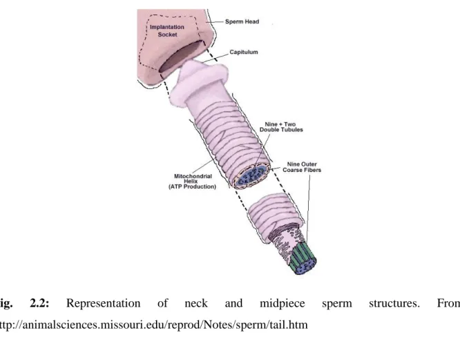
- 9 -
The tail has a filamentous and cylindrical shape and can be subdivided into three major regions, namely the midpiece, the principal piece and the terminal piece. The midpiece is 9 μm in length and 0.7 μm in diameter. The most prominent feature of the midpiece is the presence of mitochondria, organelles strongly involved in the control of many sperm functions, as well as in the generation of part of the energy needed to maintain the overall sperm function (Flesch et al., 2000; Rodríguez-Gil and Bonet, 2016).
Fig. 2.2: Representation of neck and midpiece sperm structures. From: http://animalsciences.missouri.edu/reprod/Notes/sperm/tail.htm
The principal piece is the longest segment of the spermatozoon tail, extending from the annulus or Jensen’s ring to the proximal end of the terminal piece. The principal piece is characterized by the presence of a complex fibrillar structure that maintains the proper motion characteristics
- 10 -
that needs sperm to advance into the female genital tract. The most important parts of this complex structure are the fibrous sheath, the outer dense fibres, the axoneme, and the Jensen’s ring.
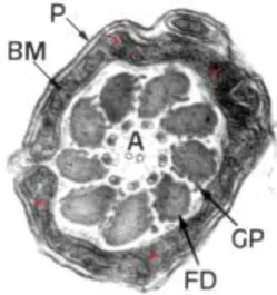
Fig. 2.3: Ultrastructural image of boar sperm mitochondria. The low development of inner
crests is noticeable (asterisks). BM: inner mitochondrial membrane. P: cell membrane. A:
axoneme. FD: dense fibres. GP: peripheral granules. From: Bonet et al., 2000. The terminal piece is the last and shorter segment of the spermatozoon tail and has no
accessory cytoskeletal structures, consisting only of the axoneme enclosed by the plasmalemma (Briz and Fabrega, 2013).
- 11 -
Fig. 2.4: Cross-sections of boar sperm tail and whole diagram of mouse spermatozoa. From: Claire et al., 2009.
- 12 -
3. Sperm journey through female genital tract: processes and molecules involved.
Spermatozoa undergo several important steps throughout their life from spermatogenesis to fusion with the oocyte. During their migration through the female genital tract, spermatozoa undergo a series of tightly controlled biochemical and membranous changes. These changes are globally known with the term capacitation, enabling sperm to reach and bind to the zona pellucida, undergo acrosome reaction, penetrate the egg vestments and finally fuse with the oocyte (Austin, 1952; Talukdar et al., 2015). In an in vivo fertilization, only a few sperm cells reach the ampulla or the site of fertilization (Sakkas et al., 2015). In boar reproduction it is estimated that from 37.5 billion of spermatozoon which are released into the reproductive tract after ejaculation, only 5000 will be able to reach the site of fertilization (Avilés et al., 2015). Thus, the number of fully capacitated sperm that can reach their final goal is actually very low.
Capacitation is a phenomenon which appears to be controlled by a complex crosstalk involving a myriad of separate molecular signalling pathways (de Lamirande et al., 1997; Fraser, 2010). Historically, Austin (1951) in rat and Chang (1951) in rabbit were the first scientists who described the capacitation process. Following these pioneering studies, capacitation has been reproduced in many other species both in in vivo and in
in vitro conditions. Despite this, many of the factors and molecular pathways involved in its
regulation remain unknown or only poorly known. In summary, after a long journey accompanied by several modifications occurring to sperm cells, the spermatozoa become able to meet the oocyte in the specific site of the oviduct named ampulla, and fertilization can takes place. The first step of this complex journey is the semen deposition into the female genital tract during the ejaculation process. Depending on the species, sperm are deposited in different sections of the female tract. Thus, in a large number of mammals, semen is ejaculated into the
- 13 -
anterior vagina during coitus (e.g. primates, cows, sheep, rabbits, dogs and cats). In other species sperm are placed into the uterus (e.g. sows) or directly spurted into the uterus (horses and many rodents; see Coy et al., 2012). In pig reproduction, this positioning in the uterus permits sperm to bypass the vaginal barrier and makes them available to the Fallopian tubes more easily (Hunter, 1981). Nevertheless, sperm meet numerous impediments during their journey, so to maintain their fertility sperm cells need to employ their limited resources and/or exploit those from the female tract (Suarez and Pacey, 2006; Mortimer et al., 2013). For this reason one of the most important parameter to value semen quality is progressive motility. Sperm motility and in particular its progressive swimming pattern, is essential in species with vaginal or cervical deposition to pass through the cervical mucus. Hence, spermatozoa with poor motility and, concomitantly, with abnormal morphology are filtered out during this passage. This is thought to be the first form of sperm selection (Sakkas et al., 2015). However, this phenomenon is less evident in species like pig, in which sperm selection starts into the uterus. This could explain the low motion characteristics that boar sperm show when compared with species with vaginal/cervical deposition like horses, dogs, bulls, or mice (Rodriguez-Gil and Bonet, 2016). Thus, after crossed all anatomical and physiological barriers found in cervix and/or uterus, spermatozoa are able to reach oviducts. Under physiological conditions, oviducts play an important role in sperm capacitation
(Immler, 2008). In fact, previously selected sperm will reach the bottom of the oviduct (isthmus) where they are stored until the moment of ovulation. This sperm storage is known as the oviductal sperm reservoir. Sperm placed inside this reservoir maintains viability for variable time lapses, depending on the species, and avoids a premature capacitation remaining in this physiological state until the time of ovulation (Brüssow et al., 2006). This break in the development of the capacitation process is needs to reach the final goal of fertilization.
- 14 -
Fig. 3: Capacitation process. A) After ejaculation, a heterogeneous population of sperm
reaches the female reproductive tract. B) Only a few sperm achieves the oviduct and forms the Sperm Reservoir (SR) in the caudal portion of the isthmus. C) During peri-ovulatory time, sperm release from the SR and those who complete a correct sperm capacitation are able to contact with the oocyte and fertilize it. Different colours indicate distinct types of sperm: dead (grey), damaged (red), normal (blue), hyperactivated (green-blue) and successfully capacitated (green). From: López-Úbeda and Matás, 2015.
- 15 - 4. Chronological events of sperm capacitation
Capacitation process is a phenomenon that starts at the same time of ejaculation and continues following a precise chronological succession of events that occurring during all the journey of sperm into the female genital tract. These events often are regrouped, in order to their timing, in two subsequent steps (Visconti, 2009):
First step, fast capacitation events: A very early event in sperm capacitation is the activation
of sperm motility. In fact, altough sperm stored in the cauda epididymis consume oxygen at a high rate, they are immotile (Visconti, 2009). The contact of spermatozoa with seminal fluid is a trigger point for activation of vigorous flagellum movement. This activation is related with the high levels of HCO3- and Ca2+ that are present not only in the seminal plasma, but also in
the secretions of the female genital tract. Specifically, the HCO3-will induce the activation of
the sperm transmembrane adenylyl cyclase SACY, which, in turn, will increase intracellular levels of cAMP, activating thus the cAMP-dependent protein kinase A activated (PKA). Finally, the PKA will be responsible for launching a very complex phosphorylation single cascade that affects a myriad of sperm proteins (Salicioni et al., 2007). Among these signals, several of these PKA-activated cascades will be responsible for the activation of sperm motion immediately after ejaculation. Other will initiate other processes that will lead to subsequent molecular changes in sperm function.
Second step, slow sperm capacitation: As a consequence of the PKA-mediated activation of
a large number of separate molecular signalling pathways a complex long-term series of changes in sperm function are launched. The complete details of these long-term changes, mainly developed during the stay of sperm into the oviductal reservoir, are not completely elucidated. Despite this, investigators have a reasonable knowledge of these processes thanks to the development of in vitro induction of capacitation. To promote the inset of capacitation,
- 16 -
spermatozoa need to be incubated in specific media with different composition depending on the species. Generally, an in vitro capacitation medium had to include some sort of energy source, a protein component that usually is bovine serum albumin (BSA), HCO3-, and both
calcium and sodium salts (Saravia et al., 2007; Ded et al., 2010). Starting from this basis, each species will have some specific requirements to yield a feasible capacitation in this in vitro environment. It is established that each species has a precise equilibrium between glycolysis and mitochondrial oxidative pathways. In fact, in species such as boars and mice, this equilibrium is greatly unbalanced in favor of glycolysis, which is the vastly majority energy-obtaining pathway in the presence of sugars such as glucose (Rodriguez-Gil and Bonet, 2016). This implies that the source of energy that must be added to the capacitation medium can vary from glucose in species like the boar to lactate in dog (Rodriguez-Gil and Bonet, 2016). Regarding the role of the other basic components of the capacitation medium, it is well known that the presence of BSA, HCO3 and Ca2+ acting through the launching of the capacitation
mechanisms started after the activation of the PKA. Thus, the BSA stimulates the replacement of the membrane cholesterol with other cholesterol-binding compounds such as β-cyclodextrins. Meanwhile, both the HCO3
−
and Ca2+ salts are involved in the regulation of SACY and in the consequent increase in cAMP levels and PKA activation (Salicioni et al., 2007). At this respect, it is noteworthy that the increase of intracellular calcium is indispensable for the launching of capacitation as well as for the induction of the acrosome reaction in previously capacitated sperm. Sperm can acquires cytoplasmic calcium thanks to the presence in plasma membrane, and in particular in the principal piece of the flagellum, of Ca2+ channels called CatSper proteins (CatSper 1 to 4) (Kirichok et al., 2006, Qi et al., 2007). In this way, the combined presence of BSA, HCO3 and Ca2+ is indispensable not
only for launching, but also to develop the capacitation process, being able these compounds to maintain and develop all of the protein phosphorylation cascades that starts as a consequence
- 17 -
of the activation of the PKA. In fact, the absence of BSA, HCO3 or Ca2+ prevents both the
overall protein tyrosine phosphorylation and the subsequent full capacitation status (Salicioni et al., 2007; Visconti, 2009). However, the importance of each of these modulators in controlling the protein phosphorylation cascades is different among species. For example, tyrosine phosphorylation in human sperm during sperm capacitation requires the presence of BSA, and HCO3-,but no Ca2+ (Muratori et al., 2010).
Meanwhile, in the case of stallion tyrosine phosphorylation requires only HCO3- but no
BSA or Ca2+ (González-Fernández et al., 2012). Another event that shows a characteristic pattern for each species is represented by different times of responsiveness to BSA, HCO3- and
Ca2+ in the inset of tyrosine phosphorylation; these differences are observed in pig, mouse, dog and bull. Thus, whereas in spermatozoa of species like boar and mice the full establishment of the tyrosine phosphorylation cascade after its induction appears at about 1 hour after the addition of HCO3- (Visconti et al., 1995; Gadella and Van Gestel, 2004), in
other species like dog the same phenomenon occurs after 90 minutes of the bicarbonate induction (Petrunkina et al., 2003; 2004) and even in bull sperm where the same event takes place only after 4 hours of the addition of heparin (Galantino-Homer et al., 1997). Regarding the precise location of the capacitation-induced increase of tyrosine phosphorylation, the flagellum seems to be the main placement (Naz et al., 1991; Carrera et al., 1996; Mahony et al., 1999; Si and Okuno, 1999; Lewis and Aitken, 2001; Urner et al., 2001), although the acrosomal area also shows an evident increase in the overall protein tyrosine phosphorylation during capacitation, especially in boar (Petrunkina et al., 2001).
- 18 -
Fig. 4: Sequence of mammalian sperm capacitation events. From: Redgrove, Aitken and
Nixon, 2012.
Another of the functional consequences of capacitation is the development of a distinct motility/kinetic pattern that is called hyperactivation. The hyperactivated motion pattern is characterized by a very vigorous motility pattern with high amplitude and asymmetrical flagellar beating (Yanagimachi, 1970). It has been suggested that the mechanical thrust due to hyperactive motility is vital for spermatozoa to penetrate the zona pellucida of the oocyte (Yanagimachi, 1969; Stauss et al., 1995; Bedford, 1998). In this way, hyperactivation is observed as the typical swimming pattern shown by most sperm retrieved from the oviductal ampulla at the time of fertilization (Suarez and Ho, 2003).As seen in mice, but not yet in boar
- 19 -
spermatozoa, these changes occur in the oviduct in in vivo conditions (Rodriguez-Martinez, 2007). Notwithstanding, hyperactivation can be also observed in in vitro conditions after a proper stimulation. It is known that the progesterone released from the ovary and secreted by oviduct epithelium at ovulation time give rise to increment of sperm intracellular [Ca2+]i, which in turn induce turning swimming with asymmetric flagellar bending (Yoshida and Yoshida, 2011; Hunter, 2012). Thus, progesterone can be utilized in many species as a feasible inductor of the hyperactivated motion pattern.
Fig. 4.1: Progressive and hyperactivated motility of mammalian sperm. Motility patterns of
mammalian sperm are displayed in time-lapse drawings. The hyperactivated mode consists of transitions to deeper and less symmetrical flagellar bending and results in a path of the sperm head that is less linear than that of activated state motility. From: Florman and Fissore, 2015. In summary, the most important changes underwent by spermatozoa after their contact with the oviductal fluid and hence with the capacitation modulators contained in this fluid are
- 20 -
modifications of lipid membrane, the loss of plasma membrane cholesterol, the activation of the cAMP/PKA-modulated pathways, the increase of Ca2+ uptake, which leads to a concomitant increase in the intracellular pH, the subsequent hyperpolarisation of cell membranes and the increase in the overall protein tyrosine phosphorylation pattern (Aitken & Nixon, 2013). In this way, it is evident the existence of a strong oviductal influence that facilitates closeness of sperm to the oocyte (Hunter & Nichol, 1986; Rodriguez-Martinez et al., 2001; Rodriguez-Martinez, 2007; Coy et al., 2010; Zumoffen et al., 2010; Kumaresan et al., 2012).
5. Acrosome reaction
The sperm capacitation process is absolutely required as a previous step in order to acquire the ability to undergo acrosome reaction in response to physiological stimuli. As mentioned above, the changes of the capacitation in the molecular organization of the sperm plasmalemma make it sensitive to the presence of zona pellucida protein ZP3. The union ZP3-outer acrosome membrane proteins combined with the stimulus of external factors like progesterone launches the acrosome reaction (AR), a phenomenon necessary for sperm cells to achieve gamete fusion (Ferramosca and Zara, 2014). Properly, the AR is the result of the fusion of the sperm plasma membrane with the outer acrosomal membrane. The acrosome content, especially in terms of hydrolytic enzymes, will be released, leaving the inner acrosomal membrane as the sperm surface membrane that will be in direct contact with the oocyte surface. The place where AR begins in mammalian sperm has been a controversial topic. On one side of the debate some researchers think that the AR takes place while the sperm advance through the cumulus, while other scientists think that AR occurs on the surface of the ZP (Coy et al., 2012). It should be noted that no one has ever followed a single spermatozoon from the beginning of the AR until
- 21 -
the end of fertilization, so the exact place where fertilizing spermatozoa begin their AR and what triggers inset the AR remain to be determined (Yanagimachi, 2011). Regarding on the myriad of chemical and physical agents reported to induce in vitro acrosome reaction, we known the existence of natural inducers, such as the zona pellucida proteins (ZP) and progesterone, or artificial inducers, like heparin and calcium ionophores (Cheng et al., 1998). In fact, the progesterone secreted by the cumulus cells that surround oocytes is one of the most important factors, if not the most, which stimulates in vivo AR (Chang and Suarez, 2010). Notwithstanding, progesterone has a double function. The first function is being a potent chemo-attractants, whereas the second function is acting as a stimulator of the in vitro acrosome exocitosys (IVAE). It is noteworthy that the progesterone-induced AR occurs only when sperm are kept in close contact with the oocyte, but not before although sperm are in previous contact with progesterone presents in the oviductal fluid. A reason that could explain this delay would probably be the differences in progesterone concentration. Thus, oviductal fluid has low concentrations of progesterone that are not able to induce the AR (Publicover et al., 2008). These low concentrations of progesterone could act instead as a chemo-attractant driving the sperm towards the oocyte. Afterwards, progesterone concentrations at the space surrounding the oocyte are much higher. These higher concentrations of progesterone would ultimately be responsible for the induction of AR in the close proximity of the oocyte (Coy et al., 2012).
- 22 -
Fig. 5: Changes in effects of Progesterone (P4) concentrations, on sperm cells, during their rute
towards oocyte. Zone A: low concentrations of P4 acting as chemo-attractant. Zone B: high P4 levels produced by cumulus cells cause acrosome reaction. From: Coy et al, 2012.
Regarding the sperm–ZP interaction, the first topic is that this interaction is a crucial point to achieve fertilization. The interaction between ZP proteins and spermatozoa induces the activation of a specific sperm phospholipase through a mechanism involving the action of a G protein. This activation induces in turn the subsequent activation of the sperm of adenylyl cyclase, which, in turn, will increase cAMP levels. These events ultimately lead to an alkalization of intracellular pH with the subsequent depolarization of sperm membrane. This last event is fundamental because it will open the Ca2+ channels that permit the entry of a flux of Ca2+ ions. These ions will in turn activate sperm phospholipases C and A2 (PLC and PLA2),
- 23 -
which, through the corresponding molecular cascade will finally activate the Protein Kinase C (PKC) (Flesh and Gadella, 2000). This activated PKC will migrate toward the membrane. This process of migration will finally initiate the fusion of sperm plasma membrane with the outer acrosomal membrane, leading thus to the final exocytosis corresponding to the AR (Doherty et al., 1995).
Fig. 5.1: Acrosome reaction (AR) triggers point: molecular events occurring after sperm- ZP
- 24 - 6. Sperm analysis
Possible fluctuations in seminal quality are associated with factors such as breed (Rijsselaere et al., 2007), age (Stone et al., 2013), seasonality (Chemineau et al., 2008; Zhang et al., 2013), temperature (Thonneau et al., 1998), photoperiod (Mazzarri et al., 1970; Kozdrowski and Dubiel, 2004) and other factors of different etiology (Petroccelli et al., 2015). Taking this into account, the definition of the precise quality of each ejaculate through an accurate analysis is of the utmost importance. Nowadays, to evaluate semen quality we can consider visual and physiological parameters. Until recent years, subjective microscope evaluation represented the only manner to predict a semen quality. However, this visual exam gave to us only a general and diffuse idea about the quality of the ejaculate and its hypothetic fertilizing potential. In this way, the addition of more precise techniques to evaluate semen quality is mandatory to optimize semen analysis. An important group of test that would be included in the routine semen analysis involves the determination of the sperm metabolic function. However, to accomplish that it is indispensable to know sperm cell physiology. This implies that microscope analysis cannot be the only system by which semen might be analyzed. Thus, besides the mandatory previous macroscopic evaluation of the ejaculate, the main parameters included in an optimal sperm quality analysis can be classified into two sections: microscope-linked parameters and physiology and metabolism parameters that cannot be only evaluated through microscope. Microscope-evaluated parameters would comprise concentration, sperm motility, sperm morphology, plasmatic membrane integrity, DNA fragmentation and osmotic resistance; while non-microscope evaluated parameters would be those related with aspects such as nuclear and acrosome structure, membrane fluidity, membrane sheath state, intracellular reactive oxygen species (ROS) levels, which comprise the evaluation of both peroxides and superoxides, and several enzymes activity (Bonet et al., 2012). It is evident how, all these assessments must be carried out using a large number of replicates because of the high
- 25 -
level of variability within and between individual samples and the subjectivity of some assays (Woelders, 1991).
6.1 Assessment of macroscopic characteristics of the ejaculate:
In boar semen, the temperature of the freshly collected ejaculate is about 36ºC±2°C, while its pH is usually between 6.85 and 7.9. Moreover osmolarity is variable, but always around 300 mOsm/Kg (Martín Rillo, 1982). The colour of the sperm-rich fraction is milky-white, although there may be colour changes due to separate alterations like contamination with urine, blood or prepucial secretion (Sancho, 2002).
6.2 Sperm volume and concentration
The total average volume of boar sperm-rich fraction of the ejaculate is about 250 mL, with variations between 50 mL and 400 mL. This volume represents the highest observed among males of the more common domestic animals (Frunză et al., 2008). The number of sperm per ejaculate varies between races and boars due to the action of a multitude of factors such as environment, temperature, nutrition, diseases, frequency of ejaculation and photoperiod (Sancho et al., 2004). Usually, a boar ejaculate contains a sperm concentration from 300x106 sperm/mL to 600x106 sperm/mL (Casas et al., 2010). To evaluate concentration we can utilize several methods like hemocytometry, colorimetry and true spectrophotometry. The hemocytomety allows practitioners to determine the number of immobilized spermatozoa that are present in a known volume. This technique uses a commercial cell counting chamber such as those from Neubauer, Thoma or Burker (Christensen et al., 2005). Related with these chambers there is also the Mackler chamber, which allows practitioners to count concentration and motility at the same time through the application of an informatics system called Computer Assisted Sperm Analysis (CASA; Roca et al., 2011). On the other hand, colorimetric and
- 26 -
spectrophotometric methods are based on the percentage of transmitted light that is trapped by a diluted semen sample (Sancho and Vilagran, 2013). However, although colorimetric and spectrophotometric methods can usually be faster, more sensitive and more objective than the hemocytometric ones, all of them are superseded by flow cytometry. In fact, at this time, flow cytometry is the most precise and sensitive method to evaluate concentration, since practitioners can count with this system a much higher number of fluorochrome-marked cells (more than 10,000) with a much higher precision. Flow cytometry, however, has a high cost, limiting thus its use to large laboratories uniquely devoted to research or analysis (Lu et al., 2006).
6.3 Sperm Motility:
Motility is recognized to be an important characteristic in predicting the fertilizing potential of the ejaculate, although its real importance in explaining the in vivo fertilizing ability of an ejaculate is not clear in species like boar. Thus, an adequate motility would be required for sperm to reach the fertilization site and penetrate the oocyte, although this parameter would not be the only needed to achieve this feat (Rodríguez, 2012). The classical method to evaluate motility is the direct, visual estimation of the parameter through an optic microscope. This system is highly subjective, especially in species like pig in which semen motion parameters are low and irregular. Thus, at this moment the use of a CASA instrument is mandatory. Actually, the development of CASA systems has enabled objective and accurate tracking of sperm movement. Modern CASA systems can capture images from 20 to more than 1,000 sperm at 50–60 frames per second, and also provides morphological measurements in addition to comprehensive motion analyses (reviewed in Amann & Waberski, 2014). The CASA measures sperm movement by establishing a centroid for each spermatozoon and evaluate cell motion based on centroid trajectory (Amann & Waberski, 2014). However, in vitro measures
- 27 -
may not reflect how the sperm moves within the female tract (Werner et al., 2007; Humphries et al., 2008). Within the female tract, sperm are spatially restricted and are therefore likely to be influenced by physical constraints such as wall effects (Humphries et al., 2008). Furthermore, natural fluids within the female tract will alter sperm movement (Curtis & Benner, 1991). Again, the information obtained through the CASA system are still subjected to the effects of external factors such as sample preparation or the type of chamber used for analysis (Tejerina et al., 2009). In this manner, the standardization of the procedure is necessary for the proper use of the system (Tejerina et al., 2009). Despite these shortcomings, CASA systems represent the best technique to evaluate sperm motility now. Motion parameters that are typically derived using automated CASA systems provide information about the velocity, linearity and lateral displacement of sperm heads as they progress along their trajectories (Holt et al., 2007). These parameters have been described many times previously (see for example: Katz and Davis, 1987; Mortimer et al., 1988; Davis and Katz, 1989, 1992, 1993a, 1993b; Kay and Robertson, 1998). Here are reported a brief description of some of these following King (King et al., 2000):
Total motility: percentage of sperm with a total velocity greater than 10µm/s.
Curvilinear velocity (VCL): distance travelled by the spermatozoa along its actual path as a
function of time. It is expressed in µm/s.
Average path velocity (VAP): distance travelled by the spermatozoa along its average path as
- 28 -
Straight-line velocity (VSL): velocity over the straight-line distance between the beginning
and the end of the sperm track. It is expressed in µm/s.
Linearity (LIN): the straight-line distance divided by the incremental deviations along the
actual path. Then, it is also the percentage relationship between the curvilinear and straight-line velocity. It is expressed in %.
Head beat-cross frequency (BCF): the frequency with which the undulatory movement of the
sperm head crosses the average path in function of time. It is expressed in Hz.
Straightness (STR): the percentage relationship between the curvilinear and the average path
velocity. It is expressed in %.
Wobbleness (WOB): the percentage relationship between the average path and curvilinear
velocity. It is expressed in %.
Amplitude of lateral head displacement (ALH): shift that makes the head in its curvilinear
trajectory of the one part and the other part of the trajectory average or linear. It is expressed in µm.
- 29 -
Fig. 6: Scheme of different velocities and parameters of sperm movement measured by CASA
system. From: http//www.mibio.org/en/metrics-used
As suggested by Garcia (Garcia et al., 2005), capacitated boar spermatozoa have a species-specific motility pattern. Despite this, there are common features among species. In this way, as a general rule, capacitated sperm show an increase in VAP values and a decrease of LIN percentage. It’s noteworthy that in an ejaculate from a healthy individual, a minimal rate of total motility of 60% would be the minimum requirement to be considered fertile (Donadeu, 2004; Broekhuijse et al., 2012).
6.4 Sperm morphology:
Some shape abnormalities of spermatozoa (AS) can be a result of pathological processes that affect testicles and the epididymis tract, whereas others AS can have a gene basis or, indeed, may be caused by unsuitable rearing conditions (Čeřovský et al., 2005). There is an abundant literature centred in the research of this field. This is logical, since AS is known to be an
- 30 -
important marker to assess semen quality (Leidl et al., 1971; Lyczynski and Pawlak, 1974; Blom and Andersen, 1975; Gamčík et al., 1976; Blom, 1977; Stemmler et al., 1982; Wekerle, 1982; Falkenberg et al., 1984; Yoshida and Kojima, 1989; Waberski et al., 1990, 1994; Itoh and Toyama, 1995; Itoh et al., 1996; Věžník et al., 2000; Louda et al., 2001; Corcuera et al., 2002; Gadea, 2002). Following the criteria of Lyczynski and Pawlak (1974) or Bach (Bach et al., 1982) the use of semen containing more than 25% of AS is not recommended for insemination. Other authors established even more restrictive conditions. Thus, Remmen and Tielen (1976) limited the AS content to 20%, whereas Gibson (1983) gave the limit up to 15% for both primary changes and secondary ones. Usually, a morphological examination of the boar ejaculate comprises a qualitative and also a quantitative classification of normal and abnormal sperm morphologies. This classification can be assessed by optical microscopy through the use of simple staining techniques such as eosin/nigrosin, Trypan Blue, Giemsa, Papanicolaou or Diff-Quick (Foxcroft et al., 2008). Currently, a contrast phase microscope can provide better resolution without requiring staining the samples and, moreover, it can be coupled to a CASA system (Sancho and Vilagran, 2013). Sperm abnormalities have traditionally been classified by location of the defect (head, midpiece, tail), or by its origin (primary: testis; secondary: epididymis; tertiary: accessory glands/post-ejaculation; see: Menon et al., 2011). Primary sperm abnormalities are due to disturbance of spermatogenesis by congenital or hereditary factors, high ambient temperature, scrotal insulation or diseases (Adeniji et al., 2010). The most important primary abnormalities are those related with head malformations, although several tail alterations are also included in this group. Since the sperm head contains the genetic material and other key effectors of fertilization, most abnormalities of head are associated with a significant impairment of fertility (Wilmington, 1981; Soderquist et al., 1991). Proximal and distal cytoplasmic droplets are considered as secondary abnormalities. The cytoplasmic droplet shows a diameter of around 1.5µm and is rich in membranous
- 31 -
vescicles that increase as the droplet displaces through the midpiece (Bonet et al., 2013). The presence of the cytoplasmic droplet on ejaculated sperm can be used as an indicator of sperm maturation (Gomez et al., 1996; Keating et al., 1997; Amann et al., 2000; Thundathil et al., 2001; Bassols, 2006). Morphological anomalies acquired by inappropriate handling of semen sample (transport, storage temperature, doses dilution etc.) are considered as tertiary abnormalities, being the most common severe morphological alterations of tail (Adeniji et al., 2010).
- 32 -
Fig. 6.1: Sperm malformations of the ejaculate of post-pubertal boars with spontaneous
- 33 -
Mature spermatozoon. b1) Bicephalic spermatozoon with two tails fused from the distal end of the midpiece (arrow). b2) Spermatozoon with two elongated heads, and two tails totally fused and folded at the midpiece. b3) Spermatozoon with two heads, one of them aberrant (arrow), and two totally fused tails. b4) Tricephalic spermatozoon with three totally fused tails. c1) Macrocephalic spermatozoon (arrow); note the presence of an elongated head spermatozoon (triangle). c2) Macrocephalic spermatozoon with folded tail at the midpiece. c3) Macrocephalic spermatozoon with folded tail at the connecting piece. d1) Roundish head spermatozoon. d2) Round head spermatozoon; note the corksrew defect affecting the principal piece (arrow). e1, e2) Pointed head spermatozoon. f) Deformed head spermatozoon due to a cephalic break (arrow). g) Spermatozoon with a transversely folded head. h) Spermatozoon with crater defect and proximal droplet (arrow). i) Macrocephalic spermatozoon with knobbed acrosome (arrow). j) Spermatozoon with acrosomal vacuolization (arrow). k) Spermatozoon with folded tail at the Jensen’s ring. l) Macrocephalic spermatozoon with mitochondrial swelling (arrow). m1) Macrocephalic spermatozoon with folded tail; note the presence of a spermatozoon with crater defect and abaxial tail (triangle). m2) Spermatozoon with coiled tail at the terminal and principal pieces (arrow); note the presence of a spermatozoon with acrosomal vacuolization (triangle). n) Immature spermatozoon with proximal droplet and abaxial tail (arrow). o1) Spermatozoon with two unfused tails abaxially attached (arrow). o2) Spermatozoon with two tails fused from the principal piece (arrow). o3) Macrocephalic spermatozoon with vacuolated acrosome (arrow) and two fused tails. ×500. From Pinart et al., 1998.
6.5 Sperm Agglutination:
Sperm agglutination is observed when a spermatozoon binds to another spermatozoon by head-to-head or tail-to-tail contact. Under standard conditions agglutination does not occur. In fact, agglutination can be an infertility cause (Sancho and Vilagran, 2013). It is well known that
- 34 -
bivalent and trivalent cations present in boar seminal plasma as well as long storage and immunological factors or bacterial contamination can induce sperm agglutination (Yeste et al., 2008; Bussalleu et al., 2011). In this manner, the presence of sperm agglutination can suggest an improper handling of the sample.
6.6 Osmotic resistance of sperm:
Boar spermatozoa are sensitive to osmotic changes in the environment and the ability to respond to osmotic stress is related to cell functionality. Thus, an osmotic shock affects the diffusion of phospholipids in the bilayer membrane of spermatozoa (Christova et al., 2002). This implies that the sperm response to environmental osmotic changes can be a good indicator of its physiological status (Yeste et al., 2010). The sperm osmotic response involves complex metabolic pathways, such as ion channels related to the Na+/K+-dependent ATPase activity or the Na+/K+-dependent antiporter one (Medrano et al., 2006). In boars, an isotonic solution is established between 300 and at 400 mOsm Kg−1 (Petrunkina and Topfer-Petersen, 2000; Petrunkina et al., 2000; Fraser et al., 2001). Several tests have been developed to measure the response of spermatozoa to osmotic changes as a parameter of semen quality. These methods are the hypo-osmotic swelling test (HOST), the hyper-osmotic resistance test (HRT) and the osmotic resistance test (ORT), all are based on the sperm tolerance to osmolarity change of medium. The most used test in boar specie is ORT. The ORT consists in checking the capacity of sperm to resist a hypoosmotic shock by challenging sperm to a hypoosmotic medium (≤150 mOsm/Kg). The evaluation is achievable with a contrast phase microscope or by optical microscopy linked to an eosin-nigrosin stain of the sample (Rodriguez- Gil et al., 1994).