Department of Pure and Applied Sciences
Dipartimento di Scienze Pure e ApplicatePhilosophy Degree in Basic Sciences and Applications
Curriculum Earth Sciences
Corso di Dottorato in Scienze di Base e Applicazioni Curriculum Scienze della Terra
XXIX Ciclo
Titolo della tesi:
Mineralogical study of the fibrous zeolites erionite and
offretite and hazard assessment
Studio mineralogico delle zeoliti fibrose erionite ed offretite e
valutazione della pericolosità
Settore Scientifico Disciplinare SSD: GEO/06
Dottorando
Matteo Giordani
Relatore
Prof. Michele Mattioli
1. GENERAL INTRODUCTION AND SUMMARY 1
Introduzione generale e Sommario 3
2. POTENTIALLY CARCINOGENIC ERIONITE FROM LESSINI MOUNTAINS, NE ITALY: MORPHOLOGICAL, MINERALOGICAL AND CHEMICAL
CHARACTERIZATION
2.1 Introduction 7
2.2 Mineralogical background 8
2.3 Materials and methods 9
2.3.1 Materials 9
2.3.2 Environmental Scanning Electron Microscope (ESEM) 9
2.3.3 Electron Micro Probe Analysis (EMPA) 10
2.3.4 X-ray Powder Diffraction (XRPD) 11
2.4 Results 11 2.4.1 Morphology 11 2.4.2 Mineralogical composition 13 2.4.3 Morphometry 14 2.4.4 Chemical data 15 2.5 Discussion 15
2.5.1 Environmental occurrence of erionite and risk assessment in Italy 19
2.6 Conclusion 20
3. GEOLOGICAL OCCURRENCE, MINERALOGICAL CHARACTERIZATION AND RISK ASSESSMENT OF POTENTIALLY CARCINOGENIC ERIONITE IN ITALY
3.1 Introduction 22
3.2 Background 23
3.2.1 Mineralogy 23
3.2.2 Erionite geology 23
3.2.3 Erionite health risks 24
3.3 Field description and materials 25
3.4 Analytical methods 27
3.4.1 Scanning Electron Microscopy (SEM) 27
3.4.2 X-Ray Powder Diffraction (XRPD) 28
3.5 Results and discussion 28
3.5.1 Morphology 28
3.5.2 Chemistry 32
3.5.3 Structure of the fibrous samples 33
3.5.4 Erionite occurrences and risk assessment in Italy 37
3.6 Concluding remarks 40
4. PRISMATIC TO ASBESTIFORM OFFRETITE FROM NORTHERN ITALY: NEW MORPHOLOGICAL AND CHEMICAL DATA OF A POTENTIALLY HAZARDOUS ZEOLITE
4.1 Introduction 42
4.2 Materials and methods 44
4.2.1 Materials 44
4.2.2 Scanning Electron Microscopy (SEM) 44
4.3.3 Chemical data 48
4.4 Discussion and conclusions 49
5. MORPHO-CHEMICAL CHARACTERIZATION AND SURFACE PROPERTIES OF CARCINOGENIC ZEOLITE FIBERS
5.1 Introduction 52
5.2 Experimental 54
5.2.1 Materials 54
5.2.2 Methods 54
5.2.2.1 SEM-EDS and EPMA 54
5.2.2.2 XRPD 54
5.2.2.3 Surface area-BET 54
5.2.2.4 EPR 54
5.2.2.5 Simulation of the EPR spectra 54
5.3 Results and discussion 55
5.3.1 Morphological data 55
5.3.2 Mineralogical data 55
5.3.3 Chemical data 58
5.3.4 Specific surface area analysis 60
5.3.5 EPR analysis 61
5.4 Conclusions 64
6. EPR AND TEM STUDY OF THE INTERACTIONS BETWEEN ASBESTIFORM ZEOLITE FIBERS AND MODEL MEMBRANES
6.1 Introduction 66
6.2 Experimental 69
6.2.1 Materials 69
6.2.2 Methods 70
6.2.2.1 Electron Paramagnetic Resonance (EPR) 70
6.2.2.2 Transmission Electron Microscopy (TEM) 70
6.3 Results and discussion 70
6.3.1 EPR analysis 70 6.3.1.1 CTAB micelles 70 6.3.1.2 Lecithin liposomes 74 6.3.1.3 DMPC Liposomes 76 6.3.1.4 TEM Results 77 6.4 Conclusion 79 REFERENCES 81 ACKNOWLEDGEMENTS 94 SUPPLEMENTARY MATERIALS
Supplementary material Chapter 3 95
Supplementary material Chapter 5 102
Chapter 1
GENERAL INTRODUCTION AND SUMMARY
Several fibrous minerals are known in nature, some of which have chemical and physical characteristics very useful in various industri-al processes and applications. However, some of them are considered highly hazardous to human health, because of their capability to divide into inhalable size fibers, together with their biopersistence in the lungs. This is the case, for example, of the well-known asbes-tos. Nevertheless, there are many other fi-brous minerals with physical and chemical characteristics very similar with those of as-bestos, which have not been sufficiently in-vestigated. For some of them, such as attapulgite, palygorskyte, byssolite, picrolite, sepiolite, thomsonite, scolecite, mesolite, nat-rolite and offretite, the dangerousness has not yet been neither confirmed nor denied. For others fibrous minerals (e.g. jamesonite, carlostauranite, wollastonite, nemalite, clinoptilolite, phillipsite, mordenite) prelimi-nary studies seem to point out some toxic ef-fects. Lastly, other fibrous phases have al-ready been well studied and are currently con-sidered carcinogenic if inhaled, such as fluo-ro-edenite, balangeroite, riebeckite, grunerite and erionite.
Several fibrous minerals belong to the zeolite group. Zeolites occurs worldwide and are widely used in materials for the construction industry, in paper, in agriculture and in other applications. Therefore, potential exposure to the fibres may occur during the mining, pro-duction and use of the fibrous zeolites (IARC, 1997). In particular, the exposure of erionite fibres to humans has been unambiguously linked to malignant mesothelioma (Baris et al., 1978), and in vivo studies have demon-strated that erionite is significantly more tu-morigenic than asbestos (Coffin et al., 1992). For these reasons, the International Agency for Research on Cancer (IARC) has referred erionite as a Class 1 carcinogen, and at pre-sent is considered as the most carcinogenic
mineral (IARC 1987; 2012). Recently, a growing concern has developed regarding the potential risks associated with environmental and occupational exposures to erionite in Turkey (Carbone et al., 2011), in the United States (Saini-Eidukat and Triplett, 2014), in Mexico (Ortega-Guerrero and Carrasco-Nùñez, 2014) and possibly in Iran (Ilgren et al., 2015). Then, it is very likely that this problem could also be extended to other coun-tries in the future.
The main mechanisms by which inhaled fi-bres of erionite, as well as other fibrous par-ticulates, induce pathological changes com-prise the following factors: (a) physical fea-tures of the fibrous mineral particles such as diameter, length and aspect ratio; (b) cal-mineralogical features (fibre type, chemi-cal composition and surface reactivity); (c) the ability to generate reactive oxygen species (ROS); and (d) the biopersistence. Despite of the great number of researches, the relation-ships among mineralogical features and bio-logical activity of erionite have not been fully understood and there are no systematic stud-ies on the distribution of erionite or other sim-ilar fibrous zeolites in the environment. Moreover, there is another zeolite, named offretite, which is closely related both struc-turally and chemically to erionite. Despite commonly occurring as prisms, offretite has also been found under asbestiform habit, meaning that the morphology of its crystals has not yet been fully known and many min-eralogical aspects are still to be discovered. Due to these similarities and to the possible intergrowth, the distinction between erionite and offretite can be hampered. To date, there are no studies regarding a potentially hazard of offretite fibers and it is unclear whether the mineralogical distinction between erionite and offretite has any health implications.
The gap of knowledge of the fibrous zeolites erionite and offretite has been the starting
point for the development of the present PhD project. This work is based on a detailed min-eralogical study with determination of mor-phological and morphometric characteristics, physical properties, chemical compositions and crystalline structures of selected samples of both erionite and offretite zeolites. Succes-sively, these samples have been used to carry out in-vitro experiments to assess the level of reactivity and transformation that such fibers may induce to micelles and membranes in contact with them, and therefore, indirectly the possibility of leading to asbestos-related lung diseases.
On the basis of this starting point, the Thesis has been organized in 6 Chapters.
This Chapter (Chapter 1) is dedicated to a general introduction illustrating the major aims of this project and the structure-organization of the Thesis. A brief summary with main results of each chapter is also pre-sented.
In Chapter 2, the occurrence and characteriza-tion of the potentially carcinogenic erionite from Lessini Mounts (NE Italy) are reported. Morphological, morphometrical, mineralogi-cal and chemimineralogi-cal investigations performed on two representative samples of fibrous erionite, which has not been examined previously, are presented. The first sample is erionite-Ca with an extremely fibrous, hair-like and flexible appearance, growth in intimate association with levyne. The second sample is erionite-Na with prismatic to acicular crystals and rigid behavior, with significant amount of K+ and Ca2+ as extra-framework cations. In both the investigated erionite samples, fibrils of inhal-able size are present. Considering that the tox-icity and the carcinogenic potential of erionite are associated with its size parameters, to-gether with its in vivo durability and very high surface area, most of the investigated fibers may also be potentially carcinogenic.
In Chapter 3, the mineralogical characteriza-tion of erionite from Northern Italy is inte-grated and completed with structural data and extended to other erionite samples from Italy. A multi-methodological approach, based on
field investigation, morphological characteri-zation, SEM/EDS chemical analysis and structure refinement through X-ray powder diffraction was applied to different samples of potentially carcinogenic erionite. The chemi-cal composition of the studied crystals ranges from erionite-Ca to erionite-Na, and they show variable morphologies, ranging from prismatic, through acicular and fibrous, to ex-tremely fibrous asbestiform habits. The fi-brous samples are characterized by an unusual preferred partition of Al at T1 instead of T2 site. Moreover, a mismatch between the a-parameter of erionite-Ca and levyne-Ca that are intergrown in the asbestiform sample has been detected. This misfit is coupled to a rel-evant microstrain to maintain structure coher-ency at the boundary. Erionite occurs in 65% of the investigated sites, with an estimated amount from 10 to 40 vol.% of the associated minerals. These amounts are not negligible for the human health, particularly if the great number of quarries and mining-related activi-ties operating in the zeolites host rocks is con-sidered. The discovery of fibrous and asbestiform erionite in the Northern Italy sug-gests the need for a detailed risk assessment in all Italian areas showing the same possible hazard, with specific studies such as a quanti-fication of the potentially airborne fibres and targeted epidemiological surveillance.
The Chapter 4 is dedicated to the morphologi-cal, mineralogical and chemical characteriza-tion of the other fibrous zeolite offretite, which has been found with asbestiform habit in the Northern Italy and is suspected to be carcinogenic. The investigated samples show different habits, from stocky-prismatic crys-tals with a solid appearance (FF102), through prismatic or acicular with rigid mechanical behavior (AD13), to extremely fibrous crys-tals (asbestiform) with rigid to flexible behav-ior (MB2287). In the acicular and asbestiform offretite samples (AD13 and MB2287), fibrils of inhalable size are certainly present. The chemical composition of the investigated offretite samples is coherent with reference data and showing EF cations K+, Mg2+ and Ca2+ in comparable proportions in all sam-ples. Despite of the lack of epidemiological
information on populations exposed to natural asbestiform minerals other than asbestos and erionite, results acquired in the present inves-tigation suggest that other mineral fibres of similar size, habit, and biopersistence may carry a risk for humans.
In Chapter 5, morpho-chemical characteriza-tion and surface properties determinacharacteriza-tions were performed on a selection of different asbestiform zeolite fibers: two erionite sam-ples (GF1, MD8), one offretite sample (BV12) and, for comparison, one scolecite sample (SC1). The specific surface area de-terminations coupled with EPR analysis indi-cated a larger availability of surface sites for the adsorption onto GF1, while SC1 showed the lowest one and the presence of large pores in the poorly fibrous zeolite aggregates. Se-lected spin probes revealed a high adsorption capacity of GF1 compared to the other zeo-lites, but the polar/charged interacting sites were well distributed, intercalated by less po-lar sites (Si–O–Si). MD8 surface was less homogeneous and the polar/charged sites were more interacting and closer to each other compared to GF1. The interacting ability of BV12 surface was much lower than that found for GF1 and MD8 and the probes were trapped in small pores into the fibrous aggre-gates. In comparison with the other zeolites, the non-carcinogenic SC1 exhibited a poor in-teracting ability and a lower surface polarity. These results helped to clarify the chemical properties and the surface interacting ability of these zeolite fibers which may be related to their carcinogenicity.
In Chapter 6, the same selection of asbestiform zeolite fibers investigated in Chapter 5 was studied by analyzing the elec-tron paramagnetic resonance (EPR) spectra of selected surfactant spin probes and TEM im-ages in the presence of model membranes - cetyltrimethylammonium (CTAB) micelles,
egg-lecithin liposomes, and
dimyristoylphosphatidylcholine (DMPC) lip-osomes - to get informations on the fiber in-ternalization in the membranes and the inter-actions occurring at a molecular level that mimicked the attack of the fibers at the cell
membrane. For the CTAB micelles, all fibers were able to enter the micelles, but GF1 mod-ified the micelle structure towards a bilayer-like organization, while MD8 and BV12 par-tially destroyed the micelles. For the lipo-somes, GF1 fibers partially entered inside the core solution, but DMPC liposomes showed increasing rigidity and organization of the bi-layer. Conversely, for MD8 and BV12, the fi-bers did not cross the membrane showing a smaller membrane-structure perturbation. As comparison, scolecite fibers (SC1) showed poor interactions with the model membranes. The carcinogenicity of the zeolites, hypothe-sized in the series SC1<BV12<MD8<GF1, may be nicely related to the structural modifi-cations of the model membranes when inter-acting with these zeolite fibers.
Introduzione generale e Sommario
In natura sono noti numerosi minerali fibrosi, alcuni dei quali possiedono caratteristiche chimiche e fisiche molto utili in vari processi industriali ed applicazioni. Tuttavia, alcuni di questi minerali fibrosi sono considerati molto pericolosi per la salute umana, per la loro ca-pacità di dividersi in fibre di dimensioni ina-labili, unitamente alla loro biopersistenza nei polmoni. Questo è il caso, ad esempio, dei ben noti minerali del gruppo degli amianti. Tuttavia, esistono molti altri minerali fibrosi con caratteristiche fisiche e chimiche molto simili a quelle dell'amianto, che non sono sta-te sufficiensta-temensta-te indagasta-te. Per alcuni di lo-ro, come ad esempio attapulgite, palygor-skyte, bissolite, picrolite, sepiolite, Thomsoni-te, scoleciThomsoni-te, mesoliThomsoni-te, Natrolite e offretiThomsoni-te, la pericolosità non è ancora stata né confermata né smentita. Per altri minerali fibrosi (ad e-sempio jamesonite, carlostauranite, wollasto-nite, nemalite, clinoptilolite, phillipsite, mor-denite) studi preliminari sembrano indicare alcuni effetti tossici. Infine, altre fibre minera-li sono già state ben studiate e sono attual-mente considerate cancerogene per inalazio-ne, come fluoro-edenite, balangeroite, riebe-ckite, grunerite ed erionite.
Diversi minerali fibrosi appartengono al gruppo delle zeoliti. Le zeoliti si rinvengono in tutto il mondo e sono ampiamente utilizzate nei materiali per l'edilizia, nella fabbricazione della carta, in agricoltura e in altre applicazio-ni. Pertanto, una potenziale esposizione alle fibre può verificarsi durante l'estrazione, la produzione e l'uso di zeoliti fibrose (IARC, 1997). In particolare, l'esposizione umana a fibre di erionite è stata inequivocabilmente collegata al mesotelioma maligno (Baris et al., 1978), e studi in vivo hanno dimostrato che l’erionite è significativamente più cance-rogena dell’amianto (Coffin et al., 1992). Per queste ragioni, l'Agenzia Internazionale per la Ricerca sul Cancro (IARC) ha classificato l’erionite come cancerogeno di classe 1, e at-tualmente è considerato il minerale più peri-coloso (IARC 1987; 2012). Recentemente, si è sviluppata una preoccupazione crescente per quanto riguarda i potenziali rischi associati ad esposizioni ambientali ed occupazionali ad e-rionite in Turchia (Carbone et al., 2011), negli Stati Uniti (Saini-Eidukat e Triplett, 2014), in Messico (Ortega-Guerrero e Carrasco-Nuñez 2014) e in Iran (Ilgren et al., 2015).
Quindi, è molto probabile che in futuro questo problema venga esteso anche ad altri Paesi. I principali meccanismi con cui le fibre di e-rionite, nonché altri particolati fibrosi inalabi-li, inducono cambiamenti patologici, com-prendono i seguenti fattori: (a) le caratteristi-che fisicaratteristi-che delle particelle fibrose come dia-metro, lunghezza e loro proporzione; (b) le caratteristiche chimico-mineralogiche (tipo di fibra, composizione chimica e reattività super-ficiale); (c) la capacità di generare reactive oxygen species (ROS); e (d) la biopersistenza. Nonostante il gran numero di ricerche, le rela-zioni tra caratteristiche mineralogiche ed atti-vità biologica dell’erionite non sono state pie-namente comprese e non ci sono studi siste-matici sulla distribuzione di erionite o di altre zeoliti fibrose nell'ambiente. Inoltre, vi è un’altra zeolite, l’offretite, che è strettamente correlata sia strutturalmente che chimicamen-te con l’erionichimicamen-te. Nonostanchimicamen-te sia comunemen-te descritta con abito prismatico, l’offreticomunemen-te è recentemente stata ritrovata anche con aspetto asbestiforme, a testimonianza del fatto che la
sua morfologia non è stata ancora pienamente studiata, e molti aspetti mineralogici sono an-cora da scoprire. A causa della somiglianza e delle possibili intercrescite, la distinzione tra erionite ed offretite può risultare molto diffi-cile. Ad oggi, non esistono studi in merito ad una potenziale pericolosità delle fibre di offre-tite e non è chiaro se la distinzione tra i mine-rali erionite ed offretite abbia qualche impli-cazione per la salute.
La lacuna nelle conoscenze delle zeoliti fibro-se erionite e offretite è stato il punto di par-tenza per lo sviluppo del presente progetto di dottorato. Questo lavoro è basato su un detta-gliato studio mineralogico, con determinazio-ne di caratteristiche morfologiche e morfome-triche, proprietà fisiche, composizioni chimi-che e strutture cristalline di campioni selezio-nati sia di erionite che di offretite. Successi-vamente, questi campioni sono stati utilizzati per effettuare esperimenti in vitro per valutare il livello di reattività e di trasformazione che tali fibre possono indurre a micelle e mem-brane a contatto con esse, e quindi, indiretta-mente la possibilità di portare a malattie pol-monari asbesto correlate.
Sulla base di questo punto di partenza, la tesi è organizzata in 6 capitoli.
Questo capitolo (Capitolo 1) è dedicato ad una introduzione generale che illustra i prin-cipali obiettivi di questo progetto e la struttu-ra-organizzazione della tesi. Inoltre, viene presentato anche un breve riassunto con i principali risultati di ogni capitolo.
Nel capitolo 2, è riportato il ritrovamento e la caratterizzazione di erionite potenzialmente cancerogena dai Monti Lessini (NE Italia). Vengono descritte le indagini morfologiche, morfometriche, mineralogiche e chimiche ef-fettuate su due campioni rappresentativi di e-rionite fibrosa, che non erano mai stati esami-nati prima. Il primo campione risulta erionite-Ca con aspetto estremamente fibroso, simile a capelli, e comportamento flessibile, cresciuta in stretta associazione con levyna. Il secondo campione è erionite-Na con cristalli da pri-smatici ad aciculari e comportamento rigido, con una significativa quantità dei cationi
ex-tra-framework K+ e Ca2+. In entrambi i cam-pioni di erionite studiati, sono presenti fibrille di dimensioni inalabili. Considerando che la tossicità e il potenziale cancerogeno dell’erionite sono associati ai relativi parame-tri dimensionali, nonché alla sua durabilità in
vivo e all’area superficiale molto elevata, la
maggior parte delle fibre indagate potrebbe essere anche potenzialmente cancerogena. Nel capitolo 3, la caratterizzazione mineralo-gica dell’erionite dal Nord Italia è integrata e completata con i dati strutturali ed estesa ad altri campioni di erionite italiana. L’approccio multi-metodologico, basato su indagini sul campo, caratterizzazione morfologica, analisi chimica con SEM/EDS e raffinamento struttu-rale attraverso diffrazione a raggi X delle pol-veri, è stata applicata a diversi campioni di e-rionite potenzialmente cancerogeni. La com-posizione chimica dei cristalli studiati varia da erionite-Ca a erionite-Na, e mostrano mor-fologie variabili, che vanno da prismatiche, passando per aciculari e fibrose, fino ad e-stremamente fibrose o asbestiformi. I cam-pioni fibrosi sono caratterizzati da una parti-zione preferenziale insolita di Al al sito T1 anziché a T2. Inoltre, è stata rilevata una mancata corrispondenza tra i parametri-a di erionite-Ca e Levyne-Ca concresciuti nel campione asbestiforme. Questo misfit è ac-coppiato ad una rilevante micro-deformazione per mantenere la coerenza strutturale. Erionite si rinviene nel 65% dei siti indagati, con una quantità stimata da 10 a 40 vol.% dei minerali associati. Questi importi non sono trascurabili per la salute umana, in particolare se si consi-dera il gran numero di cave e attività estrattive operanti nelle rocce che ospitano queste zeoli-ti. La scoperta di erionite fibrosa ed asbesti-forme nel Nord Italia suggerisce la necessità di una valutazione dettagliata dei rischi, in tutte le zone d'Italia che mostrano lo stesso possibile pericolo, con studi specifici come ad esempio una quantificazione delle fibre po-tenzialmente aerodisperse e una sorveglianza epidemiologica mirata.
Il capitolo 4 è dedicato alla caratterizzazione morfologica, mineralogica e chimica dell’altra zeolite fibrosa offretite, sospettata di essere
cancerogena, che nel Nord Italia è stata trova-ta con abito asbestiforme. I campioni esami-nati mostrano diversi abiti che vanno da cri-stalli prismatici a barilotto, con un aspetto compatto (FF102), a prismatici o aciculari con comportamento meccanico rigido (AD13), fi-no a cristalli estremamente fibrosi (asbesti-formi) con comportamento da rigido a flessi-bile (MB2287). Nei campioni di offretite aci-culare ed asbestiforme (AD13 e MB2287) so-no certamente presenti fibrille di dimensioni inalabili. La composizione chimica dei cam-pioni di offretite indagati è coerente con i dati di letteratura, mostrando quantità confrontabi-li dei cationi EF K+, Mg2+ e Ca2+ in tutti i campioni. Nonostante la mancanza di dati e-pidemiologici sulle popolazioni esposte ai minerali asbestiformi diversi da amianto ed erionite, i risultati acquisiti nel presente studio suggeriscono che altre fibre minerali di di-mensioni, abito, e biopersistenza simili pos-sono comportare un rischio per gli esseri u-mani.
Nel capitolo 5, sono state eseguite alcune ca-ratterizzazioni morfo-chimiche e sono state determinate le proprietà di superficie di diver-se fibre asbestiformi di zeoliti diver-selezionate: due campioni di erionite (GF1, MD8), un campione di offretite (BV12) e, per confronto, un campione di scolecite (SC1). Le determi-nazioni dell’area superficiale specifica, ac-coppiata con l’analisi EPR, indicano una grande disponibilità di siti superficiali per l'adsorbimento per GF1, e la più bassa per SC1, e mostrano la presenza di grandi pori negli aggregati delle zeoliti poco fibrose. Gli spin probes selezionati hanno rivelato una e-levata capacità di adsorbimento di GF1 rispet-to alle altre zeoliti, ma i siti interagenti pola-ri/carichi erano ben distribuiti, intercalati da siti meno polari (Si-O-Si). La superficie di MD8 è risultata meno omogenea e i siti pola-ri/carichi erano più interagenti e vicini tra loro rispetto a GF1. La capacità di interagire delle superfici di BV12 è risultata molto più bassa rispetto a GF1 e MD8 e le sonde sono rimaste intrappolate nei piccoli pori degli aggregati fibrosi. In confronto con le altre zeoliti, la non-cancerogena SC1 ha mostrato una scarsa capacità di interazione ed una inferiore
polari-tà superficiale. Questi risultati hanno contri-buito a chiarire le proprietà chimiche e la ca-pacità di interazione superficiale di queste ze-oliti fibrose, che possono essere correlate alla loro cancerogenicità.
Nel capitolo 6, è stata studiata la stessa sele-zione di fibre di zeoliti asbestiformi investiga-te nel Capitolo 5 analizzando gli spettri di ri-sonanza paramagnetica elettronica (EPR) di spin probes surfattanti selezionati e immagini TEM in presenza di modelli di membrane - micelle cetiltrimetilammonio (CTAB), lipo-somi di lecitina d'uovo e lipolipo-somi dimyristo-ylphosphatidylcholine (DMPC) - per ottenere informazioni sulla internalizzazione delle fi-bre nelle membrane, e le interazioni che av-vengono a livello molecolare che sono in gra-do di imitare l'attacco delle fibre sulla mem-brana cellulare. Per le micelle CTAB, tutte le
fibre sono state in grado di entrare nelle mi-celle, ma GF1, modificandole, ha prodotto strutture con un doppio strato. MD8 e BV12 hanno invece parzialmente distrutto le micel-le. Per i liposomi, le fibre di GF1 sono par-zialmente entrate, mentre i liposomi DMPC hanno mostrato un aumento di rigidità e l'or-ganizzazione del doppio strato. Viceversa, per MD8 e BV12, le fibre non hanno attraversato la membrana che mostra però una piccola per-turbazione. Come confronto, le fibre di scole-cite (SC1) hanno mostrato deboli interazioni con i modelli di membrane. La cancerogenici-tà delle zeoliti, ipotizzata nella serie SC1 <BV12 <MD8 <GF1, può essere ben correla-ta alle modifiche strutturali dei modelli di membrane quando interagiscono con queste fibre.
Chapter 2
POTENTIALLY CARCINOGENIC ERIONITE FROM LESSINI
MOUNTAINS, NE ITALY: MORPHOLOGICAL, MINERALOGICAL AND
CHEMICAL CHARACTERIZATION
2.1 Introduction
It is widely known that small particles, in par-ticular those with fibrous appearance, may be inhaled generating biological processes that lead to toxicity and carcinogenicity within the lungs (Cook et al., 1982; Wagner et al., 1985; Pott et al., 1987; Davis et al., 1991; Coffin et al., 1992; Fubini, 2001; Crovella et al., 2016). One of the most important factors that defines the hazardousness of fibrous minerals is the size of the fibers and fibrils which may origi-nate from the source crystals (Stanton et al., 1981; Lee, 1985; Oberdörster et al., 2005; Millette, 2006; Aust et al., 2011; Mossman et al., 2011; Boulanger et al., 2014; Oberdorster et al., 2015). The regulatory definition of “fi-ber” encompasses structures having a mini-mum length (typically 5µm) and an aspect ra-tio (i.e. the rara-tio of the length to the diameter) over 3:1 (NIOSH, 1994). “Fibril” means a single fiber that cannot be separated into smaller components without losing its fibrous properties or appearance (USBM, 1996). Fi-bril widths vary by mineral and geologic source, with typical values ranging from ap-proximately 0.02 µm to 0.3 µm in diameter. Fibers may thus be single fibrils or bundles of fibrils. The World Health Organization (WHO, 1986) considers as inhalable the fibers with a diameter-length ratio ≥ 1:3, a length > 5 µm and a diameter < 3 µm. However, the majority of fibers detected in the human res-piratory tract are generally < 1 µm in diameter and shorter than 5 µm in length (Dodson et al., 2003; Suzuki et al., 2005; Aust et al., 2011), and fibers ≤ 0.25 μm in diameter and > 8 μm in length seem to be the most carcino-genic (Stanton et al., 1981).
The fibers biopersistence, i.e. the length of time a particle resides within a given biologi-cal compartment, is also an important factor for fibers toxicity and carcinogenicity
(Hesterberg et al., 1998; Fubini, 2001; Maxim et al., 2006). It was postulated that a inhalable fiber might be carcinogenic if it is sufficiently durable to remain chemically and physically intact in lung tissue (Muhle et al., 1991; Oberdörster et al., 2005). The fibers biopersistence is also related to the chemical composition of the mineral fibers and some elements can play a key role to determine the carcinogenic mechanisms (e.g. the presence of iron (Fe); Bonneau et al., 1986; Wiseman and Halliwell, 1996; Fubini, 2001; Gazzano et al., 2005; Crovella et al., 2016). Other fibers characteristics such as specific surface area, interacting capability, structure, net charge, zeta potential and microtopography might al-so act as important factors in fiber-induced toxicity and carcinogenicity (Lippmann, 1988; Hochella, 1993; Pollastri et al., 2014; Oberdörster et al., 2015; Mattioli et al., 2016b).
During the late 1970s, an epidemic of malig-nant pleural mesothelioma was discovered in Karain, Tuzkoy and Sarihidir, three small vil-lages in the Cappadocia region, Central Ana-tolia, Turkey (Baris et al., 1978, 1979; Artvinli and Baris, 1979). Subsequently, through mineralogy and analysis of lung con-tent, this epidemic has been linked to the oc-currence of the fibrous mineral erionite of respirable size in the region’s bedrock (Baris et al., 1987; Emri et al., 2002; Dogan et al., 2006; Carbone et al., 2007, 2011; Metintas et al., 2010). It was reported that 78% of the deaths that had occurred in this area were due to malignant mesothelioma, and it is estimat-ed that 50% of the total deaths can be attribut-ed to erionite (Metintas et al., 1999; Emri et al., 2002). Moreover, emigrants from this re-gion were also found to have increased risk of malignant mesothelioma (Metintas et al., 1999).
Experimental studies showed that fibrous erionite of inhalable size is 300-800-fold more carcinogenic and may be 20-40-fold more active than chrysotile and crocidolite asbestos in increasing the incidence of meso-thelioma (Wagner et al., 1985; Hill et al., 1990; Davis et al., 1991; Coffin et al., 1992; Bertino et al., 2007; Dogan, 2012; Croce et al., 2013; Hillegass et al., 2013; de Assis et al., 2014; Zebedeo et al., 2014). For these rea-sons, and according to epidemiological data (Baris et al., 1978, 1979, 1987; Temel and Gundogdu, 1996; Kokturk et al., 2005; Dogan et al., 2006; Carbone et al., 2007, 2011; Kliment et al., 2009; Metintas et al., 2010; Carbone and Yang, 2012; Demirer et al., 2015), erionite was classified as carcinogenic mineral for the human health and referred to Class 1 carcinogen by the International Agen-cy for Research on Cancer since 1987 and, at present, is categorized as the most carcino-genic mineral (IARC, 1987; 2012).
In recent years, growing interest in this area produced new investigations on adverse health effects of erionite and its carcinogenic potential, mechanisms of carcinogenesis and potential genetic predispositions (Dogan et al., 2006; Metintas et al., 2010; Carbone and Yang, 2012; Ballirano et al., 2015; Ballirano and Cametti, 2015; Demirer et al., 2015; Pollastri et al., 2015). However, the sequence of events, following erionite fiber deposition leading to a fiber-type hazard to the human respiratory tract remains to be determined. Despite the growing interest in this mineral, various morphological types of erionite and the different ways to form fibers and fibrils have not yet been fully understood and many mineralogical, chemical and toxicological as-pects are still unknown.
The aim of this study was to present a detailed morphological, morphometrical, mineralogi-cal and chemimineralogi-cal characterization of two se-lected samples of potentially carcinogenic fi-brous erionite recently discovered in the Lessini Mounts (Veneto region, Northern It-aly). Data from scanning electron microsco-py-energy dispersive spectroscopy (SEM-EDS), electron probe microanalysis (EPMA) and X-ray powder diffraction (XRPD) were
combined and integrated, with the aim to im-prove knowledge of this dangerous zeolite.
2.2 Mineralogical background
Erionite is a tectosilicate belonging to the ABC-6 family of the zeolite group (Gottardi and Galli, 1985). It is hexagonal, space group symmetry P63/mmc and unit-cell parameters a = 13.19-13.34 Å, c = 15.04-15.22 Å (Staples and Gard, 1959; Alberti et al., 1997; Gualtieri et al., 1998; Bish and Ming, 2001; Deer et al., 2004; Cametti et al., 2013). The average chemical formula is Na2K2Ca3[Al10Si26O72] 30H2O and the erionite framework consists of (Si,Al)O4 tetrahedra linked together to form single-six rings and double six-rings, resulting in fibrous morphologies, to arrange three types of cages who characterize this zeolite: a six-membered double ring (empty), a can-crinite cage (preferred by K), and an erionite cage with dispersed Ca, Na, and Mg (Gottardi and Galli, 1985; Alberti et al., 1997; Passaglia et al., 1998; Bish and Ming, 2001; Deer et al., 2004; Cametti et al., 2013). Due to a signifi-cant chemical variability, three different spe-cies, erionite-K, erionite-Ca, and erionite-Na have been identified, depending on the most abundant extra-framework cation (Coombs et al., 1997; Deer et al., 2004; Dogan and Dogan, 2008; Dogan et al., 2008).
It should be noted that despite it is considered a rare mineral (Passaglia and Galli, 1974; Bish and Ming, 2001), erionite was detected in many localities globally such as Antarctica, Austria, Canada, the Czech Republic, France, Georgia, Germany, Iceland, Italy, Japan, Kenya, Korea, Mexico, New Zealand, Russia, Scotland, Slovakia, Tanzania, Turkey and USA (e.g. Passaglia et al., 1998; Dogan and Dogan 2008; Dogan et al., 2008; Van Gosen et al., 2013; Ortega-Guerrero and Carrasco-Nunez, 2014; Saini-Eidukat and Triplett, 2014), suggesting a more widespread distribu-tion than supposed. The individual crystals of erionite are about 2-200 μm long and 0.1-10 μm thick. Thus, erionite is consistently elon-gated, but is not always fibrous. This is also evidenced by the great number of terms used in literature to describe erionite crystals
in-cluding prismatic, acicular, needles, rods, fibrous, fibers, fibrils, hair-like, wooly, bun-dles, radiating clusters and sprays, as well summarized by Dogan et al. (2008) and Van Gosen et al. (2013). Anyhow, it can be re-sumed that erionite crystal habit ranges from typically prismatic to extremely fibrous (i.e. with hair-like or wooly appearance). In the prismatic habit, erionite crystals tend not to grow with parallel alignment, but form multi-directional growth patterns instead; the crys-tals tend to fracture easily, fragmenting into prismatic small particles.
Differently, the erionite with extremely fi-brous habit has also been described as being “asbestiform”, a term applied to minerals with a macroscopic habit similar to that of asbestos (NIOSH, 2011).
The asbestiform habit can be defined as a hab-it where crystals grow almost exclusively in a single dimension, in a straight line until they form long, thread-like fibers with aspect ratios of 20:1 to 100:1 and higher, and exhibit nar-row width (on the order of 0.1 μm; Case et al., 2011). Asbestiform fibers are also often flexi-ble, exhibit high tensile strength and are re-sistant to heat and chemicals.
From an environmental health prospective, erionite with asbestiform habit has been shown to be a potent carcinogen (e.g. Dumortier et al., 2001; Hillegass et al., 2013; Zebedeo et al., 2014), whereas the prismatic morphotype is typically at least 2 μm in width and would presumably be less likely to form inhalable mineral particles. However, other potential considerations are that (1) elongated forms of erionite greater than 2 μm in width (i.e. prismatic habit) may form thinner, fi-brous particles of inhalable size (e.g. Carbone et al., 2007; Ballirano and Cametti, 2015), (2) the morphology of erionite prisms that are composed of many fibers and fibrils enhances its surface-area-to-volume ratio drastically (Dogan et al., 2008; Ballirano et al., 2015), and (3) the surfaces of prismatic erionite crys-tals are less homogeneous and the po-lar/charged sites are more interacting and closed to each other compared to the asbestiform erionite (Mattioli et al., 2016b). Considering that a comprehensive study
spe-cific to erionite morphology is lacking in the literature, the carcinogenicity of all growth forms of erionite has not been established.
2.3 Materials and methods
2.3.1 Materials
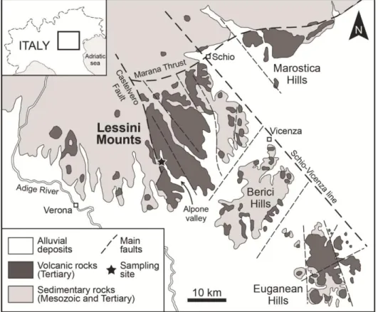
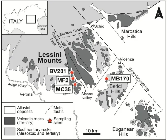
Two fibrous samples of erionite (BV201 and MF2) were investigated in this study. These samples originated from the Alpone Valley, Lessini Mounts (Figure 1), where a thick se-quence of lava flows of the Veneto Volcanic Province extensively crops out (De Vecchi and Sedea, 1995; Milani et al., 1999; Bon-adiman et al., 2001). These lavas range from poorly- to highly-vesiculated and/or scori-aceous, and vesicles are often filled by secon-dary minerals, which are dominated by zeo-lites of hydrothermal origin (Mattioli et al., 2016a). The two samples examined were se-lected according to the two main morphologic types of erionite from Lessini Mounts, which also correspond to the main morphologies de-scribed for erionite worldwide: extremely fi-brous with flexible, hair-like appearance (BV201) and prismatic to acicular crystals with rigid behavior (MF2).
2.3.2 Environmental Scanning Electron Mi-croscope (ESEM)
Morphological observations were achieved with an Environmental Scanning Electron Microscope (ESEM) FEI Quanta 200 FEG at the University of Urbino Carlo Bo. Mor-phometric data were collected by accurate size measurements (length and diameter) of all of the erionite fibers and fibrils visible in several ESEM images. These pictures were selected according to three main criteria: to (i) be representative of the total area of each in-vestigated sample, (ii) permit the measure-ments of both length and diameter of the fi-bers, and (iii) have a significant number of measurements (> 1000).
The micro-chemical characterization was per-formed using a JEOL 6400 SEM equipped with an EDX Genesis EDS system at the Uni-versity of Parma. Operating conditions were 15 kV accelerating voltage, 11 mm working
Figure 1. Geological sketch map of the volcanic rocks in the Veneto Region, NE Italy (modified from De
Vecchi and Sedea, 1995), with indication of the Lessini Mounts and sampling site (star).
distance, 0° tilt angle and 1 μm beam diame-ter. As previously suggested (Sweatman and Long, 1969; Goldstein et al., 1992; Reed, 1993; Pacella et al., 2016), in order to mini-mize the alkali metal migration, chemical data was acquired using a low counting times (up to 10 sec) and a raster scan-mode to reduce the temperature increase. With these opti-mized experimental conditions, no significant differences between the SEM-EDS and EMPA datasets were observed, indicating the reliability of the collected data.
2.3.3 Electron Micro Probe Analysis (EMPA) Pure crystals from the two samples of erionite were prepared for electron micro probe analy-sis (EMPA) by embedding a fraction of fibers in epoxy resin. Compositions were deter-mined using a Cameca SX50 EPMA equipped with wavelength-dispersive spectrometry (WDS). According to the recommended pro-tocol for the quantitative determination of zeolite compositions by EMPA (Campbell et al., 2016) the following conditions were used:
reduced counting time (up to 10 sec), beam diameter 20 μm, accelerating voltage 15 kV, low beam current and element prioritizing with the spectrometer configuration.
Chemical data were collected from several in-dividual point analyses for each sample, de-pending on the number of crystals available. In elongated crystals or crystal aggregates, several point analyses were performed along the fiber to check for chemical homogeneity. The point analyses of each sample were highly consistent, showing a variation of ma-jor elements within 2-3% of the estimated in-strumental errors and indicating a high degree of chemical homogeneity within each sample. Silicates, oxides, and pure elements were used as standards; analytical errors are 1-2% rel. and 3-5% rel. for major and minor elements, respectively. The final crystal chemical for-mula of erionite was calculated on the basis of 36 (Si+Al+Fe3+) apfu based on 72 oxygen at-oms. The reliability of the chemical analysis used to determine the erionite species was evaluated by using a charge balance error formula (E%; Passaglia, 1970; Passaglia et
al., 1998) and the Mg-content test (Dogan and Dogan, 2008; Dogan et al., 2008). Chemical analyses for zeolites are considered to be reli-able if the balance error (E%) is equal to or less than ±10%.
2.3.4 X-ray Powder Diffraction (XRDP) After preliminary screening, a sufficient amount of pure crystals was selected from each sample under a binocular microscope on the basis of the absence of any recognizable impurity phase. They were subsequently dis-aggregated and pulverized in an agate mortar, and the powder was loaded in a 0.7 mm side-opened aluminum holder. XRPD data were collected using a Philips X‘Change PW1830 powder diffractometer at the University of Urbino Carlo Bo. Analytical conditions were 35 kV accelerating voltage and 30 mA beam
current, with CuKα radiation (λ = 1.54056 Å). Data were collected in Bragg-Brentano ge-ometry from 2 to 65° 2Ɵ, with a step size of 0.01° 2Ɵ and 2.5 s counting time for each step. Semi-quantitative XRPD analyses were performed on both samples. The following software packages were used for the meas-urements and subsequent analysis: X’Pert Quantify 3.0 for data collection and instru-ment control, and X’Pert HighScore 3.0 for semi-quantitative phase analysis.
2.4 Results
2.4.1 Morphology
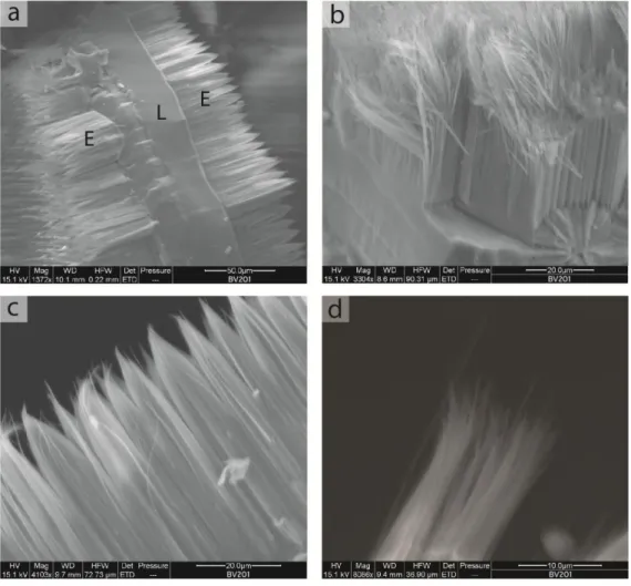
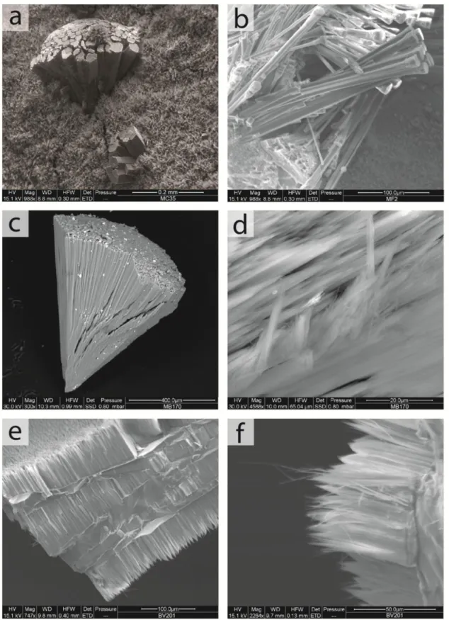
The BV201 sample is formed by alternating layers of erionite and levyne, forming a “sandwich-like” morphology (Figure 2a).
Figure 2. Electron microphotographs of erionite from BV201 sample. a) Typical erionite-levyne-erionite
(ELE) sequence; b) particular of the erionite fibers with extremely fibrous habit showing flexible behavior; c) curious “like-brushes” terminations of erionite bundles; d) bundle of erionite showing high tendency to
This erionite-levyne intergrowth is extremely similar to that described by Passaglia and Galli (1974) for some zeolites from Sardinia (Italy). In BV201 sample, levyne displays a highly tabular habit with hexagonal form and millimetric size, often made up of several overlapping crystals. Erionite grows up nor-mally to the levyne surfaces, forming a typi-cal erionite-levyne-erionite sequence (ELE, Figure 2a), which may also be repeated sever-al times. In BV201 sample, erionite is charac-terized by an extremely fibrous habit which consists of flexible, hair-like fibers having a diameter on the order of 0.1 μm and extreme-ly variable lengths. According to these char-acters, erionite of the BV201 sample can be
described as having an asbestiform habit. In some cases, erionite fibers have a uniform length of ~50 μm over the entire surface of levyne (Figure 2b), while in other cases, the length of erionite fibers is variable, ranging from < 1 μm to ~50 μm. Curiously, these fi-bers tend to intimately aggregate in parallel beams with diameter of ~10 μm and pointed ends, resulting in a “brush tip” morphology (Figure 2c). However, these bundles are al-ways characterized by a tendency to separate thin fibers and fibrils (Figure 2d). In this sam-ple, erionite demonstrated a typical wooly as-pect, which is similar in appearance to that near Durkee, Oregon (Eakle, 1898; Staples and Gard, 1959).
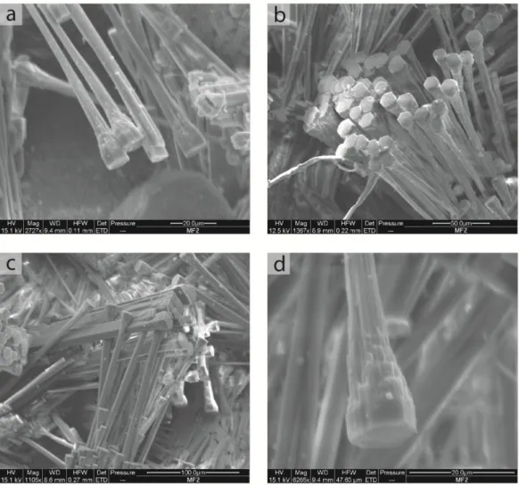
Figure 3. Electron microphotographs of erionite from MF2 sample. a) Elongated, prismatic to acicular
well-shaped, single crystals; b) aggregates of many crystals radiating from a central point with well-formed hex-agonal terminations; c) aggregates of crystals which mutually grow to generate irregular networks; d)
partic-ular of the crystal where hexagonal termination with solid appearance and the irregpartic-ular shape of the main body can be observed.
In MF2 sample, erionite is present as pris-matic to acicular crystals with diameters of ~5 μm and lengths up to 150 μm, which presents well-shaped, single crystals or discrete aggre-gates of several crystals (Figures 3a and 3b). In some cases, especially in discrete aggre-gates, crystals are joined to form packets of larger size (about 50 μm), where a clearly hexagonal shape of each crystal is recogniza-ble (Figure 3b). In other cases, crystals grow intersecting each other in directions that may be sub-parallel (in this case the crystals seem to grow one inside the other) or crossed with various angles, generating irregular networks (Figure 3c). Based on ESEM observations, the clearly hexagonal section is present only in the terminations of crystals, where they ap-pear as short, perfect hexagonal prisms of about 15 μm in diameter and 10 - 20 μm in
length. In contrast, the main body of the crys-tals is thinner (diameter ~5 μm), and any geometric basal section is recognizable (Fig-ure 3d). The short hexagonal prisms forming the ending part of the crystals show a solid appearance, whereas the main body of the crystals is of irregular shape and tend to gen-erate a number of small-size fibers with rigid behavior, partially or totally separated from main crystal body (Figure 3d).
2.4.2 Mineralogical composition
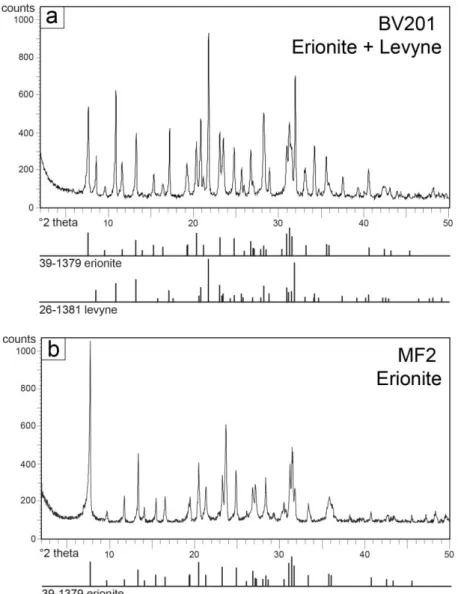
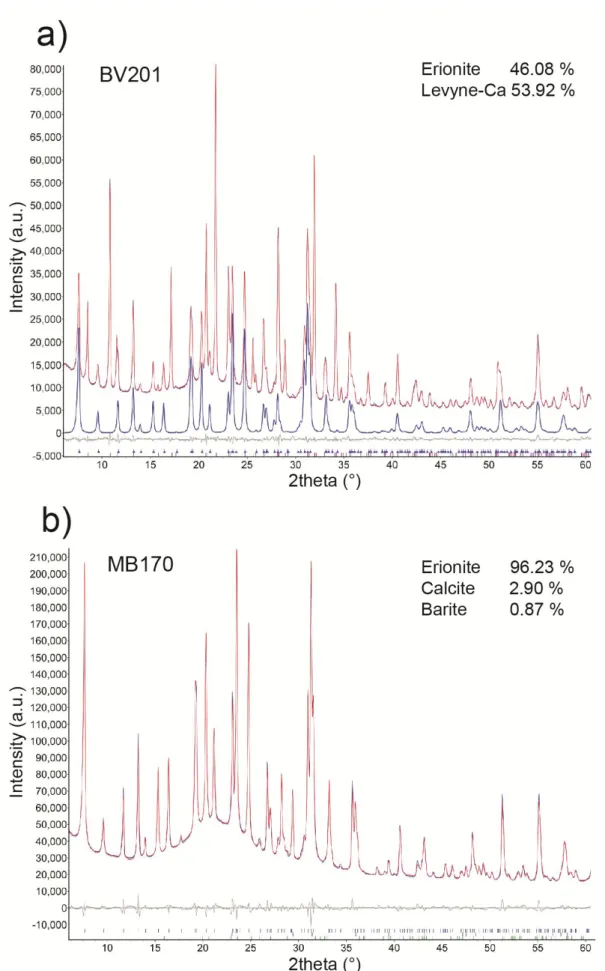
To assess the quality of separated crystals and exclude the presence of impurities, for each sample detailed, long exposures up to 24 hr X-ray diffraction analyses were performed. Experimental XRPD data collections are shown in Figure 4.
The BV201 sample showed a XRPD pattern consistent with the presence of two pure min-eral phases corresponding to erionite and levyne. Data confirmed what has already been observed in morphological analysis, i.e. the BV201 sample consists of only these two mineral phases grown in intimate association. In contrast, XRPD analysis on the MF2 sam-ple exhibited all typical diffraction peaks of pure erionite crystals. The erionite patterns of the two investigated samples illustrated nearly identical profiles. However, a significant vari-ation of the relative intensities of some peaks was observed between the two samples. This is probably due to variable amounts of Ca(II), Na(I) and Mg(II) cations which are distributed on 4 distinct cation sites in the erionite cage. 2.4.3 Morphometry
Morphometric analyses were performed on selected ESEM images in order to quantify
the size (length and diameter) of all the visi-ble fibers separated and deposited on sam-pling plate, as well as those partially separat-ed from the main crystals. These results are summarized in the histograms of Figure 5. The BV201 sample demonstrates flexible fi-bers of variable size in terms of length, but they are homogeneous (and small) as regards their diameter (Figure 5a). In term of lengths, fibers were observed in all size range from ~2 to ~85 μm with a distribution characterized by two relative modes. The main mode compris-es fibers (more than 50% of the total) with a length between ~40 and ~60 μm and clearly corresponds to the average thickness of the erionite layers in the ELE sequences, which is generally ~50 μm (Figure 2a). The secondary mode matches with shorter fibers (between ~10 and ~25 μm of length) that should be linked to the fracturing of the longer fibers, with the contribution of other fibers in some
thinner ELE sequences. In contrast, regarding the diameters, the histogram is clearly unimodal and shows the presence of a huge population of fibers (~98%) which is charac-terized by a small diameter (< 0.3 μm), and the remaining part (~2%) which also displays a small diameter (up to 0.4 μm).
The MF2 sample shows a tendency to sepa-rate fibers with rigid behavior, which are characterized by variable length and diameter (Figure 5b). Regarding the length, the distri-bution of fibers is unimodal, with the mode corresponding to fibers between ~5 and ~10 μm. Whatever, the ~95% of the total meas-ured fibers is within the range < 2 to ~30 μm, while the remaining ~5% is between ~30 and ~75 μm. In terms of diameter, more than 40% of the measured fibers possess a small diame-ter (0.1 to 0.3 μm, with an average of 0.2 μm). A significant population of fibers (~54%) is in the range ~0.3-1.5 μm, while the remaining ~6% shows a larger diameter (> ~1.5 μm).
2.4.4 Chemical data
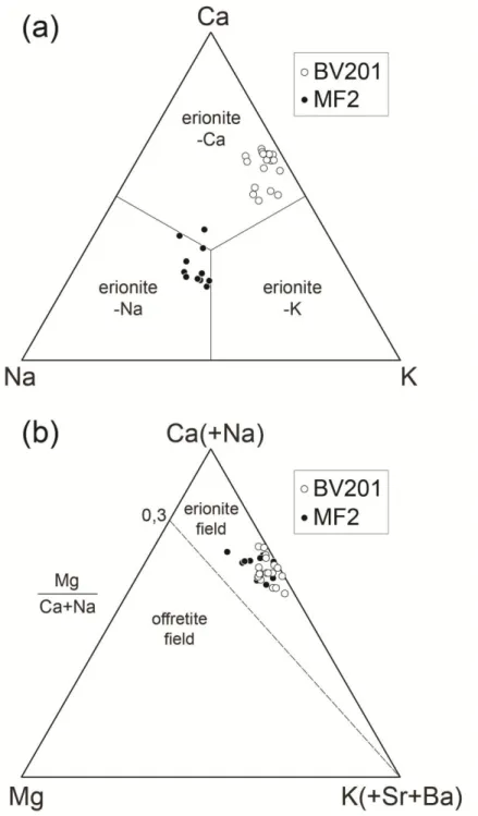
All the reliable chemical data of the investi-gated samples are reported in Tables 1 and 2, and illustrated in the diagrams of Figure 6. The average chemical composition of extreme-ly fibrous erionite from BV201 sample is (Ca3.55K2.12Na0.39Mg0.22)[Al10.37Si25.69O72]·33. 10H2O and is classified as erionite-Ca (Figure 6a). The Si/(Si+Al) ratio is in the range 0.7-0.74, that is consistent with the interval for the erionite from volcanic rocks (Passaglia et al., 1998; Passaglia and Sheppard, 2001). Ca2+ is the dominant extra-framework cation in the structure (2.96-3.83 apfu), whereas K+ and Na+ contents are lower (1.58-2.56 and 0.14-0.87 apfu, respectively). The Mg2+ content is low (0.03-0.43 apfu) and the Mg/(Ca+Na) ratio, which is considered the most significant pa-rameter for the distinction between erionite and offretite (a structurally and chemically similar zeolite), is in the range 0.01-0.11 (Figure 6b). Notwithstanding Fe3+ is not present in the ideal chemical formula, a small amount of Fe3+ is detected in the BV201 sample (0-0.15 apfu).
The average chemical composition of pris-matic to acicular erionite from MF2 sample is (Na2.39K1.92Ca1.74Mg0.39)[Al8.82Si27.18O72]·33.1 2H2O. The Si/(Si+Al) ratio varies from 0.73 to 0.77, which is numerically higher than pre-vious sample. The prevailing dominant extra-framework cation is Na+ (1.95-2.75 apfu) and, as a result, it is classified as erionite-Na. However, considering that the other extra-framework cations are also present in im-portant amounts (K+ is between 1.25-2.32 apfu and Ca2+ is between 1.3-2.44 apfu), the erionite composition of this sample tends to fall towards the central part of the classifica-tion diagram (Figure 6a). The Mg2+ content is generally low (average 0.39 apfu), although it might reach 0.7 apfu. The Mg/(Ca+Na) ratio is in the range 0.01-0.17 (Figure 6b). Similar-ly to the previous sample, the Fe3+ content is low, ranging from 0.01 to 0.3 apfu.
2.5 Discussion
The mechanisms by which an inhaled fibrous particulate induces pathological changes in-clude (1) physical features of the inhalable fi-brous mineral particles such as width, length, aspect ratio, and effective surface area availa-ble for cell contact, and (2) surface chemical composition and reactivity of the individual fiber/elongated particle.
(1) Concerning the physical features, the width and length of the fibers are the most important parameters to assess the toxicologi-cal potential. In particular, the diameter is the main factor controlling the ability of fibers to reach the lower respiratory tract, while mac-rophage phagocytosis is more dependent on fiber length (Fubini, 2001; Oberdörster et al., 2005). An elongated particle may be defined “inhalable” for humans when its diameter is < 3.5 µm (Lee, 1985). Stanton et al. (1981) cor-related the incidence of induced lung disease with dimensional distribution of elongated par-ticles, showing that fibers ≤ 0.25 μm in diame-ter and > 8 μm in length seem to be the most carcinogenic. With the aim to evaluate the se-verity induced by fibers the WHO (1986)
Figure 6. Ca-Na-K (a) and Ca(+Na)-Mg-K(+Sr+Ba) (b) ternary compositional plots of the studied erionite
samples, showing the distribution of extra-framework cations.
considers as inhalable the fibers with a diame-ter-length ratio ≥ 1:3, a length > 5 µm and a diameter < 3 µm. However, the majority of fibers detected in the lung and mesothelial tis-sues are generally < 1 µm in diameter and shorter than 5 µm in length (Suzuki and Yuen, 2002; Dodson et al., 2003; Suzuki et al., 2005; Aust et al., 2011; Mossman et al., 2011). Therefore, small-size fibers should not be excluded from those contributing to
induc-tion of human malignant mesothelioma, espe-cially in situations of high exposure. Moreo-ver, as recently reported, ultra-fine fibers are particularly suitable for penetration from the proximal area to the peripheral part in the lung, and for migration from the lung to the pleura and to the other sites within the human body (Gatti and Rivasi, 2002; Roggli et al., 2002; Dodson et al., 2003; Samet et al., 2006; Straif et al., 2009; Bunderson-Schelvan et al.,
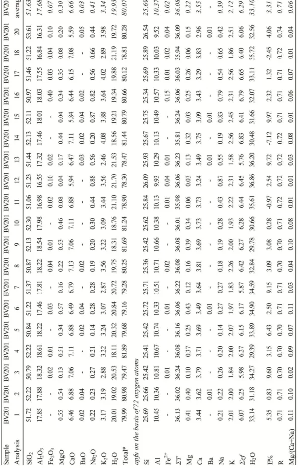
Table 1. Chemical composition of the investigated erionite fibers in the BV201 sample. Total* excludes
wa-ter content; ƩT, sum of cations in tetrahedral sites; Ʃef, sum of cations in extra- framework sites; E%, meas-ure of charge balance: E% = 100*[(Al+Fe3+)–Alkth]/Alkth where Alkth = Na+K+2*(Ca+Mg); R, Si/(Si+Al)
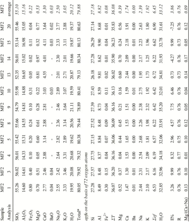
Table 2. Chemical composition of the investigated erionite fibers in the MF2 sample. Total* excludes water
content; ƩT, sum of cations in tetrahedral sites; Ʃef, sum of cations in extra-framework sites; E%, measure of charge balance: E% = 100*[(Al+Fe3+)–Alkth]/Alkth where Alkth = Na+K+2*(Ca+Mg); R, Si/(Si+Al) ratio.
2011; Boulanger et al., 2014). Thus, it is dif-ficult to exclude fibers of a particular dimen-sion from those causing disease within the lung or extra-pulmonary sites.
The erionite-Ca of BV201 sample is repre-sented by extremely thin fibers showing asbestiform habit, with ~98% of the total measured fibers having a diameter ≤ 0.3 μm. According to WHO (1986) indications, all of these fibers may be considered as inhalable. Further, these fibers are well within the range
of “more carcinogenic fibers” in terms of di-ameter (Stanton et al., 1981). The two main modes observed in the erionite fibers of BV201 sample (40-60 μm and 10-25 μm) are > 5 μm and thus considered as carcinogenic in term of lengths (Stanton et al., 1981; WHO, 1986).
Similarly, almost all fibers and fibrils from the prismatic Na-erionite of MF2 sample are thin (~94% of the measured fibers has a di-ameter <1.5 μm) and ~75% of fibers has a
length > 5 µm. Therefore, the majority of the MF2 fibers are inhalable (WHO, 1986), while only a part of these fibers are also within the range of “more carcinogenic fibers” (~42% has a diameter < 0.3 μm and ~50% has a length > 8 μm; Stanton et al., 1981).
In summary, in both the investigated samples, erionite is present as fibers of inhalable size, and most of these are potentially dangerous. However, erionite in the BV201 sample seems to be able to generate fibers dimen-sionally more homogeneous and more car-cinogenic than erionite from MF2 sample. Concerning the mechanical properties, the two studied samples displayed different be-havior having an extremely fibrous with flexible, hair-like appearance (BV201) or forming prismatic to acicular, brittle crystals (MF2). Flexibility in minerals is defined as the ability of a particle to bend without break-ing and without resumbreak-ing its original shape (Bates and Jackson, 1987). In the mineral fi-bers, this flexibility might significantly trans-form the shape of the particles, leading to var-iations of both aerodynamic properties and physical particle deposition mechanisms (Lippmann et al., 1980; Donaldson et al., 2010; Hofmann, 2011). In addition, Matassa et al. (2015) found that the flexibility of erionite fibers is related to the nanostructural features of the fibrils aggregating to form dif-ferent morphologies. The difdif-ferent dimen-sions of the fibrils, coupled with the extended defectivity, are responsible for the different mechanical behavior of the erionite samples (flexibility vs. brittleness). Therefore, particu-lar attention needs to be paid to reliably eval-uate the toxicological effects related not only to the dimension of erionite fibers, but also to their tendency to split, as the latter feature may significantly modify their surface area and, consequently, their reactivity (Matassa et al., 2015; Mattioli et al., 2016b). Bearing this in mind, to assess the potential human health effects of exposures to erionite in the Lessini area, different mechanical behavior of these two fiber varieties (flexibility instead of brit-tleness) needs to be considered.
(2) The chemical composition of the mineral fibers is also of critical importance to evaluate
the pathological changes, being related to the biopersistence and biodurability of fibers in contact with cells and tissues (e.g. in the lungs). If one considers that the main biologi-cal interactions or exchange processes devel-op at the surface of the fibers, the surface chemical composition and specific surface ar-ea are the most important elements that need to be considered. It is well known that some chemical elements in contact with cells and tissues are more effective in induction of pathological changes. In particular, the pres-ence and structural coordination of Fe are considered key factors for explaining fiber-mediated toxicity (Bonneau et al., 1986). Fur-ther, only Fe exposed at the fiber surface, es-pecially Fe(II), seems to be relevant in the re-active oxygen species (ROS) production (Fubini and Mollo, 1995; Gazzano et al., 2005).
Although erionite is a nominally Fe-free phase (Eberly, 1964; Passaglia et al., 1998; Dogan et al., 2006), Fe(II) might be present as extra-framework cation through an ion-exchange process, mainly involving Na+ (Ballirano et al., 2015). However, Fe(II) might also be found on the zeolite surface as Fe oxide nanoparticles, as a thin coating of Fe-bearing silicates, or as Fe-bearing clay minerals (Ballirano et al., 2009; Cametti et al., 2013; Croce et al., 2015; Matassa et al., 2015). In the investigated erionite samples, Fe was detected in low amounts, ranging from 0 to 0.15 apfu in BV201 sample, and from 0.01 to 0.3 apfu in MF2 sample. However, availa-ble data do not allow to discriminate if the origin of this Fe is internal (Fe(II) as extra-framework cation) or external (Fe(II) on the fibers surface).
2.5.1 Environmental occurrence of erionite and risk assessment in Italy
Although zeolites represent one of most wide-spread mineral groups in the Earth’s crust, in Europe there are no systematic studies on the distribution of erionite or other similar fibrous zeolites in the environment. This is mostly due to the fact that previous approaches to the hazard posed by fibrous zeolites were
con-ducted only in the areas with an anomalous incidence of mesothelioma, such as in Tur-key, USA and Mexico (Dogan et al., 2006; Carbone et al., 2007, 2011; Ortega-Guerrero and Carrasco-Nunez, 2014; Saini-Eidukat and Triplet, 2014).
In Italy, epidemiological and toxicological knowledge of mesothelioma linked to erionite exposure is extremely scarce, and the phe-nomenon is still little investigated, although it is known that about 20% of national cases of mesothelioma is not linked to asbestos (INAIL, 2012). In particular, if we exclude the three regions (Piemonte, Lombardia and Liguria) where the high amount of mesotheli-oma is clearly linked to the presence of asbes-tos minerals, Veneto is the region that is char-acterized by one of the highest percentages of mesothelioma in Italy (8% of the total cases) in the time interval 1993-2008. This is a sig-nificant percentage in which the contribution of exposure to erionite cannot be excluded. However, it is worth noting that epidemiolog-ical and public health data are available ac-cording to the administrative borders (e.g. provincial or regional areas) which do not correspond to the real limits of geological de-posits, i.e. erionite-containing rocks. For this reason, it is extremely difficult extrapolate the data regarding the incidence of mesothelio-mas unrelated to asbestos in the Lessinia re-gion, which could be a very useful element for the comprehension of the erionite contri-bution.
While mesothelioma cases attributed to asbes-tos exposure are well known and recorded (Crovella et al., 2016), little or nothing is known about domestic cases from exposure to airborne erionite fibers. Until a few years ago, findings of erionite specimens in Italy were limited to few localities in Sardinia (Passaglia and Galli, 1974) and Colli Euganei (Passaglia and Tagliavini, 1995). However, recently, Mattioli et al. (2016a) reported erionite and other associated zeolites within vugs and ves-icles in the Tertiary basalts of the Lessini Mounts. These basalts are extensively quar-ried and largely employed for a variety of us-es, most commonly as an aggregate. Crushed basalt is added to many construction jobs
in-cluding asphalt paving, mixed with concrete, and as a rock filtering agent in many drain fields. Basalt is also used as bedrock for rail-road tracks providing drainage and support. So, beyond of the potential environmental ex-posure produced by natural weathering pro-cesses affecting these basaltic rocks, it should also be taken into account the occupational exposure during the phases of extraction and processing of the erionite-containing rock, and a further environmental exposure due to the uses of such material.
The results acquired in the present investiga-tion provide the basis for a health and safety protection program in Italy and for a better scientific understanding of erionite and its po-tential carcinogenicity. Another outcome is to stimulate future systematic studies on the pos-sible presence and amount of such hazardous minerals in rocks and soils, suggesting at the same time a methodology to be applied to hazard identification processes in areas char-acterized by the possible natural presence of hazardous mineral phases.
2.6 Conclusions
In this study morphology, morphometry, min-eralogical and chemical composition of two erionite samples from Lessini Mounts (NE It-aly) was examined, where this carcinogenic zeolite had not apparently been found until now. On the basis of morphological and chemical data, the BV201 sample is erionite-Ca with an extremely fibrous, hair-like struc-ture, with flexible appearance, growth in in-timate association with levyne. Conversely, the MF2 sample is erionite-Na characterized by prismatic to acicular crystals with rigid be-havior, which is also enriched in K+ and Ca2+ extra-framework cations. Although erionite is a nominally Fe-free phase, Fe was detected in low amounts in both the investigated samples. Considering the size parameters, in both the investigated samples erionite was present as fibers of inhalable size, and most of these are also potentially carcinogenic. However, erionite in the BV201 sample seems to be able to generate fibers dimensionally more homogeneous and greater potential for
car-cinogenicity than erionite from MF2 sample. Further, the mechanical behavior of the erionite fibers is different (flexible or brittle) and this fact might exert some effect on the carcinogenic potential. Taking into account that the studied samples originated from bas-alts which are extensively quarried and large-ly employed, it is of great importance to eval-uate (1) potential environmental exposure
mediated by natural weathering processes of these rocks, (2) occupational exposure during the phases of extraction and processing of the erionite-containing rocks, and (3) a further environmental exposure due to the uses of such material. Finally, the results acquired in this investigation contribute to the knowledge of the erionite and provide the basis for a health and safety protection program in Italy.