Review Article
Oral Health Impact Profile in Celiac Patients: Analysis of Recent
Findings in a Literature Review
Gabriele Cervino,
1Luca Fiorillo,
1Luigi Laino,
2Alan Scott Herford,
3Floriana Lauritano,
1Giuseppe Lo Giudice
,
1Fausto Famà,
4Rossella Santoro,
2Giuseppe Troiano
,
5Gaetano Iannello,
1and Marco Cicciù
11Department of Biomedical and Dental Sciences and Morphological and Functional Imaging, Messina University,
98100 Messina, Italy
2Multidisciplinary Department of Medical-Surgical and Odontostomatological Specialties, University of Campania“Luigi Vanvitelli”,
80121 Naples, Italy
3Department of Maxillofacial Surgery, Loma Linda University, Loma Linda, CA 92354, USA
4Department of Human Pathology, University of Messina, 98100 Messina, Italy
5Departments of Clinical and Experimental Medicine, University of Foggia, 71121 Foggia, Italy
Correspondence should be addressed to Marco Cicciù; [email protected]
Received 23 May 2018; Revised 5 September 2018; Accepted 24 September 2018; Published 24 October 2018 Academic Editor: Chiara Ricci
Copyright © 2018 Gabriele Cervino et al. This is an open access article distributed under the Creative Commons Attribution License, which permits unrestricted use, distribution, and reproduction in any medium, provided the original work is properly cited.
The increment of recording atypical oral manifestation in young patients often related to systematic disease is today a challenge for the therapists. Sometime, the presence of tooth enamel lesions correlated with soft tissue lesions is just a symptom or a trigger sign
for a deeper and undetermined disease. Recently, high impact has been developed toward the influence of the diet as a controlled
and modifiable factor in patients affected by celiac pathologies. The celiac disease (CD) is a chronic immune-mediated disorder triggered by the ingestion of gluten that appears in genetically predisposed patients. Gluten is a proline-rich and glutamine-rich
protein present in wheat (gliadin), barley (hordein), and rye (secalin). The gluten-free diet (GFD) seems to better influence the
oral health status of the CD patients. For this reason, the main objective of this revision was to analyze the international data highlighting the relationship between celiac patients and the oral health impact profile. A comprehensive review of the current literature was conducted according to the PRISMA guidelines by accessing the NCBI PubMed database. Authors conducted the
search of articles in the English language published from 2008 to 2018. Thefirst analysis with filters recorded 67 manuscripts
accordingly with the selected keywords. Finally, a number of 16 appropriate published papers were comprehended in the review. The studies were different in terms of the structure, findings, outcomes, and diet quality evaluation, and for this reason, it was
not possible to accomplish a meta-analysis of the recorded data. This manuscript offers some observational evidence to justify
the advantages of gluten-free diets related to a better oral health status in the patients involved.
1. Introduction
Oral health is today considered one of the fundamental
parameters related to the patient’s general health and
behav-ior. Oral health status allows individuals to run their daily activities (mastication, articulation, and socialization) with-out any pain, discomfort, and restriction. The patients’ qual-ity of life (QoL) is a caption currently applied in the medicine
field to refer to social well-being and the effects of therapy on cancer patients. Specifically, in the dental practice, QoL, as it connected to the oral health, has only been recently
employed [1–7].
The patients’ general health condition is related to having
no problems or diseases on all the anatomical structures, even involving the oral cavity functions or aesthetics. Today, great attention is focused on the prevention and maintenance
Volume 2018, Article ID 7848735, 9 pages https://doi.org/10.1155/2018/7848735
pathologies may be connected with systemic disease and not affect the oral cavity structures directly [3, 7–10].
Nowadays, even the current standard of performing diagnosis in oral lesions like teeth enamel defects or soft
tis-sues and tongue lesions may be related to local affection or
trauma; a deep knowledge of the patient anamnesis and clin-ical history is required in order to evaluate possible hidden causes strictly related to the diet or general health status. Therefore, it is well documented how many systemic diseases are somehow related to many oral manifestations and influ-ence the individual quality of life [4, 9, 11–13].
Celiac disease (CD) is a long-term autoimmune disorder that affects the small intestine; this is caused by a constant intolerance to gluten proteins in genetically susceptible indi-viduals. CD is caused by a reaction to gliadins and glutenins found in wheat. These protein-based factors may be respon-sible for a toxic event on the intestinal mucosa in genetically receptive subjects by triggering an immune-mediated reac-tion, related to the common villous atrophy and lymphocyte infiltrate in the small intestine mucosa recorded in CD. Com-mon oral and dental manifestations of CD include mouth ulcers, in particular, recurrent aphthous ulcers, and dental enamel defects [13–16].
However, even if great important steps have been done
in the field of quick diagnosis, CD is still not promptly
diagnosed, because recently, the typical form of CD,
characterized by modified absorption and gastrointestinal
signs, is less recurrent compared with the atypical forms, often asymptomatic and involving extraintestinal clinical manifestations. A multidisciplinary evaluation and approach between clinicians, pediatricians, and gastroenterologists should be performed in order to underline all the extraintes-tinal possible manifestations of CD and to make an early diagnosis; recurrent aphthous ulcers, previously mentioned, could provide another clue to the possible presence of this
disorder [17–20].
Numerous published papers underlined how specific oral
signs and symptoms can be classified as risk factor signals for
CD; however, only the internal specialist can perform the diagnosis, evaluate the presence of specific celiac antibodies, and demonstrate intestinal mucosa damages. However, the topic is still debated, and currently, the right frequency of these oral manifestations in potential celiac patients has not yet been classified and recorded [21–23].
However, it is widely recognized that, among these atypical signs of CD, there are certain oral manifestations which are surely interwoven to CD: tooth enamel lesions and defects, frequent aphthous stomatitis, delayed tooth eruption, multiple caries, angular cheilitis, atrophic glossi-tis, dry mouth, and burning tongue. For this reason,
dentists and the first dental visits play a fundamental role
in detecting symptoms related to CD and for the next medical treatments [23, 24].
About the treatments and the prevention of such oral manifestations, recently published investigations underlined how the gluten-free diet may favor an improvement of the general health condition of celiac and no celiac patients. Moreover, numerous epidemiological and clinical studies
with the gluten-free diet of the involved patients [25–33].
The aim of the present revision is to examine the data of the last ten years’ literature about oral manifestations of celiac disease and how gluten-free diet may definitely influence the conditions of the oral health patients.
2. Methods of Screening
2.1. Protocol Development and Online Information Recording. The inclusion parameters for the current research were collected in a protocol and then submitted in advance and documented in CRD York website PROSPERO, a global pro-spective catalogue of revision manuscripts. The criteria and the formal structure of the present revision can be searched online with the CRD id and code: application number: CRD 92075.
The documents collected in the present paper followed the Preferred Reporting Items for Systematic Review and Meta-analysis (PRISMA) statement accordingly [34, 35]. 2.2. Outcome Query. The following question was sentenced and structured according to the (PICO) study design:
(i) How can the celiac disease influence the oral health status of the patients?
2.3. Searches. The PubMed-Medline resource database was applied through advanced researches. The keywords and search inquiries used during the primary stage were as
follows: “oral health gluten-free diet.” Additional manually
selected articles were included regarding the eligibility
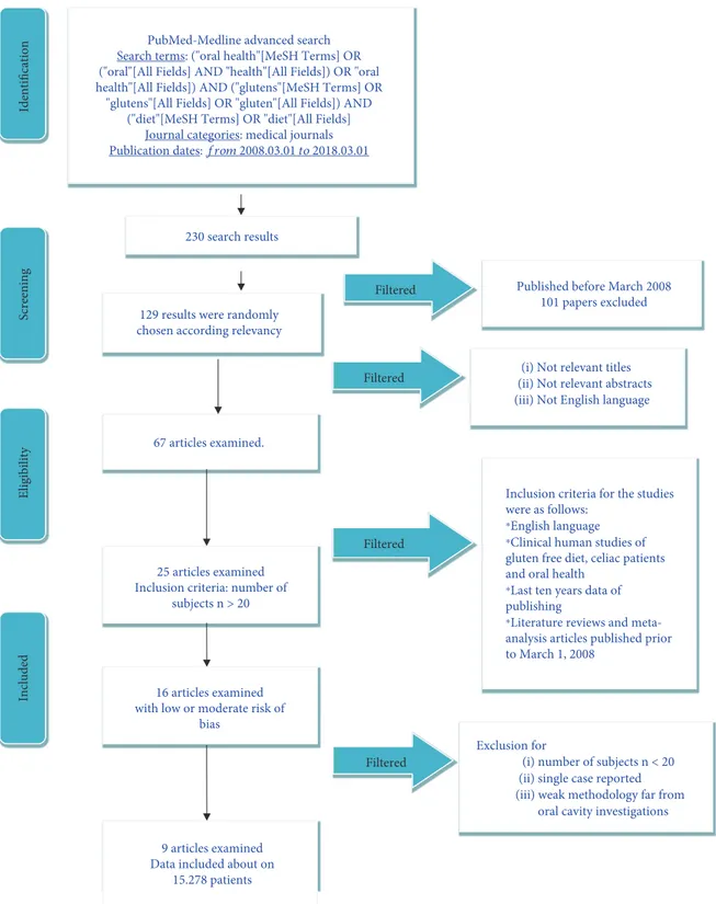
cri-teria. Figure 1 represents the flow diagram of the selected
studies according to PRISMA guidelines and following the criteria for the investigated paper choice. Web searching
and researches by hand were then executed in thefield of
gas-troenterology, dentistry, and medicine journals,finding
dif-ferent international journals. The search was restricted to English language manuscripts.
2.4. Data Recorded from the Selected Manuscripts. The
Medical Subject Headings (MeSH) was used forfinding the
keywords used in the present revision. The selected keywords (“oral health”[MeSH Terms] OR (“oral”[All Fields] AND “health”[All Fields]) OR “oral health”[All Fields]) AND
(“glutens”[MeSH Terms] OR “glutens”[All Fields] OR
“glu-ten”[All Fields]) AND (“celiac”[MeSH Terms] OR “diet”[All
Fields]) were written in the selected database.
2.5. Selections of the Papers. Three separate researchers, of three Italian universities (Messina, Foggia, and Naples Universities), singularly screened the full-text manuscripts for deciding inclusion and exclusion criteria. Reviewers com-pared decisions about criteria, parameters, and selected papers. During the step of reviewing the manuscripts, a com-plete independent two-fold revision was undertaken.
Reviewers compared their results and data. A fourth
qualified chief reviewer (Loma Linda University) was then
contacted when agreement could not be obtained at thefirst
The papers recorded in the present revision highlighted clinical researches over celiac patients printed in the English style. Letters, editorials, case reports, animal studies and degrees, and PhD thesis were not included in the revision process.
2.6. Studies Involved in the Revision. The design of recording data involved all human prospective and retrospective
clinical studies, split-mouth cohort studies, case–control papers, and case series manuscripts, published between March 2008 and March 2018, about gluten-free diet, oral dis-ease, and celiac patients.
2.7. Exclusion and Inclusion Criteria. The applied inclusion criteria for the studies were as follows:
Screening Included Eligibility Identification 230 search results 67 articles examined.
(iii) Not English language (i) Not relevant titles
25 articles examined Inclusion criteria: number of
subjects n > 20
9 articles examined Data included about on
15.278 patients 129 results were randomly chosen according relevancy
Filtered
Filtered
Filtered
Exclusion for
(i) number of subjects n < 20 (ii) single case reported (iii) weak methodology far from
oral cavity investigations
Filtered Published before March 2008
101 papers excluded
16 articles examined with low or moderate risk of
bias
PubMed-Medline advanced search Search terms: ("oral health"[MeSH Terms] OR ("oral"[All Fields] AND "health"[All Fields]) OR "oral health"[All Fields]) AND ("glutens"[MeSH Terms] OR
"glutens"[All Fields] OR "gluten"[All Fields]) AND ("diet"[MeSH Terms] OR "diet"[All Fields]
Journal categories: medical journals
Publication dates: from 2008.03.01 to 2018.03.01
⁎English language
Inclusion criteria for the studies were as follows:
⁎Clinical human studies of
gluten free diet, celiac patients and oral health
⁎Last ten years data of
publishing
⁎Literature reviews and
meta-analysis articles published prior to March 1, 2008
(ii) Not relevant abstracts
(ii) Clinical human studies of gluten-free diet, celiac patients, and oral health
(iii) Last ten year data of publishing
(iv) Literature reviews and meta-analysis articles
published prior to March 1, 2008
The following types of articles were excluded as follows: (i) In vivo/in vitro studies
(ii) Studies of testing medication and/or new treatment methodologies
(iii) Studies of cancer in locations other than mentioned (iv) Studies not relevant to our selected diagnostic
methods (v) Animal studies
(vi) No availability to the title or summary not in English words
2.8. Data Recording Design. Following the initial literature screening, all the paper titles were evaluated in order to delete irrelevant publications, case reports, and animal studies. Then studies were excluded based on data obtained from reading the summaries. The last step of screening involved reading the full texts to determine each study’s selection fol-lowing the inclusion and exclusion criteria. (Figure 1). 2.9. Risk of Bias Assessment. The quality of all involved texts was assessed during the data extraction process. The quality appraisal involved evaluating the methodological elements
that might influence the outcomes of each study.
Each reviewer evaluated the level of possible bias risk during the information taking out method. This revision work was made accordingly with the Cochrane Collabora-tion’s double tool for determining risk of bias and PRISMA rules [34, 35].
Differences in risks of bias can help explain variation in the results of the studies included in a systematic review (i.e., explain heterogeneity of results). More rigorous studies are more likely to yield results that are closer to the truth.
Risk of bias (e.g., the absence of information or selective reports on variables of interest) was assessed on the study level. The risks were indicated as lack of precise information of interest related to the keywords selected. Finally, the researches selected for the revision were then recorded in modest, moderate, significant, and unclear risk.
3. Outcomes
3.1. Paper Recording and Possible Bias. The PRISMA flow
diagram describes the revision steps for screening the papers and reaching the selected ones (Figure 1). The initial web and hand searches performed on PubMed-Medline and Oral
Sci-ences Source produced a number of 230findings. 101
refer-ences were not involved in the revision because they were
not selected for the data because they were not available on full text. 67 papers were discovered on full-text form, 25 of
which were merged in this work, and then afterfinal
screen-ing, a total of 9 full-text papers.
During the last deep screening section, from the last 16 manuscripts, some researches were excluded because they
were recorded as a unique case report (n = 2) or not
signifi-cant design study or procedures were far from the topic (n = 5). So finally, 9 papers were recorded and screened in this revision paper.
No meta-analyses could be performed due to the hetero-geneity between the studies (different study designs, control groups, and observation periods) Table 1. The possible risk
of bias was considered for each selected papers. The final
number of the selected papers was limited from 25 full-text papers to 9. The inclusion criteria were really restrictive, and for this reason also, the risk of bias was low. Ten types of research were evaluated as having minor risk of bias [36–44] whereas another seven were classified as moderate risk [45–51].
The present investigation of the data extracted from researches printed in English only could detect a publication bias. About possible bias, some of the selected papers did not specify the inclusion criteria of the patient selection. Another key parameter that can be assumed as bias is related to the evaluation of the clinical condition for selecting the patient. Moreover, data recorded from the eight studies pointed out the heterogeneity of the research methods, selections of the patients, and therapeutic options.
4. Results
The present systematic review discovered gluten-free diet is associated with oral health status of celiac patients. Due to high heterogeneity of the researches, it was not realizable to do meta-analysis for comparing the data of the selected papers. Due to poor material, it is not possible to establish specific oral health status related to diet or systemic diseases like celiac patients.
Moreover, even wide screening and research have been
performed; the inclusion criteria related to the “oral
cav-ity” was really inclusive, and for this reason, it was not
possible to state some guidelines that may significantly
increase the oral health status of the CD patients just by applying a gluten-free diet. Some papers with low risk of
bias [36–44] clearly analyzed the correlation between
gluten-free diet and oral health status of celiac patients. However, the data of those researches are not significant
and finally suggested some recommendations and not
guidelines. Specifically, because the disease involves the
gastrointestinal area, the high part of the researches firstly
investigated the microbiota related to the anatomical area far from the oral cavity area. Therefore, all the data extracted from the present revision clearly underlined how a diet associated with no gluten may favor high stan-dard of oral health quality delaying gingival oral disease due to the alteration of the oral microbiota.
Table 1: CD and OH parameters re corded in the studies with low risk of bias (CD = celiac di sease, PD = parodontal disease, OH = oral health, DDE = developmental d efects of enamel, DED = dental enamel defects, and AGA = antigl iadin antibodies). References Year Author Subjects (n ) Type of correlation Result Statistic [36] 2018 Spinell et al. 6661 PD and CD There is no signi ficant correlation between PD and CD. P =0 67 [37] 2017 van Gils et al. 5522 CD and xerostomia; CD and OH OHIP-14 and XI tests were performed. P <0 001 for both [38] 2013 Rivera et al. 20 CD and enamel hypoplasia; CD and aphthous ulcers; CD and delayed eruption Oral manifestations. In the long list of clinical signs and symptoms that have been found signi ficantly associated with CD are oral manifestations such as dental enamel hypoplasia, aphthous ulcers, and delayed eruption of teeth. Reported as signi ficant values, insu ffi cient extrapolated statistical data [39] 2013 Shteyer et al. 90 CD and enamel defects; CD and aphthous stomatitis, DMFT A high prevalence of enamel hypoplasia (66%) was found in children with CD. Plaque index was signi ficantly lower in the celiac-treated group. P <0 05 [40] 2012 Mina et al. 25 CD and OH, enamel alteration DMFT, enamel alterations, gingival index, and oral hygiene were evaluated in this study. P <0 0001 [41] 2010 Tsami et al. 35 CD and OH The periodontal treatment need of children and adoles cents with CD was high, most of them needed treatment for gingivitis (60.01%), and only a few subjects had a healthy periodontium (34.29%). P <0 05 [42] 2018 Souto-Souza et al. 2840 CD and OH; CD and enamel alterations This meta-analysis indicated a high prevalence of DDE among celiac patients with a signi ficant association of DDE with this population when compared to healthy people. P =0 014 [43] 2016 Sóño ra et al. 21 CD and enamel defects These results strongly suggest a pathological role for antibodies to gliadin in enamel defect dentition for both deciduous and permanent teeth. Insu ffi cient extrapolated statistical data [44] 2012 Muñoz et al. 64 CD and enamel defects This work describes structural similarities between gliadins and proline-rich enamel proteins and shows that pot ential cross-reactions of AGA could take place during amelogenes is in untreated patients. Further work is underway to study the biological eff ects of AGA on enamel formati on to evaluate their putative role in the pathogenesis of DED in untreated CD. P =0 045
tations in CD-affected patients are reported in literature. Oral manifestations that interest gums such as aphthous ulcers or
recurrent aphthosis are correlated to celiac disease-affected
patients. These manifestations are more frequent in CD patients than in normal population [38, 39].
4.2. Dental Manifestations. Dental hard tissue manifestations
in CD patients are various; we can find alterations on the
enamel and teeth structure. Some studies report enamel hypoplasia, enamel defect or enamel and dental structure
alterations [38–40, 42–44], this condition puts CD
patients in condition of discomfort, lowering their general conditions of oral health and expelling them to other debilitating diseases.
4.3. Oral Health. Another series of oral manifestations is present in CD patients which does not affect soft tissue or the dental structure. Some studies evaluated the oral health of patients through self-administered tests like the OHIP-14
test (Oral Health Impact Profile 14) or XI test (Xerostomia
inventory). Studies reported abnormalities in general oral health like DMFT index (Decayed, Missing, Filled Teeth); some other studies reported anomalies like delayed eruption and parodontal disease [36–42].
5. Discussion
The purpose of this review was to systematically overview published studies restricted to oral health and gluten-free diet in order to evaluate how a diet without gluten may influence the oral health status of celiac patients, following the brief
report of the 16 papers classified with moderate and low risks
of bias.
Spinell et al. [36] recently investigated whether celiac dis-ease was associated with periodontitis or periodontal disdis-eases among a population of US adult patients. In this large research, the National Health and Nutrition Examination Survey (NHANES) authors between 2009 and 2012 enrolled about 6661 subjects with full-mouth periodontal examina-tion and serological testing for antitissue transglutaminase (tTg) and antiendomysial (EMA) antibodies. CD was defined as (i) self-reported physician diagnosis while on a gluten-free
diet or (ii) tTg levels> 10.0 U/ml and positive EMA results.
Positive serology without self-reported diagnosis was defined
as undiagnosed CD (UdxCD). Authors concluded how CD is associated with modestly lower levels of mean periodontal disease but was not associated with periodontitis in a signifi-cant way. Larger studies are necessary to enhance precision and strengthen conclusions.
The oral health status and the xerostomia of celiac patients were investigated by van Gils et al. [37] in a study involving a population of about 740 patients with CD and 270 comparison participants. The Oral Health Impact Profile 14 (OHIP-14) and Xerostomia Inventory (XI) were screened and recorded. This study showed that oral health problems are more commonly experienced in adult patients with CD than in the comparison group. Collaboration between
detection of undiagnosed CD.
De Angelis et al. [45] analyzed how the oral and intestinal bacteria metabolize dietary components, affecting human health by producing harmful or beneficial metabolites, which are involved in the incidence and progression of several intestinal-related and nonrelated diseases. Moreover, the
authors stated how dietary regimens withfibers are the most
effective to benefit the metabolism profile, and a profitable
use of diet is fundamental in order to provide benefits to
human health, both directly and indirectly, through the activ-ity of the gut microbiota.
In a different revision paper, Cenit et al. [46] evaluated how in mature normal subjects, the GFD is connected with a low intake of complex polysaccharides caused shifts in the gut microbiota structure. Therefore, the authors concluded that microbiota imbalances have been recorded not only in untreated CD subjects but also in patients fol-lowing a GFD. Moreover, typical bacterial strains isolated from subjects with active and nonactive CD have been shown to increase virulence features. These alterations may be significant for increasing CD pathogenesis by con-tributing to the disease onset.
Galipeau and Verdu [47] recorded significant findings in their review underlining and effective evidence between intestinal dysbiosis and CD; however, the main limit of the present investigation was related to the analysis of the
manu-scripts classified in the revision. It was determined evidence
demonstrating causality is lacking. Therefore, it remains unclear whether general changes in microbial composition or the presence or absence of particular members of the microbiota play a direct role in CD pathogenesis, and so the diet is not fundamental in the CD developments.
Rivera et al. [38] in their research studied how CD con-tinues to be an unsolved puzzle and a much-debated topic in the recent literature. Knowing the important health implications that CD can have, not only in an individual’s health but also in the overall quality of life of these indi-viduals and their families, is of vital importance. As clini-cians, it is very important to be aware of the potential presentations, especially in terms of oral health, that CD can have and the consequences it can lead to in overall health status. When suspected, it is extremely important to refer the patient to a gastroenterologist for further eval-uation, diagnosis, and, in the case of a positive work up, initiation of treatment with a GFD.
Shteyer et al. [39] made an interesting report studying the oral health status and quality in relation to GFD in children with CD. The results showed that newly diagnosed children with CD have more dental plaque and caries than the control groups, and children receiving GFD had lower dental plaque and better oral hygiene. These results should raise pediatric
gastroenterologists’ awareness toward oral health–related
issues in children with CD. However, the data of the present investigation were not clear if the enamel defects were genetic or due to the low oral health conditions of the CD patients.
Mina et al. [40] in their study highlighted the main differ-ence among CD children who did or did not comply with a gluten-free diet and control children are the presence of
PMNs in the oral mucosa and protein salivary patterns;
these findings could be considered as markers for CD, in
conjunction with other signs and symptoms. The GFD seems to improve the oral health quality reducing the
gingival inflammation.
Tsami et al. [41] inspected the factors that influence the oral hygiene and the periodontal treatment needs of children and adolescents with celiac disease (CD). It was found that the periodontal treatment need of children and adolescents with CD correlated with factors that related to the presence of a second medical condition and to the personal oral
hygiene habits. CD seems to not have significant influence
on the oral health status of the CD adolescents. Additionally, the oral hygiene level and periodontal status of children with
CD do not have any specific characteristics, but they have
similarities to the oral hygiene level and periodontal status of the children of the general population.
da Silva et al. [48] made a brief review of the literature about CD and analyzed a clinical case, and for this reason,
this paper was not included in thefinal 9 papers. However,
the management of the case was typical. A 39-year-old woman reported the presence of many symptoms. She also noted the appearance of symptomatic lesions in the mouth. These lesions had a mean duration of a month and occurred in any region of the oral mucosa, particularly on the tongue. Topical treatment was instituted for the oral lesions with immediate relief of the symptoms. The diagnosis of celiac disease was established by means of a medical clinical exam. A multidisciplinary approach and management with the involvement of a gastroenterologist and other health profes-sionals, such as dentists, are important for diagnosing the disease and guiding the patient with celiac disease to achieve a good quality of life.
Francavilla et al. [49], thanks to advances in
understand-ing the immunopathogenesis of CD, have proposed different
kinds of treatment options alternative to the GFD. Some of these therapies try to decrease the immunogenicity of gluten-containing grains by modifying the grain itself or by applying oral enzymes to break down immunogenic peptides that usually remain intact during digestion.
Bascuñán et al. [50] evaluated how the only effective and safe treatment of celiac disease continues being the so-called gluten-free diet (GFD). Although GFD poses difficulties to patients in family, social, and working contexts, deteriorating his/her quality of life. The diet must be not only free of gluten but also healthy to avoid nutrient, vitamins, and mineral
deficiencies or excess. Overweight/obesity frequency has increased. Authors concluded how nutritional education by a trained nutritionist is of great relevance to achieving long-term satisfactory health status and good compliance.
Theethira and Dennis [51] underlined how it is impor-tant to have regular follow-up visits and lab work to detect
and treat nutritional deficiencies after initiation of the GFD.
Indisputably, the GFD is the cornerstone of treatment for CD. Keeping these nutritional concerns in mind, a patient with CD can enjoy a healthy, well-rounded diet that improves and maximizes overall health and well-being.
Souto-Souza et al. [42] investigated the relationship between developmental defects of enamel and celiac disease. In their meta-analysis, it was observed how subjects with CD
had a significantly higher prevalence of enamel defects
matched with healthy people. The most important findings
of this paper are that only developmental defects of enamel
diagnosed using Aine’s method were strictly related to CD.
In a sensitivity analysis involving the deciduous, mixed, and permanent dentitions, only individuals with deciduous denti-tion were observed to have associadenti-tion with the disease. Then patients with enamel developmental defects should be screened for the possibility of having celiac disease.
Sóñora et al. [43], based on previously reported cross-reactivity of antibodies to gliadin with the enamel proteins, amelogenin and ameloblastin, investigated the ability of anti-gliadin IgG to recognize enamel organ structures. Strong staining of the enamel matrix and of the layer of ameloblasts was observed with serum samples from women with celiac disease. The results strongly advise a pathological position for antibodies to gliadin in enamel defect dentition for both deciduous and permanent teeth, considering that IgG can be transported through the placenta during fetal tooth devel-opment. Muñoz et al. [44] in their research classified the pathogenesis of enamel anomalies in permanent teeth of sub-jects affected by CD. The studies using ELISA and western blotting, for reactivity of sera from patients with CD against
gliadin and peptides obtained from enamel, confirm that
the antibodies against gliadin generated in patients with CD can react in vitro with an important enamel protein. The involvement of antigliadin serum in the pathogenesis of enamel defects in children with untreated CD can be hypoth-esized on the basis of these new results (Table 2).
Hypoplasia of the enamel, xerostomia, and oral gingival lesions are the most common symptoms reflected in the recorded manuscripts. The tooth enamel defects can be a Table 2: Selected papers in which there is a direct correlation between CD and oral health alterations or disease.
References Author and year Subjects (n) Oral health status and symptoms
[36] Spinell et al. 2018 6661 Periodontitis
[37] van Gils et al. 2017 5522 Periodontitis and xerostomia
[38] Rivera et al. 2013 / Enamel hypoplasia; aphthous ulcers, delayed dental eruption
[39] Shteyer et al. 2013 90 Enamel defects; aphthous stomatitis
[42] Souto-Souza et al. 2018 2840 Enamel alterations
[43] Sóñora et al. 2016 21 Enamel defects
of CD, but the defect can be managed only by dental conser-vative treatment, and a GFD cannot modify these clinical conditions. After all, xerostomia and oral ulcers and gingival lesions, other clinical signs of CD disease, can be topically treated by dental care, but in those case, a GFD diet seems to favor an increase of the oral health status of the CD patients.
6. Conclusions
Reading the selected papers, it is possible to screen about 15,000 CD patients. Even if this number is large, at the same time, it is not significative and representative, because the studies presented high heterogeneity criteria and methods for evaluations.
Conflicts of Interest
The authors report no conflicts of interest related to this
study.
Authors
’ Contributions
GC is the author responsible for the paper writing. ASH is the chief reviewer for collecting data, and he is the native English speaker for the language proof and revision. LF, FF, and FL were responsible for collecting data and tables. GLG and GI were responsible for funding acquisition data. GT, RS, and LL were responsible for editing, original data, and text prep-aration. MC was responsible for the supervision.
References
[1] R. Nenna, C. Tiberti, L. Petrarca et al.,“The celiac iceberg:
characterization of the disease in primary schoolchildren,”
Journal of Pediatric Gastroenterology and Nutrition, vol. 56, no. 4, pp. 416–421, 2013.
[2] C. Catassi, I. M. Ratsch, E. Fabiani et al.,“Coeliac disease in the
year 2000: exploring the iceberg,” The Lancet, vol. 343,
no. 8891, pp. 200–203, 1994.
[3] P. Mariani, M. G. Viti, M. Montouri et al., “The gluten-free
diet: a nutritional risk factor for adolescents with celiac dis-ease?,” Journal of Pediatric Gastroenterology and Nutrition,
vol. 27, no. 5, pp. 519–523, 1998.
[4] K. Rostami, D. Aldulaimi, and M. Rostami-Nejad,“Gluten free
diet is a cure not a poison!,” Gastroenterology and Hepatology
from bed to bench, vol. 8, no. 2, pp. 93-94, 2015.
[5] F. Germano, E. Bramanti, C. Arcuri, F. Cecchetti, and
M. Cicciù, “Atomic force microscopy of bacteria from
periodontal subgingival biofilm: Preliminary study results,”
European Journal of Dentistry, vol. 7, no. 2, pp. 152–158, 2013.
[6] S. Husby, S. Koletzko, I. R. Korponay-Szabó et al.,“European
Society for Pediatric Gastroenterology, Hepatology, and Nutri-tion guidelines for the diagnosis of coeliac disease,” Journal of Pediatric Gastroenterology and Nutrition, vol. 54, no. 4, pp. 572-573, 2012.
[7] A. Fasano and C. Catassi, “Current approaches to diagnosis
and treatment of celiac disease: an evolving spectrum,”
Gastro-enterology, vol. 120, no. 3, pp. 636–651, 2001.
no. 9381, pp. 383–391, 2003.
[9] W. J. Maloney, G. Raymond, D. Hershkowitz, and G. Rochlen, “Oral and dental manifestations of celiac disease,” The New
York State Dental Journal, vol. 80, no. 4, pp. 45–48, 2014.
[10] G. Campisi, C. di Liberto, A. Carroccio et al.,“Coeliac disease:
oral ulcer prevalence, assessment of risk and association with
gluten-free diet in children,” Digestive and Liver Disease,
vol. 40, no. 2, pp. 104–107, 2008.
[11] P. Bucci, F. Carile, A. Sangianantoni, F. D'Angiò, A. Santarelli,
and L. Lo Muzio,“Oral aphthous ulcers and dental enamel
defects in children with coeliac disease,” Acta Paediatrica,
vol. 95, no. 2, pp. 203–207, 2006.
[12] L. Trotta, F. Biagi, P. I. Bianchi et al.,“Dental enamel defects in
adult coeliac disease: prevalence and correlation with symp-toms and age at diagnosis,” European Journal of Internal
Med-icine, vol. 24, no. 8, pp. 832–834, 2013.
[13] K. Cantekin, D. Arslan, and E. Delikan,“Presence and
distri-bution of dental enamel defects, recurrent aphthous lesions and dental caries in children with celiac disease,” Pakistan
Journal of Medical Sciences, vol. 31, no. 3, pp. 606–609, 2015.
[14] M. A.-A. El-Hodhod, I. A. El-Agouza, H. Abdel-Al, N. S. Kabil,
and K. A. E.-M. Bayomi,“Screening for celiac disease in
chil-dren with dental enamel defects,” ISRN Pediatrics, vol. 2012,
Article ID 763783, 7 pages, 2012.
[15] M. Bossù, A. Bartoli, G. Orsini, E. Luppino, and A. Polimeni, “Enamel hypoplasia in coeliac children: a potential clinical
marker of early diagnosis,” European Journal of Paediatric
Dentistry, vol. 8, no. 1, pp. 31–37, 2007.
[16] I. D. Hill, M. H. Dirks, G. S. Liptak et al.,“Guideline for the
diagnosis and treatment of celiac disease in children: recom-mendations of the North American Society for Pediatric
Gas-troenterology, Hepatology and Nutrition,” Journal of Pediatric
Gastroenterology and Nutrition, vol. 40, no. 1, pp. 1–19, 2005. [17] U. Krupa-Kozak, L. Markiewicz, G. Lamparski, and
J. Juśkiewicz, “Administration of inulin-supplemented
gluten-free diet modified calcium absorption and caecal
microbiota in rats in a calcium-dependent manner,” Nutrients, vol. 9, no. 7, p. 702, 2017.
[18] K. Rostami, J. Bold, A. Parr, and M. Johnson,“Gluten-free diet
indications, safety, quality, labels, and challenges,” Nutrients,
vol. 9, no. 8, p. 846, 2017.
[19] A. Avşar and A. G. Kalayci, “The presence and distribution of dental enamel defects and caries in children with celiac
dis-ease,” Turkish Journal of Pediatrics, vol. 50, no. 1, pp. 45–50,
2008.
[20] E. O. Páez, P. J. Lafuente, P. B. García, J. M. Lozano, and J. C. L.
Calvo,“Prevalence of dental enamel defects in celiac patients
with deciduous dentition: a pilot study,” Oral Surgery, Oral
Medicine, Oral Pathology, Oral Radiology, and Endodontology,
vol. 106, no. 1, pp. 74–78, 2008.
[21] L. Elli, V. Rossi, D. Conte et al.,“Increased mercury levels in
patients with celiac disease following a gluten-free regimen,”
Gastroenterology Research and Practice, vol. 2015, Article ID 953042, 6 pages, 2015.
[22] C. L. A. A. R. D. WIERINK, D. E. van DIERMEN, I. H. A.
AARTMAN, and H. S. A. HEYMANS,“Dental enamel defects
in children with coeliac disease,” International Journal of
Pae-diatric Dentistry, vol. 17, no. 3, pp. 163–168, 2007.
[23] S. Acar, A. A. Yetkiner, N. Ersin, O. Oncag, S. Aydogdu, and
with celiac disease: a preliminary study,” Medical Principles and Practice, vol. 21, no. 2, pp. 129–133, 2012.
[24] N. Tian, L. Faller, D. A. Leffler et al., “Salivary gluten
degrada-tion and oral microbial profiles in healthy individuals and
celiac disease patients,” Applied and Environmental
Microbiol-ogy, vol. 83, no. 6, article e03330, p. 16, 2017.
[25] A. Quagliariello, I. Aloisio, N. Bozzi Cionci et al.,“Effect of
bifidobacterium breve on the intestinal microbiota of coeliac
children on a gluten free diet: a pilot study,” Nutrients, vol. 8, no. 10, p. 660, 2016.
[26] M. C. Cenit, Y. Sanz, and P. Codoñer-Franch,“Influence of gut
microbiota on neuropsychiatric disorders,” World Journal of
Gastroenterology, vol. 23, no. 30, pp. 5486–5498, 2017.
[27] L. Elli, L. Roncoroni, and M. T. Bardella,“Non-celiac gluten
sensitivity: time for sifting the grain,” World Journal of
Gastro-enterology, vol. 21, no. 27, pp. 8221–8226, 2015.
[28] L. Elli, F. Branchi, C. Tomba et al.,“Diagnosis of gluten related
disorders: celiac disease, wheat allergy and non-celiac gluten
sensitivity,” World Journal of Gastroenterology, vol. 21,
no. 23, pp. 7110–7119, 2015.
[29] F. Penagini, D. Dilillo, F. Meneghin, C. Mameli, V. Fabiano,
and G. Zuccotti,“Gluten-free diet in children: an approach
to a nutritionally adequate and balanced diet,” Nutrients,
vol. 5, no. 11, pp. 4553–4565, 2013.
[30] V. M. P. Macho, A. S. Coelho, D. M. Veloso e Silva, and D. J. C.
. Andrade,“Oral manifestations in pediatric patients with
coe-liac disease—a review article,” The Open Dentistry Journal,
vol. 11, no. 1, pp. 539–545, 2017.
[31] E. Bramanti, M. Cicciù, G. Matacena, S. Costa, and
G. Magazzù, “Clinical evaluation of specific oral
manifesta-tions in pediatric patients with ascertained versus potential
coeliac disease: a cross-sectional study,” Gastroenterology
Research and Practice, vol. 2014, Article ID 934159, 9 pages, 2014.
[32] M. Moreno, A. Rodríguez-Herrera, C. Sousa, and I. Comino, “Biomarkers to monitor gluten-free diet compliance in celiac
patients,” Nutrients, vol. 9, no. 1, p. 46, 2017.
[33] L. Elli, V. Discepolo, M. T. Bardella, and S. Guandalini,“Does
gluten intake influence the development of celiac
disease-associated complications?,” Journal of Clinical
Gastroenterol-ogy, vol. 48, no. 1, pp. 13–20, 2014.
[34] D. Moher, A. Liberati, J. Tetzlaff, D. G. Altman, and The
PRISMA Group, “Preferred reporting items for systematic
reviews and meta-analyses: the PRISMA statement,” PLoS
Medicine, vol. 6, no. 7, article e1000097, 2009.
[35] J. P. T. Higgins, D. G. Altman, P. C. Gotzsche et al., “The
Cochrane Collaboration’s tool for assessing risk of bias in
ran-domised trials,” BMJ, vol. 343, article d5928, 2011.
[36] T. Spinell, F. DeMayo, M. Cato et al.,“The association between
coeliac disease and periodontitis: results from NHANES 2009–
2012,” Journal of Clinical Periodontology, vol. 45, no. 3,
pp. 303–310, 2018.
[37] T. van Gils, G. Bouma, H. J. Bontkes, C. J. J. Mulder, and H. S.
Brand, “Self-reported oral health and xerostomia in adult
patients with celiac disease versus a comparison group,” Oral
Surgery, Oral Medicine, Oral Pathology, Oral Radiology,
vol. 124, no. 2, pp. 152–156, 2017.
[38] E. Rivera, A. Assiri, and S. Guandalini,“Celiac disease,” Oral
Diseases, vol. 19, no. 7, pp. 635–641, 2013.
[39] E. Shteyer, T. Berson, O. Lachmanovitz et al.,“Oral health
sta-tus and salivary properties in relation to gluten-free diet in
children with celiac disease,” Journal of Pediatric
Gastroenter-ology and Nutrition, vol. 57, no. 1, pp. 49–52, 2013.
[40] S. Mina, C. Riga, A. I. Azcurra, and M. Brunotto,“Oral
ecosys-tem alterations in celiac children: a follow-up study,” Archives
of Oral Biology, vol. 57, no. 2, pp. 154–160, 2012.
[41] A. Tsami, P. Petropoulou, J. Panayiotou, Z. Mantzavinos, and
E. Roma-Giannikou,“Oral hygiene and periodontal treatment
needs in children and adolescents with coeliac disease in
Greece,” European Journal of Paediatric Dentistry, vol. 11,
no. 3, pp. 122–126, 2010.
[42] D. Souto-Souza, M. E. da Consolação Soares, V. S. Rezende, P. C. de Lacerda Dantas, E. L. Galvão, and S. G. M. Falci, “Association between developmental defects of enamel and
celiac disease: a meta-analysis,” Archives of Oral Biology,
vol. 87, pp. 180–190, 2018.
[43] C. Sóñora, P. Arbildi, C. Rodríguez-Camejo, V. Beovide,
A. Marco, and A. Hernández, “Enamel organ proteins as
targets for antibodies in celiac disease: implications for oral
health,” European Journal of Oral Sciences, vol. 124, no. 1,
pp. 11–16, 2016.
[44] F. Muñoz, N. Del Río, C. Sóñora, I. Tiscornia, A. Marco, and
A. Hernández,“Enamel defects associated with coeliac disease:
putative role of antibodies against gliadin in pathogenesis,”
European Journal of Oral Sciences, vol. 120, no. 2, pp. 104–
112, 2012.
[45] M. De Angelis, G. Garruti, F. Minervini, L. Bonfrate,
P. Portincasac, and M. Gobbetti,“The food-gut human axis:
the effects of diet on gut microbiota and metabolome,” Current Medicinal Chemistry, vol. 24, no. 999, p. 1, 2017.
[46] M. Cenit, M. Olivares, P. Codoñer-Franch, and Y. Sanz, “Intestinal microbiota and celiac disease: cause, consequence
or co-evolution?,” Nutrients, vol. 7, no. 8, pp. 6900–6923,
2015.
[47] H. J. Galipeau and E. F. Verdu,“Gut microbes and adverse
food reactions: focus on gluten related disorders,” Gut
Microbes, vol. 5, no. 5, pp. 594–605, 2014.
[48] P. C. da Silva, V. de Almeida Pdel, M. A. Machado et al.,“Oral
manifestations of celiac disease. A case report and review of the
literature,” Medicina Oral, Patología Oral y Cirugía Bucal,
vol. 13, no. 9, pp. E559–E562, 2008.
[49] R. Francavilla, F. Cristofori, M. Stella, G. Borrelli, G. Naspi,
and S. Castellaneta, “Treatment of celiac disease: from
gluten-free diet to novel therapies,” Minerva Pediatrica,
vol. 66, no. 5, pp. 501–516, 2014.
[50] K. A. Bascuñán, M. C. Vespa, and M. Araya,“Celiac disease:
understanding the gluten-free diet,” European Journal of
Nutrition, vol. 56, no. 2, pp. 449–459, 2017.
[51] T. G. Theethira and M. Dennis,“Celiac disease and the
gluten-free diet: consequences and recommendations for improve-ment,” Digestive Diseases, vol. 33, no. 2, pp. 175–182, 2015.
Stem Cells
International
Hindawi www.hindawi.com Volume 2018 Hindawi www.hindawi.com Volume 2018Endocrinology
International Journal ofHindawi www.hindawi.com Volume 2018 Hindawi www.hindawi.com Volume 2018
Disease Markers
Hindawi www.hindawi.com Volume 2018 BioMed Research InternationalOncology
Journal of Hindawi www.hindawi.com Volume 2013 Hindawi www.hindawi.com Volume 2018 Oxidative Medicine and Cellular Longevity Hindawiwww.hindawi.com Volume 2018
PPAR Research
Hindawi Publishing Corporation
http://www.hindawi.com Volume 2013 Hindawi www.hindawi.com
The Scientific
World Journal
Volume 2018 Immunology Research Hindawi www.hindawi.com Volume 2018 Journal ofObesity
Journal of Hindawi www.hindawi.com Volume 2018 Hindawi www.hindawi.com Volume 2018 Computational and Mathematical Methods in Medicine Hindawi www.hindawi.com Volume 2018Behavioural
Neurology
Ophthalmology
Journal of Hindawi www.hindawi.com Volume 2018Diabetes Research
Journal ofHindawi
www.hindawi.com Volume 2018
Hindawi
www.hindawi.com Volume 2018
Research and Treatment
AIDS
Hindawi
www.hindawi.com Volume 2018
Gastroenterology Research and Practice
Hindawi www.hindawi.com Volume 2018