Avogadro”
Dipartimento di Scienze del Farmaco
Dottorato di Ricerca in Biotecnologie Farmaceutiche ed Alimentari
XXVII ciclo a.a. 2011-2014
Activity of bacterial biosurfactants against Candida
albicans adhesion and biofilm formation on medical-grade
silicone
Università degli Studi del Piemonte Orientale
“Amedeo Avogadro”
Dipartimento di Scienze del Farmaco
Dottorato di Ricerca in Biotecnologie Farmaceutiche ed Alimentari
XXVII ciclo a.a. 2011-2014
Activity of bacterial biosurfactants against Candida
albicans adhesion and biofilm formation on
medical-grade silicone
Chiara Ceresa
Supervised by Dott.ssa Letizia Fracchia
Contents
CHAPTER 1
1. Introduction 1 2. Classification 2 2.1. Lipopeptides 2 2.2. Glicolipids 43. Biosurfactants as biological control agents 5
3.1. Mechanisms of action 6
3.2. Antibacterial and antifungal activity of biosurfactants 9
3.3. Biosurfactants as anti-adhesives and role in biofilms 13
3.4. Industrial application in the pharmaceutical and biomedical fields 18
4. Candida species medical device associated infections 20
4.1. Central venous catheters 21
4.2. Urinary catheters 21
5. Candida albicans biofilm 22
5.1. Standard antifungal classes 27
5.2. Candida biofilm resistance mechanisms 29
CHAPTER 2
Outline of the thesis 53
CHAPTER 3
Inhibition of Candida albicans adhesion on medical-grade silicone by a Lactobacillus-derived biosurfactant
55
CHAPTER 4
Inhibition of Candida albicans adhesion on silicone by a lipopeptide biosurfactant from Bacillus subtilis AC7
78
CHAPTER 5
Inhibition of Candida albicans biofilm formation by lipopeptide biosurfactant AC7 and farnesol
101
CHAPTER 6
Conclusions 121
1
1. Introduction
Biosurfactants (BSs) comprise a wide range of structurally different organic compounds produced by numerous prokaryotic and eukaryotic microorganisms. These molecules, generally localized on microbial cell surfaces or excreted extracellularly, are characterized by the presence of both hydrophilic and hydrophobic moieties within the same structure with allow them to exhibit surface activities [1]. Even if, in literature the terms ‘biosurfactants’ and ‘bioemulsifiers’ are often used interchangeably, molecules that reduce surface and interfacial tension at gas-liquid-solid interfaces are indicated as biosurfactants while those that are able to reduce the interfacial tension between immiscible liquids or at the solid-liquid interfaces forming stable emulsion are called bioemulsifiers [2].
The hydrophobic moiety of these compounds aggregates at the surface that represents the hydrophobic phase while the hydrophilic moiety is oriented towards the solution or hydrophilic phase. These structural orientation on the surfaces and interfases confers to these compounds a range of properties, such as the ability to lower surface and interfacial tension of liquids and the formation of micelles and microemulsions between different phases [3,4]. In heterogeneous systems, they tend to aggregate at the phase boundaries or interfaces, and form a molecular interfacial film that affects the properties of the original surface.
In the last twenty years a large amount of research activity has been dedicated to biosurfactants as potential replacement for synthetic surfactants in many environmental and industrial applications such as bioremediation, enhanced oil recovery, paint, textile, detergent, cosmetic, food, agrochemical fields and several commercial products have already been issued [5].
More recently, numerous investigations have lead to the discovery of many interesting chemical and biological properties of biosurfactants and several pharmaceutical and medical applications have been envisaged [1,2,6]. In particular, their ability to destabilize membranes by disturbing their integrity and permeability
2
leading to metabolite leakage and cell lysis [7-11], as well as their propensity to partition at the interfaces, modifying surface properties and thus affecting microorganisms adhesion, are important functions for antimicrobial and antibiofilm applications. Furthermore, some experimental results suggest that they are non-toxic or less non-toxic than synthetic surfactants [12-14], a valuable characteristic for biomedical applications.
2. Classification
BSs are mainly classified according to their chemical composition, molecular weight, and mode of action. They are divided into two groups: low molecular weight molecules (biosurfactants) that efficiently reduce surface and interfacial tension and high molecular weight polymers (bioemulsifiers) that stabilize emulsion without lower the surface tension [15,16]. Their hydrophobic moiety may be composed of an acid, peptide cations, or anions, mono-, di- or polysaccharides, and the hydrophobic moiety may include unsaturated or saturated hydrocarbon chains or fatty acids [1].
The best studied biosurfactants are lipopeptides such as surfactin and fengicin. and glycolipids such as rhamnolipids, trehalolipids, sophorolipids and mannosylerythritol lipids.
2.1.
Lipopeptides
Lipopeptides are small molecules that are formed by cyclic or short linear peptides linked with a lipid tail or other lipophilic molecules [17,18]. There are different families and each family is constituted of several variants, which can differ in their fatty acid chain and their peptide moiety [19-22]. Lipopeptides are synthesized by many species of Bacillus and other species such as Actinoplanes,
3
Lyngbya, Pseudomonas, Streptomyces, Tolypothrix and in the fungi Aspergillus nidulans [23]. A large collection of these molecules (polymyxins, polypeptins, and
octapeptins) can be classified as cyclic cationic lipopeptides. These molecules are cyclized at the C-terminus by an ester or amide bond and the lipid tail is incorporated through acylation of the N-terminal amino acid. The overall cationic charge derives from the incorporation of multiple residues of the non-proteogenic amino acid 2,4-diaminobutyric acid. Cyclic non-cationic lipopeptides include iturin, surfactin and fengycin. Surfactins (A, B and C) consist in a loop of seven amino acids, with the chiral sequence LLDLLDL, linked to a β-hydroxy fatty acid chain via a lactone bond [24]. At position 7, surfactin A presents L-Leu, surfactin B presents L-Val and surfactin C presents L-Ile [25]. Furthermore, surfactin is a mixture of isoforms characterized by a different acyl chain length (C13-C15) which confers selective properties to the biosurfactant [26,27]. Iturins (iturin A, C, AL,
mycosubtilin, bacillomycin L, D, F and Lc) are heptapeptides with the chiral sequence LDDLLDL and are cyclized by an amide bond between the N-terminal β-amino fatty acid and the C-terminus. Furthermore, acyl chain length range from 14 to 17 C atoms, resulting in a mixture of isomers. Fengycin is a mixture of two homologues differing for their amino acid sequence. Its structure consists of a β-hydroxy fatty acid linked to the N-terminus of a decapeptide, including four D-amino acids residues and the rare D-amino acid ornithine. The C-terminal residue of the peptide moiety is linked to the tyrosine residue at position 3, forming the branching point of the acyl peptide and the eight-membered cyclic lactone. The chiral sequence is LDDDLDLLLL. Fengycin A and B present at position 6 the amino acid D-Ala and D-Val respectively. A series of isoforms is present in fengycin family by varying the length of β-hydroxy fatty acid tail, linked at position 1, from 14 to 18 C atoms in both fengycin A and B.
4 2.2.
Glicolipids
Glycolipids are commonly mono disaccharides molecules in combination with long chain aliphatic acids or hydroxyaliphatic acids. Rhamnolipids, mainly produced by Pseudomonas and Burkholderia genus, consist of one or two rhamnose sugar moieties linked to one or two β-hydroxy fatty acid chains [28]. Investigations have revealed a large diversity of congeners and homologs produced by this strain following different culture conditions and by other bacterial species [29].
Figure 1. Chemical structures of the main low molecular weight microbial surface active compounds [2].
5
Trehalolipids production is associated with most species of Mycobacterium,
Rhodococcus and Corynebacterium [30]. These molecules are composed by a
trehalose, linked by an ester bond to a-branched b-hydroxy fatty acids [31]. The most reported trehalose lipid is trehalose 6,6’-dimycolate, which is a α -branched chain mycolic acid esterified to the C6 position of each glucose [31]. Sophorolipids are most commonly produced by Candida bombicola and Candida apicola, along with Rhodotorula bogoriensi, Wicherhamiella domercqiae, Pichia anomala [32,33]. They can be classified in two major groups: the acidic sophorolipids comprising of a disaccharide, sophorose, linked to the sub-terminal or terminal carbon of the fatty acid chain and the lactonicsophorolipids where the carboxylic acid portion of the fatty acid is joined to carbon 4’’ of the disaccharide unit [34]. Mannosylerythritol lipids (MELs) are generally produced by Pseudozyma yeasts species, P. rugulosa, P. aphidis and P. Antarctica [35]. These molecules contain a 4-O-b-D-mannopyranosyl-D-erythritol connected to two medium length chains of fatty acyl esters [36]. Based on the degree of acetylation MELs have been classified as MEL-A when diacetylated, MEL-B and MEL-C when monoacetylated and MEL-D when no acetylated [37].
3. Biosurfactants as biological control agents
The urgent need for new antimicrobial compounds remains a major concern nowadays because of the newly emerged pathogens and conventional others which have become almost insensitive to existing antibiotics [38,39]. Microbial metabolites are recognized as a major source of compounds endowed with potent biological activities and, among these, some biosurfactants have been described as alternatives to synthetic medicines and antimicrobial agents [6,40,41]. Moreover, thanks to their ability to modulate the interaction of cells with surfaces, biosurfactants are able to interfere with microbial adhesion and biofilm formation, an important and mostly hazardous occurrence on medical devices, especially as
6
bacteria within such biofilms usually become highly resistant to antibiotics and adverse environmental challenges [42,43]. From this point of view, it could be useful to increase the efficacy of known antibiotics and biocides with alternative strategies aimed at decreasing bacterial adhesion and reducing the biofilm populations on medical device surfaces.
3.1. Mechanisms of action
Understanding the functional mechanisms of biosurfactants is of great help for the disclosure of interesting applications. Among biosurfactants, lipopeptides and glicolipids have the most potent antimicrobial activity and represent an important source for the identification of new antibiotics.
The antimicrobial activity of lipopeptides is due to their ability to self-associate and form a pore-bearing channel or micellular aggregate inside a lipid membrane [44,45]. Thanks to these properties, lipopeptides usually cause membrane disruption, increased membrane permeability, metabolite leakage and cell lysis. Furthermore, changes in membrane structure or disruption of protein conformations alter important membrane functions such as transport and energy generation [23,46]. It has been observed that pore formation in membranes occurs after lipopeptide oligomer binding, some of which are Ca2+ dependent multimers [47]. These pores may cause transmembrane ion influxes, including Na+ and K+, leading to membrane disruption and cell death [47-49]. The bactericidal activity of lipopeptides increases with the presence of a lipid tail length of 10-12 carbons atoms whereas an enhanced antifungal activity is exhibited in lipopeptides with a lipid tail length of 14 or 16 carbon atoms [23]. In addition, due to the difficulty of the target cells to reorganize their membranes, the development of resistant strains is extremely reduced [48].
Surfactin, known as one of the most powerful biosurfactants, destabilize membranes disturbing their integrity and permeability [50]. Infact, surfactin creates
7
changes in physical membrane structure or disrupts protein conformations which alter important membrane functions such as transport and energy generation [7-11,51,52]. A key step for membrane destabilization and leakage is the dimerization of surfactin into the bilayer [44]. In vitro, the incorporation of surfactin into the membrane gives rise to dehydration of the phospholipid polar head groups and the perturbation of lipid packing which strongly compromise the bilayer stability, leading to the disturbance of the membrane barrier properties [44]. The degree of phospholipid bilayer perturbation depends on the concentration of surfactin. At low concentrations surfactin penetrates into the cell membrane, where it is completely miscible with the phospholipids and forms mixed micelles. At moderate concentrations this lipopeptide creates domains segregated within the phospholipid bilayer that may contribute to the formation ion-conducting pores in the cell membrane; at high concentrations, surfactin operates as a detergent, leading to membrane disruption and permeabilization [53,54].
Mechanisms of action and activity of other lipopeptides have been reviewed by Cochrane and Vederas [55]. Polymyxins primarily exert their strong bactericidal effect against Gram-negative bacteria through the binding of the lipid A component of lipopolysaccharide (LPS) and disruption of the outer membrane, followed by permeabilization and disruption of the inner membrane [56,57]. Octapeptins A and B display broad-spectrum activity against both Gram-positive and Gram-negative bacteria and have also antimicrobial activity against some filamentous fungi, protozoa and yeasts due to their ability to disrupt the cytoplasmic membrane [58,59]. The iturin family exerts fungicidal action through the interaction with sterol components in the fungal membrane, leading to an increase in K+ permeability [60,61]. Concerning the fengycin family, even if the complete mode of action is not known, current studies suggest it operates through a membrane disruption mechanism [45,62,63].
Concerning glycolipids mode of action, Sotirova et al. [64] demonstrated that the exposure of P. aeruginosa to rhamnolipids causes a multi-component response
8
of the bacterial cells characterized by a reduction of total cellular LPS content, an increase in cell hydrophobicity and changes in membrane proteins and surface morphology. At the same manner, the antimicrobial activity of sophorolipids involves mechanisms that cause destabilization and alteration of the permeability of the cellular membrane [65]. Furthermore, Ortiz et al. [7,8] have recently studied the interactions of bacterial biosurfactants trehalose lipids with phosphatidylserine and phosphatidylethanolamine membranes. The results demonstrated that trehalose lipids, when incorporated into the bilayers, increased hydrocarbon chain conformational disorder and decreased the hydration of the interfacial region of the bilayer, leading to structural perturbations that might affect the function of the membranes.
The ability to reduce microbial cells adhesion to surfaces, thus limiting biofilm formation, is another well-known property of biosurfactants. The initial deposition rates and numbers of microorganisms adhering to a surface are determined by a complex interplay of hydrophobicity (interfacial free energies), electrostatic interactions, the presence of specific receptor sites on the microbial cell surfaces and possible biosurfactants produced [66]. In particular, biofilm formation on solid surfaces is generally directly proportional to the hydrophobicity of the surface, as long as the suspended medium is a simple buffer [67]. Microbial adhesion on hydrophobic substrates (e.g. silicone rubber) is presumably related to the removal of interfacial water between microorganism and surface interacting surfaces, which facilitates close approach [67]. These Authors suggested that biosurfactants reduce hydrophobic interactions, resulting in a decrease in the hydrophobicity of the surface, which interferes with the microbial adhesion to the surface and consequently, alters biofilm development.
9
3.2. Antibacterial and antifungal activity of biosurfactants
Lipopeptides form the most commonly reported class of biosurfactants with antimicrobial activity. Antimicrobial lipopeptides are surfactin, fengycin, iturin, bacillomycins and mycosubtilins produced by B. subtilis, lichenysin, pumilacidin and polymyxin B produced by B. licheniformis, Bacillus pumilus and Bacillus
polymyxa, respectively, and cyclic lipopeptides such as daptomycin, from Streptomyces roseosporus and viscosin, from Pseudomonas [55]. Glycolipids, as
well, have been reported to exhibit antimicrobial activities, in particular, rhamnolipids from P. aeruginosa, sophorolipids from C. bombicola, mannosylerythritol lipids (MEL-A and MEL-B) from Candida antarctica [68].
Ghribi et al. [69] investigated the antimicrobial activity of a biosurfactant produced by the strain B. subtilis SPB1 against bacteria and fungi. The biosurfactant exhibited a broad spectrum of action, including antimicrobial activity against microorganisms with multidrug-resistant profiles. The compound showed higher activity against Gram-positive cocci than against Gram-negative bacilli and its activity was particularly significant against Enterococcus faecalis. Ding et al. [70] isolated two lipopeptide antibiotics, pelgipeptins C and D from the strain
Paenibacillus elgii B69, active against a number of positive and
Gram-negative bacteria and against pathogenic fungus strains of Candida. In particular, pelgipeptin D exhibited rapid and effective bactericidal action against a methicillin resistant strain of S. aureus and, according to acute toxicity test, the intraperitoneal LD50 value of pelgipeptin D was slightly higher than that of the structurally related antimicrobial agent polymyxin B. Tabbene et al. [71] studied three anti-Candida
albicans compounds derived from the strain Bacillus subtilis B38, designated a1,
a2 and a3, and identified as analogues of bacillomycin D-like lipopeptides. The compound a3 displayed the strongest fungicidal activity and was even more active than amphotericin B against the pathogenic strain C. albicans sp. 311 isolated from finger nail. More recently, a lipopeptide produced by B. licheniformis M104 was
10
investigated as antimicrobial agent against Gram-positive bacteria (B subtilis, B.
thuringiensis, B. cereus, Staphylococcus aureus, L. monocytogenes),
Gram-negative bacteria (P. aeruginosa, E. coli, S. typhimurium, P. vulgaris, K.
pneumonia) and C. albicans [72]. All the tested microorganisms, with the
exception of L. monocytogenes and K. pneumonia, were affected by the biosurfactant and S. aureus was the most susceptible. The antimicrobial effect the lipopeptide was time and concentration-dependent. The maximum inhibitory activity was observed at a concentration of 48 μg ml-1 after 12 h of treatment. The lipopeptide 6-2 produced by Bacillus amyloliquefaciens was found to have an interesting antifungal activity against Candida albicans, Metschnikowia
bicuspidate, Candida tropicalis, Yarrowia lipolytica, and Saccharomyces cerevisiae [73]. The authors evaluated how lipopeptide 6-2 was able to kill C. albicans cells by scanning electronic microscope, revealing the presence of
invaginations of the cell wall, the disruption of the whole cells and the disappearance of integrity of the cell wall. In addition, it was shown that the plasma membrane of the yeast cells was damaged by the treatment with lipopeptide 6-2 as well as the biosurfactant was responsible of the lysis of the C. albicans protoplast [73]. Very recently, Sharma et al. [74] purified and characterized a novel lipopeptide from Streptomyces amritsarensis sp. nov. The antimicrobial activity of biosurfactant was evaluated on a broad spectrum of bacteria and fungi. The MIC values of purified lipopeptide against B. subtilis (MTCC 619), S. epidermidis (MTCC 435), M. smegmatis (MTCC 6) and Methicillin Resistant Staphylococcus
aureus (MRSA) were found to be 10, 15, 25 and 45 μg ml-1, respectively. No
activity against any of the tested Gram-negative bacteria and against fungi was observed. The results concerning biosurfactant heat stability demonstrated that a treatment at 100 °C or 121 °C for 15 minutes reduced the antimicrobial action of 13.7% and 18.2% respectively. In addition, the lipopeptide demonstrated to be non-cytotoxic and non-mutagenic, which is an important prerequisite for the development of a drug. Liang et al. [75] analyzed the antimicrobial effect of a
11
biosurfactant obtained by cultivating the strain Paenibacillus macerans TKU029 in a medium with 2% (w/v) squid pen powder as carbon/nitrogen source. The purified TKU029 biosurfactant showed significant antimicrobial activity, which remained active after high-temperature treatment (121 °C) and at different pH (pH 4-10). TKU029 biosurfactant displayed a significant inhibitory effect on E. coli BCRC13086 and S. aureus BCRC10780 at concentrations of 2 and 1.5 mg m-1 respectively. TKU029 BS also showed a good antifungal activity against F.
oxysporum BCRC32121 and A. fumigatus BCRC30099.
Samadi et al. [76] evaluated some biological activities of mono and di-rhamnolipids produced by Pseudomonas aeruginosa MN1 isolated from oil contaminated soil. The mono-rhamnolipid containing fraction was a more potent antibacterial agent than the di-rhamnolipid fraction, in particular, against Gram-positive bacteria that were inhibited at 25 μg ml-1 concentration. Moreover, the rhamnolipids remarkably enhanced the activity of oxacillin against Methicillin-resistant Staphylococcus aureus strains and lowered the minimum inhibitory concentrations of oxacillin to the range of 3.12-6.25 μg ml-1. Abdel-Megeed et al [29] analysed the antimicrobial activity of a glycolipid produced by a strain of
Rhodococcus erythropolis isolated from contaminated sites. It exhibited high
inhibitory activity against Escherichia coli, Pseudomonas aeruginosa, Aspergillus
niger and Aspergillus flavus. Investigation of the glycolipid effects by scanning
electronic microscope, showed that bacteria were totally deformed and exhibited severe destruction. In other works Luna et al. [77] and Rufino et al. [78] demonstrated antimicrobial activity of two biosurfactants derived respectively from
Candida sphaerica UCP0995 and Candida lipolytica UCP 0988 against
Gram-positive strains such as Streptococcus agalactiae, Streptococcus mutans,
Streptococcus sanguis, Streptococcus oralis, Staphylococcus epidermidi and
against Candida albicans. In the study conducted by Lotfabad et al. [79] the antibacterial ability of rhamnolipids produced produced by two P. aeruginosa strains, isolated from oil excavation areas in south of Iran, was elucidated. MR01
12
and MASH1 biosurfactants did not affect Gram-negative bacteria growth. On the other hand, they exhibited a strain dependent inhibitory effect against Gram-positive bacteria. The MIC values of MR01 biosurfactant for S. epidermidis ATCC 12228, B. cereus PTCC1247, E. faecalis ATCC 29212, and the clinical isolated E.
faecalis were higher than the values obtained for MASH1 biosurfactant, whereas,
MR01 biosurfactant presented lower MIC values for S. aureus ATCC 29213 and for the clinical isolated M. luteus in comparison to MASH1 biosurfactant. Rhamnolipids were also examined to evaluate their antimicrobial potential against
L. monocytogenes and their combined effect with nisin against two wild-type
isolates of L. monocytogenes with different susceptibility to these biosurfactants [80]. Rhamnolipids alone inhibited the 90.6% of the tested cultures and were characterized by MIC values ranging from 78.1 µg ml-1 to 2500 µg ml-1. The combination of nisin and rhamnolipids was bactericidal at lower concentration than for the individual antimicrobials, revealing a strong synergistic effect against L.
monocytogenes isolates. Interestingly, Joshi-Navare and Prabhune [81] have paid
attention to sophorolipids (SL) ad their synergistic effect with antibiotics. Tetracycline alone, at the concentration of 15 µg ml-1, was not able to totally kill S.
aureus cells after 6 h of exposure but, when combined with sophorolipids (300 µg
ml-1), a total inhibition of the strain was achieved after 4 hour and an increase of the inhibition of 22% in comparison to SL alone was observed after 2 hours of exposure. Similarly, Cefaclor achieved almost total inhibition of E. coli after 6 h exposure, SL alone was unable to completely inhibit bacterial growth, but when administered in combination, they resulted in faster killing of the bacterium. Scanning electron microscopy revealed that the cells treated with mixtures of SL and antibiotics were characterized by cell membrane damage and pore formation, leading to leakage of the cytoplasmic contents and accumulation of cell debris. A glycolipid biosurfactant from Halomonas sp BS4, containing 1, 2-Ethanediamine N, N, N’, N’- tetra and (Z)-9-octadecenamide, showed antibacterial activity against
13
S. aureus, K. pneumonia, S. pyrogenes and S. typhi and antifungal activity against Aspergillus niger, Fusarium sp, Aspergillus flavus and T. rubrum [82].
3.3. Biosurfactants as anti-adhesives and role in biofilms
The continuous increase in the use of medical devices is associated with a significant risk of infectious complications, including systemic infections, septic thrombophlebitis, endocarditis, metastatic infections and sepsis. These microbial infections are due to the formation of biofilms, complex biological structures adhering to the medical device consisting of a sessile and multicellular community encapsulated in a hydrated matrix of polysaccharides and proteins. Once a mature biofilm is developed, the bacteria growing in the biofilm become highly resistant to both antimicrobial agents [83] and host immune response. The Gram-positive bacteria Staphylococcus epidermidis, S. aureus, Enteroccocus faecalis, constitute more than 50% of the species isolated from patients with medical device-associated infections. Candida spp., Pseudomonas aeruginosa and uropathogenic Escherichia
coli are the remaining causal agents. Catheter-associated infections (CAIs), in
particular, have become one of the most common sources of healthcare-associated infections [84,85]. Similarly, orthopaedic metallic prostheses are associated with a significant risk of infection [86,87].
Current biofilm preventive strategies are essentially aimed at coating medical surfaces with antimicrobial agents, a process not always successful [88]. Surface modification strategies based on plasma, UV and corona discharge treatment of typical catheter materials, such as silicone and polyurethanes, have been developed with the aim to increase material hydrophilicity, thus decreasing microbial adhesion and biofilm formation [89,90]. Such modifications have a temporary effect on silicone, due to the rapid rearrangement of macromolecular chains, leading to surface hydrophobicity recovery [91]. Surface coatings releasing biocides (e.g. nitric oxide, antibiotics or silver) have been developed on metallic
14
and polymer biomaterials, as short term antimicrobial strategies [92]. To obtain surfaces with permanent antimicrobial properties, polymeric and metal surfaces have been frequently modified via direct covalent coupling of antifouling (e.g. polyethylene glycole (PEG) [93]), cationic (e.g. polycarbonate copolymers [94], chitosan [95], quaternary ammonium salts [96] and cationic peptides [97]) or zwitterionic molecules (e.g. poly(sulfobetaine methacrylate) [98]). Main drawbacks of antimicrobial coatings arise from time limited effectiveness (as in the case of PEG-based coatings, which are susceptible to oxidative degradation [99]), development of microorganism resistance and potential toxicity towards human cells (as in the case of quaternary ammonium salts coatings [100]).
In this context, biosurfactants have recently emerged as a potential new generation of antiadhesive agents with enhanced biocompatibility. Biosurfactants, have demonstrated the ability to interfere with biofilm formation, modulating microbial interaction with interfaces by altering the physical and chemical condition of the environment where biofilms are developing [101-107].
Rivardo et al. [108], observed that a lipopeptide biosurfactant produced by the strain B. subtilis V9T14 in association with antibiotics synergistically increased the efficacy of antibiotics against biofilm formation of the pathogenic E. coli CFT073 and, in some combinations, led to the total eradication its biofilm. An international patent on this application has also been issued [109]; the biosurfactant composition can be used in combination with biocides, as an adjuvant, to aid in preventing formation and/or eradicating bacterial growth as planktonic cells or as a biofilm on biotic and abiotic surfaces. Janek et al. [110] investigated the role and applications of pseudofactin II, cyclic lipopeptide biosurfactant secreted by Pseudomonas
fluorescens BD5, as an antiadhesive compound for medicinal and therapeutic
applications. Pseudofactin II lowered the adhesion of Escherichia coli,
Enterococcus faecalis, Enterococcus hirae, Staphylococcus epidermidis, Proteus mirabilis and Candida albicans to glass, polystyrene and silicone. In particular,
15
bacterial adhesion by 36-90% and that of C. albicans by 92-99%. The same concentration of pseudofactin II dislodged 26-70% of pre-existing biofilms grown on previously untreated surfaces. Pseudofactin II also caused a marked inhibition of the initial adhesion of E. faecalis, E. coli, E. hirae and C. albicans strains to silicone urethral catheters. The highest concentration tested (0.5 mg ml-1) caused a total growth inhibition of S. epidermidis, partial (18-37%) inhibition of other bacteria and 8-9% inhibition of C. albicans growth. In other work, a lipopeptide biosurfactants from Paenibacillus polymyxa was able to inhibit single and mixed species biofilms [111]. This biosurfactant complex, mainly composed of fusaricidin B and polymyxin D1, reduced the biofilm biomass of Bacillus subtilis,
Micrococcus luteus, Pseudomonas aeruginosa, Staphylococcus aureus and Streptococcus bovis, and inhibited a self-assembling marine biofilm in
co-incubation assays by 99.3% and disrupted the established marine biofilm thickness by 72.4%. Biofilm inhibition and antimicrobial activity of a lipopeptide biosurfactant produced by a soil strain of Bacillus cereus resistant to the heavy metals iron, lead and zinc was described by Sriram et al. [112]. It inhibited the biofilm formation in pathogenic strains of Pseudomonas aeruginosa and
Staphylococcus aureus. The highest biofilm inhibition (57%) was observed against
S. epidermidis at a concentration of 15 mg ml-1. In another work, marine bacterial
culture supernatants of Bacillus pumilus and B. indicus significantly inhibited the initial attachment process and biofilm formation and dispersal of mature biofilms of Vibrio spp. strains [113]. The bacterial supernatants also reduced the surface hydrophobicity of Vibrio spp., which is one of the important requirements for biofilm development. Zeraik and Nitschke [114] assessed the effect of different temperatures on the anti-adhesive activity of surfactin and rhamnolipids biosurfactants on polystyrene surfaces, regarding the attachment of Staphylococcus
aureus, Listeria monocytogenes, and Micrococcus luteus. Surfactin inhibited
bacterial adhesion at all tested conditions, and its activity increased with the decrease in temperature, giving a 63-66% adhesion reduction in the bacterial strains
16
at 4 °C. Rhamnolipids promoted a slight decrease in the attachment of S. aureus but were not as effective. Prevention of Candida albicans biofilm formation on silicone disks and on acrylic resins for denture prostheses by lipopeptide biosurfactants produced by Bacillus sp. were reported by Cochis et al. [14]. Precoating with biosurfactants caused a greater reduction in biofilm cell number and viability than chlorhexidine. The antiadhesion activity of the biosurfactants was observed at low concentrations (78.12 μg ml-1
and 156.12 μg ml-1) which were noncytotoxic. In another work, the lipopeptide biosurfactant produced by Bacillus
tequilensis CH (CHBS) was able to inhibit biofilm formation of pathogenic
bacteria on both hydrophilic and hydrophobic surfaces [115]. E. coli and
Streptococcus mutans biofilms were grown with different concentrations of
biosurfactant on glass pieces or polyvinyl chloride surfaces. Biofilms of E. coli and
S. mutans were observed on the surfaces co-incubated with 0 and 25 μg ml-1
CHBS, whereas there was a complete absence of biofilm on the surfaces incubated with 50 and 75 μg ml-1
CHBS. Interestingly, CHBS did not inhibit the growth of E.
coli and S. mutans planktonic cells under all tested concentrations, demonstrating
that CHBS was not a bactericidal agent but only contrasted bacterial adhesion to different surfaces [115].
The anti-biofilm potential of a glycolipid surfactant produced by a tropical marine strain of Serratia marcescens was analyzed by Dusane et al. [116]. Pre-coating of microtiter plate wells with the surfactants effectively reduced the development of Y. lipolytica biofilms. Moreover, rhamnolipid treatment disrupted pre-formed biofilms both in microtiter plates and on glass slides in a more effective manner than chemical surfactants. Confocal laser scanning microscopy confirmed the effective removal of biofilms from glass surfaces.
Rhamnolipids and other plant biosurfactants have also recently been reported to have some role in the inhibition of complex biofilms and as adjuvants to enhance some antibiotics microbial inhibitors [43]. In another study, a glycolipid biosurfactant from Pseudomonas aeruginosa DSVP20 was evaluated for its ability
17
to disrupt C. albicans biofilm. The treatment with the di-rhamnolipid RL-2 at concentrations ranging from 0.04 to 5.0 mg ml-1 significantly reduced C. albicans adhesion on polystyrene surfaces (PS) in a dose-dependent manner. Data showed a reduction of the number of adherent cells, after 2 h of treatment, of about 50 % with 0.16 mg ml-1 RL-2, that gradually increased up to a completely inhibition of adherence at a concentration of 5 mg ml-1. Moreover, C. albicans biofilm on PS surface was disrupted up to 70 % and 90 % with RL-2 treatment at concentrations of 2.5 and 5.0 mg ml-1, respectively [117]. Very recently, Pradhan et al. [118] have studied a new glycolipid obtained from Lysinibacillus fusiformis S9. This biosurfactant showed a remarkable antibiofilm activity against pathogenic bacteria such as E. coli and S. mutans, without affecting microbial cell viability. In particular, the biosurfactant was able to completely contain the biofilms formation at a concentration of 40 μg ml-1.
Anti-adhesive activities of two BSs named Rufisan and Lunasan, respectively produced by Candida lipolytica UCP0988 and Candida sphaerica UCP0995, against Gram-positive, Gram-negative and Candida albicans pathogenic bacteria were also described by Rufino et al. [78] and Luna et al. [77]. More recently, Padmapriya and Suganthi [119] have partially purified two biosurfactant produced by C. tropicalis and C. albicans and tested their anti-adhesive activity on different types of urinary and clinical pathogens. The results showed a reduction of adherent cells on the surface of urinary catheter pre-coated with biosurfactants and a higher activity of the biosurfactant synthesized by C. tropicalis in comparison with the biosurfactant synthesized by C. albicans.
Anti-adhesive activity against two C. albicans pathogenic biofilm-producing strains was described by Fracchia et al. [120] with a biosurfactant produced by the strain Lactobacillus sp. CV8LAC, isolated from fresh cabbage. The biosurfactant significantly inhibited the adhesion of fungal pathogens to polystyrene of about 80%. No inhibition of both C. albicans planktonic cells was observed, thus indicating that the biosurfactant displayed specific anti-biofilm formation but not
18
antimicrobial activity. The effect of the Lactobacillus acidophilus DSM 20079 biosurfactant on adherence and on the expression level of the genes gtfB and gtfC in Streptococcus mutans biofilm cells were analyzed by Tahmourespour et al. [121]. The L. acidophilus biosurfactant was able to interfere with the adhesion and biofilm formation of S. mutans to glass slide and could also make streptococcal chains shorter. Moreover, several properties of S. mutans cells (surface properties, biofilm formation, adhesion ability and gene expression) were changed after treatment with L. acidophilus biosurfactant. Lactobacillus biosurfactants have also been patented as inhibitors of adherence and colonization of bacterial pathogens on medical devices, in particular for preventing urogenital infection in mammals [122]. The antiadhesive activity of a lipopeptide biosurfactant secreted by the probiotic strain Propionibacterium freudenreichii was analyzed by Hajfarajollah et al. [123]. The biosurfactant showed a significant anti-adhesive action against a wide range of pathogenic bacteria and fungi (E. coli. S. aureus, P. aeruginosa, B.
cereus). The highest adhesion reduction was obtained for P. aeruginosa (67.1 %) at
the concentration of 40 mg ml-1, whereas a lower activity was observed for S.
aureus (32.3 %), B. cereus (39.1 %) and E. coli (47.7 %), at the same
concentration.
3.4. Industrial application in the pharmaceutical and biomedical
fields
In spite of the high number of publication describing the antimicrobial activity of biosurfactants and of patents addressed to their usage for health improvement, real applications in the biomedical and pharmaceutical industry remain quite limited [5]. Some lipopeptides have reached a commercial antibiotic status, like daptomycin [124], and the echinocandins caspofungin [125], micafungin [126] and anidulafungin [127]. Daptomycin (Cubicin®, Cubist Pharmaceuticals), a branched
19
cyclic lipopeptide isolated from cultures of Streptomyces roseosporus [124], was approved in 2003 for the non-topical treatment of skin structure infections caused by Gram-positive pathogens, including methicillin-resistant Staphylococcus aureus (MRSA) and in 2006 for the treatment of bacteremia and endocarditis caused by S.
aureus strains and MRSA. Daptomycin displays strong antibacterial activity
against other important pathogens, such as vancomycin resistant Enterococci (VRE), glycopeptide-intermediate-susceptible S. aureus (GISA), coagulase-negative Staphylococci (CNS), and penicillin-resistant Streptococcus pneumoniae [128]. The echinocandins caspofungin, micafungin and anidulafungin are low-toxic synthetically modified lipopeptides, originally derived from the fermentation broths of various fungi [129]. Echinocandins can inhibit fungal cell wall formation [130,131]. In particular, they show a fungicidal activity against most isolates of
Candida spp., Aspergillus spp. and Pneumocystis carinii [132]. Caspofungin was
the first licensed echinocandin, approved since 2001 for the treatment in adults - and since 2008 in pediatric patients - of oesophageal and invasive candidiasis, invasive aspergillosis in patients refractory or intolerant to standard therapy and for empirical therapy of suspected fungal infections in neutropenic patients [125]. Micafungin is used in immune compromised children to combat invasive fungal infections by Candida and Aspergillus species [126] whereas anidulafungin in the treatment of candidemia and other forms of candidiasis [127]. Lipopeptides with antimicrobial activity suitable for the treatment and prevention of microbial infections were also described in several preparations [133-137] showing potential for pharmaceutical applications. For example, the lipopeptides viscosin and analogues have been patented as therapeutic compounds that inhibit the growth of
Mycobacterium tuberculosis, Herpes simplex virus 2 and/or Trypanosoma cruzi
20
4. Candida species medical device-associated infections
Candida species are commensal fungi, belonging to the normal microbiota of
mucosal oral cavity, gastrointestinal tract and vagina, that can be isolated from approximately 70% of the healthy population [139]. In healthy individuals their growth is localized by the action of immune system and the presence of other commensal microorganism. However, they can become opportunistic pathogens in critically ill or immunocompromised patients causing disabling and lethal infections [140,141].
Candida spp. are considered important pathogens due to their versatility and ability
to survive in various anatomical sites. Their pathogenicity is associated with a number of virulence factor, the most important of which are the ability to evade host defenses, adhere to host tissue and medical devices, form biofilm, and product tissue-damaging hydrolytic enzymes [142]. Moreover, virulence includes host recognition, the production of degradative enzymes and the ability to transit from yeast to filamentous cells [143].
Invasive candidiasis presents a high global mortality rate, ranging from 36% to 63% in different patient groups [144,145], and is a significant problem in terms of patient management and healthcare costs in the public health system [146]. In particular, Candida albicans causes more than 50% of the cases of candidaemia in Europe [147]. Recent studies revealed that the majority of disease produced by
Candida spp. is associated with biofilm formation [148]. Cells grow forming a
multicellular community, both in tissues and on prostheses, catheters and other surfaces rather than living in their planktonic free form [149].
Transplantation procedures, the use of chronic indwelling devices, immunosuppression and prolonged intensive care unit stays are specific factor risks that contribute to increase the prevalence of these fungal diseases [150]. Candida spp. causes 10% of the overall infections of intravenous catheters and cardiac
21
devices prosthetic valves as well as 21% of the total cases of urinary catheters infection with a mortality rate of 20-40% [151].
4.1. Central venous catheters
The increased use of central venous catheters has been related to a steady rise of nosocomial bloodstream infections with significant increases in hospital costs, duration of hospitalization, and patient morbidity [152]. As shown by Scanning Electron Microscopy (SEM) and Transmission Electron Microscopy (TEM), biofilms can be found on the outside or the inner lumen of virtually all central venous catheter [153]. Infections may arise at any time during hospitalization. Frequently, contamination occurs in the infusion fluid itself, or in the catheter hub, but, more often, organisms are introduced from the patient’s skin or from the hands of nursing staff. In some cases, the distal tip of the catheter is contaminated at the time of insertion but it is also possible that organisms can migrate down the catheter wound. Alternatively, Candida spp. can contaminate the catheter tip endogenously, if they are able to penetrate the intestinal mucosa and invade the bloodstream [154].
Management of Candida catheter-related infections should include catheter removal plus treatment with antifungal therapy for at least 14 days after the last positive blood culture and when signs and symptoms of infection have resolved [155].
4.2. Urinary catheters
Candida spp. are the microbial pathogens that are most frequently isolated from the
urine samples of patients in surgical intensive care units (ICUs), with about 10– 15% of nosocomial urinary tract infections being caused by this yeast [156]
22
Candida infections of the urinary tract are strongly associated with the presence of
a urinary catheter [151]. Catheterization can cause infection by introducing organisms during the catheterization process or by allowing migration of organisms into the bladder along the surface of the catheter from the external periurethral surfaces [155]. Distinction between Candida colonization of the urinary tract and infection is often problematic, and symptomatic candiduria occurs in a large proportion of catheterized ICU patients [156]. In these cases, candiduria, when treated, can be eradicated by antifungal therapy or catheter removal. Bladder irrigation with amphotericin B and oral fluconazole were equally efficient but recurrences were common with both approaches [151].
5. Candida albicans biofilm
C. albicans is the fourth and third leading cause of hospital-acquired bloodstream
and urinary tract infections, respectively [157] and represents the fungus most frequently associated with the formation of biofilms on a wide variety of medical devices [152,158-160].
Biofilms are structured microbial communities attached to a surface and surrounded by a self-produced extracellular, often slimy, matrix [161]. Cells in biofilm display altered phenotypes from those associated with planktonic counterpart. Biofilms help fungi to maintain their role as pathogen protecting cells from host defenses, withstanding competitive pressure from other organisms, and giving cells a markedly enhanced resistance to antimicrobial agents [162]. Drugs concentrations necessary for a 50% reduction of metabolic activity is 5-8 times higher in biofilms compared to planktonic cells, as well as minimum inhibitory concentrations (MICs) are increased 30- to 20,000-fold [163]. Consequently, biofilm related infections are extremely difficult, if not impossible, to eradicate, leading to surgical removal and later replacement of the infected device [164].
23
In general, C. albicans biofilm formation in vitro consists of four stages: (1) adherence of yeast cells to a surface, (2) growth of the attached yeast cells into a thin layer of cells, (3) maturation of the biofilm with the development of pseudohyphae and hyphae, excretion of matrix material and (4) dispersal of yeast cells from the biofilm leading to colonization of other location within the host [150,165].
Figure 2. Stages of Candida albicans biofilm formation. Biofilm development can be viewed as a series of sequential steps: 1) the adherence phase, 2) the initiation phase, 3) the maturation phase and 4) the dispersal phase (modified from Finkel and Mitchell [166])
Candida biofilms formed in in vivo models seem to follow the same sequence
[167]. However, maturation is faster and thickness is greater in these biofilms compared to those grown in in vitro systems. The thickness of a biofilm grown in
vitro can range from 25 µm to 450 µm [150,168,169], whereas it usually exceeds
100 µm in in vivo models [167].
The first factor that influences the fungal colonization of human tissues is the adhesion to host surfaces or biomaterials. This process is mediated by non-specific factors (hydrophobicity and electrostatic forces) and promoted by specific adhesins present on the surface of fungal cells that recognize and bind to amino acids and sugars on the surface of other cells or promote adherence to abiotic surfaces [170]. One of the most clearly defined biofilm adhesins that mediate surface binding is Eap1 [171]. This evidence is demonstrated by three observations: the expression of Eap1 in a non-adherent Saccharomyces cerevisiae strain confers adherence to
24
polystyrene; a C. albicans Eap1–/– deletion mutant is defective in adherence to polystyrene; and a C. albicans Eap1–/– deletion mutant is defective in biofilm formation, as assayed both in vitro and in an vivo catheter model [172]. The closely related cell wall proteins Als1 and Als3 also assume an important role in biofilm surface attachment [173]. It has been demonstrated that their expression in S.
cerevisiae promotes binding to several different proteins [174] while their lacking
give a C. albicans mutant defective in biofilm formation in vitro and in vivo (Nobile et al. 2008). Furthermore, catheter surfaces inoculated with a double mutant was found virtually devoid of cells after incubation in vivo [175]. Als1 expression is detectable in cells grown as either yeast or hyphal cell types, whereas Als3 is expressed primarily or exclusively in hyphae. This finding suggest that the initial adherence step that leads to biofilm formation in vivo can be carried out by either yeast-form cells or hyphae [176].
The capacity of C. albicans to switch from yeast to hyphae is a crucial step in the formation of biofilms. Genetic analyses indicate that both yeast cells and hyphae are crucial for biofilm formation, which suggests that each cell type has a unique role in the process [166]. This morphological transition is induced by many environmental factors, such as serum, a temperature of 37°C, and neutral pH and repressed by the quorum sensing molecule E,E-farnesol [177]. Biofilm formation is prevented by this molecule, if provided during adherence, because it inhibits hyphal growth and the expression of necessary morphology-specific genes. In particular, farnesol inhibits the yeast-to-mycelium conversion of C. albicans [178]. The limited biofilms that form in the presence of farnesol comprise mainly yeast and pseudohyphal cells, rather than hyphae. Furthermore, farnesol also accumulates in supernatants of mature biofilms, where it stimulates the production of yeast cells and promotes biofilm dispersal [179].
The matrix is one of the most distinctive features of a microbial biofilm. It forms a three-dimensional, gel-like, highly hydrated and locally charged environment in which the micro-organisms are largely immobilized [180]. Matrix potentially
25
serves several special functions in the growing biofilm, such as defending against phagocytic cells, providing a scaffold to maintain biofilm integrity, limiting active drug diffusion, or a combination of all these. In C. albicans biofilms, matrix consists of carbohydrate, together with small amounts of proteins, hexosamine, phosphorus and uronic acid [181]. β-1,3 glucan is one of the main extracellular carbohydrate constituent and its increased production is associated with biofilm cells rather than planktonic counterparts. Furthermore, another important elements of matrix is represented by extracellular DNA. A recent study, reported that the detection of extracellular DNA through the addition of DNase to a mature biofilm partially disrupts the biofilm. At the contrary, the addition of extracellular DNA at the beginning of biofilm development results in mature biofilms with increased biofilm biomass, confirming that extracellular DNA in the matrix contributes to the structure and stability of a mature biofilm [179,181].
Mature C. albicans biofilms, mostly present after 24–48 h, have a highly heterogeneous architecture in terms of distribution of fungal cells and extracellular material. In addition, matrix-enclosed microcolonies are separated by water channels, which provide a mechanism for nutrient circulation within the biofilm [182]. Commonly, it is formed by a basal layer composed of several thicknesses cells in the yeast form adhering to the surface and, above this, a thick heterogeneous outer layer of filamentous cells in the hyphal form and a extensive exopolymeric matrix [183].
In the last phase, cells are released from biofilm and can disseminate into host tissues and initiate the formation of new biofilms in another sides. The majority of dispersed cells are yeast cells. This finding suggests that the transition from yeast cells to hyphae that occurs during the initial phase of biofilm formation is reversed for this step. In addition, dispersed cells have a distinct phenotype when compared with planktonic cells displaying elevated adherence, filamentation capacity, and increased pathogenicity in a disseminated infection model [165].
26
Hawser and Douglas were the first to create a model to study for C. albicans biofilm development in 1994 [184]. Since then, different model systems have been developed both in vitro and in vivo by several research groups [148,185,186]. Quantification of biofilms is evaluated by a colorimetric assay that depends on the reduction of a tetrazolium salt [168,187], by [3H] leucine incorporation [184], or dry weight measurements [185]. The overall morphology and architecture of
Candida biofilms is generally examined with fluorescence microscopy, scanning
electron microscopy (SEM) or confocal scanning laser microscopy (CLSM) techniques. SEM is able to visualize detailed surface topography and morphology, whereas CLSM is used to give an image of the three-dimensional structure of biofilms and the emergence of extracellular matrix during biofilm formation [185]. Biofilm development is influenced by nature of the device surface, presence of a conditioning films, and liquid flow [150,184]. Hawser and Douglas [184] evaluated the ability of C. albicans to form biofilm on various catheter materials. The most extensive biofilm was observed for latex urinary catheters, followed by PVC and polyurethane. In contrast, 100% silicone was capable of significantly less biofilm formation. Serum, as a conditioning film, is important in the early adhesion events of biofilm formation, providing receptor binding sites for planktonic C. albicans [188], and in the interaction with Candida cells to initiate and promote biofilm formation and maturation [189]. The amount of matrix depends on incubation conditions. Gentle shaking produce a flow of liquid over the surface of the cells leading to an increase of the amount of the matrix, not observed in static condition. Similarly, matrix production is increased when conventional flow systems or perfused biofilm fermenter are used [154].
27
5.1. Standard antifungal classes
Current therapies against Candida infections may be grouped into four classes of antifungal compounds: polyenes, azoles, echinocandins and nucleoside analogues.
Figure 3. Antifungal drugs and their targets. The main classes of antifungal drugs that are in clinical use and how they exert their effects on the fungal cell.
Polyene antifungal agents bind sterols in the fungal cell membrane and cause electrolyte leakage via formation of transmembrane channels [190]. A second proposed mechanism of action involves a cascade of oxidation reactions and
28
interactions with lipoproteins that impair membrane permeability through the release of free radical. Acquired resistance to amphotericin B is relatively rare and biofilms are approximately eight-times more resistant to amphotericin B than their planktonic counterparts [191]. The specific mechanisms of resistance to polyenes are not known, but might involve alterations in the cell membrane composition [192].
Azoles target ergosterol biosynthesis via blockage of the fungal cytochrome P450-dependent enzyme lanosterol 14α-demethylase encoded by the ERG11 gene. Although acquired resistance is not common, cells in the biofilm environment are up to 1000-fold more azole resistant than their planktonic counterparts, making azoles an ineffective option [193]. The most frequently observed resistance mechanisms include alteration of the target enzyme either by overexpression or as a result of point mutations in the gene that encodes it and upregulation of membrane-bound efflux pumps [194].
Pyrimidine analogs arrest fungal DNA and RNA synthesis following their incorporation in a growing RNA/DNA strand. The group is solely composed of flucytosine. It is brought into the cell via a cytosine permease and metabolized, by a cytosine deaminase into a toxic version of uridine triphosphate. Flucytosine is also converted into a metabolite that inhibits the thymidylate synthetase, leading to decrease the availability of nucleotides for DNA synthesis [195]. The most common causes of drug resistance are mutations in the cytosine permease gene FCY2, or in the cytosine deaminase gene FCY1. Candida strains that are heterozygous for these mutations show partial resistance and can quickly acquire further mutations to gain full resistance upon drug exposure [196].
Echinocandins block the enzyme β-1,3-glucan synthase and thereby inhibit incorporation of β-1,3-glucans in the cell wall disturbing its integrity [132]. Echinocandins are the most recent advances in antifungal drug development. These fungicidal compounds are semi-synthetic amphiphilic lipopeptides composed of a
29
cyclic hexapeptide core linked to a variably configured lipid side chain [197]. Compared to planktonic cells, biofilms are approximately 2–20-fold more resistant [191]. The low resistance to echinocandins has been linked to acquired or intrinsic FKS1 point mutations in C. albicans [198,199]
Concerning biofilms, of the classes mentioned above, only echinocandins and the polyene amphotericin B lipid formulations have been shown to be effectively active both in vitro [200,201] and in vivo [202,203].
5.2. Candida biofilm resistance mechanisms
In the last years, the incidence of fungal infections has increased significantly. This is due to an increase in antimicrobial resistance and to the restricted number of antifungal drugs. The ability of Candida albicans to form drug resistant biofilms is an important factor in their contribution to human disease [163]. Recent investigations have started to elucidate the mechanisms behind the profound resistance associated with the biofilm. This resistance appears to be multifactorial, involving mechanisms of planktonic antifungal resistance (e.g. upregulation of drug efflux pumps, upregulation of target gene expression) as well as mechanisms specific to the biofilm lifestyle (e.g. presence of matrix, persister cells) [204]. The following section describes some of the main factors that play a role in fungal biofilm resistance.
Efflux pumps. Upregulation of drug efflux pumps has been described as a causative factor in biofilm drug resistance for several biofilm-forming microorganisms [205]. In C. albicans, two groups of efflux pumps have been shown to contribute to drug resistance: the ATP binding cassette (ABC) transporters encoded by the CDR-genes and the major facilitator (MF) superfamily encoded by the MDR genes [206]. It has been observed that the overexpression of efflux pumps is involved in azole resistance [207-209], but not in resistance to echinocardins and Amphotericin B
30
[210,211]. It has been demonstrated that transcription of both MDR1 and CDR1 was more abundant in 24 h C. albicans biofilms than planktonic cultures of the same age [193,208]. Furthermore several studies showed that CDR1, CDR2 and MDR1 single and double mutants are susceptible to azoles when grown planktonically but preserve their resistance when grown in a biofilm structure, suggesting that the presence of these genes is not necessary for resistance in biofilms [193,208,212]. In addition, these findings supported the hypothesis that up-regolation of efflux pumps contributed to resistance during the early biofilm developmental phase, but their role in mature biofilms appeared to be minimal.
Cell density. To examine the role of high cell density on biofilm drug- resistance, Perumal et al. [212] compared the susceptibility of planktonic C.
albicans cells with intact and disrupted biofilms. It was demonstrated that
both type of cells exhibited azoles, amphotericin B and caspofungin sensitivity at low cell numbers, but became resistant at a high cell density, indicating that the increased resistance was indeed associated with the biofilm architecture. Similar conclusions were also obtained by Seneviratne et al. [213] for the azole ketoconazole and the pyrimidine analog 5-flucytosine, suggesting that even if the high cell density influences C.
albicans antifungal resistance, this is not a feature that can be applied only
to biofilms, since a similar trend has been detected also in planktonic cells. Persister cells. The presence of persister cells in Candida biofilms was first
shown by LaFleur et al. [214]. It has been hypothesized that the inability of antibiotics to kill persister cells is a consequence of the dormant state in which persister cells are present, since antibiotics need an active target to perform their function [215,216]. Furthermore, a study conducted by Khot et al. [217] demonstrated that these cells showed differential regulation of genes involved in both ergosterol (ERG1 and ERG25) and β-1,6 glucan
31
(SKN1 and KRE1) pathways, suggesting the possibility that the transition to a persister cell involves changes in both cell membrane and cell wall.
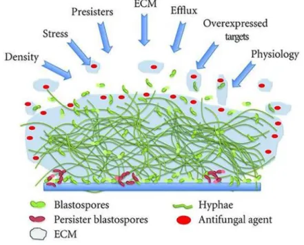
Figure 4. Schematic overview of fungal biofilm resistance mechanisms. The figure illustrates the density and complexity of the C. albicans biofilm, with different morphotypes present surrounded by extracellular matrix (ECM). The arrows represent the different factors that drive antifungal resistance within the biofilm, including density, stress, persisters, ECM, efflux, overexpressed targets, and the general physiology of the biofilm [218].
Stress. In the last years, it has been shown that drug resistance in Candida biofilms can be also promoted by the activation of several stress responses. One of the most important is the cell-wall integrity pathway. In particular, it was observed that the lacking of the mitogen-activated protein kinase (MAPK) mkc1p, which is activated by contact stress, causes the development of deficient biofilms with reduced filamentation and susceptible to MICs 100-fold lower than the sessile wild type and both planktonic strains [219]. Calcineurin, a Ca2+ calmodulin-activated serine/ threonine-specific protein phosphatase plays many critical stress roles in the
32
fungal cell and it has also implicated in mediating resistance to the azoles.
C. albicans sessile cells are up to 1,000-fold more resistant to fluconazole
than planktonic counterparts. Inhibiting calcineurin pharmacologically or impairing calcineurin function, genetically increased the azoles activity against C. albicans indicating that inhibitors could be used in combinations as novel therapeutic interventions to treat or prevent biofilms [220]. Another stress response pathway contributing to Candida biofilm resistance involves a heat shock protein HSP90. Recent studies demonstrated that genetic depletion of Hsp90 reduced C. albicans biofilm growth and maturation and interestingly impaired dispersal of biofilm cells and support azoles susceptibility [221,222]. Furthermore, a marked decrease in matrix glucan levels was observed, providing that Hsp90 might regulate biofilm azole resistance acting as a regulator of the matrix sequestration pathway [222].
Extracellular matrix. Biofilm matrix material may also impair drug delivery, either via steric hindrance or by actively binding or sequestrating antifungals. Comparing biofilms grown under continuous flow with statically grown biofilms, Al-Fattani and Douglas [181] were able to link
Candida biofilm resistance to the production of an extracellular matrix
[181]. Nett et al. [157] observed that planktonic cells surrounded by purified matrix material mimicked the biofilm drug-resistant phenotype, confirming the idea that matrix may prevent the reaching of antifungals to their intracellular target. This resistance appears to correlate with production of a matrix carbohydrate, β-1,3 glucan. Its contribution was clarified when it was shown that biofilm cell-walls bound 4- to 5 fold more azole than planktonic counterparts, thereby decreasing its potential to control biofilm-associated cells [223]. Further studies have shown that β-1,3 glucans are also responsible for sequestering echinocandins, pyrimidines,
![Figure 1. Chemical structures of the main low molecular weight microbial surface active compounds [2]](https://thumb-eu.123doks.com/thumbv2/123dokorg/4809220.49764/10.774.188.592.417.879/figure-chemical-structures-molecular-weight-microbial-surface-compounds.webp)