Submitted 2 March 2017 Accepted 27 March 2017 Published 27 April 2017 Corresponding author Adriana Gallo, [email protected] Academic editor
Vincenzo Brancaleone Additional Information and Declarations can be found on page 10
DOI 10.7717/peerj.3236 Copyright
2017 Gallo et al. Distributed under
Creative Commons CC-BY 4.0
OPEN ACCESS
Oxytetracycline induces DNA damage
and epigenetic changes: a possible risk
for human and animal health?
Adriana Gallo1,*, Rosaria Landi2,*, Valentina Rubino3,*, Alessandro Di Cerbo4,
Angela Giovazzino3
, Anna Teresa Palatucci5
, Sara Centenaro6 , Gianandrea Guidetti6 , Sergio Canello6 , Laura Cortese7 , Giuseppina Ruggiero3 , Andrea Alessandrini4,8
and Giuseppe Terrazzano3,9
1Institute of Experimental Endocrinology and Oncology (IEOS), National Research Council (CNR),
Naples, Italy
2Department of Molecular Medicine and Medical Biotechnology, University of Naples Federico II,
Naples, Italy
3Department of Translational Medical Sciences, University of Naples Federico II, Naples, Italy 4Department of Physics, Informatics and Mathematics, University of Modena and Reggio Emilia,
Modena, Italy
5PhD School of Science, University of Basilicata, Potenza, Italy 6Division of Research and Development, Sanypet SpA, Padova, Italy
7Department of Veterinary Medicine and Animal Productions, University of Naples Federico II, Naples, Italy 8National Research Council (CNR), Nanoscience Istitute, Modena, Italy
9Department of Science, University of Basilicata, Potenza, Italy *These authors contributed equally to this work.
ABSTRACT
Background. Oxytetracycline (OTC), which is largely employed in zootechnical
and veterinary practices to ensure wellness of farmed animals, is partially absorbed within the gastrointestinal tract depositing in several tissues. Therefore, the potential OTC toxicity is relevant when considering the putative risk derived by the entry and accumulation of such drug in human and pet food chain supply. Despite scientific literature highlights several OTC-dependent toxic effects on human and animal health, the molecular mechanisms of such toxicity are still poorly understood.
Methods. Here, we evaluated DNA damages and epigenetic alterations by quantitative
reverse transcription polymerase chain reaction, quantitative polymerase chain reac-tion, chromatin immuno-precipitation and Western blot analysis.
Results. We observed that human peripheral blood mononuclear cells (PBMCs)
expressed DNA damage features (activation of ATM and p53, phosphorylation of H2AX and modifications of histone H3 methylation of lysine K4 in the chromatin) after the
in vitroexposure to OTC. These changes are linked to a robust inflammatory response indicated by an increased expression of Interferon (IFN)-γ and type 1 superoxide dismutase (SOD1).
Discussion. Our data reveal an unexpected biological in vitro activity of OTC able to
modify DNA and chromatin in cultured human PBMC. In this regard, OTC presence in foods of animal origin could represent a potential risk for both the human and animal health.
SubjectsCell Biology, Allergy and Clinical Immunology, Drugs and Devices, Immunology, Pharmacology
Keywords Immune pharmacology, Drug toxicity, Inflammatory response, DNA damage, Epigenetics
INTRODUCTION
The drug (4S,4aR,5S,5aR, 6S,12aS)-4-(dimethylamino)-3,5,6,10,12,12a-hexahydroxy-6-methyl-1,11-dioxo1,4,4a,5,5a,6,11,12a-octahydrotetracene-2-carboxamide, briefly oxytetracycline (OTC) is active towards a wide range of micro-organisms (Nelson &
Levy, 2011), is efficiently absorbed in the duodenum forming complexes with metallic ions,
is unstable at acid pH and its introduction along with food reduces its serum concentrations
(Palmieri, Di Cerbo & Laurino, 2014). Moreover, such drug could accumulate within bone,
skin, fat, tendons, muscles, liver and gastrointestinal tract (Agwuh & MacGowan, 2006). OTC is commonly used in medicine and is one of the main antibiotics used in zootech-nical and veterinary practices as feed supplement to ensure wellness of farmed animals (i.e., poultry, ovine, swine and livestock) (Graham et al., 2014;Brüning et al., 2014;Di Cerbo
et al., 2014;Odore et al., 2015).
Several studies have investigated the potential toxicity of OTC ranging from teratogenic effects during pregnancy (Czeizel & Rockenbauer, 2000) to some effect on immune system
(Glette et al., 1984;Potts et al., 1983;Van den Bogert & Kroon, 1982;Myers, Farrell &
Hen-derson, 1995;Di Cerbo et al., 2016). Moreover, scientific literature suggested that the drug
is able to inhibit or reduce catalase (Chi, Liu & Zhang, 2010) and affects avian cartilage degradation (Peters et al., 2002).
We recently demonstrated that OTC: (a) induces an in vitro inflammatory response characterized by T and non-T lymphocytes activation and Interferon (IFN)-γ release
(Di Cerbo et al., 2016); (b) triggers the apoptosis of human and dog haematopoietic cells
(Di Cerbo et al., 2016;Odore et al., 2015).
The potential OTC toxicity becomes more relevant when considering the potential risk derived by the eventuality of entry and accumulation of such drug in human and pet food with possible consequences on health (Palmieri, Di Cerbo & Laurino, 2014). In this regard, animal muscle, bone and fat are known to be the elective deposit for several antibiotics
(Palmieri, Di Cerbo & Laurino, 2014;Macy & Poon, 2009) and are routinely employed for
human and pet food production (Palmieri, Di Cerbo & Laurino, 2014).
In the light of the widespread use of OTC and considering the putative risk derived by the eventuality of entry and accumulation of such drug in human and pet food chain supply (Graham et al., 2014;Brüning et al., 2014;Di Cerbo et al., 2014;Nelson & Levy, 2011;
Palmieri, Di Cerbo & Laurino, 2014), it is possible to speculate that the OTC accumulates
in these edible tissues and that this occurrence represents the contact between the drug and the humans or companion animals (dogs and cats).
Here, we addressed the study over the relevance of some molecular mechanisms of drug toxicity and, specifically, on the genotoxic effect and epigenetic modifications potentially induced by OTC. This could be relevant since many of the effects observed could affect the gene expression and represent a potential risk for human and animal health.
MATERIALS & METHODS
Cells and incubation
Peripheral blood mononuclear cells (PBMCs) were obtained, as previously described (Di
Cerbo et al., 2016). Briefly, we performed the centrifugation on Ficoll-Paque cushion (GE
Healthcare, Uppsala Sweden) gradients of buffy coats obtained from six volunteer healthy donors. In order to inform the blood donors concerning the possibility to use minimal amount of their blood donation for scientific purpose, written informed consent (model n. 5526 of Azienda Ospedaliera Universitaria ‘‘FEDERICO II’’, Naples, Italy) was obtained from each donor at the time of venous peripheral blood donation performed at Blood Trasfusional Center of Azienda Ospedaliera Universitaria ‘‘FEDERICO II’’, Naples Italy, as established by Italian Law. All the experiments were performed anonymously, without any donor biographical reference. White blood cells have never been used to create a genome database.
To test the in vitro potential biochemical toxic role of OTC (Liquid Oxytetracycline 20% R, TreI, Reggio Emilia, Italy), the PBMCs (2.5 × 10⁶/ml) were incubated in presence of RPMI 1,640 medium with 10% FCS (Invitrogen, Carlsbad, CA, USA) alone or with 2µg/ml OTC (Odore et al., 2015;Di Cerbo et al., 2016) at 37◦C for different times (6 h, 12 h, 24 h).
RNA extraction and qRT-PCR and qPCR
Total RNA was extracted using TRI Reagent (T9424, Sigma-Aldrich, St Louis, MO, USA). cDNA was synthesized in a 20µl reaction volume containing 1µg of total RNA, in accordance to the life technology protocol (High-Capacity cDNA Reverse Transcription Kit 4368814; Applied Biosystem, Thermofisher Scientific, Foster City, CA, USA). The products were stored at −20 ◦C until use. Quantitative reverse transcription polymerase chain reaction (qRT-PCR) and quantitative polymerase chain reaction (qPCR) were performed three times in six replicates on a 7,500 Real Time PCR System (Applied Biosystems) using the SYBR Green-detection system (SYBR select Master Mix, 4473369, Applied Biosystem). The following primers were used: IFN-γ mRNA, 50
-TGGAAAGAGGAGAGTGACAGA-30 and 50-CTGTTTTAGCTGCTGGCGAC-30; type 1 superoxide dismutase (SOD1) mRNA 50-CTAGCGAGTTATGGCGACGA-30and 50-GTCTCCAACATGCCTCTCTTCA-30; 18S, 50-GCGCTACACTGACTGGCTC-30and 50-CATCCAATCGGTAGTAGCGAC-30.
Chromatin Immuno-Precipitation (ChIP)
Cells were treated as indicated in Cells and incubation paragraph. The cells (∼2.5 × 10⁶for each antibody) were crosslinked with a 1% formaldeyhyde/PBS solution for 10 min at room temperature, the reaction was stopped by the addition of glycine to a final concentration of 125 mM. Fixed cells were harvested and the pellet was resuspended in 1 ml of Lysis Buffer (10 mM Tris-HCl pH 8.0, 10 mM NaCl, 0.2% NP40) containing 1× protease inhibitor cocktail (Roche Applied Science, Basel, Switzerland). The lysates were sonicated in order to have DNA fragments from 300 to 600 bp. An aliquot (1/10) of sheared chromatin was used as input DNA. Sonicated samples were processed according to the manufacturer’s protocol of ChIP assay kit (Merck Millipore, Billerica, MA, USA). Samples were subjected to qPCR using the following primers: IFN-γ Promoter, 50-GAA
CAATGTGCTGCACCTCC-30 and 50-CACAGGTGGGCATAATGGGT-30; SOD1 Pro-moter, 50-CATCATTTTGCCAATTTCGCGT-30and 50-CGAGTGGCCGGGAATGACT-30.
Real Time-qPCRs were performed using the SYBR Green-detection system (SYBR select Master Mix, 4473369; Applied Biosystem).
Western blot preparation and analysis
Aliquots of the cells collected for ChIP were used for western blot. Cells were washed twice with cold phosphate-buffered saline (PBS) and nuclei were extracted using 1 ml of Lysis Buffer (10 mM Tris-HCl pH 8.0, 10 mM NaCl, 0.2% NP40) containing 1× protease inhibitor cocktail. Nuclear lysates were obtained accordingly with Nuclear Fractionation Protocol (Abcam, Cambridge, UK). γ H2AX was detected using part of the sonicated samples collected for ChIP. Lysates were cleared by centrifugation (13,000 rpm for 20 min). Protein concentrations were measured by Bio-Rad Protein Assay Dye Reagent Concentrate #500-0006. Equal amounts of cell extracts were then resolved by SDS–PAGE, transferred to nitrocellulose membranes, and immunoblotted using specific antibodies. Blots were detected using an ECL system (Lumilight Western Blotting Substrate, 12015200001, Roche).
Antibodies
AntiDNMT1 ab87656 (Abcam, Cambridge, UK), H3K4me2 ab32356 (Abcam), -H3K4me3 ab1012 (Abcam),—Total H3 ab1791 (Abcam), -Menin sc-0200 (Santa Cruz Biotechnology); phosphoATM ab81292 (Abcam), -phospho-H2AX (07164, Merck Milli-pore), MCM7 sc-9966 (Santa Cruz Biotechnology), Normal rabbit IgG sc-2027 (Santa Cruz Biotechnology), Normal mouse IgG sc-2025 (Santa Cruz Biotechnology) and -p53 ab1101 (Abcam).
Statistical analysis
Statistical significance between groups was determined using Student’s t test.
RESULTS AND DISCUSSION
IFN-γ and SOD1 gene expression
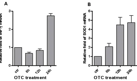
We recently demonstrated the pro-inflammatory effect of OTC in causing both the IFN-γ secretion in T and non-T lymphocytes (Di Cerbo et al., 2016). Here, we evaluated the effect of drug treatment in the up-regulation of IFN-γ gene expression.Figure 1Ashows that mRNA levels of IFN-γ robustly increased in PBMCs after 24 h of OTC incubation.
To investigate if OTC-mediated inflammatory condition could depend on oxidative stress, we evaluated whether the Cu–Zn Super Oxide Dismutase 1 (SOD1) could be increased after drug exposure. It is of note that one of the SOD1 is involved not only in oxida-tive metabolism but also in the T lymphocyte activation dependent on the accumulation of reactive oxygen species (Terrazzano et al., 2014). Our data (Fig. 1B) show that the mRNA levels of SOD1 increased from 12 to 24 h of OTC treatment.
The data reported inFig. 1showed that the enhancement of mRNA levels of IFN-γ
occurred after 24 h of OTC-treatment, whereas the induction of SOD1 mRNA appeared already in 12 h. A possible explanation might be that SOD1 is a housekeeping gene (Minc et
Figure 1 OTC induces γ and SOD1 mRNA. OTC significantly induces the increment of both
IFN-γ and SOD1 mRNA. Total RNA was prepared from PBMC stimulated with OTC for 6, 12, 24 h, as indi-cated in ‘Materials & Methods’, and analyzed by qPCR with specific primers to IFN-γ (A) and SOD1 (B) mRNA normalized to 18S RNA levels. The statistical analysis derived from 2 experiments in triplicate (n ≥ 6; Mean ± SD).
al., 1999) and its basal expression is usually higher than IFN-γ gene. Since the used in vitro
model is based on freshly isolated PBMCs that are usually resistant to natural occurring apoptosis (Miyawaki et al., 1992), the increased SOD1 level after the drug incubation could be likely associated to the hypothesis of apoptosis induction upon chromatin and DNA damages (Norbury & Zhivotovsky, 2004;Barbosa et al., 2010).
These results suggest that the drug may affect some important cellular responses as the induction of a gene expression fostering the activation of previously observed immune response by T and non-T lymphocytes after in vitro OTC exposure (Di Cerbo et al., 2016).
OTC generates genotoxic damage
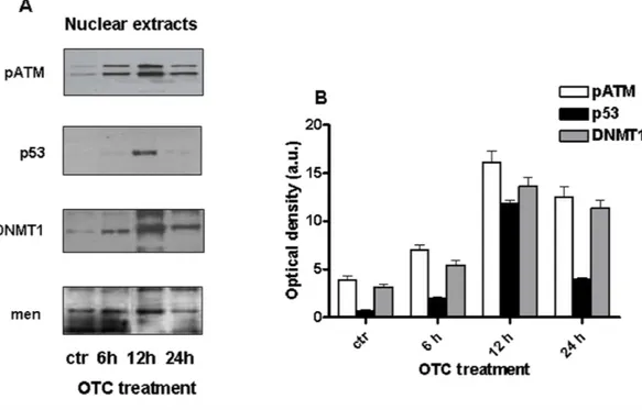
Since the OTC is able to induce apoptosis (Odore et al., 2015;Di Cerbo et al., 2016), we investigated on the potential ability of such drug in causing DNA damage and, in reason of that, in inducing apoptosis. In particular, we evaluated the presence of genotoxic markers after in vitro drug treatment of PBMCs. In this regard, it is worth noting the role for Ataxia Telangiectasia mutated protein (ATM) that is a serin/treonin kinase activated in response to the DNA double strand break to promote cell cycle arrest, DNA repair and, if necessary, the cell death by apoptosis (Canman & Lim, 1998;Lee & Paull, 2007).
As shown inFig. 2A, the phosphorylated form of ATM (pATM) is clearly increased in human PBMCs after 6 h and 12 h of OTC incubation.
Furthermore, we investigated the levels of p53, as one of principal substrates of pATM and an important marker of DNA damage (Sakaguchi et al., 1998;Williams & Schumacher, 2016).Figure 2Aindicates that p53 significantly increased after 12 h of drug exposure.
Figure 2 OTC induces genotoxic damage. Cells were treated with OTC for 6, 12 and 24 h and processed as indicated in ‘Material and Methods’. (A) the western blot for pATM, p53 and DNMT1 was performed using nuclear extract. Menin is reported as loading control; (B) quantification of the western blots nor-malized to Menin levels. Values are reported as Optical density (arbitrary units = a.u.).
These observations suggest that some DNA damage may occur after OTC incubation. One of the main epigenetic modifications involved in gene regulation is the DNA methylation (Hamidi, Singh & Chen, 2015). It is well known that DNA (cytosine-5)-methyltransferase 1 enzyme (DNMT1) is recruited to the chromatin, in response to the oxidative DNA damage, in order to inhibit gene transcription and to support DNA repair
(Ding et al., 2016). It is of relevance that DNMT1 appears to increase after OTC treatment,
following a similar kinetics of pATM and p53 (Fig. 2A).
Such evidence supports the idea that the enzymes could be recruited on the site of oxidative DNA damage occurred upon OTC incubation and cooperate each-other to induce chromatin modifications aimed to foster the DNA repair (Morano et al., 2014).
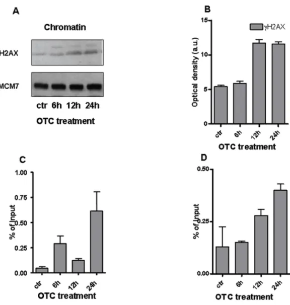
To better address the entity of DNA damage, we investigated the presence of DNA double strand break (DBS) markers. In particular, we evaluated the phosphorylated histone H2AX (γ H2AX) (Rogakou et al., 1998;Mah, El-Osta & Karagiannis, 2010) by performing western blot analysis on chromatin samples.Figures 3A and3B shows the significant increase ofγ H2AX with the highest peak from 12 to 24 h of drug treatment. Notably, the phosphorylated histone H2AX binds the regions of chromatin on the sites of DSB and DNA repair (Mah, El-Osta & Karagiannis, 2010). To better analyze the chromatin changes caused by the drug, we tested by ChIP assay the presence ofγ H2AX on the promoters of genes of our interest. We observed the accumulation ofγ H2AX at the site of promoter of IFN-γ gene (Fig. 3C). The presence ofγ H2AX was also enhanced at the level of SOD1 gene promoter after 24 h of drug incubation (Fig. 3D).
Figure 3 OTC and the chromatin changes. (A) The western blot forγ H2AX performed on chromatin extracts. MCM7 is reported as loading control; (B) Quantification ofγ H2AX normalized to MCM7 levels. Values are reported as Optical density (arbitrary units = a.u.); qChIP analysis evideces thatγ H2AX accu-mulates on IFN-γ (C) and SOD1 (D) promoters. Cells were treated with OTC as indicated, crosslinked and sonicated. The statistical analysis derived from at least 2 experiments in triplicate (n ≥ 6; Mean ± SD).
Therefore, our results strongly suggest the correlation between DNA damage occurrence and OTC administration. Moreover, the increased levels of IFN-γ and SOD1 mRNAs (Fig. 1) appear to be linked to the function ofγ H2AX, which cooperates with the induction of ATM mediated transcription (Singh et al., 2015).
Epigenetic changes
Histone modification represents an epigenetic mechanism that affects gene transcription by altering the chromatin structure and DNA accessibility. Histone methylation can be associated with the different status of chromatin (Zhang, Cooper & Brockdorff, 2015). Here, we evaluated if OTC treatment could be correlated to alterations of histone methylation. More specifically, we investigated on the methylation status of lysine 4 of Histone 3 (H3K4)
Figure 4 OTC and histone methylation. Methylation profile of histone H3K4 is induced by OTC on both the IFN-γ and SOD1 gene promoters. PMBC cells were exposed to OTC at the indicated times (0, 6, 12 and 24 h). qChIP was carried out using specific antibodies; (A) H3K4me3 and H3K4me2 occupancy at IFN-g promoter; (B): H3K4me2 and H3K4me3 occupancy at SOD1 promoter; (C) and (D); the TotalH3 occupancy at IFN-γ and SOD1 promoters respectively. The statistical analysis derived from at least 2 ex-periments in triplicate (n ≥ 6; Mean ± SD).
that is implicated in the regulation of gene activation (Barski et al., 2007;Ruthenburg, Allis
& Wysocka, 2007).
To this aim, we performed a ChIP for the promoter of the genes whose expression appeared to be modified by OTC. After 24 h of drug incubation, the increment of both the di-methylated (me2) and tri-methylated (me3) H3K4 is particularly evident for IFN-γ promoter (Fig. 4A), while the increase of H3K4 is more evident for the di-methylated form at the level of SOD1 promoter (Fig. 4B). These data are correlated with the observed activation of gene expression (Bernstein et al., 2005). Similar results are obtained from the analysis of total H3 histone levels (Figs. 4Cand4D).
Together, these data indicate that the OTC treatment can affect the status of chromatin.
CONCLUSION
Despite scientific literature that has been suggesting the potential toxicity of OTC (Czeizel
& Rockenbauer, 2000;Glette et al., 1984;Potts et al., 1983;Van den Bogert & Kroon, 1982;
Myers, Farrell & Henderson, 1995), the mechanisms of the toxic effect of such drug is still
poor understood.
We recently demonstrated that OTC induces in vitro inflammatory response (Di Cerbo
et al., 2016) and apoptosis (Di Cerbo et al., 2016;Odore et al., 2015). Therefore, this and
other suggestions open an interesting scenario on the toxicity of OTC that requires a greater understanding over the nature of observed toxic effects.
This study emphasized the toxicity of OTC, investigating over the molecular mechanisms involved in human PBMC inflammatory response. It is of note that the drug promoted a ro-bust inflammatory response as represented by the increasing of IFN-γ mRNA levels. This re-sult reflects and confirms the previously observed increment of IFN-γ production in T and non-T lymphocytes (Di Cerbo et al., 2016). In addition, OTC significantly induced SOD1 mRNA in the same experimental condition and cellular model. This evidence extends our previous observations on apoptosis induction after OTC exposure (Terrazzano et al., 2014;
Odore et al., 2015).
In addition, we observed that OTC induces genotoxic damage as well as such drug recruits some enzymes implicated in the delicate balance between cell death and survival. Indeed, our data evidenced the increased levels of pATM, p53 and DNMT1 after drug incubation. It is of note that p53 is a substrate of pATM and is crucial to the cell cycle arrest and/or to induce the cell death by apoptosis (Norbury & Zhivotovsky, 2004), while DNMT1 repre-sents a specific enzyme involved in some epigenetic changes (Hamidi, Singh & Chen, 2015), associated with the DNA damage (Rossetto et al., 2010).
Moreover, we observed the activation ofγ H2AX, as a main DSB sensor protein, and suggested an epigenetic effect of OTC on the methylation status of H3K4 that is implicated in gene expression regulation (Barski et al., 2007;Ruthenburg, Allis & Wysocka, 2007). The increase of both me2 and me3 H3K4 occurred after OTC incubation and was evident for IFN-γ and SOD1 gene promoters.
Our data represent a preliminary step in the understanding of OTC toxicity, since the knowledge of the molecular mechanisms involved in the toxic effect may help in the generation of new drugs with reduced risk for human health.
In conclusion, it could be of great relevance to ascertain the possible acute and long term effects of OTC on human health. It is worth noting that the use of antibiotics for growth promotion is prohibited in Europe and it is considered a health hazard by WHO since 2006. The use of antibiotics in agriculture for non-therapeutic purposes is allowed in United States and Canada (FaAOF, 2014). Therefore, new regulations are urgently necessary to reduce antibiotic contaminants in foods as well as the antibiotic resistance phenomenon
STUDY LIMITATIONS
Notably, the current study incurs some limitations that are not addressable without further researches. In this regard, our study did not perform chemical-pharmaceutical and pharmacological test to evaluate the molecular complexity and/or stability of the used OTC or to verify the possible presence of active sub-products generated during in vitro tests. In addition, this study did not address any chemical evaluation of the excipients (i.e., fillers, binders, dyes, flavorings, preservatives and other materials) present in the here used commercial liquid formulation of OTC drug employed in veterinary medicine. Therefore, further evaluations are required to complete the significance of OTC toxicity. In particular, the absence of in vivo experiments, able to confirm the in vitro observed OTC toxicity, represents the main relevant limitation. Therefore, clinical studies are required to ascertain the effect of the drug in inducing the inflammatory status in animals and/or in humans.
ADDITIONAL INFORMATION AND DECLARATIONS
Funding
The authors received no funding for this work.
Competing Interests
None of the authors have financial or personal relationships with other people or organisations that could inappropriately influence or bias the content of the paper. This research was performed in collaboration with some scientists from the Division of Research and Development, Sanypet SpA, Padova, Italy (as indicated in the authors’ affiliation) according to scientific and ethical principles of the scientific community. No financial funding was obtained from Sanypet Industry for this research study.
Author Contributions
• Adriana Gallo and Giuseppe Terrazzano conceived and designed the experiments, analyzed the data, wrote the paper, prepared figures and/or tables, reviewed drafts of the paper.
• Rosaria Landi conceived and designed the experiments, performed the experiments, analyzed the data, contributed reagents/materials/analysis tools, wrote the paper, prepared figures and/or tables.
• Valentina Rubino conceived and designed the experiments, performed the experiments, analyzed the data, wrote the paper, prepared figures and/or tables.
• Alessandro Di Cerbo, Sara Centenaro, Gianandrea Guidetti, Sergio Canello, Laura Cortese and Andrea Alessandrini contributed reagents/materials/analysis tools.
• Angela Giovazzino performed the experiments, contributed reagents/materials/analysis tools.
• Anna Teresa Palatucci performed the experiments, analyzed the data, prepared figures and/or tables.
Human Ethics
The following information was supplied relating to ethical approvals (i.e., approving body and any reference numbers):
The ethics committee approval is not required for this type of experimental test, as it is not a clinical trial in vivo, we do not use patients, but merely use peripheral blood leukocytes from healthy blood donors as volunteers who went at the Blood Bank of Federico II.
The experiments were only performed ‘‘in vitro’’ using peripheral blood mononuclear cells from these healthy donor’s buffy coats, derived as discarded products upon preparation of medical blood components (red cells, platelets, plasma and its derivatives).
Italian Law (L.107/1990, L.219/2005, DL25/01/01 no. 25, DL20/12/07 no.261, DM 02/11/2015) allows the use of these ‘‘remnant’’ parts of blood donation to scientific purpose if the donors are informed and if they subscribe a written consent at the time of blood donation.
The informed consent (model n. 5526 of Azienda Ospedaliera Universitaria ’’FEDERICO II’’, Naples, Italy) has been already subscribed by the 6 donors at the time of blood donation, performed at Blood Trasfusional Center of Azienda Ospedaliera Universitaria ’’FEDERICO II’’, and steated the possibility to use some blood components also for scientific purposes. The in vitro experiments were performed fully anonymous. White blood cells have never been used to create a genome database.
Data Availability
The following information was supplied regarding data availability: The raw data has been supplied asData S1.
Supplemental Information
Supplemental information for this article can be found online athttp://dx.doi.org/10.7717/ peerj.3236#supplemental-information.
REFERENCES
Agwuh KN, MacGowan A. 2006. Pharmacokinetics and pharmacodynamics of the
tetracyclines including glycylcyclines. Journal Antimicrobic Chemother 58:256–265
DOI 10.1093/jac/dkl224.
Barbosa LF, Cerqueira FM, Macedo AF, Garcia CC, Angeli JP, Schumacher RI, Sogayar MC, Augusto O, Carrì MT, Di Mascio P, Medeiros MH. 2010. Increased SOD1
association with chromatin, DNA damage, p53 activation, and apoptosis in a cellular model of SOD1-linked ALS. Biochimica et Biophysica Acta 802:462–471
DOI 10.1016/j.bbadis.2010.01.011.
Barski A, Cuddapah S, Cui K, Roh TY, Schones DE, Wang Z, Wei G, Chepelev I, Zhao K. 2007. High-resolution profiling of histone methylations in the human genome.
Cell129:823–837DOI 10.1016/j.cell.2007.05.009.
Bernstein BE, Kamal M, Lindblad-Toh K, Bekiranov S, Bailey DK, Huebert DJ,
2005. Genomic maps and comparative analysis of histone modifications in human
and mouse. Cell 120:169–181DOI 10.1016/j.cell.2005.01.001.
Brüning A, Brem GJ, Vogel M, Mylonas I. 2014. Tetracyclines cause cell
stress-dependent ATF4 activation and mTOR inhibition. Experimental Cell Research
320:281–289DOI 10.1016/j.yexcr.2013.11.012.
Canman CE, Lim DS. 1998. The role of ATM in DNA damage responses and cancer.
Oncogen17:3301–3308.
Chi Z, Liu R, Zhang H. 2010. Potential enzyme toxicity of oxytetracycline to catalase.
Sci-ence of the Total Environment 408:5399–5404DOI 10.1016/j.scitotenv.2010.08.005.
Czeizel AE, Rockenbauer M. 2000. A population-based case-control teratologic study
of oral oxytetracycline treatment during pregnancy. European Journal of Obstetrics &
Gynecology and Reproductive Biology 88:27–33 DOI 10.1016/S0301-2115(99)00112-8.
Di Cerbo A, Canello S, Guidetti G, Laurino C, Palmieri B. 2014. Unusual antibiotic
presence in gym trained subjects with food intolerance; a case report. Nutricion
Hospitalaria30:395–398 DOI 10.3305/nh.2014.30.2.7594.
Di Cerbo A, Palatucci AT, Rubino V, Centenaro S, Giovazzino A, Fraccaroli E, Cortese L, Ruggiero G, Guidetti G, Canello S, Terrazzano G. 2016. Toxicological
implications and inflammatory response in human lymphocytes challenged with oxytetracycline. Journal of Biochemical and Molecular Toxicology 30:170–177
DOI 10.1002/jbt.21775.
Ding N, Bonham EM, Hannon BE, Amick TR, Baylin SB, O’Hagan HM. 2016.
Mis-match repair proteins recruit DNA methyltransferase 1 to sites of oxidative DNA damage. Journal Molecular Cell Biology 8:244–254DOI 10.1093/jmcb/mjv050.
FaAOF. 2014. Headquarters codex alimentarius commission, maximum residue limits
for veterinary drugs in foods, 35th session. 1–40. Available atftp:// ftp.fao.otg/ codex/ weblinks/ MRL2_e_2012.pdf.
Glette J, Sandberg S, Haneberg B, Solberg CO. 1984. Effect of tetracyclines and UV
light on oxygen consumption by human leukocytes. Antimicrobial Agents and
Chemotherapy26:489–492 DOI 10.1128/AAC.26.4.489.
Graham F, Paradis L, Bégin P, Paradis J, Babin Y, Des Roches A. 2014. Risk of allergic
reaction and sensitization to antibiotics in foods. Annals of Allergy, Asthma &
Immunology113:329–330DOI 10.1016/j.anai.2014.06.029.
Hamidi T, Singh AK, Chen T. 2015. Genetic alterations of DNA methylation machinery
in human diseases. Epigenomics 7:247–265DOI 10.2217/epi.14.80.
Lee JH, Paull TT. 2007. Activation and regulation of ATM kinase activity in response to
DNA double-strand breaks. Oncogen 26:7741–7748DOI 10.1038/sj.onc.1210872.
Macy E, Poon K-YT. 2009. Self-reported antibiotic allergy incidence and
preva-lence: age and sex effects. The American Journal of Medicine 122:778.e1–778.e7
DOI 10.1016/j.amjmed.2009.01.034.
Mah LJ, El-Osta A, Karagiannis TC. 2010. gammaH2AX: a sensitive molecular marker of
Minc E, De Coppet P, Masson P, Thiery L, Dutertre S, Amor-Guéret M, Jaulin C. 1999.
The human copper-zinc superoxide dismutase gene (SOD1) proximal promoter is regulated by Sp1, Egr-1, and WT1 via non-canonical binding sites. The Journal of
Biological Chemistry 274:503–509DOI 10.1074/jbc.274.1.503.
Miyawaki T, Uehara T, Nibu R, Tsuji T, Yachie A, Yonehara S, Taniguchi N. 1992.
Dif-ferential expression of apoptosis-related Fas antigen on lymphocyte subpopulations in human peripheral blood. Journal of Immunology. 149:3753–3758.
Morano A, Angrisano T, Russo G, Landi R, Pezone A, Bartollino S, Zuchegna C, Babbio F, Bonapace IM, Allen B, Muller MT, Chiariotti L, Gottesman ME, Porcellini A, Avvedimento EV. 2014. Targeted DNA methylation by homology-directed repair in
mammalian cells. Transcription reshapes methylation on the repaired gene. Nucleic
Acids Research42:804–821DOI 10.1093/nar/gkt920.
Myers MJ, Farrell DE, Henderson M. 1995. In vitro modulation of bovine blood
neutrophils and mononuclear cells by oxytetracycline. American Journal of Veterinary
Research56:1007–1011.
Nelson ML, Levy SB. 2011. The history of the tetracyclines. Annals of the New York
Academy of Sciences1241:17–32DOI 10.1111/j.1749-6632.2011.06354.x.
Norbury CJ, Zhivotovsky B. 2004. DNA damage-induced apoptosis. Oncogen
23:2797–2808DOI 10.1038/sj.onc.1207532.
Odore R, De Marco M, Gasco L, Rotolo L, Meucci V, Palatucci AT, Rubino V, Ruggiero G, Canello S, Guidetti G, Centenaro S, Quarantelli A, Terrazzano G, Schiavone A. 2015. Cytotoxic effects of oxytetracycline residues in the bones of broiler chickens
following therapeutic oral administration of a water formulation. Poultry Science
94:1979–1985DOI 10.3382/ps/pev141.
Palmieri B, Di Cerbo A, Laurino C. 2014. Antibiotic treatments in zootechnology and
effects induced on the food chain of domestic species and, comparatively, the human specie. Nutriction Hospitalaria 29:1427–1433DOI 10.3305/nh.2014.29.6.7350.
Peters TL, Fulton RM, Roberson KD, Orth MW. 2002. Effect of antibiotics on
in vitroand in vivo avian cartilage degradation. Avian Diseases 46:75–86
DOI 10.1637/0005-2086(2002)046[0075:EOAOIV]2.0.CO;2.
Potts RC, MacConnachie A, Brown RA, Gibbs JH, Robertson AJ, Hassan HA, Beck JS. 1983. Some tetracycline drugs suppress mitogen-stimulated lymphocyte
growth but others do not. British Journal of Clinical Pharmacology 16:127–132
DOI 10.1111/j.1365-2125.1983.tb04975.x.
Rogakou EP, Pilch DR, Orr AH, Ivanova VS, Bonner WM. 1998. DNA double-stranded
breaks induce histone H2AX phosphorylation on serine 139. The Journal of Biological
Chemistry273:5858–5868DOI 10.1074/jbc.273.10.5858.
Rossetto D, Truman AW, Kron SJ, Côté J. 2010. Epigenetic modifications in
double-strand break DNA damage signaling and repair. Clinical Cancer Research
16:4543–4552DOI 10.1158/1078-0432.CCR-10-0513.
Ruthenburg AJ, Allis CD, Wysocka J. 2007. Methylation of lysine 4 on histone H3:
intricacy of writing and reading a single epigenetic mark. Molecular Cell 25:15–30
Sakaguchi K, Herrera JE, Saito S, Miki T, Bustin M, Vassilev A, Anderson CW, Appella E. 1998. DNA damage activates p53 through a phosphorylation-acetylation cascade.
Genes & Development 12:2831–2841DOI 10.1101/gad.12.18.2831.
Singh I, Ozturk N, Cordero J, Mehta A, Hasan D, Cosentino C, Sebastian C, Krüger M, Looso M, Carraro G, Bellusci S, Seeger W, Braun T, Mostoslavsky R, Barreto G. 2015. High mobility group protein-mediated transcription requires DNA damage
markerγ -H2AX. Cell Research 25:837–850 DOI 10.1038/cr.2015.67.
Terrazzano G, Rubino V, Damiano S, Sasso A, Petrozziello T, Ucci V, Palatucci AT, Giovazzino A, Santillo M, De Felice B, Garbi C, Mondola P, Ruggiero G. 2014.
T cell activation induces CuZn superoxide dismutase (SOD)-1 intracellular re-localization, production and secretion. Biochimica et Biophysica Acta 1843:265–274
DOI 10.1016/j.bbamcr.2013.10.020.
US Government Publishing Office. 2014. Tolerance for residues of new animal drugs in
food. Subpart B-specific tolerance for residues of new animal drugs. Electronic Code of Federal Regulations (e-CFR) PART 556. Available athttp:// www.ecfr.gov/.
Van den Bogert C, Kroon AM. 1982. Effects of oxytetracycline on in vivo proliferation
and differentiation of erythroid and lymphoid cells in the rat. Clinical and
Experi-mental Immunology50:327–335.
Williams AB, Schumacher B. 2016. p53 in the DNA-Damage-Repair Process. Cold Spring
Harbor Perspectives in Medicine6(5):a026070DOI 10.1101/cshperspect.a026070.
Zhang T, Cooper S, Brockdorff N. 2015. The interplay of histone modifications—writers