MANUSCRIPT TITLE:
A pattern-based approach combining tumor morphology on MRI with distinct signal patterns on diffusion-weighted imaging to assess response of rectal tumors after chemoradiotherapy.
Lambregts DM, Delli Pizzi A, Lahaye MJ, van Griethuysen JJM, Maas M, Beets GL, Bakers FC, Beets-Tan RG. A Pattern-Based Approach Combinind Tumor Morphology on MRI With Distinct Signal Patterns on Diffusion-Weighted Imaging to Assess Response of Rectal Tumors After Chemoradiationtherapy. Dis Colon Rectum. 2018 Mar;61(3):328-337. doi: 10.1097/DCR.0000000000000915.
STRUCTURED ABSTRACT:
Background:
Diffusion-weighted imaging is increasingly used in rectal cancer MR imaging to assess response after chemoradiotherapy. Certain pitfalls (e.g. artefacts) may hamper MRI assessment, leading to suboptimal diagnostic performance. Combining diffusion-weighted MRI with the underlying morphology on standard (T2-diffusion-weighted) MRI may help overcome these pitfalls.
Objective:
To evaluate the diagnostic performance of a pattern-based approach combining tumor morphology on T2-weighted MRI with distinct diffusion-weighted imaging signal patterns to assess response after chemoradiotherapy in rectal cancer.
Patients:
Design:
Response to chemoradiotherapy was scored according to four patterns:
A: cases with either a clear residual mass with corresponding high diffusion signal (A+) or completely normalized wall without diffusion signal (A-);
B: cases with circular and/irregular fibrosis with (B+) or without (B-) small foci of diffusion signal scattered throughout the fibrosis;
C: cases with semi-circular fibrosis with (C+) or without (C-) high diffusion signal at the inner margin of fibrosis;
D: polypoid tumors showing regression of the polyps and fibrosis at the site of the stalk with (D+) or without (D-) focal high diffusion signal in the stalk.
N=75 cases were re-scored by an indipendent second reader to studdy interobserver variations.
Standard of reference was histopathology or long-term outcome.
Main outome measures:
Diagnostic performance to discriminate between a complete response and residual tumor.
Results:
The pattern-based approach resulted in a sensitivity of 94%, specificity 77%, PPV 88%, NPV 87%, and overall accuracy of 88% to differentiate between tumor vs. complete response. Accuracy per pattern were 100% (A), 73% (B), 86% (C) and 92% (D). Interobserver agreement was good (k0.75).
Limitations:
No comparison with “routine” (non-pattern) diffusion-MRI assessment.
Conclusions:
A pattern-based approach combining tumor morphology with distinct diffusion-weighted imaging patterns results in good diagnotic performance to assess response.
INTRODUCTION:
In recent years diffusion-weighted imaging (DWI) has increasingly been acknowledged as a beneficial adjunt to the routine MRI protocol for assessing rectal cancer, particularly in the restaging setting after neoadjuvant treatment. DWI uses the differences in the
extracellular movement “diffusion” of water protons to discriminate between tissues of varying cellularity and is known to be a very sensitive technique to detect malignant (hypercellular) tumors. Various studies, including a meta-analysis by van der Paardt and colleagues, have shown that the addition of DWI can significantly improve the diagnostic performance of MRI for re-evaluation of the local tumor stage after chemoradiotherapy (CRT). 1-3 In particular, DWI can help discriminate patients with residual viable tumor from patients with a complete tumor response. This is an increasingly relevant clinical issue, given the recent introduction of “watchful waiting” as a potential alternative to surgical resection for complete responders. 4,5 In order to safely select patients for ‘watchful waiting’ accurate selection of the true complete responders is crucial.
The generally employed principle of assessing DWI is relatively straightforward: the absence of high signal on high b-value DWI is suggestive of a complete tumor regression, whereas a remaining high DWI-signal indicates that a tumor remnant is still present. However, there are some potential drawbacks to this approach. High DWI-signal may also be caused by ‘shine through’ effects of high signal in fluids (for example in the rectal lumen), and by the fact that other anatomical structures with a dense cellular
exhibit high signal 6. Moreover, artefacts caused by the presence of air in the rectal lumen may result in false high signal projecting over the rectal wall.7 To help overcome these pitfalls, it may be helpful to advocate a more structured, pattern-based approach combining diffusion-weighted signal patters with the underlying morphology on standard MRI. Such a pattern-based reading approach has previously also been shown beneficial for the staging and follow-up of rectal tumors on T2-weighted MRI and for the assessment of rectal cancer lymph nodes on DWI10, but has not yet been investigated for tumor response evaluation on DWI.
Aim of this study was to explore the value of a pattern-based approach to assess rectal tumor response after neoadjuvant treatment (in specific to differentiate between complete responders and patients with residual tumor) using a combination of tumor morphology on standard T2-weighted imaging with distinct signal patterns on DWI.
MATERIALS & METHODS
A retrospetive analysis of imaging data acquired as part of routine classic diagnostic imaging procedures was performed. The study was approved by the local istitutional review board and informed consent was waived.
Patients
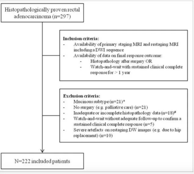
297 consecutive locally advanced rectal cancer patients treated with a long course of neoadjuvant treatment at [BLINDED] between 2008 and 2015 were considered for inclusion. Inclusion criteria consisted of: histopathologically proven non-mucinous rectal adenocarcinoma, [2] long course neoadjuvant treatment, [3] availability of a primary staging MRI and a restaging MRI including a DWI sequence, and [4] data on the final response based on histopathology after surgery or long term follow-up (in case of a clinical complete managed with watchful waiting). Seventy-five patients were excluded (see Figure 1). Mucinous tumors were excluded as due to their mucin content (causing high signal T2 shine through effects) these tumours show different characteristics on DWI.11 Patients with severe artefacts on DWI (e.g. caused by metal hip prostheses) were also excluded as in these cases the rectal wall and thus the response pattern cannot be assessed
on DWI. A final total study population of 222 patients remained (154 male; median age 66). The routine neoadjuvant treatment consisted of 50.4 Gy radiation combined with 2 x 825 mg/m2/d capecitabine. Twenty-three patients underwent an alternative neoadjuvant treatment scheme (25 Gy radiation followed by a long interval, with or without additional chemotherapy or rapamycin (Rapamune®).
Imaging
All restaging MRIs were performed at 1.5T (Intera (Achieva) or Ingenia MR system, Philips Medical Systems, Best, The Netherlands) using a phased array surface coil, routinely performed 6-10 weeks after completion of neoadjuvant treatment. To reduce bowel motility, patients received 20 mg of scopolamine butylbromide (Buscopan, Boehringer Ingelheim, Germany) intravenously, either in case of aniticipated bowel
movement artefacts on the sagittal planning scan (first part of the study period) or routinely (final part of the study period). From March 2014 patients also routinely received a micro-enema (Microlax®, McNeil Healthcare Limited, Dublin, Ireland) ±15 minutes before onset of the examination, to reduce air artefacts. The standard clinical MRI protocol included 2D-T2-weighted fast spin echo sequences in 3 planes (sagittal, transverse, coronal) and a transverse diffusion-weighted sequence (highest b-value b1000): the transverse and coronal planes were respectively angled perpendicular and parallel to the tumor axis as identified on the sagittal planning scan. Apparent Diffusion Coefficient (ADC) maps were automatically generated at the operating system. Detailed sequence parameters are given in Table 1.
Image evaluation – Response patterns:
An experienced reader (BLINDED, 7 years specific rectal MRI experience) evaluated the restaging high b-value (b1000) DWIs together with the restaging T2-weighted images, as well as the primary staging MRI to take into account the location and morphology of the tumor prior to treatment. The ADC maps were also at the reader’s disposal to confirm high signal on DWI to be caused by actual restricted diffusion (corresponding low signal on ADC) and not T2 shine though effects (high signal on ADC). The reader scored the
response according to four patterns, combining the tumor morphology on the T2-weighted images with – in case of any high signal on DWI – the specific distribution and location of the DWI-signal on the restaging images. The different patterns are illustrated schematically in Figure 2 and with MR imaging examples in Figures 3-6:
A. Cases showing either a clearly normalized bowel wall at the previous tumor site without any remaining high signal on DWI (A-), or a clear bulky residual tumor mass on T2W-MRI with corresponding focal high signal on DWI (A+); Figure 3. B. Cases with primarily circular and/or irregular tumors showing
irregular/spiculated fibrosis on T2W-MRI after CRT, either without corresponding focal high signal on DWI (B-) or with small foci of high DWI-signal scattered throughout the fibrosis (B+) ; Figure 4.
C. Cases with primarily semi-circular tumors showing semi-circular or focal fibrosis on T2W-MRI after CRT, either without any corresponding focal high signal on DWI (C-) or with focal high DWI-signal, originating specifically at the inner margin of the fibrosis (C+); Figure 5
D. Cases with primarily polypoid tumors showing a regression of the polyp after CRT with a focal fibrotic remnant at the site of the stalk on T2W-MRI, either without corresponding focal high signal on DWI (C-), or with a focal high DWI-signal specifically at the site of the stalk (C+); Figure 6.
If a high signal was found to be shine through (high on ADC) or located at a different location than the specific sites described above, this was discarded as ‘false high signal’ and not taken into account in the scoring to avoid false positives caused by for example artefacts. To study interobserver effects a second independent reader (BLINDED) with similar reading experience scored a random sample of one third (n=75) of the study cases using the same scoring system.
In 161 patients the reference standard consisted of histopathological assessment of the rectal resection specimen, including assessment of the tumor regression grade (TRG) according to Mandard .12 Response was dichotomized into a pathologic complete response (ypT0/TRG1) or residual tumor (ypT1-4/TRG 2-5). The remaining 61 patients did not undergo surgery but were followed according to a watch-and-wait policy because of strong clinical evidence (on MRI, digital rectal examination and endoscopy) of a complete tumor remission. In these patients a recurrence free follow-up period of >1 years (established by means of repeated negative MRIs and endoscopies ±biopsies) was considered a surrogate endpoint of a complete response. Median recurrence-free follow-up in these patients was 36 months (range 12-104 months).
Statistical analyses
Statistical analyses were performed using the Statistical Package for the Social Sciences (SPSS, version 22, IBM® Corps., Darmonk, NY, USA). Descriptive statistics and contingency tables were used to compare the results of the pattern-based scoring to the final response outcome (residual tumor versus complete response) and diagnostic accuracy figures were calculated. For these analyses all DWI+ patterns were considered patterns indicative of residual tumor, while all DWI- patterns were considered patterns indicative of a complete response. In the n=75 cases that were double-read by a second reader,
interobserver agreement was assessed using weighted kappa values with quadratic
weighting (taking into account all individual patterns ordered ascendingly according to the risk and extent of residual disease: A-, D-, C-, B-, D+, C+, B+, A+).
RESULTS
Patient characteristics
Baseline patient characteristics are provided in Table 2. In total 144 patients had residual tumor (16 ypT1, 40 ypT2, 78 ypT3, 10 ypT4) and 78 a complete response (17 ypT0; 61 ycT0 with a sustained clinical complete response during long-term watchful waiting). In the 78 complete responders, the initial tumor stage was cT1-2 in n=19, cT3 in n=56 and cT4 in n=3. Initial nodal stage was cN0 in n=27, cN1 in n=26 and cN2 in n=25; 2 out of the 78 complete tumor responders (3%) still had node positive disease and underwent TME
(which confirmed a pN1 stage in both patients). Median interval between restaging MRI and surgery in the operated patients was 18 days (range 1-50).
Diagnostic performance:
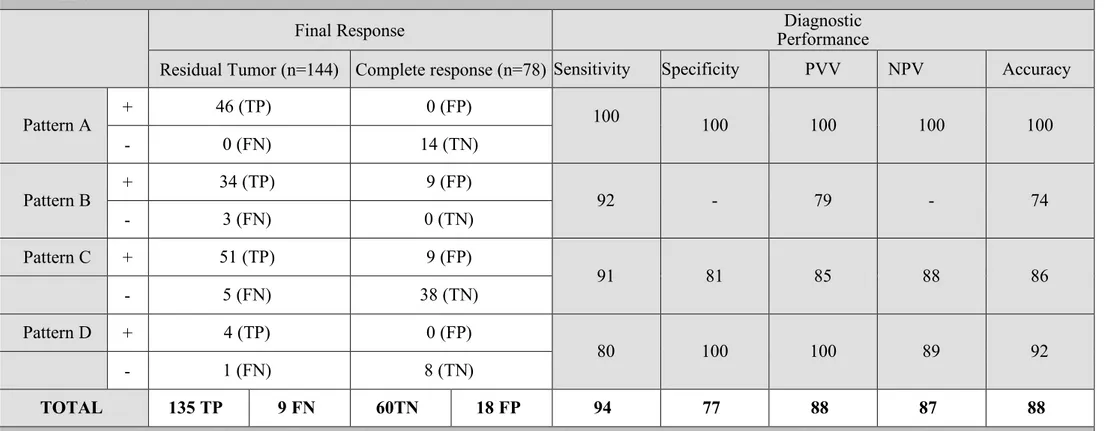
Table 3 shows the diagnostic accuracy figures for the different response patterns.
Sensitivity for the pattern-based approach to discriminate tumor from complete response was 94%, specificity 77%, PPV 88%, NPV 87%, and overall accuracy 88%. Pattern A was 100% sensitive to detect residual tumor (A+) and 100% specific to identify a complete response (A- ). Amongst the remaining patterns, results were better for pattern C (semi-circular fibrosis: accuracy 86%) and pattern D (polypoid: accuracy 92%), compared to pattern B (irregular fibrosis: accuracy 73%).
Distribution of patterns per tumour stage:
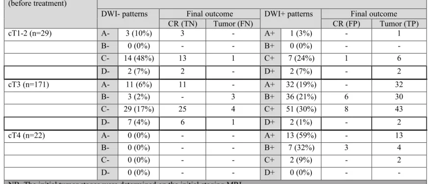
Table 4 shows the distribution of patterns according to the initial (pre-treatment) cT-stage. In patients with initial stage cT4 tumors, no DWI-negative patterns occurred and the majority (59%) showed obvious bulky residual tumor s (pattern A+) after CRT. Only 3 initial cT4 patients underwent a complete response, but all 3 had irregular fibrosis with suspected residual tumour on DWI (pattern B+). The majority of cT1-2 tumors had semicircular (pattern C) tumors, of which most underwent a complete response (pattern C-). A completely normalized rectal wall (pattern A-) occurred in 10% of cT1-2 patients, 6% of cT3 patients and in none of the cT4 patients.
False positive and false negative cases:
Eighteen false positive and nine false negative cases occurred. Apart from 1 FP and 1 FN case in patients with initial stage cT1-2 tumors, these exclusively occurred in patients with initial stage cT3/cT4 tumors. Of the nine FN cases, five were near-complete responders (TRG 2). In 2 patients, the rectal wall was collapsed at the previous tumor site on restaging MRI, making the response pattern more difficult to assess. In the remaining cases main reasons for error were small susceptibility artefacts caused by air in the rectal lumen occurring at the site of the tumor.
Interobserver agreement was good with a kappa of 0.75 (95% confidence interval 0.63-0.87).
DISCUSSION AND CONCLUSION:
Aim was to evaluate the performance of a pattern-based approach to differentiate between a complete tumor response and residual tumor after neoadjuvant treatment for rectal cancer combing the morphology on T2-weighted MRI with distinct signal patterns on diffusion-weighted MRI. In patients with a clear bulky residual tumor mass or a complete
normalization of the rectal wall, sensitivity and specificity respectively were 100%. These ‘clear cut’ patterns will typically not offer much challenge in daily practice, but only occurred in a minority of patients: 21% A+ (mainly in case of initial cT4 stage) and 6% A-. The remaining patients constitute the more clinically challenging cases, in which a pattern-based approach may be of added value. In these patients use of specific morphology-DWI patterns resulted in good diagnostic performance with overall accuracies ranging between 73% and 92%. For the whole patient group, overall accuracy was 88%, which is similar to the accuracies ranging between 82 and 90% previously reported by different groups for visual evaluation of DWI without a specific pattern-based approach.2,3,13 Sensitivity (94%) and PPV (88%) in our study are also within the same range as previously reported (81-98%).2,3,13 However, specificity and NPV in our study were considerably better. Our overall NPV was 87%, compared to 43-81% previously reported. 2,3,13 Specificity in our study was 77%,compared to 52-64% previously reported by Lambregts et al. and 33-50% by Song et al. 13 Only the group of Kim et al. reported a higher specificity of 82-91%.2 Compared to a visual DWI evaluation without any specific criteria or patterns, our
approach may thus particularly be of value to rule out tumor and help identify patients with a complete tumor response. Mainly patterns C (semi-circular or focal fibrosis after CRT) and D (polypoid tumors with fibrosis at the site of the stalk after CRT) proved beneficial as these showed the best concordance the final response outcome. The key principle of
patterns C and D is that in case of a high signal on DWI this occurs at a specific site (originating at the inner margin of the fibrosis in pattern C and in the stalk of the polyp in
pattern D). Knowing where to specifically look for a high DWI-signal apparently helps to more accurately recognize areas of residual tumor and discriminate these from high DWI-signal caused for example by artefacts, that might otherwise be falsely interpreted as suspicious for residual tumor. With this approach the risk of missing residual tumour – which would result in undertreatment and thus a risk for recurrence when selecting these patients for watch-and-wait – is relatively low (5% in pattern C and 8% in pattern D), albeit not non-negligible.
Conversely, in pattern B (circular tumors with irregular/spiculated fibrosis), results were considerably poorer. Only 3 DWI-negative pattern B cases were observed, of which none corresponded with an actual complete response. On the other hand nine false
positives occurred. Apparently when we see irregular/spiculated fibrosis after CRT, the signal pattern on DWI is less reliable. This is probably because in these cases any remaining areas of vital tumor are more scattered (as small tumor nests) throughout the fibrosis, making them more difficult to assess on DWI. In the majority (80%) of the pattern B cases, residual tumor was still present, indicating that it may in fact even be the safest option to consider all these patients to be at risk for residual tumor, albeit at the expense of overestimating tumor in 20%. The main problem of overestimating tumor is that patient will be denied the option of watch- and-wait, despite being potential suitable candidates. In the current study, 17 patients with a complete response underwent surgical resection. This is in part because in the early study period, watch-and-wait was not yet routinely offered as a treatment option. Moreover, even with an optimal clinical response assessment using a combination of MRI (including DWI), digital rectal examination and endoscopy, around 15% of complete responders will typically still be missed.14
With the current study we only focused on response assessment of the primary tumor and did not take the nodal stage into account. We however fully acknowledge that a complete response assessment should also include an evaluation of the nodes. When considering patients for watch-and-wait we need to ensure that – apart from the primary tumor - all suspicious lymph nodes have resolved. In the current cohort, 2 of the 78 complete tumor responders (3%) still had node positive disease, which was recognized on restaging MRI. Both patients were operated on, confirming a pN1 status.
There are some limitations to our study design. First, this was a retrospective study and the patterns described in this study will need to be tested prospectively. Moreover, although we have shown that in the hands of experienced readers results for the pattern- approach are reproducible with a kappa of 0.75, reproducibility of the findings will also need to established for less expert readers. Second, the percentage of complete responders in our study is relatively high (35%). This is because patients were included from a referral centre for organ-preservation, to which patients with a suspected good response are
referred for further response evaluation. Moreover, the availability of organ-preservation as a treatment option at the institution may have induced a certain preference for CRT, particularly in elderly/frail patients. Third, our study did not include a direct comparison of ‘routine’ visual interpretation of DWI and the pattern-based approach. As such, the added benefit of the pattern-approach was estimated by comparing our results to those previous reported in literature. Fourth, 10 patients had to be excluded because of severe artefacts on DWI (mainly caused by metal hip replacements) rendering the DW images uninterpretable. This is a known drawback of DWI that may limit its clinical utility in certain patients. Finally, there were some variations in the DWI acquisition and patient preparation protocols used throughout the study (as a result of protocol optimization over the years). This may have affected our study results, although we believe this effect will probably be limited given the study setting were only images with the same b-value (b1000) were assessed and no quantitative measurements were performed. Use of non-standardised imaging protocols may even be viewed as a positive factor, as it suggests that results may be generalised and will likely be less dependent on protocol variations between
centers/scanners.
In conclusion a pattern-based approach – taking into account the morphology on T2-weighted MRI combined with distinct DWI signal patterns – can be helpful to assess response after neoadjuvant treatment in patients with rectal cancer and aid in identifying patients with a complete tumor response after CRT. These patterns may serve as a basis to teach radiologists to read DWI after CRT and help improve their diagnostic performance in assessing response of rectal tumors on MRI.
REFERENCES
1. van der Paardt MP, Zagers MB, Beets-Tan RG, Stoker J, Bipat S. Patients who undergo preoperative chemoradiotherapy for locally advanced rectal cancer restaged by using diagnostic MR imaging: a systematic review and meta-analysis. Radiology
2013;269(1):101-12.
2.Kim SH, Lee JM, Hong SH, et al. Locally advanced rectal cancer: added value of diffusion-weighted MR imaging in the evaluation of tumor response to neoadjuvant chemo- and radiation therapy. Radiology 2009;253(1):116-25.
3. Lambregts DM, Vandecaveye V, Barbaro B, et al. Diffusion-weighted MRI for selection of complete responders after chemoradiation for locally advanced rectal cancer: a
multicenter study. Ann Surg Oncol 2011;18(8):2224-31.
4. Martens MH, Maas M, Heijnen LA, et al. Long-term outcome of an organ- preservation program after neoadjuvant treatment for rectal cancer. J Natl Cancer Inst 2016;108(12): pii:djw171
5. Habr-gama A, Gama-Rodrigues J, São Julião GP, Proscurshim I, Sabbagh C, Lynn PB, Perez RO.Local recurrence after complete clinical response and watch and wait in rectal cancer after neoadjuvant chemoradiation: impact of salvage therapy on local disease control. Int J Radiat Oncol Biol Phys 2014;88(4):822-24
6. Koh DM, Blackledge M, Padhani AR, et al. Whole-body diffusion-weighted MRI: tips, tricks, and pitfalls. AJR Am J Roentgenol 2012;199(2):252-62.
7. Sánchez- González J. How to identify and avoid artefacts on DWI In: Luna A, Ribes R, Soto JA, editors. Diffusion MRI outside the brain - A case-based review and clinical applications: Springer-Verlag Berlin Heidelberg; 2012. p. 17-31.
8. Taylor FG, Swift RI, Blomqvist L, Brown G. A systematic approach to the interpretation of preoperative staging MRI for rectal cancer. AJR Am J Roentgenol
2008;191(6):1827-35.
9. Lambregts DM, Maas M, Bakers FC, et al. Long-term Follow-up Features on Rectal MRI During a Wait-and-See Approach After a Clinical Complete Response in Patients With Rectal Cancer Treated With Chemoradiotherapy. Dis Colon Rectum
2011;54(12):1521-8.
10. Kim SH, Yoon JH, Lee Y. Added value of morphologic characteristics on diffusion- weighted images for characterizing lymph nodes in primary rectal cancer. Clinical imaging 2015;39(6):1046-51.
11. Nasu K, Kuroki Y, Minami M. Diffusion-weighted imaging findings of mucinous carcinoma arising in the ano-rectal region: comparison of apparent diffusion coefficient with that of tubular adenocarcinoma. Jpn J Radiol 2012;30(2):120-7
12. Mandard AM, Dalibard F, Mandard JC, et al. Pathologic assessment of tumor regression after preoperative chemoradiotherapy of esophageal carcinoma. Clinicopathologic correlations. Cancer 1994;73(11):2680-6
13. Song I, Kim SH, Lee SJ, Choi JY, Kim MJ, Rhim H. Value of diffusion-weighted imaging in the detection of viable tumor after neoadjuvant chemoradiation therapy in patients with locally advanced rectal cancer: comparison with T2-weighted and PET/CT imaging. Br J Radiol 2012;85(1013):577-86.
14. Maas M, Lambregts DM, Nelemans PJ, et al. Assessment of clinical complete response after chemoradiation for rectal cancer with digital rectal examination, endoscopy and MRI: selection for organ-saving treatment. Ann Surg Oncol 2015;22(12):3873-80
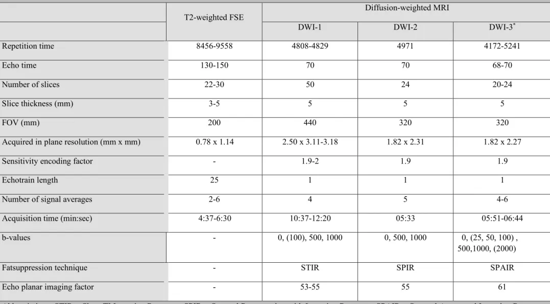
Table 1. Sequence parameters of the T2-weighted and DWI sequences used during the study.
T2-weighted FSE
Diffusion-weighted MRI
DWI-1 DWI-2 DWI-3*
Repetition time 8456-9558 4808-4829 4971 4172-5241
Echo time 130-150 70 70 68-70
Number of slices 22-30 50 24 20-24
Slice thickness (mm) 3-5 5 5 5
FOV (mm) 200 440 320 320
Acquired in plane resolution (mm x mm) 0.78 x 1.14 2.50 x 3.11-3.18 1.82 x 2.31 1.82 x 2.27
Sensitivity encoding factor - 1.9-2 1.9 1.9
Echotrain length 25 1 1 1
Number of signal averages 2-6 4 5 4-6
Acquisition time (min:sec) 4:37-6:30 10:37-12:20 05:33 05:51-06:44
b-values - 0, (100), 500, 1000 0, 500, 1000 0, (25, 50, 100) ,
500,1000, (2000)
Fatsuppression technique - STIR SPIR SPAIR
Echo planar imaging factor - 53-55 55 61
Abbreviations: STIR = Short TI Inversion Recovery, SPIR = Spectral Presaturation with Inversion Recovery, SPAIR = Spectral Attenuated Inversion Recovery. The DWI-1 sequence was applied from September 2008 – December 2011, the DWI-2 from December 2011 – June 2012, the DWI-3 from June 2012 till the end of the study period. * In 9 patients, a slightly adjusted protocol was used with a smaller FOV (180) and a reduced number of b-values (0, 1000).
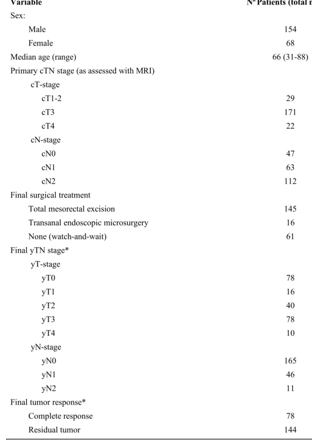
Table 2. Baseline patient and treatment characteristics
Variable No Patients (total n=222)
Sex:
Male 154
Female 68
Median age (range) 66 (31-88)
Primary cTN stage (as assessed with MRI) cT-stage cT1-2 29 cT3 171 cT4 22 cN-stage cN0 47 cN1 63 cN2 112
Final surgical treatment
Total mesorectal excision 145
Transanal endoscopic microsurgery 16
None (watch-and-wait) 61
Final yTN stage* yT-stage yT0 78 yT1 16 yT2 40 yT3 78 yT4 10 yN-stage yN0 165 yN1 46 yN2 11
Final tumor response*
Complete response 78
Residual tumor 144
* in 161 patients response and final yTN stage was assessed based on histopathology of the resection specimen after surgery. In the remaining 61 patients it was based on a sustained clinical complete response (with repeated negative findings without signs of recurrence on MRI, endoscopy and digital rectal examination) with a median recurrence free follow-up period of 36 months (minimum of 1 year).
Table 3. Distribution of response patterns and corresponding diagnostic performance to differentiate between a complete response and residual tumor
Final Response Performance Diagnostic
Residual Tumor (n=144) Complete response (n=78) Sensitivity Specificity PVV NPV Accuracy
Pattern A + 46 (TP) 0 (FP) 100 100 100 100 100 - 0 (FN) 14 (TN) Pattern B + 34 (TP) 9 (FP) 92 - 79 - 74 - 3 (FN) 0 (TN) Pattern C + 51 (TP) 9 (FP) 91 81 85 88 86 - 5 (FN) 38 (TN) Pattern D + 4 (TP) 0 (FP) 80 100 100 89 92 - 1 (FN) 8 (TN) TOTAL 135 TP 9 FN 60TN 18 FP 94 77 88 87 88
Table 4. Distribution of patterns per tumour stage
Initial tumor stage (before treatment)
Pattern of response vs final outcome
DWI- patterns Final outcome DWI+ patterns Final outcome
CR (TN) Tumor (FN) CR (FP) Tumor (TP) cT1-2 (n=29) A- 3 (10%) 3 - A+ 1 (3%) - 1 B- 0 (0%) - - B+ 0 (0%) - - C- 14 (48%) 13 1 C+ 7 (24%) 1 6 D- 2 (7%) 2 - D+ 2 (7%) - 2 cT3 (n=171) A- 11 (6%) 11 - A+ 32 (19%) - 32 B- 3 (2%) - 3 B+ 36 (21%) 6 30 C- 29 (17%) 25 4 C+ 51 (30%) 8 43 D- 7 (4%) 6 1 D+ 2 (1%) - 2 cT4 (n=22) A- 0 (0%) - - A+ 13 (59%) - 13 B- 0 (0%) - - B+ 7 (32%) 3 4 C- 0 (0%) - - C+ 2 (9%) - 2 D- 0 (0%) - - D+ 0 (0%) - -
NB. The initial tumor stages were determined on the initial staging MRI.
CR = complete response, TN = true negative, FN = false negative, FP = false positive, TP = true positive Numbers are absolute numbers, percentages are given in parentheses.
FIGURE LEGENDS Figure 1.
Flowchart of patients considered for inclusion, excluded patients and patients finally included in the study cohort.
* Mucinous tumors were excluded as these are known to exhibit different characteristics on diffusion-weighted MRI.11
Figure 2.
Four morphologic patterns were defined, which were each subdivided into positive and DWI-negative sub-patterns. Pattern A – cases with either a complete normalization of the rectal wall (typically observed in smaller tumors) with no high signal on DWI (A-) or a clear bulky residual tumor mass after CRT (A+). Pattern B – irregular and/or circular tumors that after CRT show irregular and spiculated fibrosis, either without high signal on DWI (B-) or with focal spots of high signal scattered throughout the fibrosis on DWI (B+). Pattern C – semi-circular tumors that show a focal fibrotic wall thickening after CRT, either without high signal on DWI (C-), or with a focal high signal originating specifically at the inner margin of the fibrosis (C+). Pattern D – polypoid tumors that after CRT show fibrotic changes at the site of the stalk of the polyp, either without high signal on DWI (D-), or with a focal high signal at the site of the stalk/fibrosis (D+).
Figure 3.
Examples of a two pattern A cases. In the upper patient, a large bulky tumor mass is visible on pre-treatment T2-weighted MRI (a). After CRT, the tumor has responded poorly with an obvious bulky residual tumor mass on T2W-MRI (arrows in b) with corresponding high signal on b1000 DWI (c). In the lower patient, a relatively small cT1-2 tumor is present at primary staging (arrows in d). After CRT, the rectal wall has completely normalized (arrowheads in e) and no residual mass or fibrotic changes are observed. On DWI no focal high signal is seen (f).
Figure 4.
Examples of two pattern B cases. On pre-treatment T2-weighted MRI (a and d) both patients have irregular circular tumors, in the lower case (d) with spiculation. On the T2-weighted images after CRT (b,e) both tumors show irregular fibrotic changes with spiculation. In the upper patient, no signal is visualized in the rectal wall at the site of the fibrosis (B-), only a slight area of T2 shine-through is visualized caused by fluid in the lumen (arrowhead in c). In the lower patient, several spots of high signal are visible scattered throughout the fibrosis (B+; arrows in f).
Figure 5. Examples of two pattern C cases. On pre-treatment T2-weighted MRI both patients have a
semicircular tumor originating from the anterior rectal wall (arrows in a and d). On the T2-weighted images after CRT both tumors show a similar fibrotic wall thickening (arrowheads in b and e). In the upper patient, no signal is visualized in the rectal wall at the site of the fibrosis (C-). In the lower patient, a clear high signal is visualized at the inner margin of the fibrosis (C+; arrows in f).
Figure 6.
Examples of two pattern D cases. On pre-treatment T2-weighted MRI both patients have a polypoid tumor (arrows in a and d). On the T2-weighted images after CRT both tumors show some slight fibrotic changes in the rectal wall (arrowheads in b and e). at the site of the stalk of the polyp. In the upper patient (c), no signal is visualized at the site of the fibrosis on DWI (D-). In the lower patient, a clear high signal is visualized at the site of the fibrosis, at the site of the stalk of the polyp (D+; arrow in f).