i
CONTENTS
ABSTRACT…………...………...1
1. INTRODUCTION………..………..3
1.1 The concept of “nociception”………...………4
1.1.1 Defining pain, the first step in understanding nociception………4
1.1.2 Theories on pain perception………..6
1.1.3 Measuring Pain………..7
1.2 Sensory transduction in pain……….8
1.2.1 Primary sensory neurons, the first step in nociception………...8
1.2.2 Development of primary sensory neurons……….9
1.2.3 Mechanoreceptors………...12
1.2.4 Proprioceptors……….14
1.2.5 Nociceptors: sensing pain………14
1.2.6 Signal transduction in nociception………...16
1.2.7 TRP Ion Channels and thermosensation………..16
1.2.8 Acid-Sensing Ion Channels……….19
1.2.9 Na+ channels………20
1.2.10 Piezo2………20
1.3 Nociceptive pathways……….21
1.3.1 The first synapse in nociception………...21
1.3.2 Distinct, Parallel Pathways for Pain………22
1.3.3 Ascending pathways and processing of nociceptive information………23
1.3.4 Focus on the main brain areas involved in pain processing……….25
1.3.5 Modulation of pain………..28
1.4 Neuropathic pain and sensitization……….31
1.4.1 Mechanisms for Primary Hyperalgesia………32
1.4.2 Secondary Hyperalgesia and Central Mechanisms of Mechanical Allodynia………..33
1.5 The discovery of NGF and the “Neurotrophic Hypothesis”………34
1.5.1 NGF Expression: the Levels of Neurotrophin Matter………..36
1.5.2 NGF, receptors and downstream pathways………..38
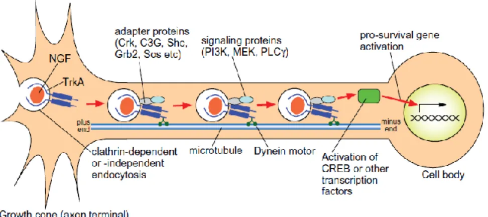
1.5.3 Signaling by retrograde transport of neurotrophin-Trk complexes……….40
1.5.4 Molecular features of retrograde transport of NGF-TrkA………41
1.5.5 Retrograde apoptotic signaling mediated by p75NTR………...……….43
1.5.6 Multiple functions for retrograde neurotrophin signaling: What is their importance?...43
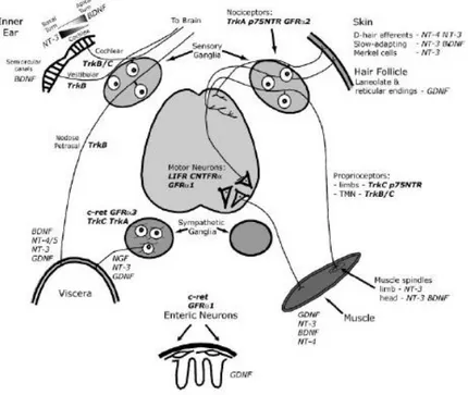
1.5.6 NGF supports the development of peripheral neurons……….44
ii
1.5.8 NGF as a therapeutic target for clinical pain states………...48
1.6 HSAN diseases: the other side of the coin of pain………...50
1.6.1 HSAN V: a complex pathology caused by a point mutation……….52
1.6.2 Processing, secretion and biochemical features of R100W protein………..53
2. AIM of the THESIS………..………..55
3. MATERIALS and METHODS……….………57
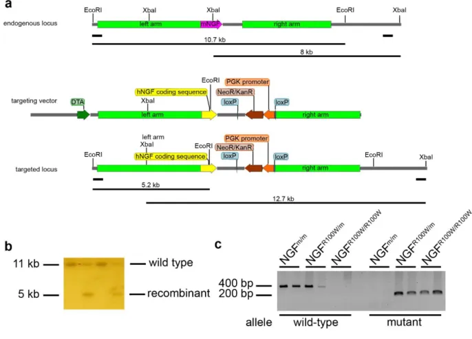
3.1 Generation of knock in human NGFh/m and NGFR100W/m mice……….58
3.2 Southern Blot analysis………58
3.3 Ethics statement on mouse experiments………..59
3.4 Behavioral analyses………59
3.5 NGF treatment………59
3.6 Object recognition test………59
3.7 Morris water maze (MWM)………60
3.8 Y-maze test……….60
3.9 Elevated plus maze……….60
3.10 Marble burying test………...60
3.11 Nest building test………..61
3.12 Hot plate test……….61
3.13 Cold sensitivity test………...61
3.14 Capsaicin injection test……….61
3.15 Tape response assay………..62
3.16 Cotton swab assay……….62
3.17 In vivo nociceptive assay………..62
3.18 Cued fear conditioning……….62
3.19 Contextual fear conditioning………63
3.20 Predator fear……….63
3.21 Immunohistochemistry……….63
3.22 Electrophysiological recordings………...64
3.23 Skin and DRG immunofluorescence………65
3.24 RNA preparation for microarray analysis……….65
3.25 Whole genome expression profiling……….66
3.26 Microarray data analysis………...66
3.27 Electron microscopy……….67
3.28 NGF immunoprecipitation and western blot……….67
3.29 Oxytocin and NGF ELISA………68
3.30 Hek293 cells culture……….68
iii
3.32 Dorsal root ganglia neurons primary cultures………...69
3.33 Behavioral tasks on human subjects………..70
3.34 fMRI acquisition and data analysis………...71
3.35 Data analysis and statistics………72
4. RESULTS………73
4.1 A new mouse model for HSAN V: characterization of homozygous mice………..74
4.1.1 Generation of a new mouse model for HSAN V………..74
4.1.2 Homozygous NGFR100W/R100W mice display an early lethality that is rescued by NGF treatment……..75
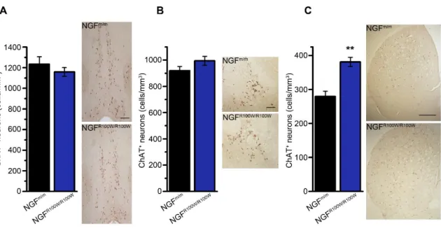
4.1.3 Effects of NGFR100W on cholinergic neurons of homozygous NGFR100W/R100W mice……….76
4.2 Characterization of heterozygous HSAN V mice……...78
4.2.1 Tactile-and pain-related behavioral characterization of heterozygous NGFR100W/m mice……….78
4.2.2 The NGF R100W mutation does not affect peptidergic and non-peptidergic nociceptors in DRGs of NGFR100W/m mice………...………..79
4.2.3 The NGF R100W mutation induces a limited number of specific transcriptomic changes in DRGs of NGFR100W/m mice...………..81
4.2.4 In vivo pain response and in vitro pain transduction pathways mediated by NGFR100W………82
4.2.5 Exploring nociceptive information routes in NGFR100W/wt mice………...84
4.2.6 Absence of cognitive deficits in NGFR100W/m mice………...88
4.2.7 The R100W mutation impairs acquired, but not innate, fear………90
4.2.8 Reduced levels of pain-related neuropeptides in NGFR100W/m mice………..97
4.2.9 Altered responsiveness to painful stimuli in HSAN V human heterozygous carriers………...98
4.3 STATISTICS SUMMARY………..101
5. DISCUSSION………...………107
5.1 NGF as a mediator of pain……….108
5.2 HSAN V and its implications for understanding pain………...109
5.3 Effect of the R100W mutation on DRG neurons………...110
5.4 Altered pain-related memory in the HSAN V model………111
5.5 The role of CGRP in mediating fear responses……….112
5.6 A parallel between human and mouse brain areas activated by pain……….114
5.7 HSAN V vs HSANs: the different role of endogenous analgesic peptides………115
5.8 A role of microglia in the HSAN V phenotype?...117
5.9 Applying NGFR100W to pain therapy……..………...117
APPENDICES………...……….……..120
APPENDIX I - Step-by-step description of the cloning strategy for the generation of targeting vectors carrying human NGFWT and NGFR100W coding sequences………121
iv APPENDIX III - Cloning strategy for the generation of HSAN IV knock-in mouse………..128 REFERENCES………...………..131
1 ABSTRACT
Pain is an important physiological function, whose primary role is to preserve an organism’s integrity. Disruption of the nociception transduction chain results in the inability to perceive pain. Among these “painlessness” pathologies, Hereditary Sensory and Autonomic Neuropathy type V (HSAN V) is caused
by the 661C>T transition in the ngf gene, resulting in the R100W missense mutation in mature Nerve Growth Factor (NGF), in keeping with the key role of this neurotrophin in the development of nociceptors and in their function in the adult . Homozygous HSAN V patients display indifference to noxious stimuli but, no cognitive deficits. In contrast, heterozygous carriers do not show an overt clinical phenotype and have been identified only through pedigree and genetic screening. Considering the particular features of HSAN V patients, I hypothesized that the R100W mutation might cause a dissociation between the actions of NGF on the central and peripheral nervous systems. To test this hypothesis and understand the mechanisms underlying the HSAN V phenotype, I generated a transgenic mouse line harboring the human 661C>T mutation in the human ngf gene. Homozygous NGFR100W/R100W mice were born normal, but failed
to reach the first month of age. This early lethality could be due to reduced NGF bioavailability and, indeed, was rescued by continuous treatment, during development and the early postnatal life, with wild type NGF. In contrast, heterozygous NGFR100W/m mice grew normally but displayed impaired nociception, despite
Dorsal Root Ganglia (DRGs) neurons being morphologically normal. On the other hand, skin innervation was reduced. The NGFR100W protein showed reduced capability to activate pain-specific signalling,
paralleling its reduced ability to induce mechanical allodynia. Surprisingly, NGFR100W/m mice, unlike
heterozygous mNGF+/- mice, showed no learning nor memory deficits, despite a reduction in secretion and
brain levels of NGF. These results prove the hypothesized dissociation between the peripheral and central actions of NGF, prompting me to investigate if the R100W mutation might affect brain elaboration of pain. To address this issue, I used the fear conditioning test and found that NGFR100W/m mice, despite normal
nociceptive responses to a painful conditioning stimulus, showed a deficit in learned fear. Strikingly, their innate fear responses were normal. This was accompanied by a reduced activation of brain regions involved in pain processing and in the generation of task-related motor responses. I also found a decreased density of CGRP-positive fibers in the amygdala, which can provide a mechanistic explanation of the reduced fear response. On the other hand, the expression of endogenous analgesic peptides, namely β-endorphin and
2 oxytocin, was decreased in NGFR100W/m mice, suggesting a different set point of the homeostatic
pain/analgesia system, as a consequence of a prolonged reduction of afferent pain signals. Consistent with these findings in mice, data collected in humans showed that heterozygous R100W carriers, despite having a normal pain threshold, had a decreased urgency to react to a painful stimulus, along with impaired ability to integrate sensory information with behavioral task requirements. Functional magnetic resonance imaging (fMRI) revealed, in accordance with mouse data, an altered processing of painful stimuli in brain areas involved in pain processing. These findings demonstrate an uncoupling of nociceptive signals from their central elaboration, leading to altered interpretation and meaning attributed to painful stimuli in human HSAN V carriers and heterozygous NGFR100W/m mice.
In addition to clarify the role of NGF in transduction of nociceptive inputs, these data also demonstrate that NGF is at the center of a regulation system linking peripheral nociception to the brain processes responsible for constructing painful perceptions and pain-related memories. Moreover, the peculiar effects of NGFR100W
3
4
1.1 The concept of “nociception”
1.1.1 Defining pain, the first step in understanding nociception
What is pain?
Researchers working on pain admit their difficulty when trying to define pain or refuse to come up with a simple definition.
In his physiology book published in 1900, Charles Sherrington defined pain as “the psychical adjunct of a
protective reflex”. His definition proposed an important separation between the protective reflex component and the sensory perception of pain (Sherrington, 1906). We generally define pain as the protective reflex that involves also a psychical adjunct, but the two processes are separate. Protective reflexes are present in all animals and were defined by Sherrington as the neural processes serving nociception (Sherrington, 1906). This last term refers to the process by which the nervous system detects and transmits injury-related information; it does not imply conscious perception of pain. On the other hand, the “psychical adjunct” term refers to a mental process of pain perception that depends on our social, religious and scientific beliefs. But what is the nature of this psychical adjunct?
In 1909, James MacKenzie defined pain as “a disagreeable sensation which everyone has experienced and
which we all recognize”. Later, Thomas Lewis wrote: “pain is known to us by experience and described by illustration”. Both Lewis and MacKenzie thought that the only way to assess pain is through communication with other human beings.
However, Sherrington’s definition is applicable only to “good pain”, i.e., the pain that protects us from
injury. Pain can be considered an alarm system, as Descartes first described it in the Traité de l’Homme, published in 1664, in which pain is represented as a line that links the stimulus – fire – with the brain (Fig.1.1).
5
Figure 1.1 Pain as an alarm system. The picture is from Descartes’ Traité de l’Homme, (published in 1664, adapted from
Cervero, Understanding Pain, MIT Press, 2012).
Descartes’s vision of pain has been very influential on modern pain research. He proposed that fire excites
the terminations of sensory fibers in the foot and that this information is transmitted to the brain, where an alarm is triggered in the form of an unpleasant sensation of pain. Thus, a “sensor” (or better, a “nociceptor”)
pulls the cord and the pain bell rings in the brain.
In addition to good pain that is helpful and protective, an “evil pain” exists, that is, pain associated with
disease. An example is the “phantom limb syndrome”, characterized by feeling pain sensations in a limb that no longer exists as a result of amputation. Similarly, chronic pain, which has no biological value as it persists after the nociceptive stimulus and beyond normal tissue healing time.
Is pain different from the five senses? Aristotle, in his De Anima, regarded as senses only the perceptions that could be associated with a specific sense organ. According to him, pain is not linked to a particular sense organ that gives us information regarding the external world, but is associated with an extreme stimulation of any other sense. Aristotle did not focus on the sensory and protective aspects of pain, concentrating instead on the emotional and behavioral components of pain and pleasure; indeed, he wrote “we measure our actions by the rule of pleasure and pain”. He considered pleasure and pain as engines of all our action and he called them “the passions of the soul”. The Aristotelian view of pain, as a behavioral
drive triggered by extreme stimulation of any sense organ, is very different from Descartes’s idea of an alarm signal (Cervero, 2012). Actually, current knowledge of pain mechanisms is a synthesis of these two theories: pain is indeed a powerful alarm signal that protects us, but pain is also a passion of our soul, an emotion that drives our behavior, and in some cases a destructive curse. Pain is a conscious experience, an
6 interpretation of the nociceptive input influenced by memories, emotional, pathological, cognitive and genetic factors. Pain is not related linearly to the nociceptive input, neither it is simply connected to vital protective functions. This is particularly true in the chronic pain state.
Pain is, therefore, a highly subjective experience, as illustrated by the accepted definition given from the International Association for the Study of Pain (Treede, 2018) “an unpleasant sensory and emotional experience associated with actual or potential tissue damage, or described in terms of such damage”. This definition covers all the various aspects of pain.
1.1.2 Theories on pain perception
The dawn of the 17th century saw the postulation of four major theories to describe the perception of pain:
specificity, intensity, pattern and gate control theories.
Intensity theory defines pain as a sensory experience occurring when a stimulus is stronger than usual (Moayedi and Davis, 2013). This theory has been ruled out by evidence for specialization of primary sensory neurons. In addition, the pattern theory, proposed by Weddell and Sinclair, posited that the response to stimuli is produced by impulses travelling in various spatio-temporal patterns, with no dedicated transmission pathways. The pain, according to the pattern theory, is produced by an intense stimulation of non-specific fibers. This theory ignored the specialized properties of fibers, such as the threshold to different stimulus intensities; for this reason it was criticized by scientists.
On the contrary, the specificity theory postulated that dedicated pathways carry specific stimuli. For example, noxious stimuli are carried to the brain by pain-sensing afferents (later named nociceptors), while non noxious stimuli are encoded by mechanoreceptors. A representation of these connections had already been elaborated by René Descartes. This theory was further supported by the identification of cutaneous organs, such as the Meissner and Pacinian corpuscles, or Merkel cells.
Finally, the Gate Control theory of pain (GCT), proposed by Wall and Melzach in 1965, is based on the presence of gates in dorsal horn of the spinal cord, which are composed of inhibitory neurons that receive afferents from A and C fibers. According to this theory, noxious stimuli, carried by pain transducing fibers, open the gate, inhibiting the interneurons and leading to pain perception, whereas the A fibers excite the interneurons, thus suppressing the output of the projection neurons (Melzack and Wall, 1965). Interestingly,
7 the existence of a gate between the brain and nerves, open by ‹A sensory cue driven by nerves, “tubular structures”›, had also been postulated by Descartes in 1664 (Perl, 2007).
1.1.3 Measuring Pain
Many physicians and insurance companies would like to have an instrument, a dolorimeter, to accurately measure if a person is in pain, and how much of it he or she feels. Indeed, we have thermometers and sphygmomanometers to measure signs of disease, but no devices to quantify the level of pain.
One of the tools that partially accomplish this goal is the visual analogue scale (VAS), that is a continuous scale comprised of a horizontal (HVAS) or vertical (VVAS) line, usually 10 centimeters long. The clinician simply asks the patient to rate the pain, with 0 meaning no pain and 10 meaning the worst pain imaginable. Another tool, developed by Ronald Melzack, is the McGill Pain Questionnaire (Melzack, 1975), that is based on the selection by the patient of the adjectives that best describe his or her pain. For instance, inflammatory pain is often described as pulsating and neuropathic pain as burning. Thus, both the VAS and the McGill Pain Questionnaire are based on patients’ verbal reports.
In the 20th century, thanks to introduction of bioelectrical signal recordings, it was possible to show that an
electrical stimulus in the nerve evoked a series of electrical waves. Using a cathode-ray oscilloscope, Hebert Gasser and Joseph Erlanger recorded electrical changes of nerve potential. This technique allowed them to describe the compound action potential of peripheral nerves, i.e., an electrical signal that was generated by the summation of the action potentials of the single fibers composing the nerve (Raju, 1999). In accordance with the latency of appearance of the compound potential with respect to the stimulus, the fibers were named A, for the fastest, and C, for the slowest type.
A further development of this approach, is the recording of electrical signals generated by the brain when the subject experiences a specific pain condition. This can be achieved by using the electroencephalogram to sample the electrical activity of different brain sites (with good spatial specificity), and can be coupled to stimulation of the body periphery (e.g., the skin of a limb) to measure the corresponding “evoked potential” (Chen and Rappelsberger, 1994; Ploner et al., 2017). It should be noted, however, that these
electrophysiological recordings often do not allow to discriminate between a deficit caused by abnormal pain perception or by other underlying neurological diseases.
8 Brain imaging, namely functional Magnetic Resonance Imaging (fMRI), allows to analyze brain activation during pain or emotional states associated with pain condition (Valet et al., 2004). The ultimate purpose of these studies is to assess objectively, by looking a brain map, if a person is in pain, and how strong his or her pain sensation is (Morton et al., 2016).
Studying pain is difficult due to the wide variety of factors (context, cognitive condition, mood etc.) that influence this perception. However, substantial help can come from the development of experimental paradigms based on measuring spontaneous and evoked pain behaviors in animal models that clearly reproduce specific acute and chronic human pain conditions (Schwei et al., 1999; Blackburn-Munro, 2004). These models can also take into account the genetic background, gender and age of the animal, thus leading to results that can be translated to the human context (Mogil and Belknap, 1997). These studies allow, for instance, to understand how nociceptors react to various forms of stimulation and how the brain responds to a pain- producing process and to identify the molecular mechanisms involved in the pain reaction.
1.2 Sensory transduction in pain
1.2.1 Primary sensory neurons, the first step in nociception
The cell bodies of primary afferent nociceptive fibers are located within the Dorsal Root Ganglia (DRGs). These cells are pseudo-unipolar neurons containing one process branching into different target regions (e.g., a specific skin receptive field), and an axon terminating in specific laminae of the spinal cord. Sensory neurons are, generally, electrically silent and transmit action potentials only upon stimulation. Indeed, their activity does not per se lead to pain perception. This requires peripheral information to reach higher center and normally depends on the frequency of action potentials in primary afferents, on temporal summation of pre- and postsynaptic signals, and is modulated by central influences.
Sensory neurons are classified into Aβ, Aδ and C-fibers, according to the propagation speed of impulses.
The speed of transmission depends from the diameter of axons and on their myelination (Zotterman, 1939). Aβ fibers are thickly myelinated and have a fast conduction velocity (16-100 m/s). Aδ-fibers (diameter
1-5 µm) are also myelinated and have a conduction velocity of 1-5-30 m/s. C-fibers have a small diameter (0.2-1.5 µm) and unmyelinated axons, bundled in fascicles surrounded by Schwann cells. They have a conduction velocity of 0.4-1.5 m/s. In addition, electrophysiological studies based on mechanical
9 stimulation of DRG neurons, have shown the existence of low-threshold and high-threshold sensory neurons (Iggo, 1985). Almost all Aβ-fibers and subsets of Aδ and C-fibers show low mechanical threshold to stimulation with von-Frey filaments, suggesting a role in detecting innocuous mechanical stimuli, whereas most Aδ and C-fibers have high mechanical threshold and respond to noxious mechanical or
thermal stimuli (Zotterman, 1939; Koerber and Woodbury, 2002; Zimmermann et al., 2009).
Generally, A-fibers mediate the initial pain sensation, that is described as lancinating; the following, longer lasting pain is caused by the activation of C fibers (Fig.1.2). In correlation with the corresponding conduction velocities, these pain sensations are often referred to as “fast” and “slow”.
Figure 1.2 First and second pain (adapted from (Bear, Neuroscience, IV edition, Wolters Kluwer, 2015).
1.2.2 Development of primary sensory neurons
Sensory neurons originate from trunk neural crest cells (NCCs) and their neurogenesis occurs in waves. The first wave is characterized by migration of neural crest cells that form large-diameter touch and movement neurons The second wave occurs after DRGs coalesce, producing small nociceptive, as well as large touch and movement neurons. A third wave of neurogenesis generates primarily nociceptive (expressing the TrkAreceptor) neurons from neural crest-derived boundary cap cells (Maro et al., 2004). The first two waves of NCC differentiation are regulated by Neurogenin 1 (Ngn1) (Ma et al., 1999) and Neurogenin 2 (Ngn2) (Fode et al., 1998). Indeed, analyses of individual Ngn1 and Ngn2 mouse mutant embryos show that Ngn2 is expressed in early migratory NCCs, whereas Ngn1 by coalesced DRGs (Ma et
10 al., 1999; Marmigere and Ernfors, 2007). Ngn1 seems to play an important role in the formation of small-diameter nociceptive (TrkA+) neurons and a minor involvement in the formation of large-diameter
mechanoceptive (TrkB+) and proprioceptive (TrkC+) neurons. In contrast, Ngn2 plays a transient role in the
formation of large-diameter mechanoceptive (TrkB+) and proprioceptive (TrkC+) neurons. Furthermore,
Ngn2+ cells contribute to a small fraction of nociceptive (TrkA+) neurons (Zirlinger et al., 2002). During
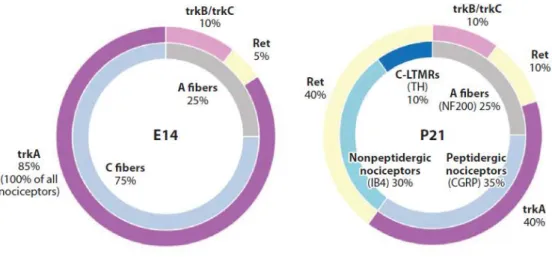
development, many factors influence the early stage of DRG differentiation. One of these is the POU-domain transcription factor Brn3a, that is expressed in differentiating sensory neurons and is important for the correct development and/or survival of proprioceptive, nociceptive and mechanoceptive neurons (McEvilly et al., 1996). Brn3a binds to regulatory elements of the ntrk1 gene, which encodes for the Nerve Growth Factor (NGF) receptor TrkA (Ma et al., 2003) and is expressed in all nociceptors during development. The expression of TrkA requires another transcription factor, Klf7 (Lei et al., 2005). After neurogenesis has completed, most DRG neurons maintain the expression of TrkA, become responsive to NGF (White et al., 1996), and some of them start to express Calcitonin Gene-Related Peptide (CGRP). A distinct, albeit small, population of TrkA- neurons expresses the Ret receptor as early as E14; these TrkA-/
Ret+ cells are classified as the “early Ret” population (Molliver et al., 1997). The majority of Ret+ neurons,
however, emerge from TrkA+ neurons during the late embryonic period. This population of neurons begins
to express Ret around E16 and gradually extinguishes TrkA expression after birth, and is classified as the “nonpeptidergic” population (Molliver and Snider, 1997). Thus, although TrkA is expressed in most DRG
neurons during development, only a subset of them, CGRP+ peptidergic neurons, continue to express TrkA
into adulthood. The segregation of peptidergic and nonpeptidergic populations is complete between 2 and 3 weeks after birth.
Another factor that plays an essential role in the differentiation of nonpeptidergic neurons is Runx1 (Chen et al., 2006; Kramer et al., 2006; Yoshikawa et al., 2007). NGF regulates expression of Runx1 that, in turn, supports the expression of Ret and of a large cohort of genes characteristic of nonpeptidergic neurons (Luo et al., 2007). In this population of neurons, the transcription factor MafA plays an important role in the differentiation of low-threshold mechanoreceptors (LTMR). At birth, the MafA/Ret population can be divided into three subsets: Ret+, Ret+/TrkB+ and Ret+/TrkC+. The specification of subsets of TrkB-expressing mechanoreceptor neurons requires Shox2 (Scott et al., 2011), that drives the development of
11 early Ret+/TrkB+ population into two different subtypes: (i) neurons expressing TrkB+ only become D-hair
mechanoreceptor; (ii) the Ret+ and TrkB+ subset of neurons might represent a class of Aβ-LTMRs, such as
Meissner’s corpuscles afferents (Abdo et al., 2011).
Finally, the maintenance of TrkC+ proprioceptive neurons depends on expression of Runx3 around
embryonic day E14.5 (Levanon et al., 2002). The Ret+/TrkC+ subset of neurons is required for the
development of Pacinian corpuscles and circumferential endings (Bai et al., 2015) (Fig.1.3).
Figure 1.3 DRG neuron subtypes and receptor distribution in embryonic (left) and early postnatal (right) development
(adapted from Denk et al., 2017).
Each subset of sensory neurons displays different combinations of receptors and ion channels. Usoskin and colleagues (Usoskin et al., 2015) (Fig.1.4), using single-cell sequencing technique, characterized the marker profile of each subset of sensory neurons. This study supports the idea that specific sensation modalities can be either generated through the selective activation of a specific neuronal type, or encoded by the integration of the activities of different neuronal types. This latter hypothesis suggests the intriguing idea that a combinatorial code might be present, in close analogy to other sensory modalities, such as color vision or olfaction.
12
Figure 1.4 Classification of sensory neuron subtypes (adapted from Usoskin et al., 2015).
1.2.3 Mechanoreceptors
Mechanoreceptors represent a class of neurons that detect innocuous mechanical forces acting on tissues and do not elicit the sensation of pain. Mechanoception is the basis of physiological processes such as the senses of touch, balance and hearing. Mechanoreceptors can be classified according the threshold as low-threshold mechanoreceptors (LTMRs) and high-low-threshold mechanoreceptors (HTMRs) and are distributed in the skin, tendons, muscles, joints and viscera.
LTMRs include Aδ D-hair fibers that innervate hairs in the skin and detect hair movement, and Aβ-fibers
that terminate on Merkel cells, Pacinian corpuscles and hair follicles and detect texture, vibration and light pressure. Finally, LTMRs also include C-fibers that terminate as free nerve endings and are associated with “tickling” sensations. LTMRs are activated by innocuous mechanical stimuli and they can be distinguished
in rapidly adapting (RA; 3-6ms) and slowly adapting (SA; 200-300 ms) based on their rates of adaptation to sustained mechanical stimuli (Mountcastle, 1957). Meissner corpuscles, Pacinian corpuscles and longitudinal lanceolate endings are RA mechanoreceptors (Iggo and Ogawa, 1977), whereas, Merkel discs are the principal SA mechanoreceptors in rodents and monkeys (Pare et al., 2002).
The mammalian skin comprises hairy and glabrous parts. Glabrous skin is found on the hands and feet; it is specialized for discriminative touch, thus allowing object recognition, along with feedback to the central nervous system to mediate proper grip control, reaching and locomotion. The glabrous skin contains four types of LTMRs with fast conduction velocity (Aβ LTMRs), with distinct terminal morphologies and tuning properties
13 I. Aβ SA1-LTMRs innervate Merkel cells in the basal epidermis and report the static nature of touch
stimuli;
II. Aβ SA2-LTMRs terminate in Ruffini corpuscles in the dermis and are sensitive to skin stretch; III. Aβ RA1-LTMRs innervate Meissner corpuscles in dermal papillae and are sensitive to movement
across the skin;
IV. Aβ RA2-LTMRs terminate in Pacinian corpuscles deep in dermis and are tuned to high-frequency vibration. (Fig1.5).
Figure 1.5 LTMR innervation of glabrous skin (adapted from Zimmerman et al., 2014).
Hairy skin covers more than 90% of the body surface and has a discriminative touch role, albeit with lower spatial acuity respect to glabrous skin. Hairy skin is composed of three main hair follicle subtypes: guard, zig-zag and awl/auchene which are innervated by different combination of LTMRs. Zig-Zag hair follicles are the most numerous and are associated with C and Aδ-LTMR lanceolate endings. The Awl/auchene type is innervated by C-, Aδ- and Aβ-LTMR longitudinal lanceolate endings, representing roughly 23% of total follicles. Guard hairs represent around 1% of the follicles and receive Aβ SA1-LTMR longitudinal lanceolate endings that innervate structures named “touch domes” (Li et al., 2011).
Different combinations of LTMRs, in association with hair follicles, confer a high specialization and complexity to hairy skin. In general, mechanotransduction influences many important processes, such as
14 embryonic development and sensory perception, and deregulation of mechanosensitivity, for example an aberrant sensitivity to mechanical forces, leads to peripheral neuropathies.
1.2.4 Proprioceptors
While cutaneous mechanoreceptors provide information about external stimuli, another important class of receptors is formed by proprioceptors (“receptors for self”), providing information about mechanical forces
arising within the body itself, in particular from the musculoskeletal system. Proprioceptors have large diameter (13-20 µm), myelinated axons and support a conduction velocity of 80-120 m/s; their main role is to give detailed and continuous information about the position of the limbs and other body parts in space. Similar to other sensory neurons, the cell bodies of proprioceptors are located in DRGs, and their projections reach lamina V of the dorsal horn of the spinal cord. In particular, group Ia and II afferent axons innervate muscle spindles, whereas group Ib afferents innervate Golgi tendon organs. Group Ia afferent axons centrally ramify in the intermediate zone of the spinal cord and terminate in the motor nucleus of the ventral horn, whereas group Ib and II afferent axons project to the intermediate zone.
Strong evidence suggests that, in DRG neurons, the transcription factor Runx3 is necessary for the specification of TrkC+ proprioceptive neurons (Inoue et al., 2002; Levanon et al., 2002; Chen et al., 2006).
Indeed, in Runx3-deficient mice, TrkC+ neurons of DRGs do not survive enough to extend their axons to
reach target cells, resulting in lack of connectivity and ataxia (Inoue et al., 2002; Levanon et al., 2002). Moreover, a mutation in the Egr3 (early growth response 3) gene impairs group Ia/II muscle spindle activation, eliminating one class of proprioceptive feedback. Analysis of walking and swimming behaviors in this mouse model demonstrated that altered proprioceptive sensory feedback causes degradation of the correct locomotor pattern (Akay et al., 2014).
1.2.5 Nociceptors: sensing pain
The word nociceptor comes from the Latin nocere “to hurt”. Nociceptors are thus defined as sensors that respond to types and intensities of energy that can produce possible damage and injury to the body. Activation of nociceptors can lead to a conscious experience of pain: nociception and pain are vital for our life, but they do not always come together. Pain is the feeling, or the perception of sensations arising from a part of the body; whereas, nociception is the sensory process providing signals that trigger pain.
15 Nociceptors are present in many organs of the periphery, including skin, bone, muscle, most internal organs, blood vessels and heart. They are absent from the brain, except for the meninges. Unlike the specialized somatosensory receptors for light touch and pressure, most of these nociceptors are simply the free nerve ending that Von Frey first described as the pain sensors (Norrsell et al., 1999). Anatomically, nociceptive afferents form extensive free nerve endings in the epidermis of the hairy and the glabrous skin (Zylka et al., 2005) and terminate within lamina I and outer lamina II(II0) of the spinal cord. Two classes of
nociceptors exist: Aδ and C nociceptors. The first class is represented by medium-diameter myelinated (Aδ)
afferents that mediate acute pain. The second class includes small-diameter unmyelinated C-fibers that convey slow pain. Most of the C-fiber nociceptors (>70%) respond to all forms of stimuli – thermal, mechanical and chemical – and are therefore called polymodal (Woolf and Ma, 2007; Basbaum et al., 2009). C-fiber nociceptors are heterogeneous, with subsets that respond preferentially to heat or chemical, rather than mechanical stimulation. For example, nociceptors that express the heat-gated ion channel TRPV1 are required for noxious thermoception and chemoception of capsaicin (Caterina et al., 1997). On the other hand, noxious cold is detected by nociceptors that express the ion channel TRPM8 (Knowlton et al., 2013). Finally, nociceptors that express the Mas-related G protein-coupled receptor D, Mrgprd, (they constitute > 90% of all nonpeptidergic IB4+ fibers) innervate the epidermis and respond to “blunt” mechanical stimuli,
such as a von Frey hair (Cavanaugh et al., 2009). In this paper, Cavanaugh et al. explored the behavioral consequences of selectively ablating two non-overlapping populations of nociceptors: one required for the response to mechanical, but not to thermal stimuli (Mrgprd+ cells), the other, conversely, required for
thermal, but not for mechanical sensitivity (TRPV1+ cells) (Fig.1.6). They also showed that combined
elimination of these two populations yields an additive phenotype with no further behavioral deficits.
Figure 1.6 Schematic illustrating modality-specific contributions of Mrgprd+ and TRPV1+ fibers to behavior and their different termination zones in the epidermis and spinal cord (adapted from Cavanaugh et al., 2009).
16 Nociceptors can also be classified in two groups, according to the expression of specific markers (Luo et al., 2007):
nonpeptidergic; this population comprises approximately 60% of total DRG neurons, expresses the tyrosine kinase receptor Ret, the signaling receptor for the Glial-Derived Neurotrophic Factor (GDNF) family of ligands (GFLs) (Dong et al., 2001); these neurons bind the IB4 lectin from Griffonia simplicifolia;
peptidergic; this population expresses TrkA and respond to the calcitonin gene-related peptide (CGRP).
These two populations show different patterns of peripheral and central axonal projections. For instance, the peripheral axons of peptidergic neurons innervate the stratum spinosum (SS) of glabrous skin, while the central axons terminate in laminae I and II0 of the dorsal horn of the spinal cord. Peripheral projection
of Ret+ neurons innervate the superficial stratum granulosum (SG) of the epidermis, while the central axons
terminate in inner lamina II (IIi) of the spinal cord.
1.2.6 Signal transduction in nociception
All sensory systems have the role of converting environmental stimuli into electro-chemical signals. In the case of vision or olfaction, primary sensory neurons detect one type of stimulus (light or chemical odorants). Nociception, to this regard, is unique because individual primary sensory neurons have the ability to detect a wide range of stimulus modalities. A variety of noxious stimuli depolarize the bare nerve endings of afferent axons and generate action potentials that are propagated centrally. How is this achieved? The membrane of the nociceptor contains different types of receptors that transduce thermal, mechanical, or chemical stimuli into a change of membrane potential.
1.2.7 TRP Ion Channels and thermosensation
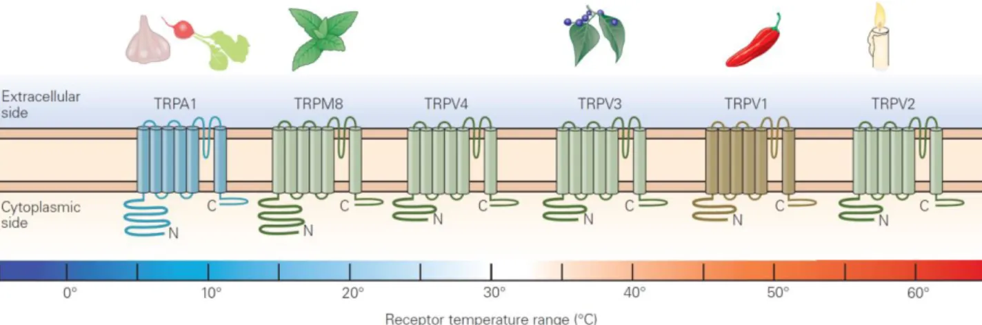
Transient receptor potential (TRP) channels make up the most abundant family of ion channels expressed in sensory neurons of mammals (Damann et al., 2008) (Fig.1.7).
17
Figure 1.7 Classification of different TRP channels expressed by dorsal root ganglion neurons, their temperature response
profiles, and some of their agonists (from Kandel, Principles of Neural Science, V edition, McG-H, 2013).
The prototype of this receptor-channel family, TRPV1, also known as VR1, was isolated in 1997 (Caterina et al., 1997). The TRPV1 transcript and protein were found to be maximally expressed in sensory neurons (Caterina et al., 1997) with predominant expression in small-diameter cell bodies, most of which give rise to unmyelinated C-fibers. TRPV1 immunoreactivity was also detected peripherally, in the sciatic nerve, and on the central terminals of afferents fibers projecting to the superficial layers of the spinal cord and the trigeminal nucleus caudalis.
This receptor is activated by elevated temperature and mediates the pain-producing actions of capsaicin, the active ingredient of hot peppers (Caterina and Julius, 2001). Interestingly, exposure of nociceptors to capsaicin not only causes their excitation, but also induces release of inflammatory mediators, such as substance P (SP) and CGRP. Another important aspect of the actions of capsaicin is that prolonged exposure of nociceptors to this chemical renders them insensitive to it, a process known as desensitization. VR1 is a non-selective plasma-membrane cation channel possessing a very steep temperature dependence and a thermal activation threshold of ~ 43°C. Thus, a correlation between heat and capsaicin sensitivity (i.e., chemoception) exists, arising from the existence of a common transducer. Additional studies demonstrated that TRPV1 could be activated at room temperature when proton concentration was increased (pH<6), indicating that protons directly gate TRPV1 (Tominaga et al., 2003). Although capsaicin is an exogenous ligand for TRPV1, it is possible that there endogenous, pain-producing, chemical regulators for this channel are also present. Indeed, different endogenous ligands have been identified, including anandamide,
18 lipoxygenase products and N-arachidonoyl dopamine. These lipid compounds have been proposed to modulate TRPV1 activity in vivo (Rosenbaum and Simon, 2007). Another endogenous ligand capable of activating TRPV1 is oxytocin. In a recent paper, it has been reported that oxytocin-induced suppression of nociceptor might be achieved directly through potentiation of the pain receptor TRPV1, causing analgesia upon desensitization of the channel (Nersesyan et al., 2017). Among inflammatory mediators, bradykinin and NGF, via bradykinin receptor and TrkA respectively, cause sensitization of TRPV1 and consequent hypersensitivity (Chuang et al., 2001). In particular, NGF, acting on TrkA, activates a signaling in which PI3 kinase plays an important early role, with Src kinase as the downstream element that binds to and phosphorylates TRPV1. These events induce insertion of further TRPV1 channel units into the surface membrane and explain the rapid sensitizing actions of NGF (Zhang et al., 2005). TRPV1 appears to be the principal molecule by which inflammatory mediators enhance sensitivity to heat, as demonstrated by absence of heat hyperalgesia in TRPV1 knockout mice (Caterina et al., 2000; Davis et al., 2000).
A protein with 49% identity to TRPV1 was isolated and called vanilloid-receptor-like protein 1 (VRL-1), and later renamed TRPV2 (Caterina and Julius, 2001). The TRPV2 channel is expressed predominantly in Aδ fiber terminals and is activated by very high temperature, with a threshold of ~ 52°C. Thus, temperatures
activating TRPV2 are more harmful to the body than those activating TRPV1. Therefore, expression of TRPV2 in myelinated sensory fibers seems reasonable, because Aδ fibers transmit nociceptive information
much faster than C-fibers, which express TRPV1.
TRPV3 and TRPV4 have been found to be activated by warm temperatures, ~ 34-38°C for TRPV3 and ~ 27-35°C for TRPV4, and to be expressed in multiple tissues, among others, sensory and hypothalamic neurons and keratinocytes (Guler et al., 2002; Peier et al., 2002; Smith et al., 2002; Xu et al., 2002). TRPV3 is also activated by the plant-derived compound camphor. TRPV3 knockout mice show impairment in thermotaxis behavior (Moqrich et al., 2005). The authors also observed behavioral deficits to acute thermal stimulation at temperatures > 50°C in TRPV3-/- mice, which indicates that TRPV3 and TRPV1 have
overlapping functions in noxious thermoception.
In addition, defects in the expression of TRPV4 affect the detection of noxious mechanical stimuli and cause mechanical hypersensitivity to pain (Suzuki et al., 2003; Alessandri-Haber et al., 2004).
19 The cooling sensation of menthol, a chemical found in mint, is transduced by a nonselective cation channel, called TRPM8 (McKemy et al., 2002; Peier et al., 2002). TRPM8 is also activated by a temperature of ~25-28°C and by other cooling compounds, such as menthone, eucalyptol and icilin (Bandell et al., 2004). TRPM8 is expressed in a subset of DRG neurons that can be classified as small-diameter C-fiber (McKemy et al., 2002; Peier et al., 2002) and is not coexpressed with TRPV1.
TRPA1, is considered to be distantly related to TRP’s, and is activated by cold with a lower threshold than
TRPM8, namely 17°C; for this reason, it is involved in cold nociception. Unlike TRPM8, TRPA1 is specifically expressed in a subset of sensory neurons that express the markers CGRP and substance P (Story et al., 2003). Furthermore, TRPA1 is also coexpressed with TRPV1, raising the possibility that TRPA1 and TRPV1 mediate the function of a specific class of polymodal nociceptors. This coexpression might also explain the paradoxical hot sensation experience when one is exposed to a very cold stimulus.
1.2.8 Acid-Sensing Ion Channels
Acid-Sensing Ion Channels (ASICs) form a large family of proton-gated ion channels consisting of 7 isoforms, most of which are expressed in mechanosensory and nociceptive neurons (Lingueglia, 2007; Moshourab et al., 2013). The expression of ASIC proteins in sensory neurons is controlled also by neurotrophin signaling. Pro inflammatory mediators, including NGF, are involved in upregulating ASICs mRNA and NGF increases the density of -mediated currents in cultured sensory neurons (Mamet et al., 2002). The ASIC3 channel is specifically expressed in nociceptors and is well represented in fibers that innervate skeletal and cardiac muscle. ASIC3 channels are thought to be responsible for muscle or cardiac pain that results from changes in pH associated with ischemia. By contrast, genetic studies indicate that the products of the MDEG (BNC1) gene (ASIC2a and 2b) do not contribute to acid sensitivity in most non-cardiac nociceptors (Price et al., 2000).
Histological experiments have shown expression of ASIC2 and 3 in lanceolate endings and nerves innervating Meissner’s corpuscles and Merkel cells. Gene knockout experiments in rodents show defects
in mechanical sensitivities and firing properties of RA-fibers in the skin, while the behavioral effects have been very modest (Price et al., 2001; Mogil et al., 2005), thereby questioning the direct effects of ASICs in mechanotransduction of noxious stimuli.
20
1.2.9 Na+ channels
The graded potentials arising from receptors in the distal branches of nociceptive fibers must be transformed into action potentials, in order to be conveyed to synapses in the dorsal horn of the spinal cord. Voltage-gated sodium channels are critical in this process, and one specific subtype of sodium channel (SCN9A, also called Nav1.7) appears to be important for the transmission of nociceptive information. Altered activity
of Nav1.7 is responsible for a variety of human pain disorders. One class of mutations inactivates the
channel and results in a complete inability to sense pain (Cox et al., 2006; Goldberg et al., 2007). Nav1.7
deletion in mouse leads to increased transcription of Penk mRNA, along with higher levels of enkephalins in sensory neurons. The analgesia associated with loss of Nav1.7 in both humans and mice is reversed by
the opioid antagonist naloxone (Minett et al., 2015). A second class of mutations in the SCN9A gene changes the inactivation kinetics of this channel: individuals with these mutations exhibit an inherited condition called paroxysmal extreme pain disorder, characterized by rectal, ocular and submandibular pain (Drenth and Waxman, 2007).
The Nav1.8 gene is expressed by most nociceptors (Renganathan et al., 2001), and is the main effector
through which activation of TrkA influences the electrophysiological properties of rapidly conducting nociceptors (Fang et al., 2005). Nav1.8 has been associated with the transmission of thermal information,
in particular in the perception of cold pain. The Nav1.8-null mice show minimal responses to noxious cold
and mechanical stimulation at low temperatures (Zimmermann et al., 2007).
1.2.10 Piezo2
Piezo2 is expressed in around 20% of all DRG cells, most of which are NF200+ mechanosensors, with some
nociceptors included (Coste et al., 2010). Piezo2 has been established as the most prominent mechanically gated ion channel in mice (Ranade et al., 2014). Moreover, mice with conditional knockout of Piezo2 in proprioceptive neurons, obtained with expression of Cre recombinase under the control of the Parvalbumin or HoxB8 promoters, show profound defects in body movements and abnormal limb positions (Woo et al., 2015). In a recent paper, a class of nociceptors, “silent nociceptors”, characterized by the expression of the nicotinic acetylcholine receptor subunit alpha-3 (CHRNA3) was studied. They are insensitive to mechanical stimuli in basal conditions, but become sensitized when exposed to NGF, which exerts a
21 proinflammatory action. This process is specifically mediated by Piezo2 (Prato et al., 2017). Prato et al. hypothesized that Piezo2 is normally inhibited by a yet unknown protein, whereas sensitization is triggered by another unknown protein that is upregulated by NGF.
1.3 Nociceptive pathways
1.3.1 The first synapse in nociception
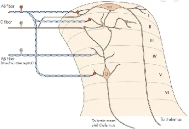
Incoming stimuli from the skin and viscera terminate in specific laminae of the dorsal horn of the spinal cord. When the axons of DRG neurons reach the dorsal horn, they branch into ascending and descending collaterals, forming the dorsolateral tract of Lissauer. At this level, axons typically run up and down for one or two spinal cord segments, before they penetrate the gray matter of the dorsal horn. Within the dorsal horn, the axons emit branches that contact neurons located in laminae I, II and V (Fig.1.8).
Figure 1.8 Neurons in lamina I of the dorsal horn receive direct input from Aδ fibers and both direct and indirect inputs
from unmyelinated fibers, via interneurons in lamina II. Lamina V neurons receive input from myelinated fibers (Aβ) of mechanoreceptors, as well as inputs from nociceptive afferent fibers (adapted from Kandel, Principles of Neural Science V edition, McG-H, 2013).
The dorsal horn, in general, can be divided into six laminae as described by Rexed using Nissl staining of spinal cord section (Light and Perl, 1979). Laminae consist of combination of neurons can be distinguished by projections, morphology and different gene expression. Lamina I receives input from Aδ and C- afferents about pain and heat/cold sensing. Because they respond selectively to noxious stimulation, they have been called nociception-specific neurons and project to higher brain centers. Another class of neurons of lamina I respond to both innocuous and noxious mechanical stimuli and are termed wide-dynamic-range neurons.
22 Lamina II, or substantia gelatinosa, can be divided into two regions: lamina II0 and IIi, which receive inputs
from neurons carrying information about pain and touch, respectively. Lamina III and IV contain both interneurons and supraspinal projection neurons. Many of these neurons receive inputs from Aβ fibers that
respond to innocuous cutaneous stimuli, such as deflection of hairs. Lamina V contains neurons that respond to a wide variety of noxious stimuli (inputs from Aβ and Aδ fibers) and send signals to the brain stem and thalamus. Neurons in lamina V also receive input from nociceptors in visceral tissues and are involved in the phenomenon called “referred pain”, a condition in which pain from injury to visceral tissues
is perceived as originating from a region on the body surface. Neurons in lamina VI receive inputs from large-diameter fibers of muscle and joints. These neurons are activated by innocuous joint movement and for this reason do not contribute to pain information.
Nearly 30-40 % of interneurons in the dorsal horn are inhibitory and use GABA and/or glycine as neurotransmitters, whereas the remaining interneurons are excitatory and use glutamate. In response to tissue injury or after intense stimulation of peripheral terminals, sensory neurons activate neurons in the dorsal horn releasing neuropeptides, such as Substance P, CGRP, somatostatin and galanin, in order to transmit the nociceptive information.
1.3.2 Distinct, Parallel Pathways for Pain
The classic pain pathway consists of a three-neuron chain that transmits pain information from the periphery to the cerebral cortex. As already mentioned, the first-order neuron has its cell body in the dorsal root ganglion and two axons, one extending distally to the tissue, while the other extending to the dorsal horn of the spinal cord, with a “T-pseudounipolar” morphology. At this level, this axon synapses with the
second-order neuron which, in turn, crosses the spinal cord through the anterior white commissure and ascends to reach the brain. Second-order fibers in the anterolateral system (the neural pathway that conveys pain and temperature information) project to a number of different structures in the brainstem and forebrain. These central destinations are likely to mediate different aspects of the sensory and behavioral response to a painful stimulus. The pain pathway is now understood to be a dual system at each level and the sensation of pain that reaches the brain is composed of sensory-discriminative and affective-motivational components (Fig.1.9). The sensory-discriminative aspects of pain include quality, location and intensity processing
23 (Andersson et al., 1997) while the affective-emotional component of pain comprises the unpleasant character of pain perception (Craig, 2003b). The cognitive component is also involved in attention, anticipation and memory of past experiences and this component can interact with components from other senses giving rise to modulation of pain (Valet et al., 2004).
Figure 1.9 Different brain region involved in parallel aspects of pain experience. (adapted from Purves, Neuroscience,
VI edition, Sinauer Associates, Sunderland, 2017).
1.3.3 Ascending pathways and processing of nociceptive information
The central processing of pain information is mediated by five major ascending pathways: the spinothalamic, spinoreticular, spinomesencephalic, cervicothalamic and spinohypothalamic tracts.
The spinothalamic tract is the most prominent ascending pathway in the spinal cord. It includes the axons of nociception-specific and wide-dynamic-range neurons in lamina I and V of the dorsal horn. These axons cross the midline of the spinal cord and ascend within the anterolateral white matter before reaching the thalamic nuclei. This tract has an important role in the transmission of nociceptive information; electrical stimulation of the tract is sufficient to induce painful sensations, whereas lesions of this tract can result in a marked reduction of pain sensation. By virtue of an
24 anatomical connection between the sensory thalamus and the lateral amygdala (LA) (Nader et al., 2000), the spino-thalamic pathway has been studied as a potential circuit for transmission of the unconditioned stimulus (US) during Pavlovian fear conditioning (Shi and Davis, 1999).
The spinomesencephalic (or spinoparabrachial) tract contains the axons of projection neurons in laminae I and V. This tract projects, within the anterolateral quadrant of the spinal cord, to the mesencephalic reticular formation and periaqueductal gray matter (PAG). Information transmitted along this tract is important because it contributes to the affective aspects of pain. Axons of this tract also project to the parabrachial nucleus (PBN) that, in turn, projects to the amygdala. Anterograde tracing studies show that most spinal lamina I projection neurons send their axons to the external lateral subdivision of the PBN (PBel) (Al-Khater and Todd, 2009), and field potential recordings in vivo show that noxious stimuli in the periphery induce activity in PBel (Bester et al., 2000) and central nucleus of amygdala (CeAl) (Neugebauer and Li, 2002). Consistently, neuronal tracing studies reveal that PBel neurons directly innervate CeAl neurons (Lu et al., 2015), and electrical stimulation of axonal fibers from the PBel induce depolarization of neurons in the CeAl (Jhamandas et al., 1996). In a recent paper, Sung Han and colleagues pursued the idea that the parabrachio-amygdaloid pathway is responsible for relaying the US pain signal to the CeAl during fear conditioning. Indeed, their findings reveal that CGRP neurons in the PBel convey the US signal to the CeAl, and that CGRPR neurons are the functional US-recipient neurons in the CeAl, thus beingcritical for establishing a threat memory (Sung et al., 2018).
The spinoreticular tract is composed of the axons of projection neurons in laminae VII and VIII. This tract ascends within the anterolateral quadrant of the spinal cord, with no decussation, and terminates in the reticular formation and thalamus.
The cervicothalamic tract contains the axons of neurons of the lateral cervical nucleus, which receives input from neurons in laminae III and IV of the dorsal horn. Many axons of this tract cross the midline and ascend within the medial lemniscus of the brain stem, terminating in midbrain nuclei and in the ventroposterior lateral and posteromedial nuclei of the thalamus. Other neurons in laminae III and IV send their axons directly into the dorsal columns, terminating in the cuneate and gracile nuclei of the medulla.
25 The spinohypothalamic tract is composed of the axons of neurons from in laminae I, V and VII of the dorsal horn. This tract projects to the hypothalamic nuclei that serve as autonomic control centers involved in the neuroendocrine and cardiovascular responses that accompany pain. Spinal projections to the brainstem are important for integrating nociceptive activity with homeostatic and autonomic processes, in addition to providing a means to indirectly convey nociceptive information to forebrain regions after brainstem processing. The capacity of projections to the brainstem to influence both spinal and forebrain activity suggests that these pathways play a direct role in affecting the pain experience.
1.3.4 Focus on the main brain areas involved in pain processing
The thalamus is one of the supra-spinal structures that receives projections from multiple ascending pathways. This structure is not only a relay centre, but is involved in processing nociceptive information before transmitting the information to various parts of the cortex (Craig, 2003a). The thalamus consists of several groups of nuclei and each group has important functions in the sensory discriminative and affective motivational components of pain. Spinal neurons of lamina I project extensively to the posterior group and to the ventrobasal complex that, in turn, project to the primary somatosensory cortex. This pathway constitutes the lateral pain system, that plays an important role in the discrimination of stimuli (Andersson et al., 1997). The affective-motivational aspect of pain is mediated by the medial pain pathway, which includes the intralaminar thalamic nuclei (Royce et al., 1989) and the posterior part of ventromedial thalamic nuclei that project to the somatosensory cortex and limbic structures (Shyu et al., 2004). For example, the amygdalostriatal area (AStr) is the main target of CGRP-projections from the posterior thalamus: this pathway plays an important role for the effects of CGRP, including induction of fear-like behaviors and antinociceptive effects (D'Hanis et al., 2007).
Given the complexity of the pain sensation, many cortical areas are involved in nociceptive processing, contributing to a large brain network together with the thalamus. For this reason, Melzack described a pain “neuromatrix”, now known as the “pain matrix” or “pain signature” (Melzack, 1999). Activation of
signature pain matrix has been related to perceived pain intensity, within and between individuals, and is now considered a candidate biomarker for drug discovery (Huang et al., 2013; Wager et al., 2013). Because different brain regions play a more or less active role in influencing pain perception, what comprises the
26 pain matrix is not unequivocally defined. However, there is a consensus in including at the core of the matrix: primary and secondary somatosensory cortices, insular cortex (IC), anterior cingulate cortex (ACC) and prefrontal cortex, amygdala, thalamus and periaqueductal gray (PAG) (Tracey and Mantyh, 2007). These areas and structures transmit and decode nociceptive information, amplify or reduce the pain sensation, and mediate the expression of defensive behavior.
The amygdala appears as a key component of the pain matrix (Simons et al., 2014). The amygdala provides an emotional value – either positive or negative – to sensory information, leading to adaptive behavioral and affective responses and contributing to emotional memory. This role has been extensively studied using the fear conditioning paradigm (LeDoux, 2000, 2014). The amygdala is, indeed, known to be a critical brain region that integrates the sensory (i.e., the conditioning stimulus, CS) and pain (i.e., the unconditioning stimulus, US) signals to create a memory that will produce a threat response when the subject will be re-exposed to the CS alone (Gross and Canteras, 2012). The amygdala is a heterogeneous structure of the limbic system, composed of a dozen of nuclei clustered in four groups: superficial, basolateral, central and medial (Sah et al., 2003). The lateral-basolateral nuclei (LA-BLA) form the input region for sensory (e.g. nociceptive) information from thalamus and cortical areas such as ACC, insula and other medial prefrontal cortical areas (Price, 2000). The BLA contains neurons that respond preferably to noxious stimuli and their projections reach the medial prefrontal cortex (mPFC), providing emotion- and value-based information to guide executive functions, such as decision-making and behavior control (McGaugh, 2004; Laviolette and Grace, 2006). The result of information processing in the LA-BLA network is transmitted to the central nucleus of the amygdala (CeA). The latter is also termed the “nociceptive amygdala” and it is considered as the output nucleus of the amygdaloid complex, projecting to pain modulatory systems through forebrain and brainstem connections (Price, 2000; Neugebauer et al., 2004). The CeA is also connected to rostral forebrain structures, such as the lateral part of the bed nucleus of the stria terminalis (BSTL) and the dorsal part of the substantia innominata. The CeA is strongly implicated in fear. Indeed, peptidergic CeA projection neurons are innervated by CGRP containing terminals from the parabrachial area and stimulate freezing behavior, whereas their silencing reduces conditioned fear responses (Campos et al., 2018). Neuronal activity changes in the amygdala are not simply a reflection of continued input from spinal cord
27 and other regions, but can also result from an imbalance between excitatory and inhibitory synaptic mechanisms internal to amygdala circuits.
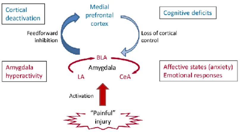
Figure 1.10 Role of amygdala in pain. Pain-producing events generate hyperactivity in the lateral, basolateral and central nuclei
(LA, BLA, CeA) of the amygdala network .This can account for the emotional-affective aspects of pain. Output from BLA deactivates the medial prefrontal cortex through feedforward inhibition;its alterations can play a role in cognitive deficits such as impaired decision making. Decreased medial prefrontal cortical output to the amygdala allows the uncontrolled persistence of amygdala hyperactivity, hence contributing to the persistence of pain (adapted from Neugebauer et al., 2004).
The insular cortex (IC) comprehends three areas conserved across different species (i.e., mouse, man and non-human Primates): the granular, dysgranular and agranular subdivisions, according to their cytoarchitecture. The granular IC has a classical six-layered structure; in the dysgranular IC, layer 4 becomes thinner and, finally, the agranular IC is tri-laminar, completely lacking layer 4. The IC is strongly connected with an extensive network of cortical and subcortical regions, by which it mediates interoception. Indeed, the IC receives from the thalamus sensory inputs regarding information about the outer environment (somatosensory, olfactory, gustatory and visual information) and the inside of the body (interoceptive information). In this regard, the insula exerts an important control on autonomic functions, for instance, regulation of the heartbeat, blood pressure, or gastric motility. It performs these actions through direct projections to the hypothalamic area, the parabrachial nucleus and the nucleus of the solitary tract (Craig, 2002). In addition, the insula presents reciprocal connections with the limbic system and, using the Pavlovian fear-conditioning paradigm, fear-induced activation of the insular cortex across different species, from mouse to human was demonstrated (Kusumoto-Yoshida et al., 2015). Lesions or pharmacological inhibition of different insula regions show a significant role for insula in the consolidation of learned fear;
28 patients with large lesions in this area show deficits in the emotional dimension of pain (Berthier et al., 1988). In particular, patients with insular lesion can recognize pain but cannot attribute negative valence to adverse experiences as a result of lack an appropriate emotional response (Gogolla, 2017), which may indicate a role for the insula in mediating empathy. Electrical stimulation of several sites within the posterior insula has been shown to evoke unpleasant pain, often with distinct sensory qualities (Ostrowsky et al., 2002). These observations suggest that this area can be involved in creating both sensory and affective components of pain.
Different studies indicate that the ACC is a critical brain region involved in pain processing (Shackman et al., 2011) and its role is more complex than IC. ACC neurons appear to integrate multiple sensory inputs and their neuronal activity is associated with escape responses, attention and response selection in monkeys (Isomura and Takada, 2004). In particular, ACC activity increases during escape from a noxious thermal stimulus but not non-noxious chemical, mechanical and thermal stimuli (Koyama et al., 2001; Iwata et al., 2005). More in detail, neurons of the rostral anterior cingulate motor area appear to be involved in the selection of appropriate motor responses, as well as in the planning of movements (Isomura and Takada, 2004). Some neurons in ACC respond during cues that signal impending pain, participating in the anticipatory response to pain, whereas others respond to anticipation of a reward. This is consistent with neuroimaging studies showing that the rostral ACC is activated during anticipation of pain (Koyama et al., 2001). Besides, the ACC coordinates input from the insula and medial thalamic nuclei with prefrontal cortex and limbic structures, in order to provide an appropriate motor response, or action planning. Furthermore, activity in the ACC, but not in the somatosensory cortex, is closely related to the affective features of pain, such as subjective feelings of unpleasantness (Rainville et al., 1997; Tang et al., 2005). Studies in mouse models, using fear conditioning, show a role of ACC in memory recall, rather than memory formation (Frankland et al., 2004), providing evidence that the ACC is preferentially involved in the recall of remote fear memories.
1.3.5 Modulation of pain
The ability of higher centers of the brain to modulate the transmission of nociceptive information had been yet demonstrated in the 1900s by Sherrington, who showed that nociceptive reflexes were enhanced