UNIVERSITA’ DEGLI STUDI DI CAMERINO
School of Advanced Studies
Doctorate course in
“LIFE AND HEALTH SCIENCES: ONE HEALTH”
XXXI cycle
“DEVELOPMENT OF AN ANIMAL CANCER REGISTRY FOR THE
MARCHE REGION AS A TOOL FOR PREVENTIVE HEALTH CARE“
PhD Student
Tutor
Dr. Silvia Scarpona
Prof. Giacomo Rossi
Coordinatore curriculum
Prof. Anna Maria Eleuteri
TABLE OF CONTENTS
Table of contents ..….………. 2 Summary ……….4 Preface ……….……….…………5 1. Introduction ……….………..………6 1.1 ‘One Medicine – One Health’ concept ……….……….6 1.2 One Health – One Oncology ……….………8 1.3 Animal sentinels ……….………….……..12 1.4 Cancer registry ………..…….…….13 1.4.1 Introduction ……….13 1.4.2 The concept of diseases registries ……….15 1.4.3 Types of registries ……….16 2. Animal Cancer Registry of the Marche region ……….……18 2.1 Introduction ……….….18 2.2 Materials and Methods ……….…………19 2.2.1 Data source ……….……….19 2.2.2 Sample collection and diagnosis ……….……..20 2.2.3 International Classification of Diseases for Oncology: ICD-O ……….……..20 2.2.4 Data analysis ……….………..22 2.3 Results ……….………..23 2.3.1 Dataset ……….………23 2.3.2 Breed distribution ……….………35 2.3.3 Age distribution ……….………43 2.3.4 Sex distribution ……….…….45 2.3.5 Lifestyle ……….……..49 2.3.6 Most common tumors types according to ICD-O ……….…….542.3.7 Most common anatomical tumor locations ………..……….……..63 2.4 Discussion ……….…..65 3. The Cuban experience: Animal Cancer Registry of Havana city ……….……….……69 3.1 Introduction ……….……. 69 3.2 Materials and Methods ………..……70 3.2.1 Data source ……….…….70 3.2.2 Sample collection and diagnosis ……….…..70 3.2.3 Data analysis ……….………..72 3.3 Results ……….………..73 3.3.1 Dataset ……….…………73 3.3.2 Breed distribution ……….…..……73 3.3.3 Age distribution ……….…………75 3.3.4 Sex distribution ……….………….76 3.3.5 Most common tumors types according to ICD-O ……….……….79 3.3.6 Most common anatomical tumor locations ..……….………..85 3.4 Discussion ……….………..88 4. General conclusions ….……….……….…………91 5. References ……….91 6. List of activities and publications ………..97
SUMMARY
Cancer registries are a key feature of any epidemiological study or prevention and control strategy. Moreover, companion animal tumor registries are intended to assist in different aspects of research on tumor development, pathogenesis, genetics and treatment. Traditionally, comparative cancer research is based on murine models, which lack many features that define human cancer, including growth over longer time periods, genomic instability, function of the immune system and a significant heterogeneity of tumor cells and tumor microenvironments. To fill this gap, spontaneous tumors in dogs and cats reflect more features of human cancer. Furthermore, sharing the living environment with humans, they are exposed to similar risk factors, therefore acting as sentinels for recognition of environmental factors implicated in oncogenesis.
Comparison of data from canine tumor registries has recently gained increasing interest in the context of the ‘One Medicine-One Oncology’ concept, part of the ‘One Health Initiative’. The One Health concept is a worldwide strategy for expanding collaborations and communications of multiple disciplines in all aspects of health care for humans, animals and the environment. It is believed that an achieved synergism will improve public health, scientific knowledge as well as biomedical research. To learn more about tumors in companion animals, such as cancer development and risks, knowledge on the occurrence of tumors in pets needs to be expanded because statistics on the incidence of cancer in pet animals are very rare. As part of ‘One Health curriculum’ of the School of Advanced Studies of the University of Camerino, this thesis was based on establishment of a canine cancer registry of the Marche region, so far lacking, focused on extensive data collection and interpretation about spontaneous tumors occurring in dogs living in the Marche region as a tool for preventive health care. Tumors were classified according to the tumor type, malignancy and physical location following the guidelines of the International Classification of Oncology for Humans (ICD-O), which subsequently allows comparisons with human cancer registries. Being a newborn cancer registry, the collected data were still insufficient to carry out an adequate statistical analysis, therefore a descriptive examination of the first available data was performed, pending further implementation in order to have a more truthful panorama of the oncological cases of dogs of the Marche region.
Moreover, this dissertation describes a similar study carried out in Cuba. The aim was to collect data from a country with socio-economical, cultural and climatically characteristics completely different from ours, and to investigate ‘if’ and ‘how’ these differences could influence tumors onset in canine population. In the same manner of canine cancer registry of the Marche region, data collected of the city of Havana were analyzed as descriptive statistic and represent a groundwork to implement further.
PREFACE
This thesis was conducted at the School of Biosciences and Veterinary Medicine of the University of Camerino, in a trans-disciplinary collaboration with IZSUM, and thanks to the School of Advanced Studies, that opened doctoral positions in ‘One Health curriculum’, and to the Marche Region, that courageously decided to take part to the ‘One Health Initiative’. The model of this research project was initiated by the Umbria Region, that successfully set up an Animal Cancer Registry 2 years early, managed by IZSUM in collaboration with the University of Perugia. In 2015, the Marche Region launched its own challenge with the goal of establish its Animal Cancer Registry that could, over time, become a useful tool to pursuit the Public Health.
My involvement in this ambitious project developed throughout my whole doctoral course and represented for me a professional and personal growth. This topic suited me really good and allowed me to move in the field I love more: pathology. At the same time, it opened my horizons, both scientifically and geographically. The trans-disciplinarily of the project introduced me to disciplines before unknown, like informatics and epidemiology, while comparative research led me to apply my new and old skills in a country diametrically opposite to ours, 9000 km far from Italy.
I would like to thank my tutor, Prof. Giacomo Rossi, who always believed in me and pushed me beyond my limits… what I am today is only his merit! My thanks go also to my work team and co-authors for their support, help and inputs. I am grateful to the School of Advanced Studies of the University of Camerino and its examiners, who choose my project 3 years ago and gave me the opportunity to go on until now. Thanks to the Marche Region that, relying on the School of Biosciences and Veterinary Medicine of the University of Camerino and on my tutor, allowed me to work and manage its worth project. Immensely thanks to the staff of the Laboratory of Experimental Pathology and Surgery of the National Institute of Oncology and Radiobiology (INOR) of the city of Havana, in particular to my Cuban tutor Prof. Juan Carlos Rodriguez Aurrecochea, that welcomed me and support in many aspects of my not easy foreign experience. Further, I would like to thank the Experimental Animal Prophylaxis Institute (IZS) of Umbria and Marche, both the Histopathology Laboratory and Epidemiologic Observatory, for the valuable contribution. Many thanks to my family, friends and colleagues outside the University, who always understood and supported me along the way.
1. INTRODUCTION
1.1 ‘ONE MEDICINE – ONE HEALTH’ CONCEPT
The origin of the One Medicine concept has been linked to the 19th century German physician and pathologist Rudolf Virchow (1821-1902) who created the field of comparative pathology. During his study on human and animal pathogens, he noted the similarity in disease processes among animals and humans stating that differ only in details and not in kind. Dr. Virchow proclaimed “between animal and human and medicine there is no dividing line, nor should there be. The object is different, but the experience obtained constitutes the basis of all medicine”. Although the One Medicine theme was continued by William Osler (1849-1919), Virchow’s student and father of modern medicine, who taught it to his medical and veterinary students, human and animal medicine were practiced separately until the latter half of the 20th century. The One Medicine concept was revived and bolstered by the American veterinarian Calvin W. Schwabe (1927-2006) who coined the term ‘One Medicine’ in his textbook Veterinary Medicine and Human Health in 1964. Today, the early term ‘One Medicine’ is commonly referred to as ‘One Health’ worldwide. This terminology evolution occurred during the first decade of the 21th century. One Health was born out of, and fueled by, fear. In 2004, there was global anxiety that a zoonotic disease, HPAI H5N1, could cause a pandemic in the human population, rivaling, and possibly exceeding, the estimated 50 million human deaths associated with Spanish influenza at the end of the First World War20. The introduction of the One Health initiative provided international agencies (FAO,
OIE, WHO and the World Bank) with a vehicle for interinstitutional and interdisciplinary collaboration to address the threat of emerging zoonotic diseases, and it enabled these international agencies and national authorities to come to the table as equal partners in the search for solutions to the threats posed by this highly virulent strain of influenza21.
The expression ‘One Health’ was proposed as a concept to foster interdisciplinary collaboration between physicians and veterinarians, but also wildlife specialists, environmentalists, anthropologists, economists and sociologists, among others, required to prevent and control zoonosis. One Health recognizes that humans do not exist in isolation, but are a part of a larger whole, a living ecosystem, and that activities of each member affect the others. Thus, One Health considers health as a whole, the humans, the animals, and the environment they exist on.
‘One Health is the collaborative effort of multiple health science professions, together with their related disciplines and institutions – working locally, nationally, and globally – to attain optimal health for people, domestic animals, wildlife, plants, and our environment.’ One Health
Commission
World-One Health’TM to embrace both medicine and ecosystem health, and listed 12
recommendations for establishing a more holistic approach to preventing epidemic/epizootic disease and maintaining ecosystem integrity for the benefit of humans, their domesticated animals, and the foundational biodiversity that supports us all (www.oneworldonehealth.org). This series of recommendations became known as the Manhattan Principles, in recognition of the fact that the meeting was hosted by Rockefeller University in New York.
The Manhattan Principles exhort the world’s leaders, civil society, the global health community and institutions of science to:
1. Recognize the essential link between human, domestic animal and wildlife health and the threat disease poses to people, their food supplies and economies, and the biodiversity essential to maintaining the healthy environments and functioning ecosystems we all require.
2. Recognize that decisions regarding land and water use have real implications for health. Alterations in the resilience of ecosystems and shifts in patterns of disease emergence and spread manifest themselves when we fail to recognize this relationship.
3. Include wildlife health science as an essential component of global disease prevention, surveillance, monitoring, control and mitigation.
4. Recognize that human health programs can greatly contribute to conservation efforts. 5. Devise adaptive, holistic and forward-looking approaches to the prevention, surveillance, monitoring, control and mitigation of emerging and resurging diseases that take the complex interconnections among species into full account.
6. Seek opportunities to fully integrate biodiversity conservation perspectives and human needs (including those related to domestic animal health) when developing solutions to infectious disease threats. 7. Reduce the demand for and better regulate the international live wildlife and bush meat trade not only to protect wildlife populations but to lessen the risks of disease movement, cross-species transmission, and the development of novel pathogen-host relationships. The costs of this worldwide trade in terms of impacts on public health, agriculture and conservation are enormous, and the global community must address this trade as the real threat it is to global socioeconomic security. 8. Restrict the mass culling of free-ranging wildlife species for disease control to situations where there is a multidisciplinary, international scientific consensus that a wildlife population poses an urgent, significant threat to human health, food security, or wildlife health more broadly.
domestic animals and wildlife. Enhanced capacity for global human and animal health surveillance and for clear, timely information-sharing (that takes language barriers into account) can only help improve coordination of responses among governmental and nongovernmental agencies, public and animal health institutions, vaccine / pharmaceutical manufacturers, and other stakeholders.
10. Form collaborative relationships among governments, local people, and the private and public (i.e.- non-profit) sectors to meet the challenges of global health and biodiversity conservation.
11. Provide adequate resources and support for global wildlife health surveillance networks that exchange disease information with the public health and agricultural animal health communities as part of early warning systems for the emergence and resurgence of disease threats.
12. Invest in educating and raising awareness among the world’s people and in influencing the policy process to increase recognition that we must better understand the relationships between health and ecosystem integrity to succeed in improving prospects for a healthier planet.
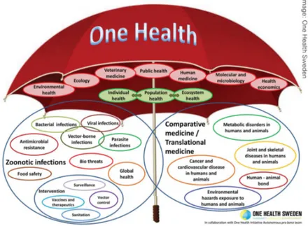
In the Manhattan Principles, the importance of education about the One Health concept is introduced, and public and private participation is encouraged. Several Universities worldwide have accepted the challenge and decided to contribute to this cause by offering dedicated didactical courses to their students. Most of them, like the Royal Veterinary College in London and the Royal (Dick) School of Veterinary Studies at the University of Edinburgh, set up specific master degrees in One Health, while very few Universities have gone beyond by offering PhD degree specifically in One Health. One of these is the Italian University of Camerino, and the present thesis is the product of such opportunity. 1.2 ONE HEALTH - ONE ONCOLOGY During the period when the focus of international agencies was on avian influenza, there was recognition that the One Health approach had a wider application21. One Health activities do not involve only zoonotic diseases, but many other topics considered to be relevant for the promotion of health in a wider context (Figure 1). Cancer in humans and animals is one of these. If we look back over the last 100 years, we realize how much studies in animals have contributed to the global health. For example, the study of avian leukosis virus led to a fundamental understanding of oncogenes in cancer82. The vaccine for cervical cancer, the second most fatal cancer in women, can be directly attributed to study done in the last fifty years on cattle infected with papillomavirus42.
Figure 1: Scope of One Health according to the One Health Initiative (www.onehealthinitiative.com). Image: One Health Sweden
Translation of this findings into human field is called comparative oncology. Comparative oncology shifts the occurring cancers seen in animals into more general study of cancer biology.
A great deal of work in comparative oncology is based on rodent models because of their low cost, short average lifespan and easiness in handling. However, despite of the unquestionable importance of rodent models in advancements in cancer research and preclinical tests, much of the data obtained from them rarely translate into human clinical practice due to the limitations of these models to better reflect the complexities of human tumors37. Mice have different anatomical, cellular and molecular features similar to humans that are known to have critical properties and functions in cancer. In addition, the percentage of murine genes with a human orthologue is 80%79, thus providing an excellent experimentally tractable model system as a research tool to investigate the basic mechanisms of cancer development and treatment responses43. Although mouse models remained a valuable tool for examining the molecular
mechanisms of carcinogenesis, the low degree of heterogeneity in mouse tumors compared to very heterogeneous human tumors is an important limitation75. Similarities in tumors expression are significantly closer between human and dog than those between human and mouse. Firstly, the recent deciphering of the canine genome provided evidence of strong similarities with humans35, 46; secondly, many gene families of canine genome, especially associated with cancer, show a greater homology to human genome than murine48 (Figure 2). Furthermore, canine tumors share evolutionarily conserved genomic changes that are found in their human counterparts63. In addition, tumors naturally occur in dogs, their initiation and progression are influenced by similar factors in both human and canine cancers, including
clinical features closely parallel to the corresponding tumors in humans63, 84. All these reasons
place dogs in a unique position to better reflect tumor development and progression than traditional rodent models. Again, the range of tumors occurring in dogs are as diverse as tumors occurring in humans. This is given by a biological complexity of canine cancers47, based on the intra-tumor heterogeneity (ITH)19, which captures the essence of tumor in humans and shares the same features. Fig. 2: Homology between dog, human and mouse for recognized cancer genes. Recent completion of the canine genome draft sequence has allowed demonstration of the strong similarities between canine and human cancer genes. VISTA graph displays (http://genome.lbl.gov/vista/index.shtml) visually compare aligned canine and murine genes with their human orthologues. The y-axis of the graph represents the percent conservation of the canine or murine sequence against the human gene target. The y-axis ranges from 50% to 100% conservation with a threshold line drawn at 70%, which is a level denoting significant similarity. The colors of the peaks describe the function of the sequence, dark blue representing exons, pink non-coding regions and light blue untranslated regions. The plots compare the entire human sequence for the following cancer-associated genes with their respective canine and mouse orthologues. a | MET, an oncogene activated in canine and human sarcomas. b | Insulin-like growth factor 1 receptor (IGF1R), a receptor for IGF, which is an important growth factor in various tumors. c | KIT, a causative oncogene in human gastrointestinal stromal tumors (GISTs) and canine mast-cell tumors and GISTs. d | Mammalian target of rapamycin (mTOR, also known as FRAP1), an integral regulator of protein translation in various tumors and a therapeutic target of rapamycin. The graphs indicate that the dog nucleotide sequences are more highly conserved with human sequences than mouse sequences are for all four candidate genes. This is especially evident at the level of similarity within the non-coding regions. Image and caption: Nature Reviews Cancer (2008) 8:150 (see reference 48).
Human disease is polygenic. Genetic manipulation on mice, exalting one or few genes, increases the gap between rodent models and humans64. On the contrary, dogs have strong analogies with humans in genetic molecular alterations that drive cancers84. Most, if not all, of
the cancer associated genetic alterations that influence cancer progression in humans have been identified in canine cancer. For example, similar mutations in KIT, a tyrosine kinase growth factor receptor, have been identified in both gastrointestinal stromal tumors (GIST) in humans and mast-cell cancers in dogs54. Additionally, statistical analysis of genomic alterations in human and
dog colorectal tumors showed the same genetic pathway in tumorigenesis in both species, and that species-specific alterations tend to localize to evolutionarily unstable genome regions as irrelevant mutation hotspots70. This suggests that alterations common to both species are more
likely to cause tumor than those found in only one.
The germline genetic diversity (frequency of single nucleotide polymorphism) of a population of dogs with a given cancer is similar to the diversity observed in a well-balanced population of human patients with a given cancer35, 48. This relatively rare nature of dogs compared to most rodent cancer models further contributes to designating the dog as an ideal animal model in comparative oncology.
Next to humans, dogs have the most phenotypic diversity and known naturally-occurring diseases of all land mammals67. For example, in the same species coexist members that differ in
size by 65-fold, as between Chihuahuas and English Mastiffs. Dogs share over ~650 Mb of ancestral sequence in common with humans, absent in mice, and DNA and protein sequence are more similar between human and dog than human and mouse35. Dogs and humans share
approximately 400 similar inherited diseases, such as tumors, heart disease, and neurological disorders49, 62. Indeed, more than 40 naturally occurring canine diseases have mutations in a homologous human gene associated with a similar disease45. The greatest advantage of dog models is the result of their evolutionary history which led to selection of breeds on the basis of morphological and behavioral traits. Today there are ~400 isolated populations or breeds. Breed creation inadvertently selected "founder" mutations that are associated with specific traits and diseases; this translates into reduced disease and genetic heterogeneity, consistent with the fact that most breeds are predisposed to a distinct set of diseases59. It has been known for many years that there are some breeds that have a high
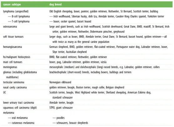
incidence and a high risk of specific cancer subtypes, sometimes even more than one subtype (Table 1). Predisposed breeds provide the platform to readily identify genes known to be linked to cancer development (i.e. oncogenes) and those whose loss trigger cancer development or progression (i.e. tumor suppressor genes). Since several genetic alterations and molecular signaling pathways are the same in human and dog cancers, studying breeds with increased cancer incidence may allow more rapid progress in the identification of new cancer-associated genes than the study of human or mouse cancers alone.
Table 1: Tumors associated with specific dog breeds (data from reference 63).
In summary, dogs are useful in multiple approaches to cancer investigation5: breed-specific risk can be used to discover disease pathways; human cancer pathways can be tested for roles, and targeted for treatment, in canine disease; and canine somatic mutations and genome alterations can be used to narrow down human mutations. Through these studies, comparative oncology confirms its value in the field of public health. 1.3 ANIMAL SENTINELS
The term “sentinel” is derived from the French sentinelle, “watch tower”. An animal sentinel system is one in which animal data are regularly and systematically collected, summarized, and analyzed in order to identify health hazard to either humans or the animals themselves from chemical or biological contaminants in the environment87. The familiar image of the canary in the coal mine remains relevant in the 21st century 8. Just as miners carried caged canaries in the early 20th century to detect exceeding levels of carbon monoxide in the air, many animals have been used over time to warn of environmental contamination effects in human populations. Pets, in particular, share the environment and are exposed to many of the same agents as their human companions. Furthermore, they suffer a similar spectrum of disease as humans and, therefore, may be sensitive indicators of environmental hazards and provide an early warning system for public health intervention. There
Much of the work involving the use of sentinels to identify environmental hazards has focused on cancers in pet animals, particularly dogs, which share the environment intimately with humans but do not indulge in activities (i.e. smoking or working) that confound interpretation of human epidemiologic studies84. Naturally occurring canine tumors provide
useful models for the study of the health effects of environmental hazards. Many canine cancers are similar to those in humans for biological behavior, histopathologic features, proportional morbidity, and recognized risk factors. A classic example of a canine cancer sentinel is the study of mesothelioma by Glickman23. The authors identified chrysotile asbestos bodies in lung tissue of dogs with spontaneous mesothelioma and linked this finding to asbestos exposure of their owner. The findings showed the importance of epidemiologic research to identify environmental health hazards for humans who share the environment with their pets. Thus, the diagnosis of canine mesothelioma is an early warning system for the human disease, because of the shorter latency period of mesothelioma in dogs than in humans, of about 8 and 30 years respectively 87. The impact of these interactions can be appreciated only by studying population effects under natural conditions over time. Herein lies the strength of the epidemiologic method which, if rigorously applied, can bring closer to the truth and provide a clear picture of what happens 87. 1.4 CANCER REGISTRY 1.4.1 Introduction 29
Cancer registry is one of the fundamental tools for epidemiological research. It has a pivotal role in cancer control. Its primary function is to record all cancer cases occurring in a defined population, collected continuously and systematically from various data sources. The registry analyses and interprets such data periodically and provides information on the incidence and characteristics of specific cancers in various segments of the resident population and on temporal variations in incidence. Such information is the primary resource not only for epidemiological research on cancer determinants but also for planning and evaluating health services for the prevention, diagnosis and treatment of the disease. Cancer registries can also be used for monitoring occupational groups and cohorts of individuals exposed to various carcinogens and as a convenient source of subjects for clinical and epidemiological studies. The value of a cancer registry depends on the quality of its data and the extent to which they are used in research and health services planning. It is obviously important that the registration of cancer cases should be as complete as possible. Epidemiological research, based on comprehensive cancer registration, remains the most valid and efficient way to plan and evaluate all aspects of cancer control.
The data collected by individual registries may vary according to local needs and availability of information but the nomenclature and definition of each item should be the same in all registries to give uniformity and facilitate international comparability of cancer data. The main objective of the cancer registry is to collect and classify information on all cancer cases in order to produce statistics on the occurrence of cancer in a defined population and to provide a framework for assessing and controlling the impact of cancer on the community. Cancer registry information may be used in a multitude of areas, and the value of the data increases if comparability over time is maintained. The data become useful for more and more purposes as they are accumulated over longer periods of time.
The cancer registry’s enumeration of cancer cases in a defined population permits assessment of the scale of the cancer problem in terms of the number of new cases and the computation of incidence rates. The type of statistics emerging from the cancer registry should be adapted to local needs and interests, bearing in mind the importance of international comparability. Ability to calculate rates depends on the availability of population denominators. Indeed, the information on cancer cases should be collected and classified so that it accords with the population statistics.
Comparison of cancer occurrence in various populations may provide clues to etiology, and the demonstration of variation in incidence has made an important contribution to the recognition of the environmental origin of many cancers, thus pointing to the possibilities for prevention. Such basic features of cancer incidence may not always be easily understood and explained, but they should provoke the epidemiologist’s curiosity and are useful in the generation of etiological hypotheses.
The contribution of cancer registries to our knowledge of international variation in cancer incidence is an important purpose of registering cancer cases. The stimulation of etiological ideas from such geographical comparisons of cancer incidence may be enhances by correlation with statistics on potential risk factors. Cancer registries, through their mission to perform public health surveillance and research in oncology, contribute to the development of public health. Just as human cancer registry, animal cancer registry takes part to epidemiological studies and represent a useful tool of comparative oncology. Quantitative comparison of tumor types may reveal unusual cancer frequencies, providing directions for research and generation of hypotheses of cancer causation in a specific area, and suggest leads for identifying risk factors.
While human cancer registries began for the first time in London in 172815, and now
numbering over 400 individual registries, animal cancer registries are more recent, small in number and often short-lived and sporadic. A review of animal cancer registry from the beginning to recent time was conducted by Brønden and others6. The review explains that many animal
collaboration between the registers, showing how their potential as information sources has not been fully exploited making them largely underutilized. The continuation of the registries, together with the collaboration between them, would increase the size of the database, allowing to evaluate temporary trends, fluctuations in cancer incidence and assessment of potential environmental and individual risk factors.
Inactive and active veterinary registries in Italy and worldwide are listed below. For inactive registries, period of passed activity was reported in brackets, while for active registries only the start year is reported: • California 1968 School of Veterinary Medicine, Davis (Jul 1963- Jun 1966) • Norway (1990-1998) • Denmark Royal Veterinary and Agricultural University – 2005 In Italy: • Genoa Animal Cancer Registry – 1985 • Ivrea Animal Cancer Registry – 2001 • Venice and Vicenza Animal Cancer Registry – 2005 • Sicily Animal Cancer Registry • Tuscany Animal Cancer Registry (RoVeT) – 2006 • Campania Animal Cancer Registry – 2010 • Lazio Animal Cancer Registry – 2010 • Emilia Romagna Animal Cancer Registry – 2012 • Umbria Animal Cancer Registry – 2014 1.4.2 The concept of disease registries A number of definitions have been suggested for the word "registry". Such definitions vary from author to author, but have the same perspective. Last34 stated that "in epidemiology, the term register is applied to the file of data concerning all cases of a particular disease or other health-relevant condition in a defined population such that the case can be related to a population base". Bellows3 defined registries as "a system of recording frequently used in the
concerned with the long-term care, follow up or observation of individual cases”. Solomon66
defined a registry as "data base of identifiable persons containing a clearly defined set of health and demographic data collected for a specific public health purpose." Finally, a complete definition of a registry was presented by the World Health Organization85 as follows: "a registry is a continuously updated file, set up for a specific purpose, of individuals with symptoms, health states disorders or diseases, or events in a defined population." Weddell80 classified all registries into seven types: registers used in preventive medicine, disease-specific registers, treatment registers, after-care registers, at risk registers, registers for prospective studies, and specific information registers. These classification systems are useful, but they are limited because they fail to recognize that potentialities of registry uses are related to their sources of registry data. Accordingly, Pedersen51 classified registries by their sources of data. He proposed three types of registries (specifically for cancer): local hospital registries, central registries, and population-based registries. 1.4.3 Types of Registry Registries are classified by their sources of data and the scope of coverage that can be achieved. A registry may be population-based, a central cancer registry or hospital-based51. A population-based registry covers the entire population in a defined geographic area1. A population-based cancer registry attempts to gather as much detailed information as possible on all new cancer cases diagnosed in a population of a known size and composition. The task of a population-based registry will obviously be much easier when there are collaborating hospital registries, which contribute in providing the information. The central registry is analogous to the local hospital registry, but includes a selected group of hospitals in a region. Its chief function is to supply data on diagnosis and treatment of the involved hospital patients and submit to the central registry. A central cancer registry is a co-ordination facility of co-operating hospital registries in a specified geographic area, which collects information on cancer patients. Such kinds of cancer registries are particularly valuable for comparing end results among different therapeutic regimens24. Unlike a population-based registry, and a central cancer registry, a hospital-based registry covers only one hospital51. The purpose of a hospital-based cancer registry is to serve the needs of hospital administration, the hospital cancer program, and above all the individual patient86. Its main function is to ensure that the information in case records is detailed enough to enable statistical analysis. Thus, some of the hospital registry data items collected will be different from those collected by a population based registry. The hospital registry alone does not contribute to the epidemiology of cancer because it cannot provide the incidence of cancer in the population51.
The key objective of a cancer registry is to produce statistics on the occurrence of cancer in a defined population, and to assess cancer survival. To perform such tasks, cancer registries need to have the capability, the computing facilities, and the statistical skill necessary for such analyses. Based on its main function registries can be classified into three groups: The first group are registries that are interested only in producing cancer incidence reports. Such reports represent basic presentation of the registry data. They allow feedback to reporting physicians, health authorities, and the public on the occurrence of cancer. The report could be annual, or based on incidence information for several consecutive years.
The second group are registries interested in numerous issues related to cancer survival. Such data once calculated can be used to represent the average prognosis in the population and provide theoretically at least, an objective index of the effectiveness of cancer care in the region concerned. The third group are population-based cancer registries, whose main task is to perform incidence data reporting, but also to have the facilities and skill for follow-up reporting. If undertaken by a population-based cancer registry, such tasks include all those cases that reside in the registry area.
2. ANIMAL CANCER REGISTRY OF THE “MARCHE” REGION
2.1 INTRODUCTION In a panorama of fragmented data on canine cancer epidemiology and following the need to acquire more and more useful tools to pursue the public health, with resolution n°627 of the 3rd August 2015 (Fig. 3), Marche Region created the Animal Cancer Registry of the Marche region (ACR-M). Fig. 3: Resolution of Marche Region, dated 3rd August 2015, that established the regional Animal Cancer Registry. The ACR-M was created to enable exhaustive and continuous recording of all cases of cancer in dogs living in the Marche region. It is a population-based registry, for this reason it is currently dealing only with canine population, for which a mandatory registry based on identification by microchip exists. This registry has been participating in epidemiological surveillance and evaluation of cancer, through the analysis of incidence data over time including more than 700 cases since December 2015.Its activity is based on the cooperation between Marche Region, School of Biosciences and Veterinary Medicine of the University of Camerino, Experimental Animal Prophylaxis Institute (Italian acronym IZS) of Umbria and Marche, and veterinary practitioners of provincial professional Orders of Ancona, Ascoli Piceno, Fermo, Macerata, and Pesaro/Urbino.
Veterinarians practicing on the regional territory are responsible for samples collection. Histopathology laboratories of the School of Biosciences and Veterinary Medicine of the University of Camerino and of the Experimental Animal Prophylaxis Institute of Umbria and Marche perform the histopathologic diagnosis of tumors through a double-blind mechanism. Epidemiologic Observatory of the Experimental Animal Prophylaxis Institute of Umbria and Marche deals with data analysis. Marche Region promotes its animal cancer registry and provides the digital platform on which this is based on.
2.2 MATERIALS AND METHODS 2.2.1 Data source
In human medicine, population-based cancer registries are maintained using hospital and death-certificate data as numerators and census data as denominators in morbidity and mortality rates. Animal cancer registries usually lack census data and so the denominators tend to be biased by non-response. Some registries reduce the non-response bias by utilizing demographic survey in specified areas. The Animal Cancer Registry of the Marche region uses demographic census data of canine population based on the SIVA information system (http://siva.regione.marche.it). SIVA (Italian acronym for Veterinary Information System and Food) is a digital platform where canine regional demographic data, based on identification microchip number of each dog living in the Marche region, are collected. In addition to the denominator, SIVA also provides numerator of incidence rates since it hosts not only the regional canine registry but also the canine cancer registry. The Animal Cancer Registry of the Marche region is entirely digitalized and developed in SIVA system. Indeed, veterinary practitioners have a dedicated SIVA section where insert exam requests and receive related histopathologic reports, and pathologists enter their diagnosis directly into the digital system.
In SIVA system, veterinarians are asked to fulfill a digital request form at the time of excisional surgery, for obtaining a numeric code identifying sample and patient and a histopathological diagnosis. The form’s items concern animal data, some automatically caught by the system from the regional canine registry thanks to microchip number (i.e. date of birth, sex, breed, ovariohysterectomy or castration status, geographical area of residence) and others added by veterinarian (i.e. gross data on lifestyle – urban or rural, and on nutrition – commercial, home-made, etc.), and details about anatomical site of the lesion, date and type of surgical excision and clinical history of the patient.
Once received the sample, pathologists carry out diagnosis and enter it into the SIVA system. Diagnosis reliability is guaranteed by a double-blind mechanism: first and second pathologists perform microscopically evaluation and report separately and, only if there is coincidence of diagnosis, the SIVA system sends the report to the veterinarian electronically. When there is no coincidence, a third pathologist who completes diagnosis is involved.
Since diagnosis are entered into a computerized system, reports cannot be only descriptive but need of a classification and coding system. Classification and coding system also answers to problems that a cancer registry is always faced: internal comparability of long time tumor series and international comparability between registries. The underlying principles of
adopted the International Classification of Diseases for Oncology (ICD-O)16, an internationally
accepted system, which easily allows to classify tumors in broad categories and to assign a code for each tumor type.
2.2.2 Sample collection and diagnosis
To promote participation of veterinary practitioners, a free courier service was established. Following the request of practitioners, courier took the sample directly from the veterinary facilities and delivered it to the pathology laboratory of the School of Biosciences and Veterinary Medicine of Camerino. Once delivered, the sample was registered with a double code: the code assigned by the SIVA information system at the time of exam request, and the internal code of the university laboratory.
The tissue samples were processed routinely through graded alcohol and xylene in automatic tissue processor to obtain paraffin–embedded tissue blocks. The blocks were cut using manual microtome to obtain 3 um thick sections. The sections were stained by hematoxylin and eosin staining method and examined under the microscope. The diagnosis of various tumor conditions was made based on the characteristic histopathological features. As previously reported in ‘data source’ paragraph, all histological slides from diagnosed tumors in the registry were examined independently by two experienced veterinary pathologists. The classification was according to the ICD-O codes. After reaching a diagnosis the results were compared and any disagreements between the two raters were solved by consensus of a third pathologist. 2.2.3 International Classification of Diseases for Oncology: ICD-O Since it was first published in 1976, the International Classification of Diseases for Oncology (ICD-O)26 has been internationally recognized as the definitive classification of neoplasms. It is used by cancer registries throughout the world to record incidence of malignancy and survival rates, and the data produced are used to inform cancer control, research activity, treatment planning and health economics. The classification of neoplasms used in ICD-O links closely to the definitions of neoplasms used in the WHO/IARC Classification of Tumors series which are compiled by consensus groups of international experts and, as such, the classification is underpinned by the highest level of scientific evidence and opinion.
ICD-O consists of two axes (or coding systems), which together describe the tumor: • the topographical code, which describes the anatomical site of origin (or organ system) of the tumor, and • the morphological code, which describes the cell type (or histology) of the tumor, together with the behavior (malignant or benign). By agreement with the College of American Pathologists, the morphology section of ICD-O is incorporated into the ‘Systematized Nomenclature of Medicine’ (SNOMED)10, 11 classification as the neoplasm section of the morphology field. The ‘International Classification of Diseases for Oncology, Second Edition’52 was published in 1990, followed by the ‘International Classification of Diseases for Oncology, Third Edition'16 in 2000. The topography section of the third edition remained the same as in the second edition, which was based on the neoplasm section of the ‘International Statistical Classification of Diseases and Related Health Problems, 10th Revision’ (ICD-10)28. However, the morphology
section was revised. New classifications, especially for lymphomas and leukemias, were introduced, and new codes assigned to accommodate them. Although one of the prime commitments of the editors was to change as few terms as possible, to add new terms at empty spaces, and not to reuse previously assigned codes, this was not always possible. In order to keep groups of similar entities together, the codes for some terms had to be changed. Furthermore, the sequence or grouping of terms may not always be as logical as possible because of the limitations of available code numbers.
In developing the previous editions and the present third of ICD-O, a particular effort was made to use the nomenclature appearing in the World Health Organization ‘International Histological Classification of Tumors’ series (WHO “Blue Books”)27. This series covers all the principal sites of cancer and includes the morphology codes of ICD-O for each neoplasm.
Since the initial publication of the third edition of ICD-O (ICD-O-3) in 2000, updates to the WHO Blue Book series have continued. During the development of the fourth edition of the Blue Book volumes, chapter authors worked with the International
Agency for Research on Cancer/International Classification of Diseases for Oncology (IARC/ICD-O) Committee for ICD-O-3 to review recently identified neoplasm entities and assign morphology codes. This updated version of ICD-O-3 (ICD-O-3 First Revision, or ICD-O-3.1) (Figure 4) includes the new terms, codes, synonyms, related terms, morphology, and behavior code changes from the WHO Blue Books published between 2007 and 2010 on tumors of hematopoietic and lymphoid tissues68, the central nervous system36, and the digestive system4.
The International Classification of Diseases for Oncology (ICD-O) is a dual classification, with coding systems for both topography and morphology.
The ‘topography’ code describes the anatomical site of origin of the neoplasm and, while it uses the same categories as in the neoplasm section of Chapter II of the International Statistical Classification of Diseases and Related Health Problems, 10th Revision (ICD-10), some of the individual codes are different. The code always has a prefix of “C”, followed by a three-digit number that indicates the site (two digits) and the subsite (one digit), separated by a decimal point. For example, in C18.4, the C18 indicates that the site is the colon and the 4 indicates that the subsite is the transverse colon.
The ‘morphology’ code describes the characteristics of the tumor itself, including its cell type and biological activity. The code is composed of four digits that indicate the cell type or histology and one digit that indicates the behavior. The first four digits are separated from the last (behavior) digit by a forward slash (/). The behavior digit can be 0 (benign), 1 (uncertain behavior), 2 (carcinoma in situ), 3 (malignant, primary site), 6 (malignant, metastatic site), or 9 (malignant, uncertain whether primary or metastatic site). 2.2.4 Data analysis Data analysis was carried out by the Epidemiological Observatory of the Experimental Animal Prophylaxis Institute (IZS) of Umbria and Marche. Similar to what happens for human population-based registries, when possible, the data were evaluated on an annual basis.
Prevalence ratio (PR) was used to quantify the relationship between tumor and independent variable (sex, age, breed, lifestyle), and reported as percentage. Given the low total number of cases collected in 32 months, crude incidence rates (CIR) and related 95% confidence intervals (CIs) of benign and malignant tumors per 100.000 dogs were calculated not per year but for the whole period of study. A still exiguous number of cases collected did not allow to have an adequate denominator for the calculation of incidence rates by race, sex, age, topography and lifestyle. In spite of this, CIR and 95% CIs calculation of tumors by sex and age was anyway carried out, forcing the calculation and obtaining results only partially comparable to reality. For all the other variables, a proportional morbidity rate was introduced. A proportional morbidity rate is the number of cases of a specific disease in a specified population during a specified time period, divided by the total number of cases of all diseases in that population during that time period, and expressed by percentage. A spatial analysis highlighting each municipality trend was also performed.
Data source for analysis of regional canine population was the canine registry of the Marche Region, contained in SIVA. Despite legal obligations for owners to register their dogs by an identification microchip and to quickly denounce the death, when occurs, data of the regional canine registry cannot be considered exhaustive. In order to obtain a more real dimension of the regional canine population, therefore a correct rates denominator, the starting data were cleaned. This screening led to exclusion from processing of: • dogs without a residence reference or residing out of the region; • dogs much older than expected average lifespan per breed. Expected average lifespan was calculated, per breed, on the basis of the available scientific literature13, 73, while per mongrel, on the 95th percentile of dead dogs’ distribution recorded in the regional canine registry. Age was categorized into 5 classes: ‘very young’, ‘young’, ‘adult’, ‘senior’, and ‘very old’ (Table 0). Table 0: Categorization based on maximum life expectancy Categories Maximum life expectancy (years) 8 10 11 12 13 14 15 16 17-18 and mongrels Very young 0-1 0-1 0-1 0-1 0-1 0-1 0-1 0-1 0-1 Young 2-3 2-3 2-3 2-3 2-3 2-4 2-4 2-4 2-5 Adult 4-5 4-6 4-7 4-8 4-8 5-9 5-10 5-11 6-11 Senior 6-7 7-8 8-9 9-10 9-11 10-12 11-13 12-14 12-15 Very old 8 9-10 10-11 11-12 12-13 13-14 14-15 15-16 16-18
Statistical analysis was performed with the Stata 11.2 software (StataCorp, College Station, TX, USA), while for maps creation the freeware program QGIS 2.4.0-Chugiak.
2.3 RESULTS 2.3.1 Dataset
The dataset was based on extraction of data from 1st January 2015 to 31st August 2018
(32 months). During this period of time, the Animal Cancer Registry of the Marche region (ACR-M) received a total of 589 requests. In the first year of activity of the ACR-M, 183 requests were recorded, in the second year the requests were 253 with a 38% increase compared to the previous year. The highest number of requests was recorded in the month of April in the first year and in October in the second; the monthly distribution during the year is shown in Table 1 and Figure 1. In 2018 the requests were st st
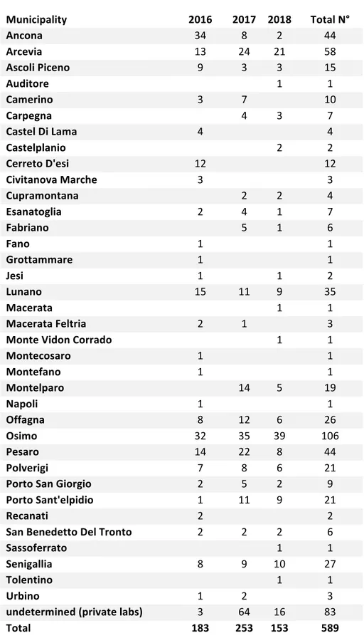
the first year, the peak was recorded in April with 35 requests followed by the month of May with 28 (Figure 5 and Table 2). Veterinary facilities conferring samples to the registry were 35 in the first year (Figure 6), of whom 17 stopped in the second year, and 26 in the second year (Figure 7), 8 of which sent samples for the first time. In 2018 (till August) participating facilities were 28 (Figure 8), of whom 5 had never collaborated to the ACR-M. Over the three years, only 15 veterinary facilities have continuously conferred. The most represented municipalities were Osimo, Arcevia, Ancona, Pesaro and Lunano (Table 3 and Figure 9). Many of 2017-2018 samples were got from private histopathology laboratories, not yet included into the ACR-M mechanism of SIVA request and diagnosis, so these data lack of some details like geographical origin and are indicated as “undetermined” in Table 3. Figure 5 and Table 2: Requests distribution (number) per month and year Month 2016 2017 2018 Total January 10 8 17 35 February 17 33 17 67 March 23 13 20 56 April 32 18 35 85 May 16 19 28 63 June 12 21 15 48 July 21 21 12 54 August 11 10 9 30 September 13 22 - 35 October 9 41 - 50 November 12 26 - 38 December 7 21 - 28 Total 183 253 153 589 0 5 10 15 20 25 30 35 40 45 Ja nu ar y Fe br ua ry Ma rc h Ap ril Ma y Ju ne July Au gu st Se pt em be r Oc to be r Nov em be r De ce m be r Nu m be r 2016 2017 2018
Table 3: Belonging municipalities of the veterinary facilities conferring to ACR-M Municipality 2016 2017 2018 Total N° Ancona 34 8 2 44 Arcevia 13 24 21 58 Ascoli Piceno 9 3 3 15 Auditore 1 1 Camerino 3 7 10 Carpegna 4 3 7 Castel Di Lama 4 4 Castelplanio 2 2 Cerreto D'esi 12 12 Civitanova Marche 3 3 Cupramontana 2 2 4 Esanatoglia 2 4 1 7 Fabriano 5 1 6 Fano 1 1 Grottammare 1 1 Jesi 1 1 2 Lunano 15 11 9 35 Macerata 1 1 Macerata Feltria 2 1 3 Monte Vidon Corrado 1 1 Montecosaro 1 1 Montefano 1 1 Montelparo 14 5 19 Napoli 1 1 Offagna 8 12 6 26 Osimo 32 35 39 106 Pesaro 14 22 8 44 Polverigi 7 8 6 21 Porto San Giorgio 2 5 2 9 Porto Sant'elpidio 1 11 9 21 Recanati 2 2 San Benedetto Del Tronto 2 2 2 6 Sassoferrato 1 1 Senigallia 8 9 10 27 Tolentino 1 1 Urbino 1 2 3 undetermined (private labs) 3 64 16 83 Total 183 253 153 589
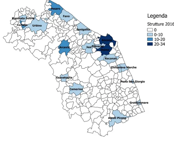
Figure 6: Municipality thematic map of veterinary facilities conferring in 2016
Figure 8: Municipality thematic map of veterinary facilities conferring in 2018
The total number of dogs involved was 569, most of them clustered in 2017 (Figure 10 and Table 4). Figure 10: Dogs distribution (number) per year Table 4: Dogs distribution (number) per year The number of dogs with confirmed tumors was 468 (82%). Of these, in 2016 represented 79% (142/179), in 2017 about 84% (202/240), and in the first 8 months of 2018 were 83% (124/150). Figure 11 and table 5 show distributions of percentage and absolute frequency. Figure 11: Percentage (%) distribution of negative/positive for tumor per year Table 5: Absolute frequency distribution of negative/positive for tumor per year Given the low total number of cases collected in less than 3 years, crude incidence rates (CIR) and related 95% confidence intervals (CI) of benign and malignant tumors per 100.000 dogs were calculated not per year but for the whole period of study. Incidence rate for all tumors was ACR-M Total N° 2016 179 2017 240 2018 150 Total 569 Year Negative
for tumor for tumor Positive number Total
2016 37 142 179 2017 38 202 240 2018 26 124 150 Total 101 468 569 179 240 150 2016 2017 2018 Nu m be r Year 21% 16% 17% 79% 84% 83% 0% 20% 40% 60% 80% 100% 2016 2017 2018 Negative for tumor Positive for tumor
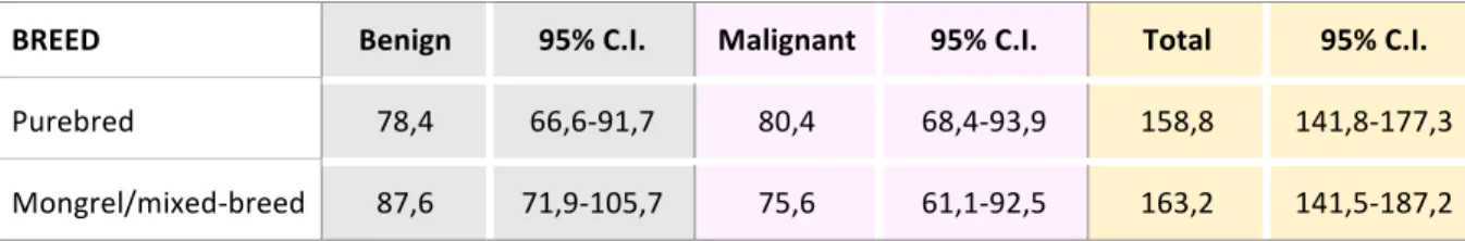
160,5/100.000, while for benign and malignant tumors were 81,9/100.000 and 78,5/100.000 respectively. These data and related 95% confidence intervals are reported in the table 6. Table 6: Incidence rates of tumors per 100.000 dogs and related lower and upper limits 95% confidence intervals (CI) in the Marche region for the period 01/01/2016-31/08/2018.
Tumors Incidence rate Lower limit
95% C.I. Upper limit 95% C.I. All tumors 160,5 147 174,9 Benign tumors 81,9 72,4 92,4 Malignant tumors 78,5 69,2 88,8
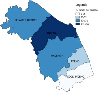
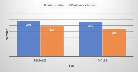
Spatial analysis indicated Ancona and Pesaro-Urbino as the provinces with the greatest number of conferred samples and of tumors (Table 7 and Figure 12). Table 7: Distribution (number) of negative/positive cases for tumor by province and year Year 2016 2017 2018
Province for tumor Negative Positive for tumor for tumor Negative Positive for tumor for tumor Negative Positive for tumor Total
Ancona 19 75 17 89 13 67 280 Ascoli Piceno 5 12 1 8 1 4 31 Fermo 5 4 29 2 13 53 Macerata 4 15 3 28 11 61 Pesaro-Urbino 9 35 13 48 10 29 144 Total 37 142 38 202 26 124 569
Figure 12: Thematic map of tumors (benign and malignant) by province in Jan 2016-August 2018 period Proportional morbidity rates calculated by provinces and year were reported in Table 8. Table 8: Proportional morbidity rates expressed in percentage (PMR 100) by province and year Variable PMR 100 2016 2017 2018 2016-2018 Province Ancona 54% 36% 59% 46% Ascoli Piceno 9% 4% 2% 5% Fermo 5% 16% 9% 11% Macerata 11% 15% 9% 13% Pesaro Urbino 22% 29% 20% 25% Municipalities conferring the greatest number of samples were Ancona (75), Osimo (53), Pesaro (33), Senigallia (21), Pergola (18), and Castelfidardo (15). The same municipalities, but in different order, had the largest number of diagnosis positive for tumor: Ancona (57), Osimo (42), Pesaro (25), Pergola (15), Senigallia (13), and Castelfidardo (13) (Table 9 and Figure 13).
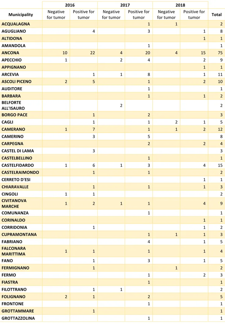
Table 9: Distribution of negative/positive cases for tumor by municipality and year
2016 2017 2018
Municipality for tumor Negative Positive for tumor for tumor Negative Positive for tumor for tumor Negative Positive for tumor Total
ACQUALAGNA 1 1 2 AGUGLIANO 4 3 1 8 ALTIDONA 1 1 AMANDOLA 1 1 ANCONA 10 22 4 20 4 15 75 APECCHIO 1 2 4 2 9 APPIGNANO 1 1 ARCEVIA 1 1 8 1 11 ASCOLI PICENO 2 5 1 2 10 AUDITORE 1 1 BARBARA 1 1 2 BELFORTE ALL'ISAURO 2 2 BORGO PACE 1 2 3 CAGLI 1 1 2 1 5 CAMERANO 1 7 1 1 2 12 CAMERINO 3 5 8 CARPEGNA 2 2 4 CASTEL DI LAMA 3 3 CASTELBELLINO 1 1 CASTELFIDARDO 1 6 1 3 4 15 CASTELRAIMONDO 1 1 2 CERRETO D'ESI 1 1 CHIARAVALLE 1 1 1 3 CINGOLI 1 1 2 CIVITANOVA MARCHE 1 2 1 1 4 9 COMUNANZA 1 1 CORINALDO 1 1 CORRIDONIA 1 1 2 CUPRAMONTANA 1 1 1 3 FABRIANO 4 1 5 FALCONARA MARITTIMA 1 1 1 1 4 FANO 1 3 1 5 FERMIGNANO 1 1 2 FERMO 1 2 3 FIASTRA 1 1 FILOTTRANO 1 1 2 FOLIGNANO 2 1 2 5 FRONTONE 1 1 GROTTAMMARE 1 1
2016 2017 2018 Municipality for tumor Negative Positive for tumor for tumor Negative Positive for tumor for tumor Negative Positive for tumor Total
JESI 2 2 LORETO 3 3 LUNANO 1 1 2 MACERATA 1 5 3 9 MACERATA FELTRIA 1 1 2 MAGLIANO DI TENNA 1 1 2 MAIOLATI SPONTINI 2 1 1 1 5 MASSIGNANO 1 1 MATELICA 1 1 2 MERCATELLO SUL METAURO 1 1 1 3 MONDAVIO 1 1 MONDOLFO 2 2 MONSAMPIETRO MORICO 1 1 2 MONSANO 2 1 3 MONTALTO DELLE MARCHE 1 1 MONTE CERIGNONE 1 1 2 MONTE URANO 1 1 MONTECALVO IN FOGLIA 1 1 2 MONTECAROTTO 1 1 2 MONTECASSIANO 2 2 MONTECICCARDO 1 1 MONTECOSARO 1 1 MONTEFANO 1 1 MONTEGIORGIO 1 1 MONTEGRANARO 2 2 MONTELABBATE 1 1 MONTELEONE DI FERMO 1 1 MONTELPARO 1 3 4 MONTEMARCIANO 1 1 MORRO D'ALBA 1 1 MUCCIA 1 1 2 NUMANA 2 3 1 6 OFFAGNA 3 2 1 6 ORTEZZANO 2 3 5 OSIMO 3 12 4 20 4 10 53 OSTRA 1 1 OSTRA VETERE 1 1 2 PERGOLA 1 5 1 5 1 5 18
2016 2017 2018 Municipality for tumor Negative Positive for tumor for tumor Negative Positive for tumor for tumor Negative Positive for tumor Total
PESARO 2 11 2 11 4 3 33 PETRIANO 1 1 PETRITOLI 4 4 PIANDIMELETO 1 1 1 3 PIETRARUBBIA 1 1 2 PIOBBICO 1 1 POGGIO SAN MARCELLO 1 1 POLVERIGI 2 3 5 10 PONZANO DI FERMO 1 1 2 PORTO RECANATI 2 2 PORTO SAN GIORGIO 1 1 2 PORTO SANT'ELPIDIO 2 1 5 1 5 14 POTENZA PICENA 1 1 RIPE SAN GINESIO 1 1 ROSORA 1 1 2 ROTELLA 1 1 SAN BENEDETTO DEL TRONTO 1 1 1 2 1 1 7 SAN GINESIO 2 2 SAN LORENZO IN CAMPO 1 1 1 6 9 SAN MARCELLO 1 1 SAN PAOLO DI JESI 1 1 SAN SEVERINO MARCHE 2 5 1 1 9 SANTA VITTORIA IN MATENANO 1 1 SANT'ANGELO IN VADO 3 2 5 SANT'ELPIDIO A MARE 3 1 1 5 SARNANO 2 2 SASSOCORVARO 3 3 SASSOFERRATO 1 1 SENIGALLIA 3 5 3 2 2 6 21 SERRA DE'CONTI 3 3 6 SERRA SAN QUIRICO 2 2 SERRA SANT'ABBONDIO 1 1 1 3 SIROLO 1 1 3 5 SPINETOLI 1 1 TAVULLIA 2 1 3
2016 2017 2018 Municipality for tumor Negative Positive for tumor for tumor Negative Positive for tumor for tumor Negative Positive for tumor Total TORRE SAN PATRIZIO 1 1 TRECASTELLI 2 2 URBANIA 1 1 1 3 URBINO 1 1 2 4 URBISAGLIA 2 2 VALLEFOGLIA 2 1 1 2 6 Total 37 142 38 202 26 124 569 Figure 13: Thematic map of tumors (benign and malignant) by municipality in Jan 2016-August 2018 period
2.3.2 Breed distribution
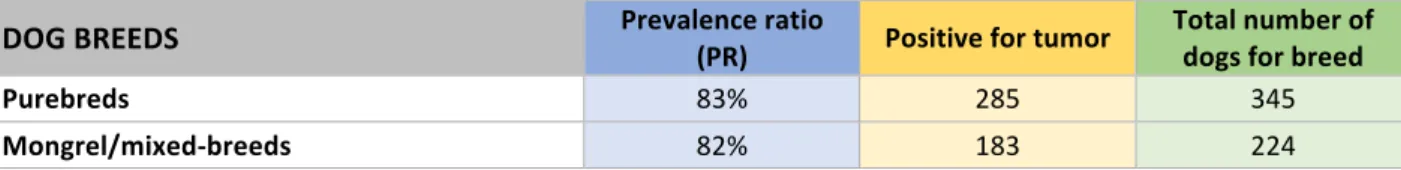
The number of dogs belonging to a given breed varied. Most dogs were purebred (61%), while mongrels were 38% (Figure 14 and Table 10). The most represented breeds were: German shepherd, Labrador retriever, Golden retriever and Miniature pinscher (Table 15). This prevalence remained constant in the whole period of time. Figure 14: Distribution in percentage (%) of mongrel and purebred dogs by year Dog breeds 2016 2017 2018 Mongrel 66 98 60 Purebred 113 142 90 Total number 179 240 150 Table 10: Distribution in number of mongrel and purebred dogs by year Table 15: Distribution (number) of dog breeds DOG BREEDS 2016 2017 2018 Total Mongrel/mixed-breed 66 98 60 224 German shepherd 9 10 10 29 English setter 11 7 8 26 Labrador retriever 6 13 4 23 Golden retriever 3 10 6 19 Miniature pinscher 10 5 4 19 Boxer 5 6 2 13 Dachshund 1 8 3 12 Jack Russell terrier 3 2 4 9 English springer spaniel 4 2 3 9 Miniature poodle 3 2 2 7 Maremma sheepdog 2 3 2 7 American pit bull terrier 3 2 2 7 Italian short haired hound 2 3 2 7 37% 41% 40% 63% 59% 60% 0% 10% 20% 30% 40% 50% 60% 70% 80% 90% 100% 2016 2017 2018 Mongrel Purebred
DOG BREEDS 2016 2017 2018 Total Beagle 2 2 2 6 French bulldog 2 3 1 6 English cocker spaniel 2 2 2 6 Italian coarse haired hound 1 3 2 6 Yorkshire terrier 1 4 1 6 Dobermann pinscher 3 2 5 Shih tzu 2 3 5 Staffordshire bull terrier 2 2 1 5 Poodle 1 1 2 4 Italian Bracco 1 1 2 4 American cocker spaniel 1 3 4 German short haired pointer dog - Kurzhaar 2 2 4 Maltese 2 1 1 4 Medium-size schnauzer 2 1 1 4 Giant schnauzer 4 4 Akita 3 3 Bichon à poil frisé 1 1 1 3 Border collie 1 2 3 Bernese mountain dog 1 2 3 Bulldog 1 2 3 Cane Corso 2 1 3 Australian kelpie 2 1 3 Pug 2 1 3 Dogo Argentino 2 1 3 Lagotto Romagnolo 1 2 3 Siberian husky 2 1 3 Volpino Italiano 1 2 3 Great Dane 1 1 2 Alaskan malamute 1 1 2 Boston terrier 1 1 2 German wired haired pointer dog - Drahataar 1 1 2 Chihuahua 2 2 Chow-chow 1 1 2 Dogue de Bordeaux 1 1 2 Brittany 2 2 White Swiss shepherd 2 2 Italian greyhound 2 2 Rottweiler 1 1 2 Old English sheepdog 1 1 Bolognese 1 1 Great Pyrenees 1 1 Briquet griffon vendeen 1 1 Bull terrier 1 1 Czechoslovakian wolfdog 1 1
DOG BREEDS 2016 2017 2018 Total Cavalier king Charles spaniel 1 1 Chin 1 1 Gordon setter 1 1 Greater Swiss mountain 1 1 Brussels griffon 1 1 Pekingese 1 1 Miniature schnauzer 1 1 Irish setter 1 1 Spinone Italiano 1 1 Miniature German spitz 1 1 Terranova 1 1 Weimaraner 1 1 Whippet 1 1 TOTAL 179 240 150 569 Most dogs with cancer involved purebreds. Over the whole period of study, dogs with tumor were 468, of which 285 purebreds (61%) and 183 mongrels (39%). Observations by year were performed and the general prevalence always resulted similar, indeed in 2016 purebreds represented 65% (93/142) of tumor cases, in 2017 were 60% (121/202), and in 2018 were 57% (71/124) (Figure 16 and Table 11). Figure 16: Distribution in percentage (%) of dogs with tumor by breed and year Dog breeds 2016 2017 2018 Mongrel 49 81 53 Purebred 93 121 71 Total number 142 202 124 Table 11: Distribution in number of dogs with tumor by breed and year 35% 40% 43% 65% 60% 57% 0% 10% 20% 30% 40% 50% 60% 70% 80% 90% 100% 2016 2017 2018 Mongrel Purebred