UNIVERSITA’ DEGLI STUDI DELLA CALABRIA
SCUOLA DI DOTTORATO DI RICERCA "LIFE SCIENCE"
INDIRIZZO: BIOLOGIA ANIMALE
XXIV CICLO
TESI DI DOTTORATO
ANGIOTENSIN II AND MECHANISMS OF OXIDATIVE
DAMAGE IN HUVECs
Coordinatore:
Prof.ssa Maria Carmela Cerra
Supervisore: Dottorando:
Ch.mo Prof. Guglielmo Martino Dott.ssa Annarita Carino
A Mamma, Pap
Mamma, Pap
Mamma, Pap
Mamma, Papà, Andrea e Claudio
à, Andrea e Claudio
à, Andrea e Claudio::::
à, Andrea e Claudio
le quattro persone più importanti della mia vita…
A zio Gianni
zio Gianni
zio Gianni e a Nonno
zio Gianni
Nonno
Nonno
Nonno che, da un mondo lontano insieme
agli angeli del cielo, mi sono stati sempre vicino
TABLE OF CONTENTS
ABSTRACT……….………..pag.6
INTRODUCTION………...…7-20
• The Endothelium and vascular homeostasis
• Endothelium dysfunction, atherosclerosis and vascular inflammation • Effects of Endothelium dysfunction
OUTLINE OF THE THESIS...21
ORIGINAL DATA
PART.1
1.1 Evaluation of Angiotensin II regulatory mechanisms involved in the production/degradation of Reactive Oxygen Species and Reactive Nitrogen Species...22-45
• CHAPTER 1. Renin- Angiotensin- System : from Angiotensin I to Angiotensin IV in
Endothelial dysfunction.
• CHAPTER 2. Radical Oxidative Stress : Reactive Oxygen Species and Reactive
Nitrogen Species in Human Endothelial Cells
• CHAPTER 3. Asymmetric Dimethyl Arginine and Oxidative Stress in Endothelial
Dysfunction
1.2 Monitoring of the protective effects of antioxidants flavonoids on membrane
damage and radical species production...46-59
• CHAPTER 1. Antioxidant networks and redox signaling
• CHAPTER 2. Flavonoids : Epigallocatechin-gallate and its effects in vitro
stress pathway...60-73
• CHAPTER 1. Angiotensin IV and Renin-Angiotensin-System in vitro models.
• CHAPTER 2. Angiotensin IV receptor and IRAP receptor
• CHAPTER 3. Pharmacologic Profile of Angiotensin IV
Materials and Methods...74-88
Results and Discussion ...89-120
CONCLUSIONS...120-122
PUBLICATIONS AND
ABSTRACTS...123-124
ACKNOWLEDGEMENTS...125
LIST OF ABBREVIATIONS
...126REFERENCES...128-157
ABSTRACT
The endothelium is essential for the maintenance of vascular homeostasis. Central to this role is the production of endothelium – derived nitric oxide (EDNO), synthesized by endothelial isoform of nitric oxide synthase (eNOS). Endothelial dysfunction, manifested as impaired EDNO bioactivity, is an important early event in the development of various vascular disease, including hypertension, diabetes, genesis of tumors and atherosclerosis. Endothelial dysfunction is an early feature of atherosclerosis vascular disease, characterized by a decrease in nitric oxide (NO) bioavailability and a concomitant increase in vascular superoxide (O2. -) formation. Loss of NO bioavailability precedes the development of overt atherosclerosis and is an independent predictor of adverse cardiovascular events. Indeed, decreased NO and enhanced production of reactive oxygen species (ROS) have been recognized as major determinants of age-associated endothelial dysfunction. The degree of impairment of EDNO bioactivity is a determinant of future vascular complications. Accordingly, growing interest exists in the pathologic mechanism involved. However it is clear that immunologic mechanisms operating in the context of common cardiovascular risk factors lead to impaired endothelial function, mainly as a consequence of decreased NO bioavailability and excessive oxidative stress.
The work submitted in this thesis describes on one side studies aimed to investigate cellular mechanisms underlying endothelial dysfunction and vascular damages driven by oxidative stress in the context of aging, hypertension and atherosclerosis using in vitro models. In addition, we desired to evaluate the efficacy of reducing agents such as flavonoid to monitor whether theyactually have an action to recover from the cellular oxidative damage by these natural compounds and how real is their action at the level of microcirculation in vitro models. On the other side, we present studies focused on the pathophysiology of microcirculation as far as functional aspects are concerned in the context to better understand the functioning of the Renin- Angiotensin-System in particular if the Angiotensin IV is involved in mechanisms of oxidative stress and in Calcium intracellular levels.
Introduction
Modern Age has witnessed major developments in cardiovascular physiology thanks to scientists like E.H. Starling in the 1920s, who described the “fundamental properties of the
heart muscle itself and then found out how these are modified, protected, and controlled under the influence of the - nervous, chemical and mechanical- mechanisms which under normal conditions play on the heart and blood vessels”, quoting his remarkable studies
[167,211]. The existence of pathological cardiovascular conditions has been recognized and described. The atherosclerotic vascular disease and its main clinical manifestation, angina
pectoris were firstly reported in the 18thcentury. In parallel, the earliest descriptions of some attempts for a pharmacological treatment appeared. Nitroglycerin, for example, was initially prescribed by physicians in the late 19th century [172]. A number of new pharmacological agents found to be of benefit followed, leading to the constitution of the currently established cardiovascular pharmacotherapy. The atherosclerotic vascular disease was hypothesized to cause ischemia and infarction of the heart and other organs. Acute cardiovascular ischemic events, such as stroke, myocardial infarction or sudden death, were frequently associated with localized arterial thrombus formation. However for many decades, these conditions remained totally mysterious and unpredictable events. Moreover, hypertension was recognized to damage large blood vessels and the microcirculation of different target organs, even if the specific mechanisms involved were unknown.
Vascular biology, as the study of vascular cells under normal and pathological conditions, began in the 1970s. This novel research discipline, has since then enjoyed exponential growth, allowing the comprehension of common pathophysiologic processes of cardiovascular diseases (CVD). In 1980, the obligatory role of endothelial cells in relaxation of arterial smooth muscle by acetylcholine (Ach) was described [76]. A seminal event in the field of vascular biology, which was later awarded with the Nobel Prize in 1998 to Robert Furchgott, Louis Ignarro and Ferid Murad. By means of in vitro experiments in organ chambers, preconstricted arterial rings were demonstrated to relax in response to the muscarinic cholinergic agonist only if endothelial cells were present. Removing the endothelium by any means abolished the vasorelaxation, which was mediated by an undefined endothelium-derived substance that was named endothelium endothelium-derived relaxing factor (EDRF). EDRF, subsequently, was shown to be, in large part, nitric oxide [153]. During the last three decades,
several studies have definitively proved that the endothelium is not only a cell monolayer covering the lumen surface of the vascular wall, but it is involved in many key regulatory functions for the homeostasis of cardiovascular system.
The endothelium and vascular homeostasis
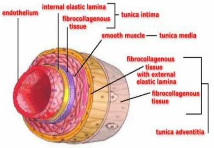
In recent years the endothelium in the literature has been simply classified as a biologically inert barrier interposed between the blood and the vessel wall. It was considered a uniform layer of cells with the task of providing a protective surface that would prevent the adhesion of blood cells but that would be able to carry or convey liquids, solutes and various substances in the blood stream from this interstitium. Since a few years, detailed studies on anatomy and physiology of endothelium have beenproviding more information that make this definition unacceptable. In fact, currently the endothelial tissue is considered a real organ with the function to modulate vascular tone and extent of blood flow [190] and has a primary role in maintaining vascular homeostasis associated with features [178], in response to humoral, nervous and mechanical stimuli[190]. The endothelium is able to perform its functions only if intact, when it establishes a balanced equilibrium between factors related to the mechanism for inducing vasodilation and vasoconstriction factors generating mechanism. When the ability of endothelial cells is compromisedto process the substances that are produced under physiological conditions the "endothelial dysfunction" is established[96]. The endothelium is the inner face of endoluminal arteries, veins, capillaries and lymphatic vessels lined by a continuous cell layer consisting of endothelial cells. The essential components of the walls, in endothelium, are classified as (Fig. 1):
• tunica intima: inner layer, delimiting the lumen of the vessel, consisting of a monolayer of endothelial cells from the underlying basement membrane and the internal elastic lamina;
• tunica media: the middle layer consists of smooth circular and longitudinal fibers;
• tunica adventitia: outer skin consists of connective tissue and fibroblasts, separated from the tunica media by the external elastic lamina.
Figure 1- Endothelial wall- Nature.com
Endothelial cells are polygonal and flattened by microscopic study (30µm x 10µm). They contain numerous vesicles and form junctional complexes with other nearby cells. Cells are welded together by means of both tight junctions,through gap junctions. The only type of cell to possess Weibel-Palade bodies, structures surrounded by membranes with a diameter of 0.1
µm and a length of 3 mM, which are organelles of the deposit of von Willebrand factor (vWF)
[178].
Endothelial function
Under physiological conditions, integrity of the endothelial cells is essential for normal function of blood vessels and maintenance of homeostasis through secretion or display on the membrane of different molecules that are responsible for the continuous regulation of vascular tone, control of blood pressure, the physiological regulation of leukocyte passage and maintenance of antithrombotic and anticoagulant effects [190,177]. Endothelial paracrine activity is directed both toward the lumen and towards the vessel wall. At the vessel lumen level, the endothelium regulates coagulation mechanisms and interactions with leukocytes and platelets [177,4]. At the vascular wall level, endothelium plays a central role in regulating blood pressure
and blood flow through a continuous modulation of vascular tone, which is under the control of local and systemic factors. Modulating vascular tone and the same structure, the endothelium plays a major role in vascular remodeling observed in hypertension [81], the stenosis after angioplasty [115] and atherosclerosis [25]. Central role in regulating endothelial cell homeostasis and thrombosis depends on the expression and release of many molecules, with autocrine and paracrine actions, such as prostacyclin PGI2, nitric oxide (NO), platelet-activating factor (PAF), von Willebrand factor (vWF), thrombomodulin, tissue factor (thromboplastin) and its inhibitor (TFPI), factor, tissue plasminogen activator (t-PA) and its inhibitor (PAI-1) [4]. In addition to being involved in the mechanism of activation and aggregation of platelets, the PAF also increases the permeability of the endothelial barrier and, together with the platelet adhesion molecule (P-selectin), promotes leukocyte adhesion to the vessel wall [155]. The adenosine diphosphate (ADP) that is released by the activation of platelets, along with the thromboxane A2, promotes platelet activation and aggregation through a more positive feedback mechanism [49,93]. The vWF factor has essential functions in homeostasis: it mediates platelet adhesion to sites of vascular injury, mean plate-plate interaction, promotes platelet aggregation in vessels with high shear-stress due to the rapid blood flow and transport coagulation factor VIII in plasma [194,193]. Endothelial cells express on their surface a protein that is the intrinsic membrane thrombomodulin and is found exclusively on the endothelial luminal surface, surface that is not damaged. It has a strong affinity to thrombin, plasma protein involved in the mechanism of the coagulation cascade. In particular, it is involved in the conversion from fibrinogen to fibrin and also in the platelet activation and aggregation [45,21]. When thrombin binds to thrombomodulin, it causes conformational rearrangements of the second compound reducing its affinity for fibrinogen and activates circulating protein C which, together with protein S, inactivates factors V and VIII, inhibiting blood coagulation [65,77]. This suggests that thrombin has an anticoagulant action in the presence of thrombomodulin [52]. Thromboplastin is a main physiological activator of coagulation. In contrast to extravascular cells, under physiological conditions, endothelial cells do not express the thromboplastin in order to preserve the fluidity of the blood [50]. However, in vitro studies have shown that endothelial cells synthesize and express on the surface in response to thromboplastin thrombin, and cytokine-activated platelets. Endothelial cells also produce tissue factor inhibitor (Tissue Factor Pathway Inhibitor, TFPI), which isthe most important physiological inhibitor of thromboplastin. This
protein is secreted by endothelial cells and is found in plasma or bound to the cell surface [197]. Under basal physiological conditions, endothelial cells do not express molecules that promote the adhesion of circulating leukocytes. However, the activation of endothelial cells by thrombin, endotoxin or inflammatory cytokines such as IL-1 and TNF-α, induces the surface expression of a series of molecules that are essential for adhesion and migration of leukocytes from the bloodstream into the damaged tissue [208]. This process is mediated by cell adhesion molecules (CAMs), glycoproteins expressed on the surface of activated cells that are involved in cell-cell and cell-matrix binding [61]. The binding of leukocytes to endothelium is mediated by immunoglobulin ICAM-1 (inter cellular adhesion molecule-1) and VCAM-1 (vascular adhesion molecule-1) that interact with integrins on the surface of circulating leukocytes and cause a stable relationship with the endothelium [19,230]. ICAM-1 is over-expressed in response to various stimuli such as inflammatory cytokines (IL-1, TNF-α, interferon γ). VCAM-1 is expressed by endothelial cells and smooth muscle cells of the vessel wall, promotes cell-cell adhesion and subsequent migration of inflammatory cells [62]. Locally, vascular tone is self-regulated mainly in response to mechanical stimuli by the power button and the shear stress affecting the vessel walls as a result of changes in blood flow [249]. This regulation is modulated by the synthesis and secretion of two potent vasodilators: prostacyclin (PGI2) and nitric oxide (NO). PGI2 is a prostaglandin deriving from arachidonic acid and is synthesized by the action of prostaglandin PGI2 synthase from H2 (PGH2) which is produced by the hydrolysis of arachidonic acid through cyclooxygenase-2 (COX-2). Nitric oxide is a diatomic free radical generated by nitric oxide synthase hetero (NOS) through the oxidation of L-arginine to L-citrulline [84]. NO is produced in the endothelium and is released as a free radical and as a nitrosyl compound, pre-eminent importance in controlling the tone of the arteries and the microcirculation, both in basal conditions and after various kinds of stimulation [74,84]. Nitric oxide has been identified as a major anti-atherosclerosis factor [37] because ofits protective action on blood vessels [159]. Nitric oxide induces vessel relaxation activating the guanylate cyclase enzyme. In addition, we found that nitric oxide inhibits the oxidation of low density lipoprotein (LDL). It is also an antagonist of platelet aggregation by inhibiting platelet activation [159]; moreover, it represses the expression of kB dependent nuclear factor of adhesion molecules that regulate the recruitment of leukocytes in the endothelium, in the early stages of atherosclerosis. Endothelial cells synthesize NO and PGI2 in response to various substances to agonist action,
such as bradykinin, acetylcholine, serotonin, thrombin, adenosine triphosphate (ATP) and adenosine diphosphate (ADP) that increases the concentrations of cytoplasmic calcium, and also in response to mechanical stimuli, such as deformation of the plasma membrane due to shear stress [74,84]. The increased shear stress across the endothelium promotes the release of other agonists, vasodilators, such as ATP and phosphate leading to increased cytoplasmic calcium in adjacent cells, thus, stimulating the synthesis of other NO. These observations are in agreement with previous hypotheses that required calcium levels to activate the synthesis of NO, calcium levels are lower than those who need to activate the synthesis of PGI2 [35]. The endothelium also acts in the process of modulation of the mechanism of vasoconstriction by means of certain substances and effector molecules such as angiotensin, endothelin, prostaglandin H2 and thromboxane A2 [47].
The Endothelium dysfunction, atherosclerosis and vascular inflammation
"Endothelium dysfunction" (Fig.2) refers to the condition in which the endothelium loses its physiological properties [179] and is determined by an imbalance in the production of mediators that regulate vascular tone, platelet aggregation, the coagulation and fibrinolysis .
Figure.2 Endothelial dysfunction
Endothelial dysfunction has been associated with a variety of processes including hypertension, atherosclerosis, aging, heart block and failure, coronary syndrome, obesity, infections, sepsis, arthritis rheumatoid diseases, thrombosis, cigarette smoking, and type 1 and 2 diabetes [72]. Oxidative stress plays a relevant role in atherosclerosis and cardiovascular diseases by promoting cellular dysfunction, inflammation and lipids and lipoproteins peroxidation, and is reducing the bioavailability of NO [259]. The mechanisms of endothelial dysfunction shows many unclear details, but what is definitely known is that the reduction in the bioavailability of NO is a relevant aspect of endothelial dysfunction [175]. This reduction of NO is followed by high levels of endothelial vasoconstrictive substances such as endothelin-1 [50]. The mechanisms by which NO bioavailability acts are essentially two: the first mechanism involves the down-regulation of the expression of eNOS (endothelial NO synthase) which synthesizes nitric oxide in the endothelium through the oxidation of nitrogen contained in the L-arginine, which is converted to L- citrulline. The reaction requires the presence of cofactors, such as nicotinamide adenine dinucleotide phosphate (NADPH), calcium / calmodulin (CaM), flavin adenine dinucleotide (FAD), flavin mononucleotide (FMN) and tetrahydrobiopterin (BH4) [74,84] that are integrated in the structure of NOS, once the enzymes are expressed in the cell [249]. The other mechanism involves the up-regulation of vascular levels of ROS . It is known that high levels of ROS damage the vascular tissue, as is known to react with NO to form peroxynitrite (ONOO.-), thus removing NO. In fact, it was demonstrated that the increased expression of superoxide may lead to the "scavenging" of NO and its reduced bioavailability [114,236]. The shear stress signal and activity of eNOS are regulated by mechanisms of Ca2+ / calmodulin-dependent [29] and Ca2+ / calmodulin-independent [15]. The eNOS is a homodimer consisting of two monomers, each of which contains a reductase (C-terminal) domain that binds NADPH, FAD and FMN, and a domain which carries a heme oxygenase (N-terminal). These heme groups are required for dimerization of both monomers to form the active NOS dimer and electron transfer from flavin to heme of the opposite monomer. To become a fully functional enzyme complex, it binds to the oxygenase domain (6R) -5,6,7,8-tetrahydrobiopterin (BH4), molecular oxygen and substrate L-arginine. Finally, a zinc ion binds at the interface of the dimer of NOS, and has mainly a structural function [15]. The eNOS catalyzes an electron transfer from NADPH reductase domain in the heme oxygenase
domain, by means of FAD and FMN. This electron transfer is favored by the binding of calmodulin induced by calcium, the domain reductase. The O2 is reduced and L-arginine is oxidized by 2-step, through N-hydroxy-L-arginine to L-citrulline and NO. Once produced NO, a short half-life gas (6-7 seconds), readily crosses the plasma membrane of smooth muscle cells of the tunica media and activates the enzyme guanylate cyclase resulting in the production of intracellular cyclic GMP, which causes smooth muscle relaxation and vasodilation through a series of mechanisms that reduce the levels of intracellular free calcium . The activity of eNOS is regulated by the concentration of intracellular calcium and by phosphorylation of the reductase domain and the calmodulin binding domain. Depending on the stimulus, the specific kinase can phosphorylate eNOS, such as Akt / protein kinase B, protein kinase A, protein kinase 5'-adenosine monophosphate activated and calmodulin-dependent kinase II. If the electron flow is disturbed in eNOS (in vitro, this may be due to the absence of the cofactor BH4 or substrate L-arginine), it changes from a coupled eNOS (dimer), which leads to the formation of NO, to a decoupled state, which generates oxygen. the enzyme moves from an oxidant state to a reduced one [133]. During the normal catalytic function of eNOS, BH4 acts as an electron donor. Under conditions of excessive production of oxidants, oxygen can react with NO, that formed ONOO.-
(peroxynitrite) [88]. It has been
shown that ONOO.-(peroxynitrite) oxidizes BH
4 in biologically inactive products that cannot be further recycled . The formation of peroxynitrite, through a process of nitration of protein affects the function of proteins and thus endothelial function;it also mediates the oxidation of LDL, contributing to the pro-atherogenic conditions [73]. Functions of nitric oxide include an important role in maintaining vascular endothelial integrity and stability, preventing platelet aggregation and leukocyte adhesion and maintenance of blood flow [31,179]. It is not so surprising that the damage to NO synthase, which is produced by the endothelium dysfunction, has deleterious effects on blood pressure and platelet count.Thus the damage to NO synthase allowschanges to the vessel wall, that are known to be associated with vascular pathogenesis in general, and atherosclerosis in particular . The reduced concentration of NO causes an "up-regulation" of VCAM-1 in endothelial cells through the expression of NF-kB ]. ROS, C-reactive protein (CRP) and oxidized LDL receptor 1 (LOX-1) increase expression of adhesion molecules (Fig.3), such as VCAM-1, ICAM-1 and E-selectins that are important in the early stages of the inflammatory process . VCAM-1 is responsible for ties with monocytes and T cells represent the first stage ofthe invasion of the vessel wall by inflammatory cells . Endothelial dysfunction allows the infiltration and retention of LDL in the serum of the tunica intima of the vessel starting an inflammatory response. Within the intima, LDL particles are modified by oxidation or by promoting the enzymatic release of phospholipids that stimulate endothelial cells to express VCAM-1 and ICAM-1 and produce growth factors, such as granulocyte-macrophagecolony stimulating factor (GM-CSF) [144]. The CAMs mediate the entry of specific leukocytes into the vessel wall, monocytes and lymphocytes at the site of endothelial damage [90,138]. Within the intima, GM-CSF stimulates monocytes to become macrophages, which plays an important role in local inflammatory response by producing inflammatory cytokines, chemokines and oxygen free radicals [138,191]. In addition, the secretion of extracellular matrix metalloproteases (MMPs) by macrophages contributes to the remodeling of the vessel wall and eventual plaque rupture. These molecules amplify the cellular response and begin promoting the low-intensity vascular inflammation, thrombosis, progressive thickening of intima and hence the formation and development of atherosclerotic plaque that may break possibly causing clinical manifestations [191].
Figure 3- Different molecules involved in endothelial dysfunction-
Science.com
In 1997, the isolation of putative endothelial progenitor cells (EPCs) from human peripheral blood has been reported [8]. Subsequently, evidence has accumulated documenting the presence of a population of endothelial precursor cells and/or adult stem cells, deriving from bone marrow, with a specific role inthe maintenance of endothelial integrity against vascular injury [34].These cells are able to home areas of injury and ischemia-induced myocardial and peripheral neovascularization [109], healing endothelial integrity .
Effects of Endothelial Dysfunction
Endothelial function has largely been investigated through the assessment of endothelium dependent vasomotion. Indeed, an impaired endothelium-dependent relaxation reflects an extensive loss of endothelial function. Actually it was important to characterize how endothelial dysfunction relates to the pathogenesis of atherosclerosis. Studies focusing on these issues continue to provide remarkable evidence that the endothelial interface between the vascular wall and the circulation is the primary site to trigger off cardiovascular events [55].Under pathologic conditions, the endothelium has a reduced availability of vasodilating factors, in particular NO, and an augmented production of vasoconstricting factors, leading to impaired endothelium-dependent vasodilation. Furthermore, NO has demonstrated to exert a major anti-inflammatory effect and can therefore be considered the most important endogenous antiatherogenic molecule. Endothelial dysfunction promotes arterial inflammation and vice versa, chronic inflammation maintains a pro-inflammatory phenotype of the endothelium [55]. Therefore EC dysfunction seems to participate in atherosclerotic process from its inception onwards till ultimate complications with a complex and pleiotropic involvement of inflammation sustained by humoral and cellular inflammatory elements. Not only these effects are present in endothelial dysfunction but other factors are involved in its genesis.
Cardiovascular Disease and Endothelial Dysfunction
Endothelial dysfunction was first described in human hypertension in the forearm vasculature in 1990 [171]. Impaired vasodilation in hypertension was confirmed by many studies in different vascular beds, including small resistance vessels [173,201]. In stage I essential hypertension, we
showed that ~ 60% of patients exhibit impaired small artery vasodilatation when this is
studied in vitro in vessels dissected from gluteal subcutaneous biopsies [174]. Impairment of
vasodilation was also described in type 1 [14] and type 2 diabetes [189,154], coronary artery disease [122], congestive heart failure [24], and chronic renal failure [225,255]. Moreover, this manifestation of endothelial dysfunction not only is associated with cardiovascular disease but may also precede its development, as shown in a study on offspring of hypertensive patients. The study subjects displayed endothelial dysfunction despite being normotensive. Another study showed endothelial dysfunction in symptom-free children and young adults at high risk for atherosclerosis [12]. Also, in normotensive, normoglycemic, first-degree relatives of patients with type 2 diabetes, endothelial dysfunction was correlated with insulin resistance [64]. Endothelial dysfunction has been demonstrated in the metabolic syndrome and in dyslipidemia and may be associated with obesity [247], hyperhomocysteinemia , sedentary lifestyle, and smoking, in the absence of overt cardiovascular disease.
The
pathophysiology of endothelial dysfunction
is complex and involves multiple mechanisms.NO
One of the most important vasodilating substances released by the endothelium is NO, which acts as a vasodilator, inhibits growth and inflammation, and has anti-aggregant effects on platelets. Reduced NO has often been reported in the presence of impaired endothelial function. It may result from reduced activity of endothelial NO synthase (eNOS; as a result of endogenous or exogenous inhibitors or reduction in the availability of its substrate, L-arginine) and from decreased bioavailability of NO. ROS are known to quench NO with formation of peroxynitrite [119], which is a cytotoxic oxidant and through nitration of proteins, theywill affect protein function and therefore endothelial function. Peroxynitrite is an important mediator of oxidation of LDL, emphasizing its proatherogenic role . Moreover, peroxynitrite leads to degradation of the eNOS cofactor tetrahydrobiopterin (BH4) [148], leading to "uncoupling" of eNOS. Using a novel peroxynitrite decomposition catalyst, FP15, endothelial and cardiac dysfunction could be prevented in diabetic mice . Oxidant excess will also result in reduction of BH4 with increase in BH2. When this occurs, formation
of the active dimer of eNOS with oxygenase activity and production of NO is curtailed (uncoupling of eNOS). The reductase function of eNOS is activated and more ROSs are formed, so NO synthase goes from its oxygenase function producing NO to its reductase function producing ROS, with the consequent exaggeration of oxidant excess and deleterious effect on endothelial and vascular function [222]. Oxidative excess is linked to a proinflammatory state of the vessel wall. ROS upregulate adhesion (VCAM-1 and ICAM-1) and chemotactic molecules (macrophage chemoattractant peptide-1 [MCP-1]) [87]. Inflammation decreases NO bioavailability. Indeed, C-reactive protein (CRP) has been shown to decrease eNOS activity [244]. The main source for oxidative excess in the vasculature is NAD(P)H oxidase [245,89]. Other sources include xanthine oxidase [88], the mitochondria [234] and uncoupled NOS.
Asymmetric Dimethylarginine
A relatively new and attractive mechanism that leads to reduced NO is asymmetric dimethylarginine (ADMA), an endogenous competitive inhibitor of eNOS, that has been linked to endothelial dysfunction. In human endothelial cells, which were stimulated with plasma from patients with chronic renal disease, inhibition of eNOS correlated with plasma ADMA levels [60]. ADMA levels were inversely related to endothelium-dependent vasodilation [254] in subjects with hypercholesterolemia, and infusion of L-arginine, the substrate of eNOS and competitor of ADMA, normalized endothelial function. It has been suggested that accumulation of this endogenous eNOS inhibitor leads to reduced effective renal plasma flow and increased renovascular resistance and BP [20]. Intravenous low-dose ADMA reduced heart rate and cardiac output and increased mean BP . ADMA is a product of protein turnover and is eliminated by excretion through the kidneys or metabolism to citrulline by the enzyme dimethylarginine dimethylaminohydrolase (DDAH). Recently, overexpression of DDAH was shown in transgenic mice to decrease ADMA, increase eNOS activity, and reduce BP [2], underlining the pathophysiologic importance of ADMA. Because ADMA is eliminated through renal excretion and degradation by DDAH, it is not surprising that it is increased in patients not only with chronic renal failure [51,111] but also with other diseases such as hepatic dysfunction [240]. New interest is focusing not only on the elimination but also on the generation of ADMA. Protein-arginine methyltransferases, which
produce methylated arginines, namely protein-arginine methyltransferase-1, were shown to be upregulated by shear stress, and this upregulation was associated with enhanced ADMA generation . Hypercholesterolemia is a risk factor for atherosclerosis, associated with endothelial dysfunction [168], and there is now also evidence that elevated ADMA levels are associated with hypercholesterolemia . Plasma ADMA levels were also increased in elderly hypertensive patients [20] and correlated with age and BP [110]. ADMA levels have been associated with increased cardiovascular risk factors in renal failure, such as CRP, carotid intima-media thickness, concentric left ventricular hypertrophy, and left ventricular dysfunction [110,150]. Moreover, it was found to be a predictor of acute coronary events [262], overall mortality of patients with chronic renal failure[239], and mortality of critical ill patients [240].
Oxidative Excess
In animal models of hypertension, oxidative excess leads to endothelial dysfunction as evidenced by improvement of the impaired endothelium-dependent relaxation after use of antioxidants [260]. Oxidative excess in hypertensive patients leads to diminished NO [57] and correlates with the degree of impairment of endothelium-dependent vasodilation and with cardiovascular events [226]. In patients with chronic renal failure, markers of oxidative excess also correlated with endothelial dysfunction [94]. Findings in animal models of chronic renal failure suggest that enhanced generation of ROS leads to decreased NO bioavailability and endothelial dysfunction, which may be improved by antioxidant pretreatment [5,92]. In humans with chronic renal failure, the administration of vitamin C improved endothelial dysfunction of resistance arteries but not of conduit arteries [243]. In animal models of diabetes, increased oxidative excess also led to endothelial dysfunction [46]. ROS also seem to be involved in the mediation of endothelial injury leading to programmed cell death or apoptosis and to a form of apoptosis characterized by detachment of endothelial cells called anoikis [76]. Apoptosis is induced by the loss of cell-matrix interactions, but its exact mechanisms and pathophysiological role in cardiovascular disease are not fully understood. Eicosapentaenoic acid, a polyunsaturated fatty acid contained in fish oil, was shown to protect endothelial cells from anoikis [113], which may contribute to the antiatherogenic and cardioprotective effects of fish oil.
Ang II
Ang II has been implicated in the pathophysiology of hypertension and chronic renal failure. Ang II infusion induces endothelial dysfunction in rats [227], increases ROS by stimulating NAD(P)H oxidase [89,182], and promotes vascular inflammation [233]. In hypertensive humans, interruption of the renin-angiotensin system with angiotensin-converting enzyme inhibitors or angiotensin receptor blockers restores endothelial function in contrast to a similar degree of BP lowering with a -blocker, which has no effect on endothelium-dependent vasodilation [201,233,203].
Hyperhomocysteinemia
A non traditional cardiovascular risk factor that leads to endothelial dysfunction is hyperhomocysteinemia. This has been evidenced by animal models of hyperhomocysteinemia [202]. Normotensive patients with hyperhomocysteinemia display endothelial dysfunction . Folic acid supplementation was able to reduce homocysteine levels and improved endothelial dysfunction in children with chronic renal failure . Cellular [248], animal [202], and human studies suggest that homocysteine reduces NO bioavailability by oxidative excess. There is now also evidence that homocysteine may cause ADMA accumulation by inhibition of DDAH . Experimental studies in humans have confirmed that hyperhomocysteinemia may lead to endothelial dysfunction via accumulation of ADMA [257,218]. However, not all studies support this link [219]. These mechanisms may explain the increased cardiovascular risk of patients with hyperhomocysteinemia. This is of special importance for patients with chronic renal failure, who often have increased homocysteine levels, which were shown to predict cardiovascular outcomes in a recent study .
Diabetes
In diabetes, additional mechanisms may trigger endothelial dysfunction. In states of insulin resistance, such as in type 2 diabetes, insulin signaling is altered, differently affecting the two major pathways emerging from the insulin receptor. The pathway leading via phosphoinositide 3-kinase, phosphoinositide-dependent kinase-1, and Akt/protein kinase B to phosphorylation and activation of eNOS is dramatically downregulated, whereas the pathway leading via mitogen activated protein kinases to mitogenic effects and growth is unaffected
[141,251]. Moreover, hyperglycemia leads to advanced glycation end products (AGE), which were shown to quench NO and impair endothelial function, as evidenced by inhibition of advanced glycosylation with aminoguanidine [48]. AGE induce ROS and promote vascular inflammation, with enhanced expression of interleukin-6, VCAM-1, and MCP-1. This turns into a vicious circle in diabetic nephropathy, because in renal failure, clearance of AGE is delayed, which further promotes vascular and renal injury [28]. Finally, acute hyperglycemia itself can reduce NO [258] and attenuate endothelium-dependent vasodilation in humans in
vivo. Endothelial dysfunction has been proposed to be an early event of pathophysiologic
importance in the atherosclerotic process and provides an important link between diseases such as hypertension, chronic renal failure, or diabetes and the high risk for cardiovascular events that patients exhibit with these conditions. Low NO bioavailability can upregulate VCAM-1 in the endothelial cell layer via induction of NF-κB expression. ROS, CRP, CD40 ligand, and lectin-like oxidized LDL receptor-1 (LOX-1) also upregulate endothelial expression of adhesion molecules. The expression of VCAM-1, ICAM-1, and E-selectin plays a role in the initiation of the inflammatory process. VCAM-1 binds monocytes and T lymphocytes, the first step of invasion of the vessel wall by inflammatory cells. NO inhibits leukocyte adhesion .Reduction in NO results in induction of MCP-1 expression, which recruits mononuclear phagocytes. Monocytes are transformed into lipid-loaded foam cells. Oxidized LDL, for example, is scavenged through LOX-1, which is highly expressed in blood vessels in hypertension, diabetes, and dyslipidemia. Oxidized LDL uptake by LOX-1 triggers a variety of actions: it reduces eNOS expression and further stimulates adhesion molecule expression. LOX-1 expression can be stimulated by Ang II and endothelin-1. As the atherosclerotic plaque progresses, growth factors secreted by macrophages in the plaque stimulate vascular smooth muscle cell growth and interstitial collagen synthesis. The event that initiates a majority of myocardial infarctions is the rupture of the fibrous cap of the plaque, inducing thrombus formation. Decreased NO and oxidative excess may activate matrix metalloproteinases (MMP) [82,140], namely MMP-2 and MMP-9, which weaken the fibrous cap. Because NO inhibits platelet aggregation , reduced NO contributes to thrombogenicity and to the severity of the event. Thus, endothelial dysfunction with reduced NO bioavailability, increased oxidant excess, and expression of adhesion molecules contributes not only to initiation but also to progression of atherosclerotic plaque formation and triggering of cardiovascular events [220].
Part.I:
Evaluation of Angiotensin IIregulatory
mechanisms involved in the production/degradation of
Reactive Oxygen Species and Reactive Nitrogen
Species
Renin – Angiotensin- System :
from Angiotensin I to Angiotensin IV
concerning
endothelial dysfunction
.
1.1 Renin Angiotensin System (RAS)
New components and functions of the renin-angiotensin system (RAS) have already been completely revealed and described in full details. The classical RAS as it appeared in 1970 included renin, which acts on angiotensinogen for the production of Angiotensin I, which in turn is converted into Angiotensin II by Angiotensin converting enzyme (ACE ) [78].
For decades, the Angiotensin II was regarded as the final product and the only bioactive peptide of the renin-angiotensin system (RAS) [224]. Angiotensin II was still considered the main effector of RAS, it was considered only as a circulating hormone that acted via angiotensin receptors, Angiotensin type 1 (AT1) and Angiotensin type 2 (AT2) receptors, on endothelial cells. Since then, a broad view of the RAS has gradually emerged. The RAS (local) tissue is identified in most organs. Recently an intracellular RAS was reported. Therefore, the RAS endocrine function, and paracrine, intacrine [78] were described. Other peptides of RAS denote those biological actions, the heptapeptide Angiotensin 2-8 [78] or heptapeptide Des Asp 1-Ang II (Ang III) possess actions similar to those of Ang II. In addition, the hexapetideAngiotensin 3-8 [78], or hexapeptide Des Asp1-Des Asp 2-Arg Ang II (Ang IV), exerts its action through the receptor insulin-regulated aminopeptidase. Finally, Angiotensin 1-7 or hheptapeptide Des Phe 8-Ang II (Ang 1-7), act through the receptor Mas [78]. Among these fragments of Angiotensin II, Ang IV has attracted more attention since it was unveiled to exercise a wide variety of effects, including, the ability to enhance learning and preserving the memory, anticonvulsant and anti-epileptogenic properties, protection against cerebral ischemia, vascular activity and involvement in atherogenesis. Some of these effects are mediated by AT1 receptor but others are more likely by binding of Ang IV to the insulin-regulated aminopeptidase (IRAP), although the exact mechanism that mediates these actions is not yet well known [224]. In fact, three hypotheses have been proposed:
I. since Ang IV is an inhibitor of the catalytic activity of IRAP, its effects in vivo could result from an accumulation of the peptide substrates of IRAP;
II. IRAP is co-located with the glucose transporter GLUT4 in several kinds of tissue and therefore, Ang IV may also interact with the uptake of glucose;
III. a final and more interesting hypothesis ascribes a receptor function to IRAP and thus an agonist role to Ang IV [224].
The discovery of ACE2 receptors and renin induced the perception of the RAS system as an unexpectedly complex one.The importance of this system in cardiovascular disease has been demonstrated by clinical benefit of ACE inhibitors and AT1 receptor antagonists. Great expectations were created by the introduction of the inhibitors, currently renin. In fact, the RAS regulates many more functions than previously thought [78].
1.2 The "Classical RAS"
Many researches on the renin-angiotensin system paved the way for a better understanding of its physiology and pathophysiology. In early 1970, the main components of the "classical" RAS assets were identified and there was no convincing evidence for important functions in the systemic water balance and blood pressure homeostasis. At that time, however, there was widespread skepticism about the role that the RAS could play in cardiovascular diseases. Only after the discovery of ACE inhibitors acting by oral administration, among which the first one was captopril, the fundamental importance of the RAS in cardiovascular homeostasis was understood. The introduction of Losartan, the first Angiotensin II type 1 receptor active and effective antagonist, further strengthened this concept [78].
1.3 Formation of Angiotensin ligands
The vision of the relatively simple "classical RAS" circulating with the angiotensinogen (AGT) synthesized by the liver, renin by the kidneys and the main effector peptide, Angiotensin II (Ang II) generated by the ACE in the vascular system, was completed with the cloning of AT1 and AT2 receptors [78, 121]. The Angiotensinogen protein serves as a precursor of Angiotensin peptides and is primarilyformed and constitutively secreted by liver cells into the circulation. Following its release, plasma active renin [170] is an aspartil-protease [224] which hydrolizes Angiotensinogen [170] at the amino terminal to form the decapeptide, Ang I [9]. The circulating renin and its precursor, pro-renin, are released mainly by juxtaglomerular cells located in the afferent glomerular arterioles. However, other tissues secrete pro-renin into the bloodstream, and can be converted into pro-renin renin by limited proteolysis [170]. Ang I is a substrate for both the Angiotensin converting enzyme (ACE) (dipeptidyl carboxypeptidase) , a zinc metal-protease, the serine protease chymotrypsin-like chimase [58], that hydrolyzes the carboxy terminal dipeptide His-Leu away to form Ang II (Fig.4).
The circulating Angiotensinogen is plentiful (500-600 nM), 1,000 times higher than Ang I (50-150 pM) and Ang II (50-100 pM ) ones. Therefore, renin activity determines the rate of formation of Angiotensins from the plasma enormous concentration of Angiotensinogen reservoir. That is, even a small change in plasma renin activity can make a big difference in circulating levels of Ang I and Ang II [170]. Ang III is converted by glutamyl aminopeptidase A (AP-A) that cleaves the N-terminal Asp residue of Ang II. The alanyl aminopeptidase N membrane (AP-N) divides the terminal N-Arg of Ang III to form Ang IV .Although Ang III, Ang IV and Ang 1-7 have biological activities, their plasma levels are much lower than those of Ang II [170].
Ang IV may be further converted in Ang (3-7) by carboxypeptidase P (Carb-P) and propyl oligopeptidase (PO) that break the link Pro-Phe. Endopeptidases such as chymotrypsin are able to separate the residue of Val, Tyr, and together with the dipeptidyl carboxypeptidase that cuts the link His-Pro reduces the Ang IV and Ang (3-7) to inactive peptide fragments and individual amino acids (4,40). Ang II can also be converted to Ang (1-7) by cutting the Carb-P Carb-Phe, the mono-peptidase ACE2 recently discovered or ICA, which splits the dipeptide Carb- Phe-His from Ang (1-9), and can be further converted to Ang (2-7) by AP-A acting on the bond Arg-Asp. Over time it was revealed that in addition to the "RAS assets", there is a local "tissue RAS" in several organs and studied tissues. In fact, it has also been reported to generate intracellular Ang II [127]. In addition, Ang I is inactive , while both the heptapeptide Angiotensin 2-8 (Ang III) and the hexapeptide 3-8 (Ang IV) have been shown to be biologically active. In particular, the heptapeptide angiotensin 1-7 (Ang 1-7) seems to play an important role in counteracting many of the actions of Ang II . There are three recognized subtypes of receptors for Angiontensin, two structurally similar, and a third that is different. Subtypes AT1 and AT2 are coupled to protein G. Instead, since the subtype AT4 is a much larger proteinand is insensitive to the guanine nucleotide,it is not associated with a G protein. The actions of Ang II and III are mediated only by receptors AT1 and AT2 [78]. New findings indicate that Ang IV binds with low affinity AT1 and AT2 receptors, but with high affinity and specificity with AT4 receptor. Nowadays there is a dispute about the identity of insulin-regulated amino peptidase of AT4 (IRAP) or growth factor receptor c-Met. A specific binding site for Ang (1-7) wasreported but not fully defined and even more surprisingly a receptor for renin / pro-renin. For all these properties, today our vision gives a quite articulate overview of the RAS [253].
1.4 Angiotensin II
Angiotensin II exerts its action through AT1 and AT2 receptors, which in principle but not always, exert opposing functions. AT1 receptors mediate actions with potentially damaging consequences, if not adequately balanced. AT2 receptors mediate protective action, whose clinical relevance has not yet been clearly established [78]. Most actions are attributed to hypertensive Ang II AT1 receptor [121]. AT1 receptor is widely expressed in various tissues of kidney and mediate cardiovascular diseases. On the other hand, AT2 receptor is highly expressed in the mesenchyme during fetal life and decreases dramatically after birth [170]; in fact, in adulthood, an increase in receptor activity (up-regulation) may occur in most tissues after injury [224]. Angiotensin II is the main regulator of water balance and salt-hemodynamics, but also cell growth and cardiovascular remodeling. Thus, AT1 receptors mediate vasoconstriction process, thirst and the release of vasopressin and aldosterone, growth and cell migration [78], the cycle of reproductive hormones and sexual behavior. Ang II causes the generation of oxidant radicals through the same receptor and is involved in the processes of inflammation, including atherosclerosis vascular aging [78]. The prevention of the hypertensive action and trophic Ang II, has proven to be among the most successful strategies for the treatment of hypertension and congestive heart failure. To this end, ACE inhibitors were administered to reduce plasma levels of Ang II and, at a later stage, non-peptide antagonists were developed to selectively block the AT1 receptor [224]. AT2 receptors are used for the vasodilatation, the release of nitric oxide (NO) and normally the vascular growth inhibition [78] as well as apoptosis and neuronal differentiation [224].
1.5 AT
1and AT
2Receptors
AT1 receptor is a G protein-coupled receptor with the signaling pathway of phospholipase C and calcium. Thus, the ligand binds to the Angiotensin AT1 receptor and induces a conformational change of the receptor protein that activates G proteins, and in turn, mediates signal transduction. This involves several transduction mechanisms associated with the plasma membrane such as phospholipase C, A2 and D-adenylate cyclase, more channels as T and L-type voltage-sensitive calcium [53] ones. This receptor (designated AT1A) is also coupled to intracellular signaling cascades that regulate gene transcription and expression of proteins involved in cell proliferation and growth in many target tissues. Cloning in
expression vectors was adopted to isolate the c-DNA encoding this receptor protein, and it was discovered that ithas 7 trans-membrane domains and consists of 359 amino acids with a mass of about 41KDa. Subsequently, a second subtype of AT1 was discovered and designated as AT1B and has been cloned in rats, mice and humans. This subtype, has for about 92-95% homology with the amino acid sequence, is AT1A subtype. Of these two isoforms, AT1A subtype appears to be responsible for the classic features associated with the brain angiotensin system [241]. AT2 receptor has been cloned and sequenced using an expression library made from fetal rat. In common with the AT1 subtype, this receptor protein has seven trans-membrane domains characteristic of G protein-coupled receptors, however, it shows only 32-34% identity with the amino acid sequence of rat AT1 receptor . The AT2 receptor protein includes a sequence of 363 amino acids (40kDa) that about 99% is common in rats and mice, and 72% homologous in humans [53]. Although this AT2 receptor has structural features in common with members of the family of receptors with 7 trans-membrane domains, it shows little or no functional similarities with this group, although it seems to be coupled to the G protein.
1.6 New functions mediated by the AT
1and AT
2receptors
Infusion of Angiotensin II, due to the decrease in plasma adiponectin, an insulin sensitizer, apparently acts via AT1 receptors in rats [78]. The deletion of adiponectin may represent a mechanism by which Ang II causes impaired glucose tolerance [188]. Other metabolic actions of Ang II include the pro-inflammatory modulation of increased insulin secretion , apoptosis of β cells, reduced gluconeogenesis, hepatic glucose production [78] and increased plasma triglycerides [186]. Angiotensin type 2 receptor may also mediate neurotrophic effects in the central nervous system. In addition, the AT2 receptor up-regulated in the ischemic brain, may exert protection against such damage [132]. The authors speculate that the latter effect is mediated by the AT2 receptor and may in part explain the superior protection against stroke in patients treated with Losartan compared to those treated with atenolol (β-blocker) [78]. Another explanation may be that Losartan lowers blood pressure (BP) more effectively in the middle of atenolol [205]. In rat kidney, Ang III but not Ang II, has been reported to induce natriuresis, mediated by the AT2 receptor [176]. The increased natriuresis blocks the aminopeptidase N, an enzyme that metabolizes Ang III to Ang IV. The authors speculated that amino-peptidase blockers weredeveloped for the treatment of
diseases characterized by fluid retention and sodium, such as hypertension and heart failure. In theory, these inhibitors may also exert beneficial actions by reduced tissue levels of Ang IV. Interestingly, the renal interstitial fluid contains higher concentrations of about 1000 times the plasma Ang II and Ang III ones [78]. The effects of AT2 receptor stimulation are slightly controversial [33]. Therefore, the beneficial effects include the effects of bradykinin-NO vasodilatory, natriuretic and antifibrotic effects. Potentially harmful effects are apoptosis, signal transduction of nuclear factor-kappa B (NF-kB) and the induction of chemokine [78]. Although many experimental results suggest positive actions of the AT2 stimulation [33], the results at the clinical level are lacking. Although treatment with AT1 receptor blockers (ARBs) substantially increase plasma levels of Ang II and probably cause increased stimulation of AT2 receptors [78].
1.7 Alternative generation of Ang II
Angiotensin II can be generated enzymatically by chimase in some pathological conditions. Chimase is stored in a macromolecular complex with heparin proteoglycan in secretory granules of mast cells. In order to become enzymatically active, complexed chimase must be released from the granules of mast cells following example to vascular damage. Therefore, the chimase is inactive in normal vascular tissue and can produce Ang II only in atherosclerotic or damagedarterial walls. Note that inhibitors of endogenous serine proteases present in the interstitial fluid, are potent inhibitors of chimase [78]. Inhibitors of chimase prevent injuries and later vein graft and arterial dilation in dogs, while ACE inhibitors are ineffective [160]. However, the effects of inhibitors may depend on the effects of other compounds, such as decreased synthesis by transforming growth factor-β (TGF-β), the stabilization of the granules of mast cells and not on decreasing the formation of Ang II. In addition, ARBs, block the actions of Ang II regardless of the enzyme that creates this hormone, but are not superior to ACE inhibitors, as demonstrated in several clinical trials [78]. Although the results of the experimental animal with inhibitors are attractive [160], the possible importance of the synthesis of Ang II through chimase is unclear and the inhibitors that are safe and useful for human experimentation were not yet developed .
1.8 Angiotensin III (2-8 heptapetide)
Angiotensin III is well known, since 1970, to cause vasoconstriction and release of
aldosterone. It is generated from Ang II between amino-peptidase A. Ang III exerts actions similar to those of Ang II, AT1 and AT2 receptors pathways. While Ang II is considered the main effector of RAS, Ang III may be equally and even more important in some actions mediated by the AT1 receptor, such as the release of vasopressin [78].The systemic infusion of Ang II or Ang III in conscious dogs at the same concentration, produced equipotent effects on Blood pressure (BP), on the secretion of aldosterone, sodium excretion and plasma renin activity, indeed all inhibited by Candesartan (AT1 agonist) [106]. This study showed that Ang II plays a dominant role as an effector of the classical "RAS assets" .
1.9 Angiotensin IV (3-8 hexapeptide)
Angiotensin IV can be generated by aminopeptidase M by Ang III. This biologically active peptide, has attracted a growing interest in the discovery and cloning of the insulin receptor-regulated aminopeptidase (IRAP), a binding site and likely Ang IVreceptor (AT4) [78]. Many studies demonstrate that Ang IV produces functions in thecentral nervous system suggesting its role as a neuropeptide or neuromodulator. The initial interest of Ang IV arises from its ability to improve memory recall and learning [224] in rodents (role models). It even pours out the memory deficits caused by scopolamine, mecamylamine, alcohol abuse, or destruction of ischemia on hippocampal piercing. In vitro and in vivo improve long-term potentiation (LTP) . In addition to its role in favour of memory, Ang IV (depending on the dose) [224] attenuates seizures induced by pentilentetrazolium (PTZ) and pilocarpine [246] and plays an antiepileptic effect. It is also skilled in influencing dopaminergic neurotransmission in the striatum and in protecting brain ischemia caused by neurological damage. It also promotes cell survival in the hippocampus [224]. Ang IV increase of renal blood flow is blocked by the antagonist of AT4, divalinal - Ang IV, and is not affected by AT1 receptor antagonists. This effect is also accompanied by an increase in urinary excretion of sodium. In other conflicting findings, Ang IV decreases renal blood flow and increases blood pressure. These effects are AT1 dependent and in fact are repressed by the antagonists of this receptor [246]. This peptide also regulates cell growth of cardiac fibroblasts, endothelial cells and vascular smooth muscle cells. It seems that the Ang IV is involved in vascular and inflammatory response so that it could play a role in cardiovascular pathophysiology [78]. Ang IV participates in various
stages of atherogenesis including the initial and later stages of plaque such as the plaque rupture and thrombus formation [195]. In atherosclerotic lesions, the average Ang IV binding increases in the neointima and in the layer of re-endothelialization cells, suggesting a role for the hexapeptide in vascular remodelling in response to damage [224]. In particular, the angiotensin 3-8 hexapeptide decreases superoxide interaction with AT2, and therefore, plays an important role in reducing oxidative stress, it increases the expression of nitric oxide synthase by endothelial post-transcriptional modulation, it increases the release of NO, which in turn stimulates solubleguanylciclase by accumulation of cGMP (effector molecule). It also gives increments of the endothelium-dependent vasodilation. The phenomena just mentioned, derived from the interaction with the AT2 receptor and AT4. Consequently, the Ang IV evokes an obvious vasoprotective effect [253]. Ang IV up-regulates several pro-inflammatory factors. In vascular smooth muscle cells, Ang IV increases the production of chemotactic protein for the monocytes-1 (MCP-1), the main chemokine used in the recruitment of monocytes and the expression of ICAM-1 (intracellular adhesion molecule) responsible for attachment and transmigration of circulating cells into the damaged tissue. Ang IV also increased cytokines such as interleukin-6 and tumor necrosis factor-α and stimulates the production of the prothrombotic factor plasminogen activator inhibitor-1 (PAI-1) and could thus participate in the perpetuation of the inflammatory response and thrombus formation. Finally, Ang IV activates NF-kB, a transcription factor implicated in inflammatory diseases and central immune responses. The mechanism by which Ang IV produces these effects in vascular smooth muscle cells is unclear but the involvement of the AT1 receptor was excluded, since the same effects occur in Knock-out mice for the AT1 receptor [224].
Radical Oxidative Stress
and cellular mechanisms of regulation
2.1 Oxidative stress and Endothelial Dysfunction
The endothelium is essential for the maintenance of vascular homeostasis. Central to this role is the production of endothelium-derived nitric oxide (EDNO), synthesized by endothelial isoform of nitric oxide synthase (eNOS). Endothelial dysfunction represents impaired EDNO bioactivity and it isan important early event in the development of many vascular diseases as hypertension, diabetes and atherosclerosis. Considerable evidence supports a casual role for enhanced production of reactive oxygen species (ROS) by vascular cells. ROS directly inactivate EDNO act as cell – signaling molecules and promote protein dysfunction, events that contribute to the initiation and developments of endothelial dysfunction [1]. Oxidative stress is defined as an imbalance between oxidants and antioxidants that favors the former, whichis a prominent feature of vascular disease states [30,91,213,232] in the role of oxidative stress in vascular disease. Sies, who introduced the term in the title of the book he edited in 1985, Oxidative Stress, defined it in 1991 as a disturbance in the prooxidant-antioxidant
balance in favor of the former, leading to potential damage. Such damage is often called
oxidative damage. Original interest in the role of oxidative stress in vascular disease stems from the “oxidative – modification hypothesis” of atherogenesis [212]. This hypothesis proposed that a critical initiating event of atherogenesis is the oxidation of low-density lipoprotein (LDL). In recent times, it has become increasingly apparent that oxidative reactions, in addition to oxidative modification of lipoproteins, are important for the initiation and progression of vascular disease. The generation of reactive oxygen species (ROS) by cells of the blood vessel wall is currently an area of intense research focus. Several enzymatic sources appear responsible for the elevated production of ROS noted in vascular disease: NADPH oxidase, xanthine oxidase, mithocondria and “uncoupled” endothelial
nitric oxide synthase (eNOS) [232].This increased flux of ROS modulates the function and phenotype of vascular endothelial and smooth muscle cells that together contribute to vascular dysfunction.
REDOX SIGNALING
The generation of ROS is inevitable for aerobic organisms. Originally, ROS were considered to be random and destructive agents produced as unnecessary byproducts of aerobic metabolism or components of the innate immune defense against microorganisms. The initiation of ROS formation requires electrons ( e-), frequently generated by the mitochondrial electron-transport chain or by NADPH oxidase, that mediate the univalent reduction of
molecular oxygen (O2) to form O2
.-
, which is subject to spontaneous or enzyme-catalyzeddismutation into hydrogen peroxide (H2O2) (Reaction 1).
O2+ 2 e- O2
.-
+ O2.-
+ 2 H+ H2O2 + O2 [1]Although the term ROS refers to all reactive species derived from the one-electron reduction of O2, it is important to note that different ROS exhibit distinct chemical properties that have important implications from their biologic actions.Instead, ROS appear to act as signaling molecules that stimulate protease release [70]. These important findings add to the rapidly growing body of evidence that reduction and oxidation (redox) reactions control cell-signaling pathways that govern an array of physiological processes, including cell growth, transformation, senescence and apoptosis. Endogenous ROS and RNS with signaling potential are frequently synthesized by NADPH oxidase, mitochondria, or NOS and act by reversibly altering the function of a variety of target proteins, including phosphatases, kinases, small GTPases, transcription factors, ion channels, structural proteins and metabolic enzyme (Table 1). Exposure of cells to select ligands (e.g., growth factors, cytokines, Angiotensin II) or hemodynamic force (i.e., shear stress or cycling strain) induces cellular H2O2 production that stimulates cell signaling.
Molecular target of redox signaling: essential to characterize the importance and aim of action of redox cell signaling is the identification of the cellular targets that
sense and transduce the redox signal; another important aspect underlying redox signaling is the reversible, covalent modification of specific cysteine thiol residues that reside within active and allosteric sites of proteins, which results in alteration of protein function. Under physiological conditions, selected proteins contain cysteine thiolate anions that are obtained through the formation of salt bridges with surrounding positively charged amino acid residues and exhibit pKa values ~ 5.0.Thus, an important aspect underlying the signalingproperties of H2O2 is its ability to target proteins containing oxidation-susceptible deprotonated cysteines, critical for protein function [209]. Several classes of signaling proteins capable of conveying a broad spectrum of cellular signals and that contain conserved redox-sensitive cysteines have been identified. These latter include phosphatases, protein kinases, transcription factors, ion channels, structural proteins and metabolic and antioxidant enzyme.
2.2 Vascular homeostasis and endothelium –derived nitric oxide
The endothelium is critical for the maintenance of cardiovascular homeostasis. Important for this function is EDNO, synthesized by eNOS, which controls vascular tone [129], arterial pressure [187], and inhibits platelet aggregation [10] and smooth muscular cells growth [79]. With respect to vascular tone, the extent to which EDNO mediates local vasodilatation differs among blood vessels of differing sizes [ i. e., although under physiological conditions, EDNO is a primary vasorelaxant in mid- to large-sized conduit and resistance arteries, its contribution to vasodilatation in smaller vascular beds (e.g., coronary arterioles) is comparatively less [125]. The importance of EDNO in vascular homeostasis is highlighted by observations with eNOS gene knockout mice that exhibit spontaneous hypertension, defective vascular remodeling plus enhanced vascular thrombosis and leukocyte interactions [75,102,128]. Moreover, deficiency of eNOS results in accelerated arterial lesion formation in atherosclerosis-prone mice [118]. However, EDNO is not always beneficial. A recent study showed that eNOS gene deficiency protects mice against anaphylactic shock, highlighting that eNOS derived NO is a principal vasodilator in the acute hypotensive response [36]. Studies in endothelial cells indicate that cellular Snitrosylation is concentrated at the primary site of active eNOS [85,105].This support that, in addition to mediating changes in cGMP, EDNO bioactivity also relates to S-nitrosylation and activation of plasma membrane transientreceptorpotential ion channels (TRP) and potentiated Ca2+