World Journal of
Gastroenterology
World J Gastroenterol
2019 May 28; 25(20): 2402-2538
ISSN 1007-9327 (print) ISSN 2219-2840 (online)
W J G
World Journal of
Gastroenterology
Contents
Weekly Volume 25 Number 20 May 28, 2019
OPINION REVIEW
2402 From 2-dimensional to 3-dimensional: Overcoming dilemmas in intestinal mucosal interpretation Charlesworth RP, Marsh MN
REVIEW
2416 Optimizing radiotherapy with immune checkpoint blockade in hepatocellular carcinoma Choi C, Yoo GS, Cho WK, Park HC
MINIREVIEWS
2430 Contribution of pancreatic enzyme replacement therapy to survival and quality of life in patients with pancreatic exocrine insufficiency
Layer P, Kashirskaya N, Gubergrits N
2442 Hepatocellular adenoma: An unsolved diagnostic enigma Renzulli M, Clemente A, Tovoli F, Cappabianca S, Bolondi L, Golfieri R
ORIGINAL ARTICLE
Basic Study2450 Trimethylamine N-oxide attenuates high-fat high-cholesterol diet-induced steatohepatitis by reducing hepatic cholesterol overload in rats
Zhao ZH, Xin FZ, Zhou D, Xue YQ, Liu XL, Yang RX, Pan Q, Fan JG
2463 Brucea javanica oil emulsion improves the effect of radiotherapy on esophageal cancer cells by inhibiting cyclin D1-CDK4/6 axis
Qiu ZH, Zhang WW, Zhang HH, Jiao GH
Case Control Study
2473 Interleukin-22 receptor 1 is expressed in multinucleated giant cells: A study on intestinal tuberculosis and Crohn's disease
Yu ZQ, Wang WF, Dai YC, Chen XC, Chen JY
Retrospective Study
2489 Prognostic significance of lymphovascular invasion in colorectal cancer and its association with genomic alterations
Contents
World Journal of Gastroenterology
Volume 25 Number 20 May 28, 2019
Observational Study2503 Rational arrangement of measuring shear wave speed in the liver
Yokoo T, Kanefuji T, Suda T, Nagayama I, Hoshi T, Abe S, Morita S, Kamimura H, Kamimura K, Tsuchiya A, Takamura M, Yagi K, Terai S
2514 Effect of Blumgart anastomosis in reducing the incidence rate of pancreatic fistula after pancreatoduodenectomy
Li YT, Zhang HY, Xing C, Ding C, Wu WM, Liao Q, Zhang TP, Zhao YP, Dai MH
SYSTEMATIC REVIEWS
2524 Systematic review: Renin-angiotensin system inhibitors in chemoprevention of hepatocellular carcinoma Barone M, Viggiani MT, Losurdo G, Principi M, Leo AD
Contents
World Journal of Gastroenterology
Volume 25 Number 20 May 28, 2019
ABOUT COVER
Editorial board member of World Journal of Gastroenterology, Kentaro Yoshioka, MD, PhD, Professor, Department of Liver, Biliary Tract and Pancreas Diseases, Fujita Health University, Toyoake 470-1192, JapanAIMS AND SCOPE
World Journal of Gastroenterology (World J Gastroenterol, WJG, print ISSN 1007-9327, online ISSN 2219-2840, DOI: 10.3748) is a peer-reviewed open access journal. The WJG Editorial Board consists of 642 experts in gastroenterology and hepatology from 59 countries.The primary task of WJG is to rapidly publish high-quality original articles, reviews, and commentaries in the fields of gastroenterology, hepatology, gastrointestinal endoscopy, gastrointestinal surgery,
hepatobiliary surgery, gastrointestinal oncology, gastrointestinal radiation oncology, etc. The WJG is dedicated to become an influential and
prestigious journal in gastroenterology and hepatology, to promote the development of above disciplines, and to improve the diagnostic and therapeutic skill and expertise of clinicians.
INDEXING/ABSTRACTING
The WJG is now indexed in Current Contents®/Clinical Medicine, Science CitationIndex Expanded (also known as SciSearch®), Journal Citation Reports®, Index
Medicus, MEDLINE, PubMed, PubMed Central, Scopus and Directory of Open Access Journals. The 2018 edition of Journal Citation Report® cites the 2017 impact
factor for WJG as 3.300 (5-year impact factor: 3.387), ranking WJG as 35th among 80
journals in gastroenterology and hepatology (quartile in category Q2).
RESPONSIBLE EDITORS
FOR THIS ISSUE
Responsible Electronic Editor: Yan-Liang Zhang Proofing Editorial Office Director: Ze-Mao GongNAME OF JOURNAL World Journal of Gastroenterology ISSN
ISSN 1007-9327 (print) ISSN 2219-2840 (online)
LAUNCH DATE
October 1, 1995
FREQUENCY
Weekly
EDITORS-IN-CHIEF
Subrata Ghosh, Andrzej S Tarnawski
EDITORIAL BOARD MEMBERS
http://www.wjgnet.com/1007-9327/editorialboard.htm
EDITORIAL OFFICE
Ze-Mao Gong, Director
PUBLICATION DATE
May 28, 2019
COPYRIGHT
© 2019 Baishideng Publishing Group Inc
INSTRUCTIONS TO AUTHORS
https://www.wjgnet.com/bpg/gerinfo/204
GUIDELINES FOR ETHICS DOCUMENTS
https://www.wjgnet.com/bpg/GerInfo/287
GUIDELINES FOR NON-NATIVE SPEAKERS OF ENGLISH
https://www.wjgnet.com/bpg/gerinfo/240
PUBLICATION MISCONDUCT
https://www.wjgnet.com/bpg/gerinfo/208
ARTICLE PROCESSING CHARGE
https://www.wjgnet.com/bpg/gerinfo/242
STEPS FOR SUBMITTING MANUSCRIPTS
https://www.wjgnet.com/bpg/GerInfo/239
ONLINE SUBMISSION
https://www.f6publishing.com
© 2019 Baishideng Publishing Group Inc. All rights reserved. 7041 Koll Center Parkway, Suite 160, Pleasanton, CA 94566, USA E-mail: [email protected] https://www.wjgnet.com
W J G
World Journal of
Gastroenterology
Submit a Manuscript: https://www.f6publishing.com World J Gastroenterol 2019 May 28; 25(20): 2524-2538
DOI: 10.3748/wjg.v25.i20.2524 ISSN 1007-9327 (print) ISSN 2219-2840 (online)
SYSTEMATIC REVIEWS
Systematic review: Renin-angiotensin system inhibitors in
chemoprevention of hepatocellular carcinoma
Michele Barone, Maria Teresa Viggiani, Giuseppe Losurdo, Mariabeatrice Principi, Alfredo Di Leo
ORCID number: Michele Barone(0000-0001-8284-5127); Maria Teresa Viggiani (0000-0002-8689-9103); Giuseppe Losurdo (0000-0001-7038-3287); Mariabeatrice Principi (0000-0003-0545-5656); Alfredo Di Leo (0000-0003-2026-1200).
Author contributions: Barone M and Di Leo A conceived and planned the study. Viggiani MT, Barone M, Losurdo G and Principi M collected the data. Barone M and Losurdo G wrote the manuscript. Viggiani MT and Principi M revised the draft. All authors read and approved the final version of the manuscript.
Conflict-of-interest statement: All authors declare no conflict of interest (e.g., employment, consultancies, honoraria, stock ownership or options, expert testimony, grants/patents received, or royalties).
PRISMA 2009 Checklist statement:
The manuscript was prepared according to the PRISMA 2009 Checklist.
Open-Access: This is an open-access article that was selected by an in-house editor and fully peer-reviewed by external reviewers. It is distributed in accordance with the Creative Commons Attribution Non Commercial (CC BY-NC 4.0) license, which permits others to distribute, remix, adapt, build upon this work non-commercially, and license their derivative works on different terms, provided the original work is properly cited and the use is non-commercial. See: http://creativecommons.org/licen
Michele Barone, Maria Teresa Viggiani, Giuseppe Losurdo, Mariabeatrice Principi, Alfredo Di Leo, Gastroenterology Section, Department of Emergency and Organ Transplantation, University of Bari Aldo Moro, Bari 70124, Italy
Corresponding author: Michele Barone, MD, PhD, Adjunct Professor, Gastroenterology Section, Department of Emergency and Organ Transplantation, University of Bari Aldo Moro, Piazza Giulio Cesare 11, Bari 70124, Italy. [email protected]
Telephone: +39-80-5593514 Fax: +39-80-5593177
Abstract
BACKGROUND
Neoangiogenesis is one of the key pathogenetic mechanisms in hepatocellular carcinoma (HCC). Modulation of the renin-angiotensin system (RAS) by angiotensin-converting enzyme inhibitors (ACE-Is) and angiotensin receptor blockers (ARBs) seems to be a possible adjuvant therapy for HCC, due to the anti-angiogenic and anti-fibrogenic activity of these drugs.
AIM
To elucidate the role of ARBs and ACE-Is in HCC.
METHODS
We performed an electronic search of the literature using the most accessed online databases (PubMed, Cochrane library, Scopus and Web of Science), entering the query terms "angiotensin-converting enzyme inhibitors" OR "ACE inhibitors" OR "ACE-I" AND "hepatocarcinoma*" OR "hepatocellular carcinoma; moreover "angiotensin II type 1 receptor blockers" OR "ARBs" AND
"hepatocarcinoma*" OR "hepatocellular carcinoma". Eligibility criteria were: (1) prospective or retrospective clinical studies; (2) epidemiological studies; and (3) experimental studies conducted in vivo or in vitro. Abstracts, conference papers, and reviews were excluded a priori. We limited our literature search to articles published in English, in peer-reviewed journals.
RESULTS
Thirty-one studies were selected. Three interventional studies showed that ACE-Is had a significant protective effect on HCC recurrence only when used in combination with vitamin K or branched chain aminoacids, without a significant increase in overall survival. Of six retrospective observational studies, mainly focused on overall survival, only one demonstrated a prolonged survival in the ACE-Is group, whereas the two that also evaluated tumor recurrence showed
ses/by-nc/4.0/
Manuscript source: Unsolicited manuscript
Received: January 23, 2019
Peer-review started: January 23, 2019
First decision: February 21, 2019
Revised: March 6, 2019
Accepted: March 24, 2019
Article in press: March 25, 2019
Published online: May 28, 2019
P-Reviewer: Ikura Y, Ocker M, Streba LAM
S-Editor: Ma RY
L-Editor: A
E-Editor: Zhang YL
conflicting results. All experimental studies displayed beneficial effects of RAS inhibitors on hepatocarcinogenesis. Numerous experimental studies, conducted either on animals and cell cultures, demonstrated the anti-angiogenetic and antifibrotic effect of ACE-Is and ARBs, thanks to the suppression of some
cytokines such as vascular endothelial growth factor, hypoxia-inducible factor-1a, transforming growth factor-beta and tumor necrosis factor alpha. All or parts of these mechanisms were demonstrated in rodents developing fewer HCC and preneoplastic lesions after receiving such drugs.
CONCLUSION
In humans, RAS inhibitors - alone or in combination - significantly suppressed the cumulative HCC recurrence, without prolonging patient survival, but some limitations intrinsic to these studies prompt further investigations.
Key words: Cirrhosis; Renin; Angiotensin; Survival; Cancer prevention; Hepatocarcinoma ©The Author(s) 2019. Published by Baishideng Publishing Group Inc. All rights reserved.
Core tip: All experimental studies strongly support the ability of renin-angiotensin
system (RAS) inhibitors to counteract carcinogenesis essentially by inhibiting angiogenesis and possibly fibrogenesis in chemically-induced or xeno-transplanted hepatocarcinomas, whereas additional pathogenetic mechanisms using animal models that mimic the progression of liver disease in humans and have been barely considered. In all randomized clinical trials, RAS inhibitors in combination with other therapies -significantly reduced cumulative hepatocarcinoma recurrence. However, the data on patient survival obtained by these studies and several observational studies are not conclusive. The beneficial effect of the early use of RAS inhibitors on the progression of cirrhosis toward hepatocarcinoma cannot be excluded.
Citation: Barone M, Viggiani MT, Losurdo G, Principi M, Leo AD. Systematic review: Renin-angiotensin system inhibitors in chemoprevention of hepatocellular carcinoma. World
J Gastroenterol 2019; 25(20): 2524-2538
URL: https://www.wjgnet.com/1007-9327/full/v25/i20/2524.htm
DOI: https://dx.doi.org/10.3748/wjg.v25.i20.2524
INTRODUCTION
Liver cancer is the fifth most common cancer and the second most frequent cause of cancer-related death globally[1]. In particular, it is much more common in men than in
women, and if we refer solely to more developed countries, it is the sixth leading cause of cancer death in men[2].
Hepatocellular carcinoma (HCC) represents about 90% of primary liver cancers and usually develops in patients with underlying liver cirrhosis[1]. Etiologic factors are
most frequently represented by chronic viral hepatitis [hepatitis B virus (HBV) and hepatitis C virus (HCV)], alcohol intake, and non-alcoholic steatohepatitis (NASH), which all together are responsible for about 80% of HCC[3].
One of the major cancer hallmarks of HCC is the process of neoangiogenesis, which is thought to occur early in the carcinogenetic process. Even if the pathogenetic mechanisms responsible for the neoangiogenesis remain to be clarified, it is plausible that the intervention of proangiogenic growth factors occurs as a result of signals mediated by hypoxia-inducible transcription factors[4].
As a result of the involvement of angiogenesis, clinical studies have been focused on anti-angiogenic biological agents in HCC treatment[5-7]. However, this therapeutic
approach to cirrhotic patients has been frequently associated with severe side effects, and it is burdened by high costs and contraindications to their long-term adminis-tration[8]. Inexpensive and easily manageable drugs could represent an alternative
therapy with a proven safety profile on long-term administration. From this point of view, angiotensin-converting enzyme inhibitors (ACE-Is) and angiotensin II type 1 receptor blockers (ARBs) seem to be a possible candidate for adjuvant therapies for HCC, as a consequence of their well-known anti-angiogenic and anti-fibrogenic activity[9-11]. Theoretically, ACE-I and ARBs could also have a role in HCC primary
prevention since they reduce both neovascularization and the development of liver fibrosis[10].
We decided to perform this study as the possible role of renin-angiotensin system (RAS) inhibitors in the prevention of HCC is still an attractive field of research, as suggested by the recently published experimental and clinical studies. In order to better clarify this issue, in this systematic review, we performed a search of studies in this field. Using this method, we selected 31 studies, mainly focused on experimental data.
MATERIALS AND METHODS
Data sources and searches
Literature was selected in August 2018, focusing on studies regarding ACE-Is, ARBs and HCCs. For this purpose, an electronic search of the literature was conducted using the online databases PubMed, Cochrane library, Scopus and Web of Science, entering the query terms "angiotensin-converting enzyme inhibitors" OR "ACE inhibitors" OR "ACE-I" AND "hepatocarcinoma*" OR "hepatocellular carcinoma; moreover "angiotensin II type 1 receptor blockers" OR "ARBs" AND "hepatocarcinoma*" OR "hepatocellular carcinoma" were used. Further relevant articles were hand-searched using the references of the selected studies.
Study selection
Eligibility criteria were: (1) prospective or retrospective clinical studies; (2) epidemiological studies; and (3) experimental studies conducted in vivo or in vitro. Abstracts, conference papers, and reviews were excluded a priori. We limited our literature search to articles published in English, in peer-reviewed journals.
Data extraction and quality assessment
We conducted the articles selection using a two-step approach, to evaluate whether they matched the eligibility criteria. The first phase was based on titles and abstracts, and the second on full-text examination. This procedure was performed by two of the authors (Viggiani MT and Principi M), and the percentage of agreement demonstrated high inter-rater reliability (97.4%). Lastly, discrepancies in data extraction were resolved by introducing a third, experienced arbitrator (Losurdo G).
In cases of studies analyzing overlapping periods from the same registry/database, we considered only the study that examined the more extended period and the more significant number of patients.
RESULTS
As shown in Figure 1, the combined search of electronic databases identified 231 articles. Three additional articles were hand-searched using references of the selected studies. After removing duplicates, 86 articles were screened. On the basis of the abstract evaluation, 33 articles were excluded since they did not match the eligibility criteria. The evaluation of full-text articles led to exclude 15 additional manuscripts. We found a meta-analysis on the beneficial effects of ACE-I/ARBs against several tumors[12]; this study was excluded since it took in consideration only a trial on HCC
that we had already included as a single study[13]. Finally, ten reviews were excluded
since they evaluated the same studies on the relationship between ACE-Is or ARBs already selected. At the end of our selection, only 31 studies were included in the present review.
Clinical studies
Table 1 reports the characteristics of all clinical studies selected. We found only three interventional studies, all performed by the same authors, which examined the influence of a putative antitumoral treatment after randomization of the patients[13-15].
These studies evaluated the influence of ACE-Is, given alone or in combination with either vitamin K or branched chain aminoacids (BCAA), on HCC recurrence, showing a significant protective effect of ACE-I only when used in combination with vitamin K or BCAA. Patients were enrolled when they had a low number and low size tumors susceptible to radiofrequency ablation (RFA) treatment and were excluded in the event of early recurrence (within 3 mo). No significant increase in overall survival was observed in any of the three studies.
Six more recent retrospective observational studies evaluated the effect of ACE-Is or ARBs on overall survival (OS) and/or time to recurrence in patients who
Figure 1
Figure 1 PRISMA flow diagram describing the process of study selection. HCC: Hepatocellular carcinoma.
underwent different HCC treatments (Table 1). Ho et al[16] evaluated the effect of
ACE-I or ARB exposure within the first six months after initiating antiviral agents in a high-risk HBV and HCV large cohorts of patients and did not find any protective effect of these drugs on HCC development adjusted for potential confounders. Surprisingly, in HCV patients without cirrhosis, diabetes, and hyperlipidemia, they found an increased HCC risk associated with ACE-I or ARB use in (HR = 4.53, 95%CI: 1.46-14.1). Hagberg et al[17] restricted the study population to patients with hypertension
(both cases and controls). Also, in this case, no protective effect was observed with ACE-I or ARB use. However, patients were already defined exposed (cases) after receiving at least two prescriptions for ACE-I and/or ARBs before the index date. Walker et al[18], included subjects that were exposed to ACE-I treatment 12-24 mo
before the first recorded liver cancer diagnosis without finding any protective effect. Differently from what observed by the previous authors, Pinter et al[19] found a
prolonged overall survival in HCC patients treated with sorafenib/supportive therapy and already receiving ACE-I or ARBs for a median duration of treatment of 12.7 mo. Also, Facciorusso et al[20] reported a prolonged overall survival in HCC
patients treated with RFA and already receiving ARBs (but not ACE-I) for at least the previous two years. In this study, a significantly longer disease-free survival in patients receiving ARBs (HR = 0.47; 95%CI: 0.27-0.82, P = 0.009)[20] was reported. In the
last clinical study, by Karibori et al[21] the treatment of hypertension with ACE-Is or
ARBs did not improve overall survival after tumor resection in HCV-HCC patients with or without cirrhosis compared to the related controls. Interestingly, HCC-HCV cirrhotic patients with hypertension treated with other antihypertensive drugs had a significantly reduced overall survival compared to HCC-HCV cirrhotic patients receiving either ACE-Is or ARBs or no antihypertensive treatment (P = 0.029). Finally, hypertension had no influence on the prognosis of inpatients with HCC related to other etiologies[21].
An additional study, not listed in Table 1, analyzing the association between the use of statins in HBV-infected patients and the risk of HCC, reported that the use of ACE-I for more than one year did not significantly influence the protective effect of statins against HCC development[22]. However, in this study, a large number of
Table 1 Clinical studies on the effect of angiotensin-converting enzyme inhibitors or angiotensin II type 1 receptor blockers exposure in patients affected or at high risk of hepatocellular carcinoma
Interventional studies
Authors Patients Treatment HCC recurrence after 42-54 mo
P value
Yoshiji et al[13] 87 with RFA for prior HCC I. Control 18/25 I vs II; < 0.01
II. ACE-I + vit. K2 9/25
19/19 I vs III, n.s.
III. ACE-I 18/18
IV. vit. K2 I vs IV; n.s.
Yoshiji et al[14] 89 with Insulin resistance and RFA for prior HCC
I. Control 16/26 I vs II; < 0.01
II. ACE-I + BCAA 9/28 I vs III, n.s.
11/19
III. ACE-I 9/16
IV. BCAA I vs IV; n.s.
Yoshiji et al[15] 54 with HCC randomized in 2 groups, before treatment
I. HCC treatment 77% I vs II < 0.01
II. HCC treatment + ACE-I and Vit. K
40%
Observational studies
Authors Patients Treatment OS, HR, OR P value
Ho et al[16] 7724 HBV-patients ACE-I or ARB (46.3% in HBV and 42.5% in HCV) within 6
mo after initiating DAAs
HCC risk after ACE-I and ARB exposure n.s.1 7873 HCV- patients HR = 0.97, 95%CI: 0.81-1.16 at high-risk of HCC development HR = 0.96, 95%CI: 0.80-1.16 respectively
Hagberg et al[17] 490 HCC ACE-I or ARBs OR = 1.14, 95%CI: 0.85-1.55 n.s.2
1909 controls users vs non- users
Walker et al[18] 224 HCC 7% ACE-I users in HCC
group
OR = 1.29, 95%CI: 0.88-1.88 n.s.3
5.9% ACE-I users in control group
2313 controls unexposed vs exposed
Pinter et al[19] 156, with Sorafenib or supportive therapy
ACE-I or ARBs in 43 pts. OS = 11.9 mo vs 6.8 mo P = 0.014 P = 0.011 HR= 0.6, 95%CI: 0.4-0.9 P = 0.043 P = 0.038 76, (confirmation cohort) with
sorafenib or supportive
therapy ACE-I or ARBs in 38 pts. OS = 19.5 movs 10.9 mo HR= 0.5, 95%CI: 0.3-1.0
Facciorusso et al[20] 153 with RFA for prior HCC I: Control,73 pts OS = 48 mo I vs II, n.s.
II: ACE-I, 49 pts OS = 72 mo
OS = 84 mo
I < III, P < 0.002
III. ARBs, 31 pts HR4= 0.39, 95%CI: 0.22-0.66 Kabori et al[21] 185 HCV-HCC pts. without
cirrhosis
I. No hypert. OS at 5 yr (76/106) I vs II, n.s. II. Hypert. + ACE/ARB OS at 5 yr (30/37)
I > III, P < 0.001 OS at 5 yr (11/42)
II > III, P < 0.001 III. Hypert. + other
anti-hypertensives 141 HCV-HCC pts. with
cirrhosis
I. No hypertension OS at 5 yr 51.6% I and II > III, P = 0.029 OS at 5 yr 76.7%
II. Hypert. + ACE/ARB
OS at 5 yr 37.3% III. Hypert. + other
143 pts. with HCC related to other etiologies
I. No hypertension OS at 5 yr 59%-74% n.s.
OS at 5 yr 60%-62% II. Hypertension
1Adjusted for sex, age, liver cirrhosis, diabetes mellitus, alcohol consumption, hyperlipidemia, malignancies other than hepatocellular carcinoma (HCC), chronic obstructive pulmonary disease, end-stage renal disease, transplantation, aspirin, metformin, and statins.
2Adjusted odds ratio for HCC risk factors (body mass index, smoking status, alcohol abuse, hepatitis B virus and/or hepatitis C virus, rare metabolic disorders, liver disease, diabetes, and use of statins and acetaminophen) and duration of hypertension. The analysis was also repeated to cases and controls without diabetes and to cases and controls without liver disease demonstrating no significant difference.
3Adjusted for alcohol use, smoking, cirrhosis, and hepatitis.
4Adjusted for age, gender, Child-Pugh class and α-fetoprotein. Time to recurrence was significantly reduced in patients receiving angiotensin II type 1 receptor blockers (P = 0.009). RFA: Radiofrequency ablation; OS: Overall survival; OR: Odds ratio; HR: Hazard ratio; ACE-Is: Angiotensin-converting enzyme inhibitors; ARBs: Angiotensin II type 1 receptor blockers; Vit.: Vitamine; CI: Confidence interval; HCC: Hepatocellular carcinoma; HBV: Hepatitis B virus; HCV: Hepatitis C virus; Pts.: Patients.
metformin or thiazolidinediones, which have been demonstrated to profoundly reduce HCC risk[23], and therefore could have also overcome the effect of
ACE-I/ARBs.
To our knowledge, the only case report that matched our selection criteria describes the protective effect of an ACE-I used in combination with vitamin K2 in a patient with a dysplastic nodule on liver cirrhosis. After one year of treatment, the authors describe a reduction of α-fetoprotein (AFP) and AFP-L3 and the disappearance of the dysplastic nodule[24].
Experimental data
The most consistent data on the potential role of ACE-Is and ARBs in HCC prevention/cure come from experimental in vitro and in vivo studies.
Diethylnitrosamine model
Table 2 summarizes all studies performed either in mice or rats using the diethylnitrosamine (DEN) model. Saber et al[25] showed that sorafenib or fosinopril,
perindopril and losartan, prolonged overall survival of mice. Both ACE-Is and losartan brought about an improvement in the histological situation, similar to that determined by sorafenib, with regression of malignant histological features. However, only sorafenib and perindopril significantly reduced α-fetoprotein levels as compared to controls. Additionally, sorafenib and anti-hypertensive drugs significantly decreased the transforming growth factor (TGF)-β, tumor necrosis factor (TNF)-α, matrix metalloproteinase (MMP)-2 and vascular endothelial growth factor (VEGF) serum levels compared to controls; however, the administration of perindopril was even more effective than sorafenib in the reduction TNF-α level. The authors conclude their study suggesting that the reduction of inflammatory cytokines, in particular, TNF-α, could have been mediated by the inhibition of phosphorylation-induced NFκBia degradation and phosphorylation of NFκBp65. Therefore, the RAS inhibition may have a dual anticancer effect mediated by the suppression of inflammation and angiogenesis. The same group repeated the same experiment[26] in mice and confirmed
the data about the regression of cellular atypia in HCC, but they did not observe a positive effect on animal survival using ACE-Is or ARBs alone, or in combination with sorafenib. In another study[27], albino DEN-treated mice received perindopril,
leflunomide, curcumin alone or in combination. While all DEN-treated mice developed HCC, the prevalence of HCC decreased to 37.5% under curcumin, and 25% under leflunomide and perindopril and no mice developed liver dysplastic lesions in the combination arm. This finding paralleled with the reduction of angiogenesis, immunohistochemically evaluated by CD31, an antigen widely used as a marker of neovascularization[28]. Finally, the authors also demonstrated a dramatic reduction of
hypoxia-inducible factor (HIF)-α. Mansour et al[29] tested captopril, perindopril, and
losartan in male albino rats treated with DEN. Some of the rats receiving DEN plus an active drug also received carbon tetrachloride to stimulate cell proliferation. The administration of captopril and losartan induced a marked reduction of α-fetoprotein and almost halved the levels of VEGF, fibroblast growth factor (FGF) and TGF-β1 only in rats in which hepatocarcinogenesis was promoted by DEN. On the other hand, MMP-2 activity was inhibited by all three drugs in HCC-rats while the levels of tissue inhibitor of metalloproteinase (TIMP)-1, a negative regulator of MMP2, were increased only by the treatment with losartan and perindopril. This study provided exciting results since it demonstrated a prophylactic effect on the mechanisms of hepatocarcinogenesis when ARBs and ACE-Is were given two weeks before DEN injection and not at the same time.
In another model, in addition to DEN treated standard rats, BALB-c mice underwent an injection of HCC cell line. In these mice, the administration of perindopril or 5-fluorouracil alone was not able to reduce the size of HCC[30], while the
combination of the two drugs produced much better results thanks to the increased apoptotic activity and the reduction of both the CD31-mRNA and VEGF expression in HCC tissue. Interestingly, the number of preneoplastic lesions was decreased only by the ACE-I, and the addition of 5-fluorouracil enhanced this effect. Lastly, on the basis of additional in vitro studies, they concluded that ACE-I and 5-fluorouracil might reduce the tumor growth by inhibiting neovascularization, which in turn may induce apoptosis of the tumor cells, rather than via direct inhibition of tumor cell proliferation.
Table 2 Studies on the effect of angiotensin-converting enzyme inhibitors or angiotensin II type 1 receptor blockers in diethylnitrosamine-induced hepatocellular carcinoma animal models
Animal model Treatment Results Comments
Saber et al[25] DEN-induced HCC in mice I. Sorafenib Both treatments improved liver histology; II. reduced
α-feto-protein and VEGF level
Inhibition of proliferation by involvement of NFкB pathway and cyclin D1 vs
II. ACEIs or ARBs
Saber et al[26] DEN-induced HCC in mice 1 Sorafenib ACE-Is and ARBs
monotherapy or plus sorafenib improved liver histology with regression to grade 1, almost restoration of
lobular architecture
No survival improvement
2 ACE-I ± Sorafenib No additional effect when
combination therapy was used 3 ARB ± Sorafenib
Nasr et al[27] DEN-induced HCC in mice Leflunomide Perindopril Curcumin
All drugs abrogated: hepatic microvessel density, elevated
VEGF; only curcumin reduced HIF-1α. Nodules
reduced or absent
Combination of these agents: further inhibited neovascularization
Mansour et al[29] DEN + carbon tetra-chloride in rats
ACE-I ARBs Significant reduction of tumor markers and hepatic
growth factors
Liver histology amelioration correlated with VEGF, CD31
and FGF Yanase et al[30] DEN-treated rats combined effect of ACE-I and
5-fluorouracil
Inhibition of HCC growth, neovascularization (VEGF
and CD31+ vessels suppression), and marked
increase of apoptosis
In vitro studies on EC tubular formation confirm the
anti-angiogenetic effect Male BALB/c mice with
injections of BNL-HCC cells.
Yoshiji et al[31] DEN-treated mice Vit. K and ACE-I used singularly or in combination
Inhibitory effects by each compound on hepato-carcinogenesis, more potent
when used in combination
Increased apoptosis in the tumor, w/o any effect on tumor cell proliferation; CD31 mRNA suppression Male BALB/c mice with
injections of BNL-HCC cells Yoshiji et al[32] DEN-treated rats and human
HCC cell lines
Vit. K or ACE-I alone or in combination
Chemopreventive effect on pre-neoplastic foci formation
by single compounds, more potent when combined.
Inhibition of endothelial cell proliferation and tubular formation; reduction of CD31
mRNA expression. Yoshiji et al[33] DEN-treated rats Interferon, ACE-I used
singularly or in combination
IFN or PE, when used singularly, significantly attenuated, in combination
nearly abolished HCC
CD31 and VEGF mRNA expression were reduced; apoptosis was also reduced; no change of cell proliferation Yoshiji et al[34] DEN-treated rats Perindopril (ACE-I) Inhibition of
neo-vascularization and VEGF expression
Suppression of VEGF-induced tubular formation; no effect on endothelial cell
proliferation in vitro Endothelial cells in vitro
ACE-Is: Angiotensin-converting enzyme inhibitors; ARBs: Angiotensin II type 1 receptor blockers; Vit.: Vitamine; DEN: Diethylnitrosamine; HCC: Hepatocellular carcinoma; VEGF: Vascular endothelial growth factor; HIF: Hypoxia-inducible factor.
Yoshiji et al[31] analyzed the effect of perindopril, vitamin K or their combination,
both in DEN model and in mice injected with BNL-HCC cell line. They showed that HCC was significantly inhibited by the treatment with vitamin K and ACE-I, and their combination produced an even more potent inhibitory effect compared to the single agents. These results were observed in both models and were associated with mRNA-CD31 suppression and promotion of apoptosis (the number of TUNEL-positive cells was significantly increased by treatment with vitamin K and ACE-I compared to the control group), while the proliferative activity was not influenced. The same group replicated this protocol[32] both in rats and HepG2 cells with the aim
of studying proliferation and angiogenesis. They found that vitamin K and ACE-I inhibited endothelial cell tubule formation and endothelial cell proliferation in a dose-dependent manner while only vitamin K inhibited tumor cell proliferation. In another similar experience[33], BALB/c mice treated with DEN and receiving either perindopril
or interferon-beta or a combination of them experienced a substantial reduction in the number of nodules/liver that was maximal (less than one nodule/liver) in the group receiving both drugs. These findings paralleled the reduction of CD31-mRNA and the increase of the apoptotic index within nodules. In another study of the same group[34]
perindopril, given at low or high doses, did not influence the production of VEGF in cultured endothelial cells but was able to suppress their migration and formation of tubules. In the same study, perindopril hampered the development of preneoplastic liver lesions in DEN-treated animals, probably induced by the suppression of CD31, which paralleled the reduction of the number of lesions.
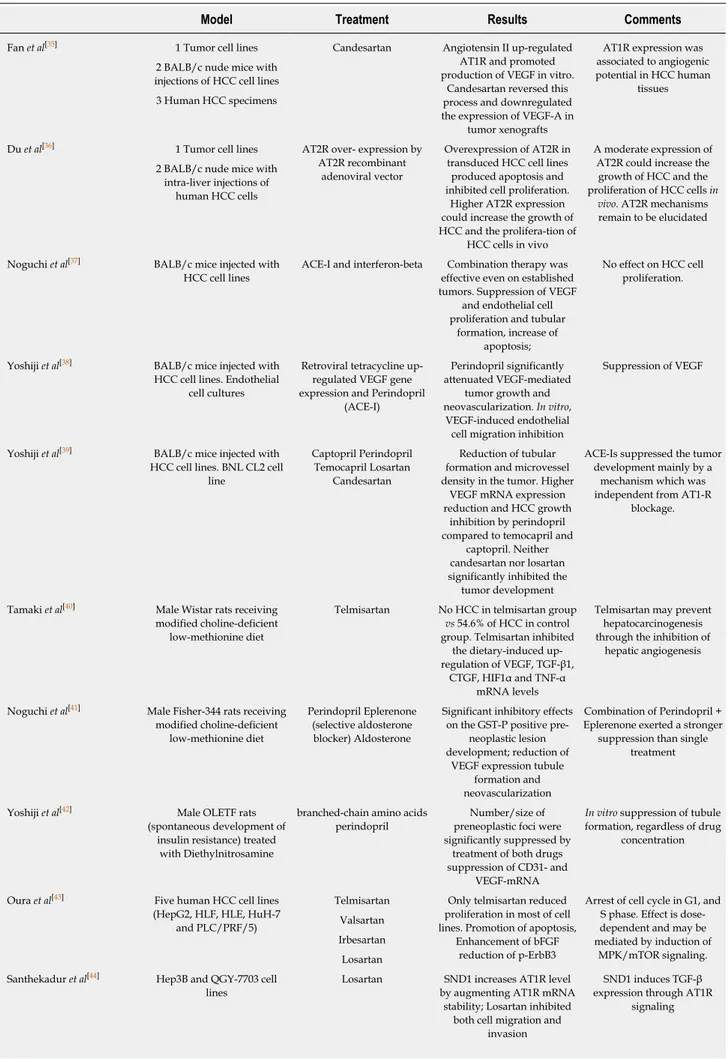
Other models
Several studies (Table 3) adopted a xenograft model in which in BALB/c mice received an injection of HCC cell lines. Using this model, Fan et al[35] demonstrated an
enhanced immunohistochemical expression of angiotensin receptor 1 (AT1R) in cancer tissue, which correlated with VEGF receptor A. The administration of candesartan reduced the mean diameter of tumors and suppressed AT1R expression and decreased microvascular density. The authors then examined the effects of angiotensin II and candesartan on the expression of AT1R protein in SMMC-7721 and HepG2 cell lines using Western blotting and found that candesartan down-regulated the expression of AT1R protein induced by angiotensin II with a dose-dependent relationship, as well as angiotensin II-induced production of VEGF-A. On the other hand, when angiotensin receptor 2 was transduced by an adenoviral vector in HCC cell lines and murine orthotropic liver graft, it inhibited cell proliferation with a significant decrease of S-phase cells and an increase of G1-phase cells, probably mediated by the alteration of expression of CDK4 and cyclinD1[36]. Therefore, it was
hypothesized that apoptosis was secondary to activation of p38 MAPK, pJNK, caspase-8 and caspase-3 and inactivation of pp42/44 MAPK (Erk1/2). In another model, using the BNL-HCC cell line transferred into BALB/c mice[37], both perindopril
and interferon-beta reduced the volume of HCC, with an even higher magnitude when administered together. Additionally, the authors showed that this effect was mediated by the promotion of apoptosis, the reduction of cell proliferation and the inhibition of neo-angiogenesis proven by the reduction of CD31 positive cells and the suppression of VEGF protein within the tumor. Yoshiji et al[38] used a retroviral vector
to enhance the expression of VEGF in the BNL-HCC cell line. In this way, they found that perindopril reversed the production of VEGF and that the ability of cells to migrate was directly dependent on VEGF levels. When these manipulated cells were injected in BALB/c mice, they discovered that VEGF over-expression within HCC did not modify the angiotensin-2 expression, thus suggesting that, in this model, VEGF-mediated tumor growth was not influenced by angiotensin 2. Lastly, similarly to previous studies, it was reported that perindopril inhibited CD31 expression. The same group[39] used the BNL-HCC allograft model in BALB/c mice to show that
perindopril, temocapril, and captopril had an inhibitory effect on total HCC volume burden, although the effect of perindopril was much stronger than other ACE-Is. Finally, they showed that perindopril reduced mRNA-VEGF expression and ACE enzyme activity, suppressed microvessel density and inhibited tubule formation in endothelial cells.
The long-term administration of a choline-deficient/low-methionine diet is another way of inducing steatohepatitis, liver cirrhosis, and HCC in rodents. Using this model, Tamaki et al[40] showed that none of the male Wistar rats receiving telmisartan
developed HCC, while 54.6% of the control rats developed the tumor. The latter condition was associated with an increase of HIF-1α and protein and VEGF mRNA. Furthermore, telmisartan inhibited the up-regulation of TGF-β1, connective tissue growth factor (CTGF), and TNF-α mRNA levels. The same diet-induced HCC model was adopted by Noguchi[41]. In this case, male Fisher-344 rats were treated with
perindopril and eplerenone, a selective aldosterone blocker. Such drugs reduced both the liver fibrosis index and TGF-β. When administered in combination, they reduced the density of hepatic stellate cells and inhibited their proliferation through the reduction of aldosterone and angiotensin 2. Finally, both eplerenone and aldosterone attenuated the CD31 positive vessels in the liver along with inhibition of pre-neoplastic lesions (less than 4 foci/cm2vs about 8 foci/cm2 in the control group) and,
even in this report, a suppression of VEGF was recorded. Therefore, authors concluded that the effects of eplerenone and ACE-I were not simply due to the suppression of RAS, but other pathways, notably the hampering of the fibrosis process in this steatohepatitis model, could be involved.
Since insulin resistance status promotes neovascularization and has been associated with an increased risk of HCC, Yoshiji et al[42] initiated hepatocarcinogenesis by DEN
in obese diabetic Otsuka Long-Evans Tokushima Fatty (OLETF) rats undergoing partial hepatectomy and used BCAAs and ACE-I, alone or in combination, at clinically comparable low doses, to assess the antineoplastic activity of these compounds. Both BCAAs and ACE-I significantly attenuated the development of dysplastic lesions, along with the suppression of both angiogenesis and VEGF expression. Used in combination, these two agents exerted an even more potent inhibitory effect. Finally, BCAAs and ACE-I induced an inhibitory effect on tubule formation in cultured endothelial cells showing a similar efficacy[42].
In a further three studies[43-45], the effect of ACE-Is or ARBs was tested only on cell
lines. In particular, in HLF, HLE, HuH-7 and PLC/PRF/5 lines, it was demonstrated that telmisartan, but not valsartan, losartan and irbesartan, was able to arrest cell proliferation[43]. Similarly, only telmisartan blocked the cell cycle in phase G1 and
Table 3 Experimental studies on the effect of angiotensin-converting enzyme inhibitors or angiotensin II type 1 receptor blockers in different hepatocellular carcinoma animal models and in hepatocellular carcinoma cell lines
Model Treatment Results Comments
Fan et al[35] 1 Tumor cell lines Candesartan Angiotensin II up-regulated
AT1R and promoted production of VEGF in vitro.
Candesartan reversed this process and downregulated the expression of VEGF-A in
tumor xenografts
AT1R expression was associated to angiogenic potential in HCC human
tissues 2 BALB/c nude mice with
injections of HCC cell lines 3 Human HCC specimens
Du et al[36] 1 Tumor cell lines AT2R over- expression by AT2R recombinant
adenoviral vector
Overexpression of AT2R in transduced HCC cell lines
produced apoptosis and inhibited cell proliferation.
Higher AT2R expression could increase the growth of HCC and the prolifera-tion of
HCC cells in vivo
A moderate expression of AT2R could increase the
growth of HCC and the proliferation of HCC cells in
vivo. AT2R mechanisms remain to be elucidated 2 BALB/c nude mice with
intra-liver injections of human HCC cells
Noguchi et al[37] BALB/c mice injected with HCC cell lines
ACE-I and interferon-beta Combination therapy was effective even on established tumors. Suppression of VEGF
and endothelial cell proliferation and tubular
formation, increase of apoptosis;
No effect on HCC cell proliferation.
Yoshiji et al[38] BALB/c mice injected with HCC cell lines. Endothelial
cell cultures
Retroviral tetracycline up-regulated VEGF gene expression and Perindopril
(ACE-I)
Perindopril significantly attenuated VEGF-mediated
tumor growth and neovascularization. In vitro,
VEGF-induced endothelial cell migration inhibition
Suppression of VEGF
Yoshiji et al[39] BALB/c mice injected with HCC cell lines. BNL CL2 cell
line
Captopril Perindopril Temocapril Losartan
Candesartan
Reduction of tubular formation and microvessel density in the tumor. Higher
VEGF mRNA expression reduction and HCC growth
inhibition by perindopril compared to temocapril and
captopril. Neither candesartan nor losartan significantly inhibited the tumor development
ACE-Is suppressed the tumor development mainly by a
mechanism which was independent from AT1-R
blockage.
Tamaki et al[40] Male Wistar rats receiving modified choline-deficient
low-methionine diet
Telmisartan No HCC in telmisartan group vs 54.6% of HCC in control group. Telmisartan inhibited
the dietary-induced up-regulation of VEGF, TGF-β1,
CTGF, HIF1α and TNF-α mRNA levels
Telmisartan may prevent hepatocarcinogenesis through the inhibition of
hepatic angiogenesis
Noguchi et al[41] Male Fisher-344 rats receiving modified choline-deficient
low-methionine diet
Perindopril Eplerenone (selective aldosterone
blocker) Aldosterone
Significant inhibitory effects on the GST-P positive
pre-neoplastic lesion development; reduction of
VEGF expression tubule formation and neovascularization
Combination of Perindopril + Eplerenone exerted a stronger
suppression than single treatment
Yoshiji et al[42] Male OLETF rats (spontaneous development of
insulin resistance) treated with Diethylnitrosamine
branched-chain amino acids perindopril
Number/size of preneoplastic foci were significantly suppressed by
treatment of both drugs suppression of CD31- and
VEGF-mRNA
In vitro suppression of tubule formation, regardless of drug
concentration
Oura et al[43] Five human HCC cell lines (HepG2, HLF, HLE, HuH-7
and PLC/PRF/5)
Telmisartan Only telmisartan reduced proliferation in most of cell lines. Promotion of apoptosis,
Enhancement of bFGF reduction of p-ErbB3
Arrest of cell cycle in G1, and S phase. Effect is dose-dependent and may be mediated by induction of
MPK/mTOR signaling. Valsartan
Irbesartan Losartan Santhekadur et al[44] Hep3B and QGY-7703 cell
lines
Losartan SND1 increases AT1R level by augmenting AT1R mRNA
stability; Losartan inhibited both cell migration and
invasion
SND1 induces TGF-β expression through AT1R
signaling
Cook et al[45] H4-II-E-C3 rat hepatoma cells transfected with Ang II
Losartan Candesartan Losartan inhibits Angiotensin II-induced proliferation
Both losartan and candesartan are equally effective in competing with
Ang II/AT1 receptor interactions on the cell
surface
Ang II: Angiotensin receptor; AT2R: Angiotensin II type 2 receptor; ACE-Is: Angiotensin-converting enzyme inhibitors; ARBs: Angiotensin II type 1 receptor blockers; HCC: Hepatocellular carcinoma; VEGF: Vascular endothelial growth factor; TNF: Tumor necrosis factor; TGF: Transforming growth factor; OLETF: Otsuka Long-Evans Tokushima Fatty.
reduced the population of cells in phase S, possibly by reducing the expression of cyclin D1 and cyclin E. Additionally, it was demonstrated that the block of proliferation could be secondary to the induction of AMPK/mTOR signaling in HCC cells. Finally, telmisartan increased the levels of phospho-p53 and decreased the levels of survivin in HLF cells, thus promoting HCC apoptosis, and it also inhibited angiogenesis by increasing bFGF levels in HLF cells as detected by the protein array. In Hep3B and QGY-7703 cells[44], it was shown that staphylococcal nuclease domain
containing-1 (SND1) could play a role in promoting hepatocarcinogenesis and cell migration by triggering TGF-β signaling, a mechanism that was mediated by increasing activation of angiotensin receptor 1. In this experiment, losartan reduced the migration and invasive potential of HCC cells. Cook et al[45] transfected
Ang(-S)Exp/pSVL plasmid, which causes the production of a stable, non-secreted form of angiotensinogen, in H4-II-E-C3 rat hepatoma cells. Thus, they discovered that this novel cell clone exhibited enhanced proliferation, which was blocked by losartan, but not by candesartan, although they were equally effective in competing with Ang II/AT1 receptor interactions on the cell surface. Furthermore, the addition of phenylarsine oxide, a molecule that blocks angiotensin receptor internalization, abolished the effect of losartan. Therefore, the authors concluded that losartan could be effective mainly on intranuclear angiotensin receptor.
DISCUSSION
The interest in the potential beneficial effects of RAS inhibitors against HCC started about two decades ago. Since then, several in vitro studies, using either different tumor cell lines or cell lines manipulated through adenoviral vectors, and numerous
in vivo models have been used to demonstrate the antitumoral effect of RAS inhibitors on hepatocarcinoma development. We had to wait about 10 years for the first clinical study on the possible role of ACE-Is administration in the recurrence of HCC[13]. This
study, designed as a randomized trial, was followed within a couple of years by two similar randomized trials from the same authors, all demonstrating the efficacy of the ACE-I perindopril in reducing tumor recurrence when used in combination with other drugs[13-15]. Interestingly, this effect was associated with a significant reduction of
VEGF[13-15] and alpha-fetoprotein[13,14] serum levels. However, a significant effect on
survival was not observed in any of these studies. The latter result requires some comments: only the first of these three studies reported a survival analysis (reported as “data not shown” in the other studies), which was interrupted at 42 mo, although the survival rate in the control group and the group receiving a combined treatment was 84% and 96%, respectively[13]. In consideration of this trend, we do not really
know whether a longer period of follow-up would have led to significant survival differences. Moreover, it is plausible that a delayed recurrence in patients prevalently in Child A class (75%-87.5%) could also prolong their survival.
The more recent human studies are all observational retrospective cohort or case-control studies[7,16-21]. All these studies evaluated the effect of ACEIs and/or ARBs on
overall survival. Among them, only one took into consideration tumor recurrence whereas another evaluated disease-free-survival[20,21]. With regard to tumor
recurrence, the study of Facciorusso et al[20] was performed on patients with
characteristics very similar to those reported in the randomized trials (patients with a low number and small-size tumors susceptible to RFA treatment). In addition, only patients undergoing antihypertensive therapy for at least two years before RFA were included to allow a reasonable induction period for the antiangiogenic and antifibrotic effects of such drugs. This study demonstrated a significant beneficial effect of ARBs, not only on time to recurrence but also on the overall survival after a period of observation of 96 months, which is twice the one reported in the randomized trials.
obesity, diabetes, and hypertension, on patients undergoing HCC resection[21]. In this
case, patients had a more advanced tumor disease, as demonstrated by the presence of pulmonary and bone metastasis at the follow-up, and underwent a liver resection including either one segment, more than one segment or even hemihepatectomy, plus removal of additional contiguous segments. In this study, neither overall survival nor disease-survival was improved by the anti-hypertensive treatment. However, the authors underline the fact that patients receiving angiotensin II-blocking agents had a better disease-free and overall survival rate than hypertensive patients not treated with angiotensin II-blocking agents.
The other observational studies conducted only on overall survival rates did not produce conclusive results since the treatment with RAS inhibitors was beneficial in one study[19], did not influence survival in two studies[17,18], and resulted either
ineffective or even associated with an increased HCC risk in non-cirrhotic HCV patients in another study[16].
Among the possible explanations for these discrepancies, the different time of exposure to antihypertensive drugs assumes particular importance if we consider that RAS inhibitors would require a reasonable induction period to counteract the carcinogenetic process through their antiangiogenic effects. In the various studies, the antihypertensive treatment preceded for a variable period the first recorded liver cancer diagnosis or started at the time of HCC diagnosis, and the comparison of exposure during the follow up was difficult. Other explanations are the characteristics of the tumor (susceptible to surgical resection or locoregional therapy or advanced), the different stages of the underlying liver disease, and the concomitant pharmacological therapies/comorbidities potentially influencing tumor biology.
All experimental studies lead to concordant conclusions on the beneficial effects of RAS inhibitors on hepatocarcinogenesis, with some difference within the same drug class. However, it should be highlighted that this beneficial effect was mainly based on the demonstration that ACE-Is or ARBs inhibit the process of angiogenesis, the most studied and extensively exploited biological mechanism. The main aspects explored included: the involvement of VEGF serum levels or VEGF protein or mRNA expression[25,28-30,33-35,37-42] in endothelial cell growth and differentiation[46]; the
contribution of HIF-1[28,40] as a transcription factor for VEGF[47]; the use of CD31[29-33,42]
[also known as platelet endothelial cell adhesion molecule 1 (PECAM-1)] to recognize endothelial cells[48]; endothelial cell proliferation and tubular formation in vitro and/or in vivo[30,32,34,37,39,41,42], a multi-step process triggered by VEGF[49].
Inhibition of tumor cells proliferation and induction of apoptosis are two mechanisms that have also been studied to explain how RAS inhibitors could counteract hepatocarcinogenesis. Some authors report an inhibitory effect of RAS inhibitors on tumor cell proliferation[25] or describe a block of proliferation secondary
to the induction of AMPK/mTOR signaling[43], all studies performed by Yoshiji et al[30-33] and by others[35,37] do not confirm these findings, and one study even describes a
pro mitogenic effect of angiotensin II[45]. Moreover, a complete overview of this aspect
requires a better definition of the role of VEGF as a potential autocrine growth factor on tumor cells[46] and the relationship between angiotensin receptor 1 (AT1R) and
angiotensin receptor 2 (AT2R)[36,50,51].
More consistent data seem to demonstrate the proapoptotic effect of RAS inhibitors on hepatic cancer cells[31,33,36,37,43].
Finally, the last possible antitumor mechanism related to the use of RAS inhibitors could consist of the suppression of inflammation by the reduction of TNF-α[25,40],
TGF-β[25,29,40,41] and MMP-2[25,29], and an increase of tissue inhibitor of metalloproteinase
(TIMP)-1, a negative regulator of MMP2[29].
All mechanisms mentioned above supporting the anti-tumor activity of RAS inhibitors were studied in HCC models, mostly induced by the carcinogenetic agent DEN or in rodents with xenotransplanted tumors, except in two studies[40,41] that used
choline-deficient/low-methionine diets to induce steatohepatitis related HCC. Viral hepatitis still represents the principal cause of HCC in humans. The demonstration of a similar protective effect of ACE-I or ARBs in an animal model that mimics the progression of viral-related liver disease in humans, such as the HBV transgenic mouse model[52], would strongly support previous experimental data. In addition,
since ACE-Is have been able to produce a prophylactic effect on hepatocarcino-genesis[29,32,33], this model could be used to test the RAS inhibitor influence on the
expression of genes that are involved early on in the carcinogenesis[53]. In fact, the
adoption of transgenic mouse models is widely used to test the effect of drugs or nutrients to counteract the process of carcinogenesis[54]. Finally, to clarify if RAS
inhibitors could influence the progression toward overt HCC in humans, it is necessary to design randomized, controlled trials in which the treatment with these anti-hypertensive drugs is initiated in patients at high risk of HCC. Cirrhotics with a low number (≤ 3) of small tumors (≤ 3 cm) or a single larger tumor (≤ 5 cm), with a
complete tumor ablation would be the ideal patients for this study considering that HCC recurrence at 3 years is observed in 48%-53% and 28%-33% in the presence of HCV infection or after HCV eradication, respectively[55,56].
In conclusion, all experimental data strongly support the ability of RAS inhibitors to counteract HCC development. In humans, this protective effect was confirmed when RAS inhibitors were used, prevalently in combination with other drugs, to prevent HCC recurrence. On the other hand, even if the results of RAS inhibitors on patient survival seem to be inconsistent, these findings are less conclusive. Further studies are necessary to evaluate whether their prolonged use could influence the progression of cirrhosis toward HCC.
ARTICLE HIGHLIGHTS
Research background
Hepatocellular carcinoma (HCC) represents about 90% of primary liver cancers and usually develops in patients with underlying liver cirrhosis. One of the major cancer hallmarks of HCC is the process of neoangiogenesis, which is thought to occur early in the carcinogenetic process. Theoretically, angiotensin-converting enzyme inhibitors (ACE-Is) and angiotensin II type 1 receptor blockers (ARBs) could have a role in HCC primary prevention since they reduce both neovascularization and the development of liver fibrosis.
Research motivation
The possible role of renin-angiotensin system (RAS) inhibitors in the prevention of HCC is still an attractive field of research, as suggested by the recently published experimental and clinical studies.
Research objectives
To elucidate the role of ARBs and ACE-Is in HCC.
Research methods
Literature was selected in August 2018, focusing on studies regarding ACE-Is, ARBs and HCCs. For this purpose, an electronic search of the literature was conducted using the online databases PubMed, Cochrane library, Scopus and Web of Science. Eligibility criteria were: (1) prospective or retrospective clinical studies; (2) epidemiological studies; and (3) experimental studies conducted in vivo or in vitro. We conducted the articles selection using a two-step approach, to evaluate whether they matched the eligibility criteria.
Research results
Thirty-one studies were selected. Three interventional studies showed that ACE-Is had a significant protective effect on HCC recurrence only when used in combination with vitamin K or branched chain aminoacids, without a significant increase of overall survival. Among six retrospective observational studies, mainly focused on overall survival, only one demonstrated a prolonged survival in ACE-Is group, whereas the two of them that also evaluated tumor recurrence showed conflicting results. All experimental studies displayed beneficial effects of RAS inhibitors on hepatocarcinogenesis. Numerous experimental studies, conducted either in animals and cell cultures, demonstrated the anti-angiogenetic and anti-fibrotic effect of ACE-Is and ARBs, thanks to the suppression of some cytokines such as vascular endothelial growth factor, hypoxia-inducible factor-1a, transforming growth factor-beta and tumor necrosis factor alpha. All or part of these mechanisms were demonstrated in rodents developing less HCC and preneoplastic lesions after receiving such drugs.
Research conclusions
Our systematic analysis of the literature allowed us to partially reinterpret the data supporting the ability of RAS inhibitors to counteract HCC. While there is reasonable research supporting their ability to reduce the HCC recurrence rate, we think that the conclusions of the randomized clinical trials on overall survival underestimated the potential use of these compounds. These conclusions are further supported by the solid experimental data on the anti-angiogenetic activity of RAS inhibitors, which all suggest their clinical use in the prevention of HCC development.
Research perspectives
We also proposed enlarging the future research on the relationship between RAS inhibitors and HCC to other possible anti carcinogenetic mechanisms.
REFERENCES
1 European Association for the Study of the Liver. EASL Clinical Practice Guidelines: Management of
hepatocellular carcinoma. J Hepatol 2018; 69: 182-236 [PMID: 29628281 DOI:
10.1016/j.jhep.2018.03.019]
Cancer J Clin 2015; 65: 87-108 [PMID: 25651787 DOI: 10.3322/caac.21262]
3 Global Burden of Disease Liver Cancer Collaboration; Akinyemiju T, Abera S, Ahmed M, Alam N,
Alemayohu MA, Allen C, Al-Raddadi R, Alvis-Guzman N, Amoako Y, Artaman A, Ayele TA, Barac A, Bensenor I, Berhane A, Bhutta Z, Castillo-Rivas J, Chitheer A, Choi JY, Cowie B, Dandona L, Dandona R, Dey S, Dicker D, Phuc H, Ekwueme DU, Zaki MS, Fischer F, Fürst T, Hancock J, Hay SI, Hotez P, Jee SH, Kasaeian A, Khader Y, Khang YH, Kumar A, Kutz M, Larson H, Lopez A, Lunevicius R,
Malekzadeh R, McAlinden C, Meier T, Mendoza W, Mokdad A, Nagel G, Nguyen Q, Nguyen G, Ogbo F, Patton G, Pereira DM, Pourmalek F, Qorbani M, Radfar A, Roshandel G, Salomon JA, Sanabria J, Sartorius B, Satpathy M, Sawhney M, Sepanlou S, Shackelford K, Shore H, Sun J, Mengistu DT, Tran B, Ukwaja KN, Vlassov V, Vollset SE, Vos T, Wakayo T, Weiderpass E, Werdecker A, Yonemoto N, Younis M, Yu C, Zaidi Z, Zhu L, Murray CJL, Naghavi M, Fitzmaurice C, Moradi-Lakeh M, Topór-Mądry R. The Burden of Primary Liver Cancer and Underlying Etiologies From 1990 to 2015 at the Global, Regional, and National Level: Results From the Global Burden of Disease Study 2015. JAMA Oncol 2017; 3: 1683-1691 [PMID: 28983565 DOI: 10.1001/jamaoncol.2017.3055]
4 Yüksel Ş, Boylu Akyerli C, Cengiz Yakıcıer M. Angiogenesis, Invasion, and Metastasis Characteristics of
Hepatocellular Carcinoma. J Gastrointest Cancer 2017 [PMID: 28785955 DOI:
10.1007/s12029-017-9962-5]
5 Dhanasekaran R, Venkatesh SK, Torbenson MS, Roberts LR. Clinical implications of basic research in
hepatocellular carcinoma. J Hepatol 2016; 64: 736-745 [PMID: 26450813 DOI:
10.1016/j.jhep.2015.09.008]
6 Berretta M, Rinaldi L, Di Benedetto F, Lleshi A, De Re V, Facchini G, De Paoli P, Di Francia R.
Angiogenesis Inhibitors for the Treatment of Hepatocellular Carcinoma. Front Pharmacol 2016; 7: 428 [PMID: 27881963 DOI: 10.3389/fphar.2016.00428]
7 Facciorusso A, Licinio R, Carr BI, Di Leo A, Barone M. MEK 1/2 inhibitors in the treatment of
hepatocellular carcinoma. Expert Rev Gastroenterol Hepatol 2015; 9: 993-1003 [PMID: 25915713 DOI:
10.1586/17474124.2015.1040763]
8 Raoul JL, Gilabert M, Adhoute X, Edeline J. An in-depth review of chemical angiogenesis inhibitors for
treating hepatocellular carcinoma. Expert Opin Pharmacother 2017; 18: 1467-1476 [PMID: 28893090
DOI: 10.1080/14656566.2017.1378346]
9 Corey KE, Shah N, Misdraji J, Abu Dayyeh BK, Zheng H, Bhan AK, Chung RT. The effect of
angiotensin-blocking agents on liver fibrosis in patients with hepatitis C. Liver Int 2009; 29: 748-753 [PMID: 19220742 DOI: 10.1111/j.1478-3231.2009.01973.x]
10 Kaji K, Yoshiji H, Ikenaka Y, Noguchi R, Aihara Y, Shirai Y, Douhara A, Fukui H. Possible involvement
of angiogenesis in chronic liver diseases: interaction among renin-angiotensin-aldosterone system, insulin resistance and oxidative stress. Curr Med Chem 2012; 19: 1889-1898 [PMID: 22376037]
11 Paizis G, Gilbert RE, Cooper ME, Murthi P, Schembri JM, Wu LL, Rumble JR, Kelly DJ, Tikellis C, Cox
A, Smallwood RA, Angus PW. Effect of angiotensin II type 1 receptor blockade on experimental hepatic fibrogenesis. J Hepatol 2001; 35: 376-385 [PMID: 11592599]
12 Song T, Choi CH, Kim MK, Kim ML, Yun BS, Seong SJ. The effect of angiotensin system inhibitors
(angiotensin-converting enzyme inhibitors or angiotensin receptor blockers) on cancer recurrence and survival: a meta-analysis. Eur J Cancer Prev 2017; 26: 78-85 [PMID: 27158979 DOI:
10.1097/CEJ.0000000000000269]
13 Yoshiji H, Noguchi R, Toyohara M, Ikenaka Y, Kitade M, Kaji K, Yamazaki M, Yamao J, Mitoro A,
Sawai M, Yoshida M, Fujimoto M, Tsujimoto T, Kawaratani H, Uemura M, Fukui H. Combination of vitamin K2 and angiotensin-converting enzyme inhibitor ameliorates cumulative recurrence of hepatocellular carcinoma. J Hepatol 2009; 51: 315-321 [PMID: 19501932 DOI:
10.1016/j.jhep.2009.04.011]
14 Yoshiji H, Noguchi R, Ikenaka Y, Kaji K, Aihara Y, Yamazaki M, Yamao J, Toyohara M, Mitoro A,
Sawai M, Yoshida M, Morioka C, Fujimoto M, Uemura M, Fukui H. Combination of branched-chain amino acids and angiotensin-converting enzyme inhibitor suppresses the cumulative recurrence of hepatocellular carcinoma: a randomized control trial. Oncol Rep 2011; 26: 1547-1553 [PMID: 21874260
DOI: 10.3892/or.2011.1433]
15 Yoshiji H, Noguchi R, Ikenaka Y, Kaji K, Shirai Y, Aihara Y, Yamao J, Toyohara M, Mitoro A, Sawai M,
Yoshida M, Morioka C, Fujimoto M, Uemura M, Kawaratani H, Tsujimoto T, Fukui H. Soluble VEGF receptor-2 may be a predictive marker of anti-angiogenic therapy with clinically available safe agents.
Oncol Lett 2011; 2: 69-73 [PMID: 22870131 DOI: 10.3892/ol.2010.196]
16 Ho CM, Lee CH, Lee MC, Zhang JF, Wang JY, Hu RH, Lee PH. Comparative effectiveness of
angiotensin-converting enzyme inhibitors and angiotensin II receptor blockers in chemoprevention of hepatocellular carcinoma: a nationwide high-risk cohort study. BMC Cancer 2018; 18: 401 [PMID:
29631561 DOI: 10.1186/s12885-018-4292-y]
17 Hagberg KW, Sahasrabuddhe VV, McGlynn KA, Jick SS. Does Angiotensin-Converting Enzyme
Inhibitor and β-Blocker Use Reduce the Risk of Primary Liver Cancer? A Case-Control Study Using the U.K. Clinical Practice Research Datalink. Pharmacotherapy 2016; 36: 187-195 [PMID: 26846893 DOI:
10.1002/phar.1704]
18 Walker AJ, West J, Grainge MJ, Card TR. Angiotensin converting enzyme inhibitors and hepatocellular
carcinoma incidence in the General Practice Research Database. Cancer Causes Control 2011; 22: 1743-1747 [PMID: 21909951 DOI: 10.1007/s10552-011-9837-1]
19 Pinter M, Weinmann A, Wörns MA, Hucke F, Bota S, Marquardt JU, Duda DG, Jain RK, Galle PR,
Trauner M, Peck-Radosavljevic M, Sieghart W. Use of inhibitors of the renin-angiotensin system is associated with longer survival in patients with hepatocellular carcinoma. United European Gastroenterol
J 2017; 5: 987-996 [PMID: 29163965 DOI: 10.1177/2050640617695698]
20 Facciorusso A, Del Prete V, Crucinio N, Muscatiello N, Carr BI, Di Leo A, Barone M. Angiotensin
receptor blockers improve survival outcomes after radiofrequency ablation in hepatocarcinoma patients. J
Gastroenterol Hepatol 2015; 30: 1643-1650 [PMID: 25974743 DOI: 10.1111/jgh.12988]
21 Kaibori M, Ishizaki M, Matsui K, Kitade H, Matsui Y, Kwon AH. Evaluation of metabolic factors on the
prognosis of patients undergoing resection of hepatocellular carcinoma. J Gastroenterol Hepatol 2011; 26: 536-543 [PMID: 21332549 DOI: 10.1111/j.1440-1746.2010.06439.x]
22 Tsan YT, Lee CH, Wang JD, Chen PC. Statins and the risk of hepatocellular carcinoma in patients with
hepatitis B virus infection. J Clin Oncol 2012; 30: 623-630 [PMID: 22271485 DOI:
10.1200/JCO.2011.36.0917]
23 Singh PP, Singh S. Statins and risk reduction in hepatocellular carcinoma: fact or fiction? J Clin Oncol