A
A
l
l
m
m
a
a
M
M
a
a
t
t
e
e
r
r
S
S
t
t
u
u
d
d
i
i
o
o
r
r
u
u
m
m
–
–
U
U
n
n
i
i
v
v
e
e
r
r
s
s
i
i
t
t
à
à
d
d
i
i
B
B
o
o
l
l
o
o
g
g
n
n
a
a
DOTTORATO DI RICERCA IN
Chimica
Ciclo XXX
Settore Concorsuale: 03/C1Settore Scientifico Disciplinare: CHIM06
TITOLO TESI
Design, synthesis and characterizations of hybrid
nanosystems: nanomedicine applications in theranostics
Presentata da:
Ilaria Monaco
Coordinatore Dottorato
Supervisore
Prof. Aldo Roda
Prof. Mauro Comes Franchini
Index
Abstract ... 1
Table of acronyms ... 2
1. Introduction ... 5
1.1 State of the art in Cancer Nanomedicine ... 5
1.2 Drug Delivery and theranostic nanosystems ... 8
1.2.1 Polymeric nanoparticles as drug delivery and theranostic nanosystems ... 13
1.2.2 Poly(d,l-lactic-co-glycolic acid)-b-poly(ethylene glycol) ... 16
1.2.3 Methods of preparations for polymeric nanoparticles ... 18
1.3 Hyperthermia therapy ... 21
1.3.1 Metallic nanoparticle as photothermal agents in hyperthermia therapy ... 25
1.3.2 Gold nanorods as photothermal agents ... 28
1.3.3 Magnesium nanoparticles as photothermal agents ... 31
1.4 Nanomaterials as diagnostic and imaging tools in nanomedicine ... 35
1.4.1 Photoacoustic imaging (PAI) ... 37
1.4.2 Contrast agents for photoacoustic imaging (PAI) ... 40
1.4.3 Gold nanostructures as PAI contrast agents ... 42
1.4.4 Magnetic Resonance Imaging (MRI) ... 45
1.4.5 Super paramagnetic nanoparticles as MRI contrast agents ... 48
1.5 Surface chemistry of metal nanoparticles ... 51
1.5.1 Self-assembled monolayer (SAM) ... 51
1.5.2 Silica shell as coating of metallic nanoparticles ... 53
2. Aim ... 57
3. Discussion ... 59
3.1 Synthesis of the precursors ... 59
3.1.1 Synthesis of the organic ligand 11-(4-mercaptobenzamido)undecanoate (ligand 1) ... 59
3.1.2 Organic functionalization of chitosan... 61
3.1.3 Preparation of copolymers for polymeric nanocarriers ... 62
3.2. Polymeric nanoparticles as drug delivery systems against Glioblastoma Multiforme ... 64
3.2.1. Synthesis of chlorotoxin functionalized polymeric nanovectors for the treatment of Glioblastoma Multiforme in a combined approach with radiation therapy... 65
3.2.2. Synthesis of aptamer functionalized polymeric nanovectors for the treatment of Glioblastoma thought the BBB ... 74
3.3.1 Gold nanorods as tools for photothermal therapy of Barrett Oesophagus ... 84
3.3.2 Magnesium nanoparticles as a highly biocompatible photothermal agent for hepatocellular carcinoma treatment ... 96
3.4 Synthesis of multicomponent nanosystems as dual imaging and theranostic agents against cancer ... 104
3.4.1. Synthesis of Dumbbell-Like Gold-Iron Oxide Nanoparticles (dl-AuFe NPs) as multifunctional nanosystems ... 106
3.4.2. Synthesis of a novel magnetic resonance−photoacoustic dual imaging nanosystem based on core−shell Fe3O4@SiO2@Au NPs ... 116
4. Conclusions ... 127
5. Experimental section ... 129
5.1 Synthesis of ethyl 12-(4-mercaptobenzamido)dodecanoate (ligand 1) ... 129
5.1.1 Synthesis of 4,4’-disulfanediyldibenzoic acid ... 129
5.1.2 Synthesis of ethyl 11-aminododecanoate ... 130
5.1.3 Synthesis of bis-ethyl 11-(4-benzamido)dodecanoatedisulfide ... 131
5.1.4 Synthesis of ethyl 12-(4-mercaptobenzamido)dodecanoate (ligand 1) ... 132
5.2 Synthesis of poly(lactic-co-glycolic acid)-block-poly(ethylene glycol) (PLGA-b-PEG) copolymers ... 133
5.2.1 Synthesis of PLGA-b-PEG-NH2 ... 133
5.2.2 Synthesis of PLGA-NHS ... 134
5.2.3 Synthesis of PLGA-b-PEG-COOH ... 135
5.3 Synthesis of modified chitosan chains ... 136
5.3.1 Synthesis of Chitosan-Thioglycolic Acid (Chitosan-TGA) ... 136
5.3.2 Synthesis of Chitosan- Hydrocaffeic Acid (Chitosan-HCA) ... 137
5.4 Synthesis of chlorotoxin functionalized polymeric nanovectors for the treatment of Glioblastoma Multiforme in a combined approach with radiation therapy ... 138
5.4.1 Synthesis of AgNPs ... 138
5.4.2 Synthesis of AgNPs-1 ... 139
5.4.3 Synthesis of AgNPs-1-PNPs ... 140
5.4.4 Synthesis of AgNPs-1-PNPs-Cltx ... 141
5.5 Synthesis of aptamer functionalized polymeric nanovectors for the treatment of Glioblastoma thought the BBB ... 142
5.5.1 Synthesis of NVP-BEZ235@PNPs ... 142
5.5.2 Synthesis of [email protected] and NVP-BEZ235@PNPs-SCR ... 143
5.5.3 Synthesis of BODIPY@PNPs ... 144
5.5.2 Synthesis of [email protected] and BODIPY@PNPs-SCR ... 145
5.6 Gold nanorods as tools for photothermal therapy of Barrett Esophagus ... 146
5.6.1 Preparation of GNRs-CTAB ... 146
5.6.2 Preparation of GNRs-1 ... 147
5.6.3 Synthesis of GNRs-1@PMs ... 148
5.6.5 Preparation of GNRs-1/Curc@PMs ... 150
5.7 Magnesium nanoparticles as a highly biocompatible photothermal agent for hepatocellular carcinoma treatment ... 151
5.7.1 Synthesis of Chit–Mg MPs ... 151
5.8 Synthesis of Dumbbell-Like Gold-Iron Oxide Nanoparticles (dl-AuFe NPs) as multifunctional nanosystems ... 152
5.8.1 Synthesis of dl-AuFe3O4 NPs ... 152
5.8.2 Synthesis of dl-AuFe3O4@Chit ... 153
5.9 Synthesis of a novel magnetic resonance−photoacoustic dual imaging nanosystem based on core−shell Fe3O4@SiO2@Au NPs... 154
5.9.1 Synthesis of Native Iron Oxide Nanoparticles, Fe3O4 NPs. ... 154
5.9.2 Synthesis of Core−Shell Iron Oxide−Silica Nanoparticles Fe3O4@SiO2 NPs ... 155
5.9.3 Synthesis of Core−Shell Iron Oxide−Silica−Gold Nanoparticles, Fe3O4@SiO2@Au NPs ... 156
5.9.4 Synthesis of Lipophilic Fe3O4@SiO2@Au NPs by Ligand Exchange ... 157
5.9.5 Synthesis of Fe3O4@SiO2@Au@PMs. ... 158
5.9.6 Conjugation of Folic Acid on Fe3O4@SiO2@AuNPs@PMs ... 159
1
Abstract
The research project discussed in this PhD thesis concerns the development of drug delivery and theranostic systems for nanomedicine applications in cancer treatments.
In the first part, polymeric nanoparticles have been exploited as drug delivery systems for the treatment of Glioblastoma Multiforme. For this purpose, two different biomolecules, in one case Cholotoxin, a small peptide, and in the other case an antiPDGFRs-aptamer (GINT.4), have been employed as targeting agents conjugated on the polymeric nanoparticles surface. The second section of the project has been focused on the development of metallic nanoparticle for photothermal therapy, a cancer treatment consisting in the exposition of body tissue to an increasing of temperature (42-45°C) to induce cytotoxic effects on cancer cells. For this purpose, gold nanorods (GNRs) have been investigated as photothermal agent for the treatment of Barrett’s esophageal. On the other hand, one pot-synthesis of chitosan coated magnesium nanoparticle has been fine-tuned in order to obtain high biocompatible nanoparticles characterized by a promising unexpected photothermal behaviour.
In the last part of the project, the synthesis of multicomponent nanosystems suitable for multimodal imaging or theranostic agents have been investigated. Two different multifunctional nanomaterials based on the incorporation of iron oxide and gold nanoparticles in the same nanosystem, have been realized. The first system consisted in dumbbell-like gold-iron oxide nanoparticles coated with chitosan that showed photothermal behaviour and also good capabilities as photoacoustic imaging contrast agents. The second system consisted in core−shell Fe3O4@SiO2@Au NPs entrapped in polymeric nanoparticles decorated with folic acid, have been tested in vivo for photoacoustic and magnetic resonance imaging detection of ovarian cancer.
2
Table of acronyms
Acronym
Significance
AAS atomic absorption spectroscopy
Ag-nps silver nanoparticles
Ag-nps-1 silver nanoparticles coated with 11-(4-mercaptobenzamido)undecanoate
Ag-nps-PVP silver nanoparticles coated with polyvinylpyrrolidone
Ag-PNP silver nanoparticles
Ag-PNP-CTX silver nanoparticles entrapped in polymeric nanoparticles functionalized with chlorotoxin
APTMS (3-aminopropyl)trimethoxysilane
ATP adenosine triphosphate
BAR-T Barrett’s esophageal toumor
BBB blood−brain barrier
BODI-PY BODIPY505-515, lipophilic dye
[email protected] polymeric nanoparticles containing the lipophilic dye BODIPY505-515 functionalized on the surface with the aptamer Gint4.T
BODI-PY@PNPs-SCR polymeric nanoparticles containing the lipophilic dye BODIPY505-515 functionalized on the surface with the scrambled aptamer SCR
BODY-PY@PNPs polymeric nanoparticles containing the lipophilic dye BODIPY505-515
CDI 1,1-carbonyldiimidazole
Chit-Mg MPs chitosan coated magnesium microparticles
CT computed tomography
CTAB cetyltrimethylammonium bromide
CTX chlorotoxin
Curc@PMs polymeric nanomicelles containing curcuma molecules
cw continuous wave
DCC N,N’-dicyclohexylcarbodiimide
DCM dichloromethane
DIPEA N,N-diisopropylethylamine
dl-AuFe NPs dumbbell-like gold–iron oxide nanoparticles
dl-AuFe3O4@Chit chitosan coated dumbbell-like gold–iron oxide nanoparticles
DLS dynamic light scattering
DMF dimethylformamide
DPSSL diode pumped solid state laser
EAC esophageal adenocarcinoma
EC50 half maximal effective concentration values
EDC 1-ethyl-3-(3-dimethylaminopropyl)carbodiimide
EMA European Medicine Agency
EPR enhanced permeability and retention
FA folic acid
FDA US Food and Drug Administration
Fe3O4 NPs iron oxide nanoparticles
3
Fe3O4@SiO2@Au@PMs core−shell iron oxide−silica−gold nanoparticles coated with the 11-(4-mercaptobenzamido)undecanoate entrapped in polymeric micelles
Fe3O4@SiO2@Au@PMs−FA core−shell iron oxide−silica−gold nanoparticles coated with the 11-(4-mercaptobenzamido)undecanoate entrapped in polymeric micelles functionalized with folic acid
Fe3O4@SiO2@Au@PMs−FA/fluo core−shell iron oxide−silica−gold nanoparticles coated with the 11-(4-mercaptobenzamido)undecanoate entrapped in polymeric micelles functionalized with folic acid and fluorescein
Fe3O4@SiO2@Au-1 core−shell iron oxide−silica−gold nanoparticles coated with the 11-(4-mercaptobenzamido)undecanoate
FTIR fourier transform infrared spectroscopy
GBM glioblastoma multiforme
Gint4.T aptamer able to recognize platelet-derived growth factor receptors β on the blood−brain barrier
GNPs gold nanoparticles
GNRs gold nanorods
GNRs-1 Gold nanorods coated with
11-(4-mercaptobenzamido)undecanoate)
GNRs-1/Curc@PMs polymeric nanomicelles containing lipophilic gold nanorods and curcuma
GNRs-1@PMs polymeric nanomicelles containing liphophilic GNRs-1 GNRs-CTAB gold nanorods coated with cetyltrimethylammonium bromide
Gy gray
HAADF-STEM high angle annular dark field scanning transmission electron microscopy
HCA hydrocaffeic acid
HCA hydrocaffeic acid
MES Buffer 2-(N-morpholino)ethanesulfonic acid Buffer
Mg MPs magnesium microparticles
Mg NPs magnesium nanoparticles
MMP-2 matrix metalloproteinase-2
MPS mononuclear phagocytic system
MRI magnetic resonance imaging
MTEOS methyltriethoxysilane
NaOH sodium hydroxide
NHS N-hydroxysuccinimide
NIR near-infrared spectroscopy
NMR nuclear magnetic resonance
NPs Nanoparticles
NVP-BEZ235@PNPs polymeric nanoparticles containing the lipophilic drug NVP-BEZ235
[email protected] polymeric nanoparticles containing the lipophilic drug NVP-BEZ235 functionalized on the surface with the aptamer Gint4.T
NVP-BEZ235@PNPs-SCR polymeric nanoparticles containing the lipophilic drug NVP-BEZ235 functionalized on the surface with the scrambled aptamer
4
OI optical imaging
PAI photoacoustic imaging
PDGFRβ platelet-derived growth factor receptors β
PDI polydispersity index
PET positron electron tomography
PLGA-PEG poly(d,l-lactic-co-glycolic acid)-b-poly(ethylene glycol)
PMs polymeric micelles
PNPs polymeric nanoparticles
PTT photothermal therapy
PVP polyvinylpyrrolidone
RES reticuloendothelial system
RT-qPCR real-time quantitative polymerase chain reaction analysis
SAM self-assembled monolayer
SCR scrambled aptamer
SELEX systematic evolution of ligands by exponential enrichment method
SPECT single-photon emission computed tomography
SPIONs super paramagnetic iron oxide nanoparticles
TEM transmission electron microscopy
TGA thioglycolic acid
TGA thermogravimetric analysis
THF tetrahydrofuran
US ultrasound
VSM vibrating-sample magnetometer
5
1. Introduction
1.1 State of the art in Cancer Nanomedicine
Nanotechnology is the science that concerns the study, the development and the production of systems characterized by any dimension between 1-100 nanometres. Indeed, small dimension confers to nanomaterials unique chemical-physical properties different from the bulk materials, and for this reason, in the last decades the use of nanomaterials in different area of researches such as electronic, engineering, biotechnology, biology and medicine, allowed a real technological revolution. Although IUPAC definition of nanoparticles is a particle with any shape of dimension in the 1-100 nm range, the term “nanoparticle” is commonly applied to structures that are up to several hundred nanometres in size.1
Because cancer is set to become a major cause of morbidity and mortality in the coming decades in every region of the world, there is the necessity to better understand its biology in order to improve diagnostic devices and treatments.2,3
Nowadays, the main cancer therapies consist of surgical intervention, radiation and chemotherapeutic drugs, which are often not efficacy and implicate different side effect as the death of healthy cells that cause the toxicity to the patients. In chemotherapy, the main complication is represented by the pharmaceutical properties of therapeutic molecules, such as the stability and the solubility, which can addict the circulating half-life and tumour accumulation making less efficient the therapy.
The nanotechnologies innovations applied to medicine allowed the development of nanomedicine and led to the possibility to overcome some of the main concerns of traditional cancer treatments by taking advantage by the interaction between the nanoparticles and biological systems but also discovering the therapeutic nature of some nanomaterials themselves.4 Cancer nanomedicine provides not only the use of nanoagents as drug delivery systems or diagnosis tools to improve the efficacy of traditional methodologies, but also the development of medical devices such as micro/nanoelectromechanical (MEM/NEM) device-based drug delivery or nanothechnology-device-based sensors for the detection of biomarkers. 5 In the last years, nanotherapies have been developed by employing the use of several nanoparticles platforms such as liposomes, albumin nanoparticles and polymeric micelles as drug delivery systems, and developed innovative technologies based on nanomaterials able to
6
be used in hyperthermia, radiation, gene and RNA interference (RNAi) therapy and immunotherapy (Figure 1).
Figure 1. Historical timeline of major developments in the field of cancer nanomedicine. EPR, enhanced permeability and retention; FDA, US Food and Drug Administration; nab, nanoparticle albumin-bound; NP, nanoparticle; PLGA-PEG, poly(d,l-lactic-co-glycolic acid)-b-poly(ethylene glycol); PRINT, particle replication in non-wetting template; siRNA, small interfering RNA adapted from Nat. Rev. Cancer 17, 20–37 (2016).
At now, few nano-drugs and nano-therapies have been available in clinic. Doxil® and Myocet®, consistent in doxorubicin encapsulated into liposomes, were the first class of therapeutic nanosystems to receive the clinical approval for cancer treatments. These formulations showed the possibility to reduce the dose-related cardiac toxicity concerning the doxorubicin and allowed the enhancement of its anticancer efficacy by enhancing patients tolerability.6
The second class of commercialized nanodrugs are represented by the Abraxane which consists in nanoparticles made of albumin-bound paclitaxel able to overcome the problems associated with administering hydrophobic taxol that induced hypersensitivity reactions.7 The mentioned examples of nanodrugs show as the nanotechnologies can deal with the challenges of cancer, improving the pharmacokinetic and the biodistribution of a chemotherapeutic drug or reducing the side effect associated to a therapy. However, clinical trials results revealed that the nanoformulations did not show an enhancement of efficacy compared with the conventional chemotherapeutic drug.8 In order to obtain an improvement of clinical efficacy of cancer treatment, the enhancement of the nanosystems design and nanomedicine strategy is necessary.
7
Polymeric micelles (as Genexol PM and NK105) and polymeric nanoparticles (CRLX101 and BIND-014) represent the new classes of cancer nanotherapeutic agents under clinical investigations.9,10
However, recently discordant clinical trials results showed the necessity to improve the delivery strategies and also the possibility to involve a patients selection to identify those most likely to respond to the nanotherapy.11
On the other hand, nanoparticles are largely investigated in the developing of “molecular imaging” in order to visualize and quantify biological processes at molecular and cellular levels in a non-invasive manner.12 The possibility to combine in the same nanosystem therapy and the diagnostic functionalities led to the development of theranostic systems. This represents a promising strategy to monitor the pharmacokinetic, the accumulation of therapeutics and the progression of disease, giving important insights into heterogeneities both within tumour and between patients for potential personalized therapies.13
8
1.2 Drug Delivery and theranostic nanosystems
Drug delivery nanosystems are nanoparticles able to encapsulate a therapeutic agent in order to improve their therapeutic index by increasing its efficacy and reducing the toxicity. For this reason, a nanocarrier can represent a “second chance” for rejected chemodrugs, bringing out several advantages such as:
the protection of the encapsulated drug from degradation due to the interactions with the biological environment;
the enhancement of the pharmaceutical properties of therapeutic molecules (for example solubility, stability, circulating half-life and tumour accumulation);
transocytosis of drugs across tight epithelial and endothelial barriers;
Because the accumulation of drug delivery nanosystems in the cancer site can be achieved by two main mechanisms: the passive targeting and active targeting.
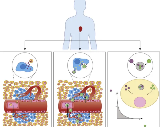
To explain these mechanisms, the development of an efficient drug delivery system needs to take in account biological features of the tumour tissue (Figure 2). Generally, tissue in advanced cancer grade tends to develop new blood vessels from existing ones in order to transport oxygen and nutrients to the new cells in a process defined angiogenesis, which is the principal cause of the rapid growth of a tumour.14 The formed tumour vessels are characterized by a disorganized vasculature and numerous openings in the walls such as vesicles and transcellular holes, inter-endothelial junctions, and a discontinuous basement spaces between endothelial cells.15 These factors lead to the enhanced permeability and retention (EPR) effect, responsible of an enhancement of the permeability of tumour vessels compared to normal tissue. This phenomenon allows the passage of blood plasma components and other macromolecules into the interstitial space of the tumour, contributing to longer retention of these molecules.
The passive targeting, a mechanism involved in the drug delivery system pharmacokinetic, is a consequence of the enhanced permeability and retention (EPR) effect. This mechanism induces nanoparticles accumulation in the tumour tissue by the extravasation from their leaky vasculature and allows the release of the encapsulated agents into the vicinity of the tumour cells. Consequently, nanoparticles build up reaching higher concentration due to lack of efficient lymphatic drainage in solid tumour. Therefore, through passive targeting, a drug
9
delivery nanosystem can reduce the side effects caused by the diffusion of the conventional small-molecule in normal tissue, improving the accumulation of nanoparticles in the tumour tissue at a concentration five to ten times higher than in normal tissue in 1-2 days.16 The nanoparticles size has a great influence on the accumulation with the EPR effect; as reported in literature, nanoparticles with a diameter between 10 - 500 nm exhibit a good tendency to extravasate and accumulate within the tumor interstitium but other studies showed that particles with diameters lower than 200 nm are more effective.17 However, passive targeting showed significant drawbacks caused by the tumour heterogeneity, vascular permeability and different pharmakinetic of each nanomaterials.18 The main problem consists in the clearance of nanoparticles from the bloodstream ascribable to the reticuloendothelial system (RES), which consists of phagocytic cells like monocytes and macrophages that are located in the spleen, lymph nodes, and Kupffer cells in the liver.
Figure 2. Passive targeting, active targeting, and combinatorial delivery. In passive targeting (left), nanoparticles (NPs) passively extravasate though the leaky vasculature via the enhanced permeability and retention (EPR) effect and preferentially accumulate in tumours. In active targeting (middle), targeting ligands on the surface of the NP trigger receptor-mediated endocytosis for enhanced cellular uptake. In combinatorial delivery (right), two or more therapeutic agents inhibit different or identical disease pathways for a synergistic effect from Trends Mol. Med. 21, 223–232 (2015).
10
This phenomenon cause the high accumulation of the nanoparticles in the liver or spleen and can increase the toxicity of the nanomaterials due to the high dose.19
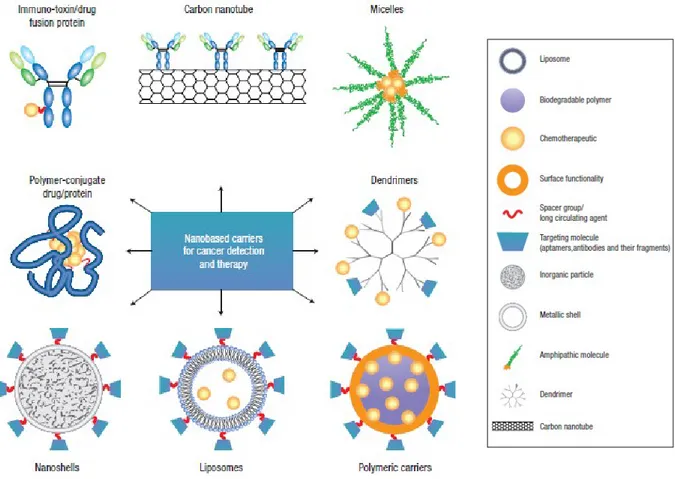
For this reason, the use of targeting agents able to be conjugated on the surface of nanosystems and to guide them to bind specific cells after extravasations, can improve the efficacy of the therapy by increasing their accumulation into the cancer site and by decreasing the collateral effect as toxicity in the health tissue.20 To maximize the unique characteristic of the active targeting mechanism, the use of an active agent able to bind with high selectivity a surface marker (antigen or receptor) overexpressed on target cells relative to normal cells is required. Several classes of bio-agents such as, antibodies, engineered antibody fragments, 21 proteins,22 peptides,23 and aptamers,24 have been employed as targeting agents, also different chemistry reactions have been developed to conjugate the these agent on nanoparticles surface. In the last years, the development of several nanocarriers for nanomedicine applications have been reported in literature (Figure 3).25
Figure 3.Images of nanocarriers for targeting cancer adapted from Nat.Nanotechnol. 2, 751-760 (2007). The main components of delivery nanosystems typically include a nanocarrier, a targeting moiety conjugated to the nanocarrier, and a cargo (such as the desired chemotherapeutic drugs).
11
The main research regarded the chemical components employed in the synthesis and formulation of these systems, that might be biocompatible and biodegradable materials and able to form nanocarriers with high efficiency of encapsulation of the therapeutic agents and with good robustness against degradation.
Carrier materials are classified in two main classes: the organic nanocarriers and inorganic nanocarriers. The first class includes nanocarriers constituted by smaller organic molecules, synthetic or natural bio-polymers, lipids and prefabricated dendrimers.26 Among these, liposomes constituted by lipids, or polymeric micelles obtained with the self-assembled of amphiphilic polymers, represent common organic nanocarriers. These systems are constituted by an inner hollow space where a hydrophilic or hydrophobic therapeutic agent can be loaded (depending on the degree of hydrophilicity of the inner surface), and an outer hydrophilic shell able to stabilize the systems in the biologic environment. However, the major drawbacks of organic based- nanocarriers consist in their premature degradation in the body, which can cause undesired side effects due to drug loss. On the other hand, because their robustness, inorganic nanomaterials result less easily degraded during delivery compared to organic nanoparticles, and in addiction, could provide additional functions, such as imaging contrast agent or therapeutic abilities, due to their peculiar physical properties. For example, optical properties of gold nanomaterials, based on surface plasmon resonance, have been investigated for the development of therapeutic agents in photothermal ablation therapy (paragraph 1.3) but also as contrast agents for photoacoustic imaging (paragraph 1.4.1). Magnetic properties of iron oxide nanoparticles have been largely studied in order to find efficient contrast agents for magnetic resonance imaging (MRI) (paragraph 1.4.4), but also as therapeutic agents in hyperthermia therapy after the application of an alternating magnetic field. In particular, these two examples show the possibility to integrate diagnostic and therapeutic functions into a single nanoparticle formulation, led to the development of theranostic nanosystems. By adopting Ferrari’s classification, a theranostic nanoparticle can be dissected into at least three components: biomedical payload, carrier, and surface modifier, depending on both their roles and their physical locations, as shown in Figure 4.27
12
Figure 4. Schematic illustration of a multifunctional nanocomposite from Nat. Rev. Cancer 5, 161–171 (2005).
The biomedical payload includes organic molecules or inorganic nanoparticles characterized by therapeutic or diagnostic functions. In particular, for specific materials, inorganic nanoparticles can be use their-self to make the therapy and the diagnosis of a specific pathological site, after an opportune surface functionalization able to improve their biocompatibility. Theranostic nanomedicine show the possibility to develop innovative strategies to investigate the pharmacokinetic and accumulation of nanocarriers, but also to monitor the progression of disease with imaging techniques. This represents the opportunity to achieve important insights into heterogeneities both within tumours and between patients for potential personalized treatment.
13
1.2.1 Polymeric nanoparticles as drug delivery and theranostic
nanosystems
Polymeric nanoparticles represents a platform able to load chemotherapic drug or active molecules in order to improve their physiochemical and pharmacological properties; in addition, they can be chemically modified on the surface by introducing an active targeting able to guide the final systems to a specific pathological site.
Several materials have been investigated in order to realize an ideal polymeric based drug delivery system characterized by an efficient drug load ability and, at the same time, chemical physical properties able to give stability and to improve biodistrubution in vivo of the final system.
At the beginning, non-biodegradable polymers such as poly(methyl methacrylate) (PMMA), polyacrylamide, polystyrene, and polyacrylates have been used as polymeric materials for medicine applications, including drug delivery, wound healing and antimicrobial activity. These systems have been designed in order to exhibit a rapid and efficiently clearance through faeces and urine and do not accumulate or distribute in tissues at toxic level because they cannot be easily degraded and excreted.28 However, biological studies observed chronic toxicity and inflammatory reactions with the use of non-biodegradable materials. After these evidences, nanomedicine research has been focused in selecting polymer materials for drug delivery, which showed bioavailability, biocompatibility, straightforward production, sustained release and tunable degradation rate.29 For these purposes, synthetic biodegradable polymers, such aspoly(lactide) (PLA), poly(lactide-co-glycolide) copolymers (PLGA), poly (ε-caprolactone) (PCL), and poly(amino acids), and natural polymers such as chitosan, alginate, gelatine and albumin, have been extensively explored.
Beyond the research in the materials, nanoparticles biodistribution and internalization in tumour cells are deeply influenced by their size and surface properties.30 Once injected in the body, nanoparticles are subjected to a mechanism of recognition and clearance by the reticuloendothelial system (RES) or mononuclear phagocytic system (MPS). These mechanisms consist of three steps: opsonisation, phagocytosis and clearance.
The opsonisation involves the coating of a foreign organism or particleby proteins called opsonins, which allows recognition of the particles by phagocytic cells and subsequent clearance from the bloodstream. Particle size can affect the mechanism of elimination, influencing on opsonin adsorption and therefore on phagocytosis.31 It is reported in literature that larger nanoparticles are usually cleared by the RES organs (lung, liver, or spleen), while
14
the smaller nano-objects, characterized by a retention time in the bloodstream, are removed by the renal system. In particular,nanoparticles with a diameter between 10–20 nm, show wide spread in various organs by crossing the tight endothelial junctions and are rapidly excreted through the glomeruli of kidney. Nanoparticles larger than 20 nm in diameter avoid filtration by the kidney, and smaller than 100 nm avoid a specific sequestration by sinusoids in spleen and fenestra of liver, which are approximately 150–200 nm in diameter.31,32 On the other hand, particles characterized by a diameter of 100–200 nm are usually taken up by receptor-mediated endocytosis while larger particles are internalized by phagocytosis (Figure 5).
Figure 5. Size of the spherical nanoparticles determines the mechanism and rate of clearance. Spheres that are smaller than 20 nm readily pass through the tight endothelial junctions, resulting in a relatively rapid rate of clearance from the circulation. For large particles (e.g., > 1 μ m), momentum forces begin to dominate and wall collisions become more common, resulting in rapidly uptake by the mononuclear phagocytic system. However, when the particle size falls between two extremes, all of these clearance mechanisms are minimized and circulation times are prolonged (the thickness of arrows represents the strength of the force) from Small 9, 1521– 1532 (2013).
The size can affect also the adhesion and the interaction with tumour cells and several studies in literature tried to identify the “right size” for a drug delivery system. Nevertheless, controversial results have been obtained. This outcome can be explained by considering the heterogeneity of tumour tissue that can diverge for cancer type, stage of disease, site in the body, causing different vasculature fenestrations for each tumour.
For this reason, it is hard to generalize the effect of the size on the internalization in the tumour cells that can be affected also by other nanoparticles parameters such as surface properties. Indeed, surface charge of nanoparticles can influence the absorption of plasma proteins (opsonins), leading to their recognition by macrophages, followed by phagocytosis and elimination. However, also in this case, the studies regarding the effect of surface charge on the circulation and biodistribution of nanoparticles led to inconsistent results due to the difference of nanoparticle types, the nature of charged groups, and other confounding factors
15
such as inhomogeneous particle size.7,33,34 In order to establish stability of colloidal suspension, might take in account Zeta Potential value, which consists in the measurement of the electric potential at the surface of the hydrodynamic layer. Nanoparticles with a zeta potential value higher than (+ /−) 30 mV are stable in suspension, because high negative or positive zeta potential values tend to repel each other reducing aggregation.35
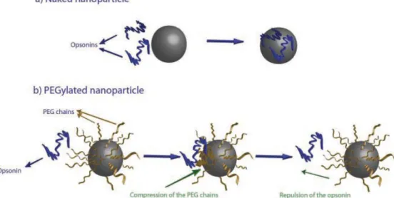
Surface hydrophobicity is the other parameter that might be considered in the design of drug delivery nanosystems. Indeed, the hydrophilic–lipophilic balance of nanomaterials influences the mechanism of opsonisation that results faster forthe particles more hydrophobic due to the enhanced “adsorbability” of blood serum proteins onto their surface.
In order to reduce the clearance by opsonisation, shielding groups can be introduced onto the nanoparticles surface. Polyethylene glycol (PEG) represents the most widely material used in the constitution of a hydrophilic outer shells of drug delivery systems.36 The “PEGylated” process can be achieved by grafting, entrapping, adsorbing or covalently binding to nanoparticles these molecules. Due to their flexibility and hydrophilicity, PEG molecules are able to stabilize nanoparticles by steric and not ionic effects especially, adopting an extended conformation able to repel the opsonins away from the nanoparticles (Figure 6). PEG molecules characterized by a molecular weight higher or equivalent to 2000 Da showed the optimal chain length necessary for reducing the RES clearance.37
16
1.2.2 Poly(d,l-lactic-co-glycolic acid)-b-poly(ethylene glycol)
In the last years, polymeric nanoparticles constituted by the di-block copolymer poly(d,l-lactic-co-glycolic acid)-b-poly(ethylene glycol) (PLGA-b-PEG) have been one of the most studied drug delivery systems. PLGA-b-PEG is constituted by a lipophilic portion made of PLGA, which confers it biodegradability and biocompatibility properties, and a hydrophilic portion made of PEG, which confers it the stealth behaviour.
Poly(lactic-co-glycolic acid) (PLGA) is a linear aliphatic polyester approved in 1989 by the FDA for drug delivery application, synthetized through the random ring-opening copolymerization of two cyclic dimers of glycolic and lactic acid, linked together by ester linkage in a casual order during copolymerization (Figure 7).
Figure 7. Chemical structure of poly(lactic-co-glycolic acid) (PLGA) constituted by m units of lactic acid and n units of glycolic acid.
It has been showed that the molar ratio of the individual monomercomponents (lactide and glycolide) in the polymer chain influences many properties of PLGA, such as degree of crystallinity, mechanical strength,swelling behaviour, and capability to hydrolyze.38
Because these properties can affect the biodegradability of the material, an investigation of the optimal molar ratio has been carried out. The results showed that PLGA copolymer with a 50:50 molar ratio of the two monomer is hydrolysed much faster in comparison with the one containing higher quantity of either of two monomers.39
Once the PLGA chains are released in aqueous media, such as in the body, undergo a hydrolytic degradation of the ester linkages. This process led to the formation of the two original monomers, lactic and glycolic acids that in body can be metabolized and eliminated as carbon dioxide and water or excreted unchanged in the kidney.40
On the other hand, poly(ethylene glycol) (PEG) is a non-ionic hydrophilic polyether synthesized in a wide range of molecular weight from 300 to 100,000 Da, through polymerization of the monomer ethylene glycol (Figure 8). PEG is a biocompatible polymer but not biodegradable, indeed in the body, PEG chains are excreted unchanged in the kidney and does not undergo biodegradation process.
17
As discussed in the previous paragraph, PEG polymer is characterized by high hydrophilicity properties and for this reason is used to stabilize nanoparticles in aqueous media, to increase solubility, and to avoid aggregation of them by steric hindrance in production, storage, and applications.41
Figure 8. Chemical structure of poly(ethylene glycol) (PEG).
Beyond the biodegradability and biocompatibility of its components, PLGA-b-PEG assumes amphiphilic properties that make it suitable for the formation of polymeric nanoparticles and micelles. In the formation of polymeric nanoparticle, the PLGA-b-PEG chains undergo to a self-assemble generating a system in which the hydrophobic PLGA remains inside the micelles and the hydrophilic PEG goes outside creating a stabilizing shell.
Because the PEG molecules can be characterized by several terminal function groups (such as carboxylic ammine, maleimide, thiol groups), it is possible to obtain a functionalized copolymer PLGA-b-PEG-X (Figure 9). In this way, the obtained polymeric nanoparticles will characterize by the PEG functional groups on the surface able to conjugate specific target agents.
Figure 9. Chemical structure of poly(lactic-co-glycolic acid)-block-poly(ethylene glycol) (PLGA-b-PEG) with indicated the characteristic final functional groups.
18
1.2.3 Methods of preparations for polymeric nanoparticles
The main strategies developed for the synthesis of drug delivery systems based on polymeric nanoparticles (PNPs) are divided in two different approaches, methods in with preformed polymers are used or methods that involve the direct polymerization of monomers using classical polymerization or polyreactions.42
The common techniques based on the use of preformed polymers are solvent evaporation, salting-out, dialysis and supercritical fluid technology.
In order to choose the suitable method for the synthesis, must be consider the chemical properties of polymeric material and/or of the agent that should be encapsulated, such as solubility and stability in organic solvent or in water.
PLGA-b-PEG is soluble in several organic solvents and due its amphiphilic properties, the synthesis of polymeric nanoparticles not required large amount of surfactant agents, able to stabilize in water the formed polymeric nanoparticles characterized by high hydrophobic properties. In particular, solvent evaporation and nanoprecipitation methods have been widely used for PLGA-b-PEG nanoparticles synthesis. 41
The solvent evaporation technique consists in the preparation of emulsion with a not-miscible organic solvent and water. Based on the solubility properties of the agents that should be encapsulated, two main strategies for the formation of emulsions have been developed: the preparation of single-emulsions (oil-in-water (o/w)) or double-emulsions ((water-in-oil)-in-water, (w/o/w)).43
In the oil-in-water (o/w) technique, an organic solution is prepared by dissolving polymer and lipophilic agents, which should be encapsulated, in volatile and water-immiscible organic solvents. The common solvents used for the solvent evaporation methods are dichloromethane, chloroform and ethyl acetate. Once prepared, the organic phase is added to a large amount of water, and the obtained biphasic solution is emulsionated with high-speed homogenization or ultrasonication, followed by evaporation of the solvent, by continuous magnetic stirring at room temperature or under reduced pressure (Figure 10).
In order to stabilize the formed polymeric nanoparticles, surfactant agents are usually solubilized in the aqueous phase, such as sodium cholate, polyvinylalcohol (PVA), polyvinylpyrrolidone (PVP), poloxamers, or other molecules. During the emulsion process, the strong energy provided from homogenizator or ultrasonicator allows the formation of organic nanosized droplets in the surrounding water phase.
19
Figure 10. On the left, a picture of tip probe sonicator using for the solvent evaporation method, on the right a schematic representation of oil-in-water technique.
Because of the amphiphilic properties, PLGA-b-PEG molecules dissolved in the organic phase can form micelles without the presence of surfactant agents, constituted by an inner core, made of the hydrophobic PLGA portion, and surrounded by an external shell made of hydrophilic PEG chains, able to stabilize the nanomicelles in aqueous environment. Depending on the lipophilic agents entrapped, this method allows the obtainment of nanospheres with good index of polydispersity (PDI) and a diameter in the range 100–500 nm. In order to encapsulate hydrophilic agents, the solvent evaporation can be used by following the double-emulsions ((water-in-oil)-in-water, (w/o)/w) strategy, which involves two emulsion processes. First, a water phase is prepared by dissolving the hydrophilic agents in a small volume of water (W1). The first emulsion process consists in the emulsion of the prepared aqueous phase W1 with an organic phase prepared by dissolving the polymer in an organic solvent, as in the oil-in-water technique. In this case, the organic phase is in the majority compared to the aqueous phase, so an opposite situation respect to the oil-in-water emulsion is created and the water phase with the drugs dissolved-in remains inside the micelles core, while the oil phase constitutes the external environmental. After that a bigger amount of a second aqueous phase (W2) is added to the previous emulsion and the entire system is re-emulsified to obtain the w1/o/w2 double emulsion; in this case, the secondary aqueous phase always contains surfactants or stabilizing agents. During this secondary emulsion, the organic phase remains entrapped between the two aqueous phases, the W1 in the inner core and the W2 that constitutes the external ambient. The organic solvent can be now removed by evaporation and the final system, dispersed in water with water in the inner core, can be obtained. In addition, when lipophilic agents are dissolved with the polymer in the organic phase, the w/o/w technique allows entrapping two different agents in the same polymeric nanoparticles, suitable for the development of theranostic nanosystems.
20
On the other and, in nanoprecipitation method the organic phase in prepared by dissolving the polymer and lipophilic agents, in organic solvent miscible with water such as ethanol, tetrahydrofuran, acetone, hexane, dimethyl sulfoxide or dimethylformamide. This organic solution is mixed under vigorous stirring into water, which may contain surfactant or stabilizing agents. This method is based on the interfacial deposition of a polymer after displacement of a semipolar solvent, miscible with water, from a lipophilic solution. Rapid diffusion of the solvent into water results in the decrease of interfacial tension between the two phases, which increases the surface area and leads to the formation of small droplets of organic solvent (Figure 11).44 The polymer dissolved in these droplets forms small macromolecules, that begins the nucleation process based on the aggregation of this nuclei which leads to the formation of polymeric nanoparticle.41 Particles formed with this technique can be obtained with small diameter (50–250 nm) and good polydispersity index (PDI).45
21
1.3 Hyperthermia therapy
Hyperthermia therapy (defined also thermal therapy) is a cancer treatment that consists in the exposition of body tissue to an increasing of temperature, just above the physiological temperature (42-45°C) to induce cytotoxic effects on cancer cells.
Indeed, cancer cells are more sensitivity to heat compared to healthy cells, and at those temperature, tend to undergo in apoptosis caused by denaturation of membrane and cytoplasmic proteins.46 This phenomenon can be explained by considering the physiological differences between normal and tumour tissues; the disorganised and abnormal vasculature caused by angiogenesis, leads to a lower vessel density in tumour tissues, that makes heat dissipation hindered in comparison to healthy tissues.47 For this reason, when hyperthermia is applied at temperatures over 42 °C, the temperature of tumour rises higher than that in normal tissue, because during heating tumour blood flow tends to decrease while in normal tissue it significantly increases. On the other hand, hyperthermia treatment at lower temperature than 42°C, tends to generate an increasing in the tumour blood flow, making cancer cells more sensitive to radiotherapy (radiosensitivity is favoured by good tissue oxygenation) and chemotherapy (drug delivery is increased by higher perfusion).48 The synergic effect of hyperthermia with other traditional therapies has been investigated in clinical trials in the treatments of many types of cancer, showing a significant reduction in tumour size.49 However, not all of these studies have shown increased survival in patients receiving the combined treatments.
There are three main types of hyperthermia treatments, whole-body, regional or local, which depend on the location, the depth and the stage of malignancy. Whole-body hyperthermia is used to treat metastatic cancer that has spread throughout the body and consists in the heating of the entire body through hot water baths, thermal chambers or infrared radiators. However, because the temperature increasing is not selective, this treatment can lead to major side effects due to regional differences in tissue characteristics, causing the production of dangerous temperatures in normal tissue. Regional hyperthermia consists to heat large areas of tissue (as a body cavity, organ or limb), generally affected by an advanced stage of tumour, by using external applicators or arrays of multiple applicators (microwave antennas). On the other hand, local hyperthermia is applied to treat small area, in particular localized tumours either superficially or in accessible body cavities, using various techniques that deliver energy to heat the cancer cells. Depending on the tumour location, there are several approaches to
22
local hyperthermia, which involve different types of energy used to apply heat, including microwave, radiofrequency, and ultrasound.50
Because the limited spatial and temporal control, traditional hyperthermia treatments can cause burns to surrounding healthy tissues. For this reason, nanotechnology advantages have been largely investigated, in order to develop nano-sized heat agents able to promote localized temperature increasing in cancer cells.
The first approach of nanomedicine in hyperthermia therapy consisted in the use of magnetic nanoparticles as generators of heat, leading to the development of magnetic fluid hyperthermia (MFH).51 This technique, based on the conversion from magnetic energy into thermal energy in the supermagnetic nanoparticles subjected to an external alternative magnetic field, led to the development of the first hyperthermia clinical nanotherapy. In 2011, the nanomedicine company MagForce launched NanoTherm®, a clinical therapy based on ultrasmall (~12 nm) iron-oxide magnetite (Fe3O4) coated by aminosilane for hyperthermia therapy of solid tumours (Figure 12).
Figure 12. Image of NanoTherm® produced by MagForce.
The treatment consists in the directly injection of NanoTherm® into the tumour and the subsequently application of an alternating magnetic field to generate heat, combined with fractionated stereotactic radiotherapy. Clinical trials results conducted in the treatment of multiform glioblastoma, an aggressive brain tumour, showed that thermotherapy using magnetic nanoparticles in conjunction with a reduced radiation dose is safe and effective and leads to longer over survival of patients, compared to conventional therapies.52
Despite the good results obtained by using MFH, nanotechnology is also exploring other alternatives. In particular, the recent development of nanoparticles capable of efficient heat generation under illumination with laser radiation has attracted much attention for the last few years. Photothermal therapy (PTT) is an hyperthermia therapy based on the use of
near-23
infrared (NIR) laser photoabsorbers to generate heat in order to induce thermal ablation of cancer cells upon NIR laser irradiation.53
PTT offers solutions to the limitations affecting other thermal therapies, for example, the possibility of eradicating tumours located nearby intrabody cavities by the use of low-loss and flexible optical fibers.54 However, PTT of sub-tissue tumours non accessible by optical fibers is very restricted, due to the fact that human tissues show strong extinction coefficients in the optical range of the electromagnetic spectrum. This fact limits PTT only to the treatment of superficial tumours. In order to overcome this limitation, PTT must be performed by using specific excitation wavelengths at which human tissues are partially transparent. This can be achieved by using laser excitation wavelengths lying in the so-called biological windows (BW): the first Biological Window (I-BW, which extends from 700 up to 950 nm) and the second Biological Window (II-BW, 1000–1350 nm) (Figure 13).55
Figure 13. Extinction coefficient of a representative tissue. The different effects leading to light attenuation (such as the presence of haemoglobin, water and optical scattering) have been indicated. The spectral extensions of the two biological windows are also indicated. Figure adapted from Nanoscale, 2014, 6, 9494.
Therefore, nanoparticles able to absorb laser excitation wavelengths lying in the biological windows are suitable candidates as photothermal therapy agents, reducing the non-selective heating of healthy tissue and, at the same time, allowing a deep tissue penetration.
For these purpose, several studies have been conducted in order to design a nanosystems able to be used in clinical photothermal therapy. In 2008, Nanospectra Biosciences developed AuroLase® Therapy (Figure 14), a thermal ablation therapy of solid tumours based on the use of silica nanoparticles coated by a gold metal shell able to absorb near infrared laser
24
energy delivered by a fibre. This therapy is under clinical investigational at this current time for the treatments of head and neck cancer treatments.56
25
1.3.1 Metallic nanoparticle as photothermal agents in hyperthermia
therapy
Metal nanoparticles have been largely studied as agents in photothermal cancer therapy (PTT) because of their ability to absorb light and rapidly convert it to heat.
This ability is ascribed to the resonant collective oscillation of the free electrons on the metal surface of the nanostructure in the presence of luminous radiation, a phenomenon called localized surface plasmon resonance (LSPR), which is common to all the nanoparticles but is particularly pronounced in those derived from transition metals (Figure 15).
Figure 15. Schematic diagrams of localized surface plasmon (LSPR)
In PTT, when a nanoparticle is irradiated by laser beam, some of the incident photons are scattered by the nanoparticle while others are absorbed. The absorbed photons are responsible for heat production and luminescence. The total number of photons interacting with the metallic nanoparticles is determined by its extinction coefficient (αext), that is given by the:
αext = αabs + αsct
where αabs is the absorption coefficient and αsct is the scattering coefficients
The extinction coefficient depends on the concentration of illuminated nanoparticles and on the extinction cross section (σext = σabs + σsct, where σabs and σsct denote the absorption and scattering cross-sections, respectively).
26
The absorption efficiency, Φ abs, of a given nanoparticle is traditionally defined as the number of absorbed photons divided by the total number of incident photons interacting with the nanoparticle (being either absorbed or scattered).
Φ abs = αabs/αext
The energy absorbed by the nanoparticles (energy of incident photons multiplied by the total number of absorbed photons) can be released by either the emission of photons of different energy from that of incident photons (luminescence) or by the emission of phonons (i.e. by generating heat) (Figure 16).53
Figure 16. Schematic representation of the different processes activated when a light beam interacts with a nanoparticle. The presence of scattering, luminescence and heat generation are included. Heat and luminescence occur as a result of light absorption. Figure from Nanoscale 6, 9494–9530 (2014).
However, the optical properties of metallic nanoparticles are strongly influenced by size, shape and environment. Indeed, the phenomenon of the LSPR can be described in details thank to the theory of Gustav Mie (1908), who solved the Maxwell equations giving a quantitative explanation of the resonance. Generally, simplified equations deriving from Mie theory are used to explain the importance of many parameters that strongly affects LSPR intensity and wavelength.57 By way of illustration, for many metals in the bulk state free electron behaviour is predominant and the wavelength of the plasmon absorption peak depends on the equation:
27
λ2 = λp2 (ε∞ + 2εm)
where
λp2 = (2πc)2/ωp2
is the metal's bulk plasma wavelength, ε∞ is the high frequency dielectric constant due to interband and core electrons’ transitions, εm is the dielectric constant of the surrounding medium and the resonance frequency (ωp) is given by:58
ωp= (N*e2/ε0*me)1/2
where N is the concentration of free electrons in the metal, e is the charge of the electron, m is the mass of the electron and ε0 is the vacuum permittivity.
The equations shows that the LSPR is strongly affected by several factors such as size and shape of the nanoparticles and most of all by the nature of the surrounding environment due to a direct dependence on the dielectric constant of the medium (εm) in which the nanoparticles are dispersed. Therefore, every modification in the interface with the surrounding environment of these particles leads to significant shifts to the LSPR wavelength and intensity.
28
1.3.2 Gold nanorods as photothermal agents
Since from 5th B.C., the use of colloidal gold as a colorant has been known for its use in making ruby glass and providing reddish tinge to ceramics.59
In 1857, the most important article in both the history of colloidal science and in the discussion of how light interacts with matter has been published by Faraday, who reported that the ruby glass was coloured because of the presence of finely dispersed gold particles. Nowadays, it is well known that gold nanoparticles assume a peculiar red colour in solution due to the localized surface plasmon resonance (LSPR) at visible range of UV-Vis spectrum. In the last decades, several studies have been focused on the development of gold nanostructures characterized by different size and the shape, in order to move the spectral location of surface plasmon resonance wavelength from the visible to the near infrared region. At macroscopic level, the change in colour of colloidal solution of gold nanoparticles is an evidence of the increase of nanoparticles size or change of nanoparticles shape. For example, an aqueous dispersion of spherical gold nanoparticles with a diameter around 10 nm is characterized by a red colour; otherwise, when gold nanoparticles size increases to nearly 100 nm or changes in shape, such as a rod-like shape with aspect ratio of 3 (length 30 nm, diameter 10 nm), the colloidal dispersion appears bluish.
This phenomenon can be explained by considering the movement of λSPR in the UV-Vis spectra of gold nanostructures characterized by a different size and shape.
The possibility of tailoring the λSPR in a very wide range (from visible to infrared) by the adjustment of their dimensions, led to the development of GNPs with a great variety of geometries (nanocages, nanostars, nanoshells, and nano- hexapods).
Their unique optical properties, in addition to their biocompatibility, make gold nanomaterials promising candidates as light-activated heating nanoparticles in PTT applications. For this purpose, gold nanorods (GNRs) have been the most studied nanoparticles among the different non-spherical metallic nanoparticles.
Indeed, GNRs are characterized by the presence of two main absorption bands in the UV-Vis spectrum (Figure 17a):
An intense absorption band at near infrared wavelengths, corresponding to electron oscillations along the longest dimension of the GNRs;
A weak absorption band at visible wavelengths, corresponding to surface electron oscillations along the short dimension of the GNRs;
29
Figure 17. (a) Diagram depicting the conduction band electron oscillation (gray arrows) upon transverse and longitudinal localized surface plasmon resonances of gold nanorods. (b) Visible/near infrared extinction spectra of gold nanorods with different aspect ratios (ARs) showing transverse (★) and longitudinal (◆) extinction peaks. TEM images of gold nanorods (c) AR 1.1, (d) AR 2.0, (e) AR 2.7, (f) AR 3.7, and (g) AR 4.4. Scale bars are 50 nm. Figure adapted fromLangmuir 32, 9905–9921 (2016).
Several methodologies have been fine-tuned in order to obtain gold nanorods characterized by a rational control over the aspect ratio (AR). The aspect ratio, corresponding to ratio between length and width of GNRs, is primarily responsible for the change in their optical properties. By increasing the aspect ratio, the longitudinal plasmon resonance of GNRs moves towards high wavelengths up to 1200 nm (Figure 17 b and c).60
This phenomenon is very important for the developing of photothermal agents based on gold nanostructures, which might be characterized by λSPR close to 800 nm, in other words placed in the first biological window in order to lead the minimum collateral heating effect on the healthy surrounding tissue.
In order to study the absorption efficiency of this particular gold rod geometry and size, several simulation studies have been carried out. The studies of El-Sayed et al. reported that GNRs usually show absorption efficiencies above 90% except when the GNR radius exceeds 20 nm. In particular, the results showed that the heating efficiency remains above 90%, when the GNR width (diameter) is kept close to 15 nm independently of the GNR length (at least for lengths as large as 160 nm). However, the absorption efficiencies predicted decrease down to 60% for GNRs characterized by a diameter greater than 15 nm.61
30
Usually, GNRs synthesis is based on the two-step ‘‘nuclear’’ method discovered by Zsigmondy, combining his synthesis technique with the Faraday’s method. Later this method has been fine-tuned and was defined ‘‘seed-mediated’’ method.
The method consists the synthesis in aqueous medium with the assistance of various surfactants, which act as both template and stabilizer for the growing nanoparticles against aggregation phenomena. Once the process is completed, the surfactant molecules remain adsorbed or deposited onto the surface of the nanoparticles, avoiding the post- synthesis collapse of the created nanoparticles. However, most of these surfactants are strongly toxic or simply not suitable for biomedical applications because they do not allow further synthetic medications. The removal of surfactants requires the development of specific ligands that would be able to replace them, to prevent the aggregation phenomena, and at the same time ensure that the specific desired final properties of GNRs are retained (paragraph 1.5). 62
31
1.3.3 Magnesium nanoparticles as photothermal agents
Magnesium is one of the most abundant element in the human body, in particular its form as divalent cation (Mg2+) is involved in more than 300 known enzymatic reactions, playing an important role in many cellular processes.63 In addition magnesium has role in many reactions involving ATP, protein and nucleic acid synthesis, mitochondrial activity and integrity, ion channel modulation, plasma membrane stabilisation and translational processes, as well as many other cellular functions.64
Due to its biocompatibility, the use of magnesium in biomedical application has been largely investigated. The modern applications of magnesium-based biomaterials are similar to those that were attempted historically. At the beginning of 1900, numerous physicians studied the application of magnesium and magnesium alloy devices in vascular, orthopaedic and general surgery. However, without the ability to accurately alloy magnesium, and before the common application of coatings on implants, the corrosion could not be controlled sufficiently to promote its use as biomaterial.
Indeed, magnesium-based materials revealed a quickly degradation that affects their mechanical integrity for long time and showed the production of hydrogen gas in the organs able to cause tissue disruption. For these reasons, the most effective results were obtained from non-orthotopic applications, such as the suturing of organs.65
In the late 1930s, Earl McBride conducted studies regarding the use of magnesium alloy as orthopaedic implants. During his studies, he found that corrosion of magnesium based materials resulted in a tissue reaction, could represent an advantage for specific applications in which the rapid corrosion of the implant materials utilized would outweigh the effect on the surrounding tissue.66 Based on these results, the field of magnesium biomaterials has been growing exponentially, especially in the fields of investigation regarding the development of magnesium alloys for vascular applications, and the field devoted to identifying appropriate magnesium based materials for orthopaedic applications.
In the last decades, the properties of magnesium nanoparticles have been investigated in nanotechnology research, fascinated by the high biocompatibility of this nanomaterial related of its biodegradation into magnesium ions, which are completely absorbable by the human body. In particular, recent studies demonstrated that magnesium nanoparticles exhibit a pronounced plasmonic response that is tunable throughout the whole visible wavelength range.67,68 In particular, Sterl et al. reported that plasmonic response of magnesium nanodisks can be tuned by adequate choice of the disk diameter throughout the visible wavelength range,
32
from resonance wavelengths below 500 nm up to 800 nm and further into the near-infrared (Figure 18).69
Figure 18. Magnesium nanodisks as a model system for magnesium plasmonics. (a−c) Colorized SEM images of magnesium nanodisks with average diameters of 80, 160, and 220 nm, respectively. (d) The resonance wavelength of such nanodisks can be tuned throughout the visible wavelength range by varying the diameter. Figure adapted from Nano Lett. 15, 7949–7955 (2015).
Due to their plasmonic properties, magnesium nanoparticles has been studied as suitable agents for photothermal tumour therapy.70,71,72,73
In 2012, Wang et al., tested the direct injection of the magnesium nanoparticles into the tissues to measure the temperature changes of the materials subjected to laser heating (Figure 19).72 The obtained results showed the high ability of magnesium nanoparticles in hyperthermia applications and the related advantages comparing to other popular nanosystems. Compared to gold-based nanomaterials, the density of magnesium is much lower and therefore more particles can be loaded into the target tissues leading a more efficient treatment. Moreover, magnesium is beneficial to human health and its high abundant in nature makes magnesium cost-effective for clinical utilization.
33
Figure 19. Thermal images of the pig tissue injected with water (A) 0.01 g/mL fresh magnesium nanoparticle aqueous solution (B) and 0.02 g/mL fresh magnesium nanoparticle aqueous solution (C) taken after 1 minute of laser irradiation at 1W. Figure from Int. J. Nanomedicine 7, 4715–4725 (2012).
Nevertheless, the results highlighted that there are still many problems to overcome in order to use magnesium nanoparticles as photothermal agents in clinics. First of all, the heating ability of magnesium nanoparticles is affected by variable sizes, structures of these particles and oxidation grade, therefore the influence of each parameter must be deeper investigated. Regarding the oxidation grade, they showed that only pure magnesium nanoparticles, which are often black in colour, are able of absorb light differently from oxidized magnesium nanoparticle (MgO nanoparticles), which are often white in colour. Because of their high reduction potential and high affinity toward oxidation, the synthesis of magnesium nanoparticles is quite challenging, and several synthetic methodologies have been developed in order to improve the process.
In addition, the electrode potential of magnesium can affected also heat generation ability of nanoparticles and their stability in vivo. Once magnesium nanoparticles are injected into the tissue, heat energy is produced because of chemical reaction between magnesium nanoparticles and water contained in the tissue.
However, magnesium nanoparticles showed a low reactivity with water below a certain temperature, such as the body core temperature of 37°C. Therefore, a suspension fluid containing the nanoparticles with an initial temperature around room temperature (25°C) can be easily injected into the target sample or tissues.
Because this phenomenon leads to a certain level of corrosion of the material, the control of the degradation rate of the magnesium nanoparticles used in hyperthermia is an important requirement. Indeed, the rapid corrosively degradation of the material represents an advantage considering the rapid absorption of magnesium ions into the body; however, if the rate of material degradation is too fast, the heating enhancement effect may not be maintained for long or fully occur. Regarding this, a protective coating on the nanoparticles might be a
34
feasible way to avoid their reacting with water as well as to control the degradation rate and further improve biocompatibility. Moreover, the surface coating formation can allow the possibility to introduce an active targeting on the nanoparticles surface, making the final system suitable for diagnostic and theranostic applications. For example, the study of Jin et al., developed a laser scanning based thermography strategy for detecting the skin cancer by using magnesium nanoparticles conjugated with biomolecules (for example antibody) as active targeting.73
35
1.4 Nanomaterials as diagnostic and imaging tools in
nanomedicine
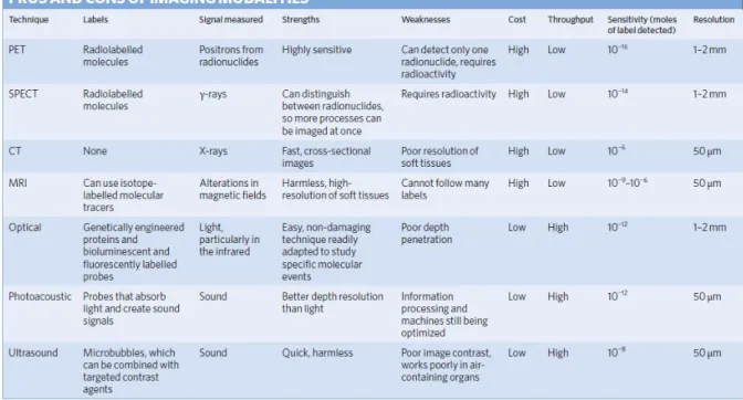
For a non-invasive diagnosis of patients, in vivo imaging techniques are powerful tools to visualize the abnormal state of the body and monitor biological situations at the target site. Several imaging techniques have been developed for this purpose, such as magnetic resonance imaging (MRI), positron electron tomography (PET), computed tomography (CT), single-photon emission computed tomography (SPECT), optical imaging (OI), ultrasound (US) and photoacoustic imaging (PAI). Because of the several physical processes behind each of these techniques, they are characterized by different depth of penetration and spatial resolution abilities.74
Figure 20. Table dealing the properties of imaging modalities adapted from Chem. Soc. Rev. 41, 2656–2672 (2012).
As shown in Figure 20, microscopy and other techniques that use un-scattered photons provide high-resolution (~1 μm) images, but only to a maximum depth of ~1 mm in most tissues. On the other hand, diffuse optical techniques such as fluorescence and near-infrared optical tomography, that exploit multiply scattered photons, can reach a depth of several centimetres but a lower spatial resolution (>1 mm).75
For this reason, the combination of different imaging techniques in multimodality approach, showed the possibility to accomplish high sensitivity and high resolution simultaneously, providing more detailed anatomical or biological information about the target disease. The first example of multimodalities techniques is represented by PET/CT in 1998, while the first