Copyrightq 1997, American Society for Microbiology
Interplay of the E Box, the Cyclic AMP Response Element,
and HTF4/HEB in Transcriptional Regulation of the
Neurospecific, Neurotrophin-Inducible vgf Gene†
GIULIANA DI ROCCO,1‡ MARIA PENNUTO,1BARBARA ILLI,1NADIA CANU,2GESSICA FILOCAMO,1§
EUGENIA TRANI,2\ ANNA MARIA RINALDI,2ROBERTA POSSENTI,2GEORGIA MANDOLESI,1
M. ISABELLA SIRINIAN,1RICHARD JUCKER,1ANDREA LEVI,2ANDSERGIO NASI1*
Centro Acidi Nucleici CNR, Universita` “La Sapienza,” 00185 Rome,1and
Istituto di Neurobiologia CNR, 00156 Rome,2Italy
Received 7 August 1996/Returned for modification 30 September 1996/Accepted 5 December 1996
vgf is a neurotrophin response-specific, developmentally regulated gene that codes for a neurosecretory poly-peptide. Its transcription in neuronal cells is selectively activated by the neurotrophins nerve growth factor (NGF), brain-derived neurotrophic factor, and neurotrophin 3, which induce survival and differentiation, and not by epidermal growth factor. We studied a short region of the rat vgf promoter which is essential for its regulated expression. A cyclic AMP response element (CRE) within this region is necessary for NGF induction of vgf transcription. Two sites upstream of CRE, an E box and a CCAAT sequence, bind nuclear protein complexes and are involved in transcriptional control. The E box has a dual role. It acts as an inhibitor in NIH 3T3 fibroblasts, together with a second E box located downstream, and as a stimulator in the NGF-responsive cell line PC12. By expression screening, we have isolated the cDNA for a basic helix-loop-helix transcription factor, a homolog of the HTF4/HEB E protein, that specifically binds the vgf promoter E box. The E protein was present in various cell lines, including PC12 cells, and was a component of a multiprotein nuclear complex that binds the promoter in vitro. The E box and CRE cooperate in binding to this complex, which may be an important determinant for neural cell-specific expression.
Nerve growth factor (NGF) is the prototype of neurotro-phins, a small family of structurally related proteins playing a crucial role in differentiation and survival of neurons and in maintenance of neuronal function (5, 28). Induced changes in gene expression are critical for their activity. Transcriptional activation of immediate-early genes, like c-fos, is rapid, tran-sient, and independent of protein synthesis, but it is not specific for the neurotrophin response, since it is stimulated by various growth factors. Induction of delayed-early genes, like vgf (27), can instead be neurotrophin specific. vgf transcription is in-duced by brain-derived neurotrophic factor (BDNF) and rotrophin 3 (NT3) in cultures of cortical or hippocampal neu-rons and by NGF in PC12 cells (3, 38). This cell line, widely used as a paradigm for neuronal differentiation, has receptors for NGF and epidermal growth factor; while NGF stimulates cell cycle withdrawal and neuronal differentiation, epidermal growth factor does not induce PC12 cell differentiation and is unable to activate vgf transcription effectively. The study of vgf regulation is therefore expected to give insight into the process of neural differentiation. vgf is developmentally regulated: its expression is detected in embryonic central and peripheral nervous tissues, peaks in the early postnatal critical period of nervous system development, and is maximal during synapto-genesis of the visual cortex (30, 41). In adult animals, vgf is expressed in subsets of brain neurons, in sensory and
sympa-thetic neurons, and in endocrine cells from the adrenal me-dulla, adenohypophysis, and gut (14, 49). The VGF protein is stored in secretory vesicles, released in response to stimuli causing membrane depolarization, and proteolytically pro-cessed in smaller peptides that may play a role in intercellular communication (39, 47).
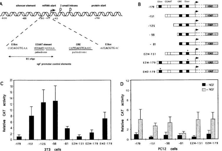
An 800-bp region upstream of the vgf transcription start site acts as a NGF-inducible promoter in PC12 cells and harbors important determinants of neural specificity, since it drives gene expression to neural and endocrine tissues in transgenic mice (36, 38). The promoter is under negative control in non-neuronal cells (38). Many of the properties of the vgf promoter are encoded by 180 nucleotides, proximal to the transcription start site, that are essential for expression in the NGF-respon-sive rat pheochromocytoma cell line PC12 and involved in negative regulation in nonneuronal cells (38). A cyclic AMP
(cAMP) response element (CRE) (position281 to 274)
me-diates vgf transcriptional induction by binding a member of the CRE binding protein (CREB) family of transcription factors, activated by cAMP and by NGF (17, 19). Other elements are also required for vgf transcriptional regulation, as suggested by the observations that mutating the CRE consensus sequence does not affect promoter strength in the absence of NGF and that although both cAMP and NGF activate CREB, NGF activates vgf much more strongly than cAMP does (19, 38). Moreover, CREB is activated by cAMP in many cell types, while vgf transcription is induced only in neuronal cells, thus indicating the presence of transcriptional inhibitors in nonneu-ronal cells. Important elements for transcriptional regulation are present within a region of about 100 nucleotides just up-stream of CRE (38). In this study, the binding of nuclear protein complexes to this regulatory region and its function in
vgf expression were analyzed. The dual role of an E-box
ele-ment in transcriptional regulation was determined, and a
* Corresponding author. Mailing address: Centro Acidi Nucleici C.N.R., Universita` “La Sapienza,” P.le A. Moro 5, 00185 Rome, Italy. Phone: (396) 49912227. Fax: (396) 49912500. E-mail: [email protected].
† This work is dedicated to the memory of Enrico Calef. ‡ Present address: Istituto S. Raffaele, DIBIT, 20132 Milan, Italy. § Present address: IRBM, 00040 Pomezia, Rome, Italy.
\ Present address: Dip. Medicina Sperimentale, Universita` “Tor Vergata,” Rome, Italy.
1244
on October 3, 2016 by ISTITUTO di NEUROBIOLOGIA e MEDICINA MOLECOLARE
http://mcb.asm.org/
cDNA coding for a basic helix-loop-helix (bHLH) transcrip-tional regulator, a homolog of the HTF4/HEB E protein, that specifically binds the E site was isolated. E and CRE sites in the vgf promoter cooperate in binding a multiprotein complex containing a product of the HEB gene.
MATERIALS AND METHODS
Cell culture and CAT assays.All cell lines were grown according to standard protocols. NIH 3T3 cells were transfected (33 105cells [2mg of DNA] per
experiment) by liposome fusion with Lipofectamine reagent (Gibco BRL) ac-cording to the manufacturer’s instruction. PC12 cells were transfected with calcium phosphate (23 106cells [15mg of DNA] per experiment) or
Lipo-fectamine with analogous results and induced by NGF as previously described (38). The various chloramphenicol acetyltransferase (CAT) reporter plasmids were cotransfected with 0.3 to 3mg of pRSV-bgal in order to assess transfection efficiencies. In the first set of experiments, CAT activity was assayed by thin-layer chromatography and quantified by counting the radioactivity of the spots corre-sponding to [14C]chloramphenicol and its acetylated derivatives with a Betascope
Image Analyzer. Subsequently, CAT activity was assayed by the diffusion method of Neumann et al. (34), with comparable results. CAT values were corrected for transfection efficiency by normalization tob-galactosidase values. All assays were performed at least three times; average values and standard deviations are reported. The data reported were obtained with CAT constructs containing 710 nucleotides of untranslated leader sequence following the transcription start site. Since these sequences may affect reporter gene expression, the experiments were repeated with shortened promoter CAT constructs, obtained by removal of sequences 39 of the PvuII site at position 140. Although the absolute CAT activities of the short constructs were lower, as already shown (38), their relative strengths, as well as their induction by NGF, were unaffected by removal of the 140 to 1710 leader sequence.
Plasmid construction.CAT reporter gene fusions from position2179 to 1710 and281 to 1710 in pEMBL8 CAT were already described (38). Deletions with endpoints between positions2179 and 281 were generated by PCR and fused to the CAT gene of pEMBL8 CAT in the following way: starting from the KpnI site (1455) in the vgf gene, fragments with different end points (2151, 2125, and 298) in the vgf promoter were amplified by PCR and used to replace the
BamHI-KpnI (2179 to 1455) restriction fragment of the 2179 to 1710 CAT plasmid. In the E2M- constructs, the256 E box (CACGTG) was mutated to CTCGGG by PCR and successive cloning of the amplified fragment. EM2-CAT and EM5-CAT vectors, with CATTTG and CAGGTT mutations in the2170 E box (CAGGTG), were generated by means of the Quick Change site directed mutagenesis kit (Stratagene). Dideoxy sequencing by the Sanger method (Se-quenase II kit; U.S. Biochemicals) was used to verify all of the constructs.
Library screening and production of recombinant proteins.Phages of a ran-dom-primedlgt11 expression library from the baboon lymphoid cell line 594S (22) and from an oligo(dT)-primed PC12 cell cDNA library inlZAPII were screened with a radiolabeled, double-stranded, nonconcatamerized EC oligonu-cleotide probe (2178 to 2132 vgf promoter sequence), as described by Singh et al. (45). A total of 53 105plaques were screened, and positive plaques were
rescreened three times; to ensure their binding specificity, positive phages were tested with an unrelated oligonucleotide probe. cDNAs were excised from the phage vector as NotI-EcoRI fragments, cloned in the same sites of pBluescript KS1, and sequenced by the dideoxy method.
The ubo cDNA was excised from pBluescript as a NotI-HindIII fragment and cloned in frame with the polyhistidine tag of pQE11 (from Qiagen) and with the glutathione S-transferase (GST) gene of the pGEX expression vector. The re-combinant protein produced by pQE11, named UBO, was purified to.90% homogeneity by affinity chromatography on an Ni-nitrilotriacetic acid column and eluted under nondenaturing condition with 1 M imidazole at pH 7 according to the manufacturer’s protocol. The fusion protein GST-UBO was purified by binding to glutathione-agarose beads and elution with reduced glutathione.
Electrophoretic mobility shift assays (EMSA).Nuclear extracts were prepared as described by Schreiber et al. (44) with minor modifications. The binding probes were terminally labeled with T4 polynucleotide kinase (double-stranded oligonucleotides and PCR-generated promoter fragments) or Klenow enzyme (restriction fragment). One to five micrograms of nuclear extracts or 20 to 100 ng of recombinant protein was incubated for 15 min on ice with 3 to 6 fmol (corresponding to 10 to 15,000 cpm) of labeled oligonucleotide. Each binding reaction mixture contained 20 mM HEPES (pH 7.9), 100 mM NaCl, 0.1 mM dithiothreitol, 0.05 mM EDTA, 0.4% Ficoll, and 2mg of poly(dI-dC) (50 ng in the case of the recombinant protein) as a nonspecific competitor in a total volume of 20ml. Competition experiments were performed by adding unlabeled oligonucleotides (100- to 200-fold molar excess) before the probe. For supershift assays, 0.5 to 1 ml of specific and control antisera were used. DNA-protein complexes were resolved by 5% polyacrylamide gel electrophoresis in 0.253 Tris-borate-EDTA buffer at 48C and autoradiographed.
DNase I footprinting.Reaction mixtures were prepared as for EMSA, with 30,000 cpm of DNA, 300 ng of purified UBO protein, and 100 ng of poly(dI-dC); binding reactions were carried out for 30 min on ice. After incubation, the concentrations of MgCl2and CaCl2were adjusted to 10 and 2 mM, respectively.
DNase I (3 ng) was then added, and the incubation was continued for 1 min at 208C. The digestion was stopped by addition of 200 ml of stop buffer (5 mM EDTA, 50 mM Tris [pH 8], 0.2% sodium dodecyl sulfate, 50mg of proteinase K per ml); after incubation at 458C for 30 min, DNA was ethanol precipitated and loaded onto 15% polyacrylamide sequencing gel.
Methylation interference.An end-labeled EC oligonucleotide (2178 to 2132) or a2178 to 280 vgf promoter restriction fragment (approximately 500,000 cpm) was partially methylated with dimethyl sulfate in the presence of 0.5 mg of sonicated salmon sperm DNA. Binding reactions were performed, under the same conditions as for EMSA, either with 250 ng of purified UBO protein and 250 ng of poly(dI-dC) or with 12mg of PC12 cell nuclear extract and 15 mg of poly(dI-dC). After electrophoresis, the gel was subjected to autoradiography for 3 h; free and bound DNA bands were excised and eluted in 0.2 M NaCl–10 mM Tris (pH 7.4)–1 mM EDTA. Recovered DNA fragments were then purified on ion-exchange minicolumns (NACS; Bethesda Research Laboratories), ethanol precipitated, and cleaved with 10% piperidine for 30 min at 908C. Samples were resolved on 15% polyacrylamide sequencing gels. The G1A lanes result from Maxam-Gilbert sequencing reactions. Quantitation of autoradiograms was per-formed with an imaging densitometer (Bio-Rad model GS670) with Molecular Analyst software.
Antisera and Western blots.The rabbit anti-UBO serum was raised by re-peated injections with purified protein produced by the pQE11 expression vec-tor. The specificity of the antiserum was tested by Western analysis of UBO-expressing bacteria compared to Escherichia coli UBO-expressing a different polypeptide (amino acids 207 to 442 of the v-CRK protein). Reactivity of the antiserum was abolished by preincubation with lysates of UBO-expressing, but not v-CRK-expressing, bacteria. The HEB antiserum, raised against a synthetic peptide encompassing the region of lowest homology between HEB and the other E proteins, and the relative preimmune serum were provided by D. Litt-man; the antiserum specifically recognizes HEB gene products and does not cross-react with E proteins derived from E2A and ITF2 genes. Nuclear proteins were extracted as described for EMSA. Western blots were performed according to standard protocols and probed with anti-UBO serum (1/5,000 dilution), fol-lowed by protein A-horseradish peroxidase and enhanced chemiluminescence detection with reagents from Amersham.
Nucleotide sequence accession number.The EMBL accession number for the
ubo cDNA sequence is X97234.
RESULTS
Binding of nuclear protein complexes to vgf promoter CCAAT and E-box sites.vgf promoter regulatory elements are
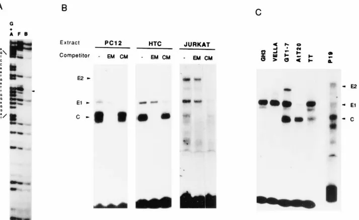
present in a 100-bp restriction fragment upstream of CRE (2179 to 281), whose deletion strongly impairs vgf expression in PC12 cells (38). In gel shift assays with PC12 cell nuclear extracts, two distinct protein complexes bind this restriction fragment, with the one of higher mobility being stronger (38). In order to determine the sequences involved in binding to the stronger complex, we performed a methylation interference analysis and identified a CCAAT motif (2140 to 2136) as the binding site (Fig. 1A). This motif is part of a dyad, and pro-tected bases were observed above and below the CCAAT se-quence; the results of microdensitometric analysis of the inter-ference experiments, defining the nucleotides involved in binding to the complex, are reported in Fig. 2. CCAAT motifs in other developmentally regulated promoters are targets of leucine zipper proteins of the C/EBP family of transcription factors (37). We utilized a synthetic peptide which competes for DNA binding of all known members of this family (37) in order to determine if the complex bound to the vgf promoter CCAAT was made of C/EBP-like proteins. Since the peptide was unable to compete for binding of this complex to the vgf promoter, this possibility was excluded (data not shown). In-spection of the sequence of the 100-bp promoter restriction fragment showed the presence of another consensus site, the E-box CAGGTG (2170 to 2165). A 47-bp oligonucleotide
including both motifs (EC oligonucleotides, 2178 to 2132)
efficiently competed for the gel shifts observed with the 100-bp restriction fragment and was used for further studies, together with control oligonucleotides (EM and CM) mutated in the E-box or CCAAT consensus sequence. The sequences of all probes used in DNA binding studies are reported in Table 1. In PC12 cell nuclear extracts, two complexes (E1 and C) bound
on October 3, 2016 by ISTITUTO di NEUROBIOLOGIA e MEDICINA MOLECOLARE
http://mcb.asm.org/
the EC oligonucleotide in a specific and competable way (Fig. 1B). The E1 complex was unaffected by competition with an excess of E-box-mutated oligonucleotide (Fig. 1B) and was undetectable when the mutated oligonucleotide was used as a binding probe, demonstrating its binding to the E box. Similar arguments confirmed that the stronger, higher-mobility com-plex (comcom-plex C) bound the CCAAT element, in agreement with the methylation interference. The same pattern of gel shifts was observed with the human neuroblastoma cell line SK-N-BE (data not shown). In order to assess a correlation between binding complexes and vgf expression, nuclear ex-tracts of several other cell lines were analyzed by EMSA (Fig. 1B and C); the binding specificity of each complex was verified by competition with mutated oligonucleotides. Among all cell lines tested, vgf mRNA is detectable in the three neuronal cell lines (PC12, SK-N-BE, and GT1-7), two endocrine cell lines
(GH3 and AtT20), and TT cells (reference 38 and data not shown). The pattern of binding complexes failed to show a correlation with vgf expression. The CCAAT binding complex appeared as a doublet only in PC12 and SK-N-BE (38) cells; it was a singlet in AtT20, HTC, GT1-7, and P19 cells, was weaker in TT and Jurkat cells, and was undetectable in Vella and GH3 cells. The E1 complex was present in all cell lines at various intensities; in two cell lines, Jurkat and P19, a second E-box binding complex, E2, was detected.
Role of E boxes in transcriptional regulation. E-box and CCAAT motifs in other promoters are involved in transcrip-tional regulation and differentiative responses (24, 48). The function of the two motifs in vgf transcriptional regulation was assessed by transient-expression assays with a neuronal (PC12) and a nonneuronal (NIH 3T3) cell line (Fig. 3). A deletion
removing the E box at position 2170 (2151 promoter)
in-FIG. 1. Binding of cellular nuclear complexes to E-box and CCAAT sites in the vgf promoter. (A) Methylation interference with PC12 cell nuclear extracts. The 2178 to 280 vgf promoter restriction fragment was labeled on the noncoding strand and used as a binding probe. F, free probe; B, bound probe; G1A, control sequencing reaction on G and A residues. The sequence shown on the left (noncoding strand) goes from position2127 (top) to 2145 (bottom). The arrowhead indicates the guanine (G) residue whose methylation most strongly interferes with nuclear protein binding. This G corresponds to the second C residue of the CCAAT motif on the coding strand. (B and C) EMSA with cell extracts from various cell lines. Nuclear extracts from the PC12 (rat pheochromocytoma), HTC (rat hepatoma), Jurkat (human T lymphocytes), GH3 (rat somatotroph cells tumor), Vella (human lymphoblasts), GT1-7 (mouse hypothalamus), AtT20 (murine corticotroph cells tumor), TT (human medullary tyroid carcinoma), and P19 (murine embryonal carcinoma) cell lines were utilized for EMSA with an end-labeled EC oligonucleotide. Competitions (shown only in panel B) were performed with excess amounts of E-box-mutated (EM) or CCAAT-mutated (CM) cold oligonucleotides, as indicated; the oligonucleotides sequences are shown in Table 1. E1 and E2, E-box binding complexes; C, CCAAT binding complexes. The uppermost shift detected with the GT1-7 cell extract is different from E2, since it was competed by both mutated oligonucleotides, and it may represent simultaneous binding of E1 and C complexes.
FIG. 2. Nucleotides involved in binding to the C complex. Guanine residues whose methylation interferes with binding of the C complex are underlined. The residual binding when the G is methylated is indicated; only values below 80% were considered. The palindrome harboring the vgf promoter CCAAT site is in boldface.
on October 3, 2016 by ISTITUTO di NEUROBIOLOGIA e MEDICINA MOLECOLARE
http://mcb.asm.org/
creased vgf expression in 3T3 cells by eightfold, indicating the
presence of a strong negative element between positions2151
and2179 (Fig. 3B and C). Longer deletions (2125 and 298
promoters), affecting the CCAAT motif, weakly potentiated the effect of the E-box deletion. Since cooperation between
different E boxes present on the same promoter is a common finding in transcriptional regulation of tissue-specific genes (16, 50), the role of a second E box, CACGTG, which is
present in the vgf promoter at position 256 (Fig. 3A), was
tested. Mutation of the second E box (E2M-179 reporter) also
FIG. 3. Role of E boxes in vgf transcriptional regulation. CAT reporter plasmids carrying vgf promoter deletions and mutations were transfected into NIH 3T3 and PC12 cells. (A) Diagram of the vgf promoter. The two E boxes (at positions2170 and 256), the CCAAT motif (at position 2140), and the CRE consensus sequence (at position281) are in boldface. The silencer element is a region with sequence homology to silencers of other neurospecific genes, like type II sodium channel and synapsin I. oligo, oligonucleotide. (B) vgf promoter CAT reporter plasmids. Deletion endpoints are indicated at the left. The E2M constructs have two point mutations (CTCGGG) in the E box at position256; the EM2-179 plasmid has point mutations (CATTTG) in the 2170 E box. (C and D) CAT assays with 3T3 cells (C) and PC12 cells (D). CAT activities are normalized for transfection efficiency withb-galactosidase values obtained by cotransfection with pRSV-bgal. The CAT values are relative to those for the2179CAT reporter.
TABLE 1. DNA sequences utilized in the study of DNA-protein interactions
Name Sequencea E...2183 ---CGCAGGTGCA--- 2158 EM2...2183 ---CGCATTTGCA--- 2158 EM3...2183 ---TTACCGTGCA--- 2158 EM4...2183 ---CGCCGGTGCA--- 2158 EM5...2183 ---CGCAGGTTCA--- 2158 EC...2178 ---CGCAGGTGCA---CCTCCAATCATT2132 EM...2178 ---CGTCACATCA---CCTCCAATCATT2132 CM...2178 ---CGCAGGTGCA---AATATAATCATT2132 vgf CRE... 290---CATTGACGTCAATG--- 265 BA/CRE2...2178 ---CGCAGGTGCA---CCTCCAATCATT---CATTG 280 PCR EM2 ...2183 ---CGCATTTGCA---CCTCCAATCATT---CATTGACGTCAATG---265 PCR WT ...2183 ---CGCAGGTGCA---CCTCCAATCATT---CATTGACGTCAATG---265 aThe three vgf promoter consensus sequences E box, CCAAT, and CRE are in boldface; mutations are underlined. All sequences were utilized as the
double-stranded form.
on October 3, 2016 by ISTITUTO di NEUROBIOLOGIA e MEDICINA MOLECOLARE
http://mcb.asm.org/
increased, although at a lower level (about threefold), expres-sion from the vgf promoter in 3T3 cells. When both E boxes were missing (E2M-151 reporter), transcription was at low levels (Fig. 3B and C). In PC12 cells, deletion of the upstream E box or mutation of both E boxes caused instead a 50% decrease of transcription activity compared to that with the 2179 promoter (Fig. 3D). To conclusively establish that the
effect of the2151 deletion on vgf transcription was due to the
loss of the E box at position2170, point mutations within this
element were utilized in NIH 3T3 transfection. A CAT re-porter (EM2-179) with mutations in the two central Gs of the consensus CAGGTG sequence had a high level of activity,
comparable to that for the2151 deletion (Fig. 3C). Mutation
of the two Gs was suggested by work on the immunoglobulin heavy-chain enhancer showing that mutation of the central Gs
in themE5 site relieved repression acting via this E box (40).
Another reporter plasmid, with a point mutation in the last G of the consensus sequence (EM5-179), showed a less strong increase of transcription (between two and three times less strong) (data not shown). These findings clearly establish a role of the upstream E box in vgf transcriptional regulation; its function is strongly negative in 3T3 cells and weakly positive in PC12 cells. Inducibility by NGF in PC12 cells was not signifi-cantly altered by removal of CCAAT and E boxes or by a
deletion extending up to position298 (Fig. 3D), while it was
severely compromised by a281 deletion, whose endpoint is at
the nucleotide preceding the CRE consensus sequence (Fig. 3D and reference 38).
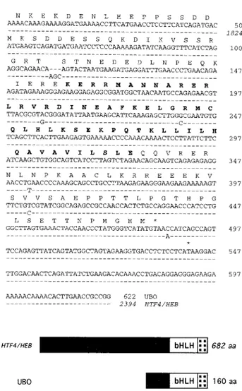
Isolation of the cDNA for the vgf promoter binding protein UBO, a homolog of the HTF4/HEB E protein.To better un-derstand the nature of the vgf promoter E-box and CCAAT binding complexes, we employed the EC oligonucleotide as a probe to screen for binding-protein cDNA expression libraries from PC12 cells and from the baboon lymphoid cell line 594S (22). The use of a nonneuronal cDNA library was justified by the finding that cell protein complexes bound to this oligonu-cleotide were not neural tissue specific (Fig. 1B and C) and by the evidence that the vgf promoter was the target of complexes endowed with repressor activity in nonneuronal cells (Fig. 3 and reference 38). Probably due to technical reasons, no pos-itive clone was identified in the PC12 library. Three pospos-itive cDNA clones were isolated from the lymphoid cell library, and two of them had the same sequence. A computer-generated alignment with sequences in nucleotide sequence databases showed an almost complete identity between one of the two clones, named ubo8 or simply ubo, and human cDNAs coding for the HTF4/HEB E protein (21, 51). E proteins (class A bHLH proteins) are a small family of widely expressed bHLH transcription factors that participate in developmental deci-sions in different tissues (29, 31). The ubo cDNA codes for the 160 carboxy-terminal amino acids, including the complete bHLH domain, and contains 136 nucleotides of the 39 untrans-lated region. Comparative analysis (Fig. 4) revealed four nu-cleotide substitutions in the simian cDNA (three of which, including the two in the bHLH domain, represent conservative changes) and the deletion of a serine close to the HLH domain (amino acid 558 in HTF4/HEB). To assess expression in neu-ronal cells, a homology screening of a human neuroblastoma (SK-N-BE cell line) cDNA library with the ubo probe was performed. Several positive clones were found, and sequencing of their cDNA inserts showed their derivation from the HTF4/
HEB gene, thus indicating its expression in neuroblasts.
The third cDNA was found to be homologous to the cDNA of the large subunit of replication factor C, a multisubunit DNA polymerase accessory protein complex (6). Since the encoded protein binds DNA with no sequence specificity (not
shown), we assume that it has no specific role in vgf transcrip-tion.
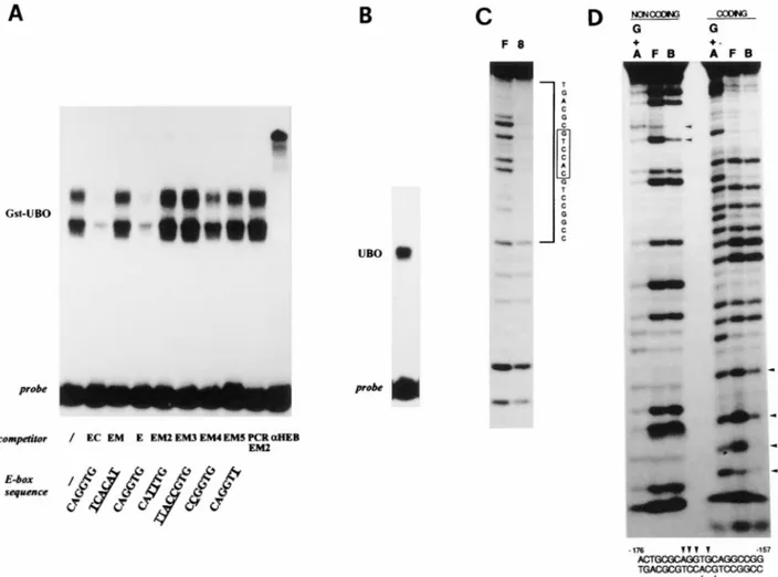
Specific binding of UBO to a vgf promoter E box.Since the bHLH domain directs protein dimerization and DNA binding to the consensus sequence CA--TG, or E box (2, 13), and since E proteins, in contrast to tissue-specific bHLH proteins, can bind DNA efficiently as homodimers, the UBO E protein was expected to specifically bind the EC oligonucleotide E-box CAGGTG in vitro. To study DNA binding, the cDNA was subcloned in the bacterial expression vectors pQE11 and pGEX, the proteins were purified, and their binding activities were tested by EMSA. As expected, UBO and GST-UBO efficiently and specifically bound the CAGGTG E box (Fig. 5A and B). The binding sites on the vgf promoter were more precisely defined by DNase protection, methylation interfer-ence, and competition with a series of E-box-mutated oligonu-cleotides (Fig. 5A, C, and D; Table 1). Following binding of radiolabeled EC oligonucleotide to purified UBO protein, a 20-bp region (2176 to 2157) encompassing the E box was
FIG. 4. Sequence comparison of cDNAs coding for UBO and HTF4/HEB. (A) Nucleotide and deduced amino acid sequences of the simian UBO cDNA are aligned with the corresponding region of the human HEB cDNA (21). Only the nucleotides differing from UBO are shown in the HEB cDNA; nucleotide changes in the bHLH domain do not determine a variation in the amino acid sequence. The bHLH region is in boldface; stop codons are indicated by aster-isks. (B) Schematic diagram of the two polypeptides. aa, amino acids.
on October 3, 2016 by ISTITUTO di NEUROBIOLOGIA e MEDICINA MOLECOLARE
http://mcb.asm.org/
protected from DNase digestion (Fig. 5C). The interference assay (Fig. 5D) indicated that methylation of two guanosines at the middle of the E-box consensus on the top, (coding) strand or of a single guanosine and the nearby A on the bottom strand strongly depressed binding to the oligonucleotide; residues 2169 and 2165 of the top strand (an A and a G, respectively) gave a weaker binding interference. All of the contacted nu-cleotides, except one immediately adjacent, are part of the E box; UBO has a contact pattern similar to that seen with
E12/myogenin in the cardiac a gene (46). Competition with
different oligonucleotides (Fig. 5A) confirmed the protection and interference data. In particular, we showed that a 26-bp oligonucleotide centered on the E box (E oligonucleotide [2183 to 2158]) was sufficient for UBO binding and that mutation of the two central Gs of the E box had the strongest effect on binding. Purified UBO was able to bind, although with a lower affinity, to the E-box CACGTG present in the vgf
promoter at position256.
Protein products of the HTF4/HEB gene and its homologs are widely expressed.Different mRNAs of HTF4/HEB and its homologs are produced by tissue-specific RNA processing
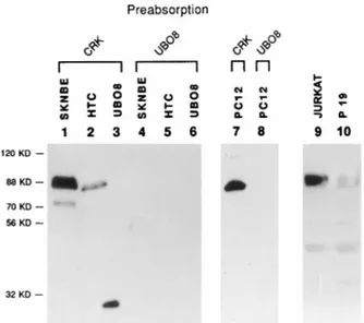
events (25, 33, 35). They have a widespread expression pattern and are present at high levels in thymus, lymphoid cell lines, and developing and mature nervous system tissue (21, 25, 33, 35). A UBO antiserum was employed in Western blots for analyzing E-protein expression in the cell lines utilized in this study; experiments with an anti-HEB serum were also per-formed, with similar results (data not shown). A nuclear pro-tein of about 88 kDa was recognized by the antiserum in nuclear extracts of SK-N-BE, PC12, HTC, P19, and Jurkat cells (Fig. 6) and 3T3 cells (not shown). At least two different isoforms were evident in shorter exposures of the Western blots. The apparent molecular weight of the HTF4/HEB pro-tein in the human cell line SK-N-BE is higher than that of its homologs in the HTC and PC12 rat cell lines; this may reflect species variation in protein size, since the coding sequence of the rat HTF4/HEB homolog, REB (25), is 80 amino acids shorter.
Cooperation of vgf promoter E box and CRE in binding to a widely expressed nuclear complex that contains products of the HEB gene.In order to clarify the relationship between the two previously described nuclear complexes that bind the vgf E
FIG. 5. The UBO polypeptide specifically binds the2170 E box. (A) E-box-specific binding of GST-UBO fusion protein (EMSA). The end-labeled, double-stranded EC oligonucleotide was incubated with 100 ng of purified GST-UBO protein. Excess amounts (1003) of cold, wild-type oligonucleotides (EC and E), a CCAAT-mutated oligonucleotide (CM), DNA sequences with various E-box mutations (EM, EM2-5, and PCR EM2), and an HEB antiserum were used as competitors as indicated; all DNA sequences are listed in Table 1. (B to D) DNA binding of UBO on the EC oligonucleotide. (B) Gel shift of the UBO protein employed in footprinting and methylation interference. (C) DNase I footprinting. The EC oligonucleotide was end labeled on the noncoding strand, incubated with purified UBO protein, digested with DNase, and resolved on a sequencing gel. F, free probe; 8, UBO8-bound probe. The region protected from DNase digestion (2176 to 2157) encompasses the E box; its sequence is shown on the right (39 on top). (D) Methylation interference. The partially methylated EC oligonucleotide was end labeled on the coding or the noncoding strand and used for EMSA with purified UBO. Free (F) and bound (B) probes were excised, cleaved with piperidine, and resolved on a sequencing gel. Those residues whose methylation interferes with binding are indicated by arrowheads. G1A, control sequence on G and A residues.
on October 3, 2016 by ISTITUTO di NEUROBIOLOGIA e MEDICINA MOLECOLARE
http://mcb.asm.org/
box (Fig. 1) and HTF4/HEB E-protein homologs, we per-formed binding competition experiments with a set of E-box mutations and gel supershift experiments with an HEB anti-serum on nuclear extracts of PC12, Jurkat, and NIH 3T3 cells (Fig. 7). The competition experiments showed that the binding properties of the E2 complex, detected in Jurkat and P19 cells only, were very similar to those of purified UBO protein (com-pare Fig. 5 and 7). Those of the E1 complex, in contrast, were clearly different: the E site was necessary for E1-complex bind-ing, since binding was abolished by mutation of the central Gs of the E box (probe PCR EM2), but was not sufficient. The E1 complex, in fact, was not competed by (Fig. 7), nor could bind (data not shown), the E oligonucleotide (Table 1), indicating
the requirement of sequences downstream of the E box. Gel supershift experiments demonstrated that a factor specifically recognized by the HEB antiserum was present in the E2 com-plex of Jurkat cells, but not in the ubiquitous E1 comcom-plex (Fig. 7); the P19 E2 complex was supershifted as well (not shown). These data indicate that E2 is not the vgf repressor. The pres-ence of the E2 complex was not strictly correlated to a larger amount of the E protein in nuclear extracts of different cell lines, as indicated by an estimate of protein amounts obtained by Western analysis via comparison with known quantities of recombinant UBO. We believe that E2 is the same as the lymphoid-cell-specific complex named CD4-3A by Sawada and Littman (43) (P19 cells were not examined in that study); that complex was a heterodimer between HEB and an E12-related protein which binds the CD4 promoter E-box CAGGTG in T cells. E2 and CD4-3A are similar in binding properties, gel mobility, and cell type specificity. The tissue specificity of E2 could be explained by our hypothesis. The finding that HEB family E proteins could not be detected in a ubiquitous com-plex bound to the EC oligonucleotide was in contrast with the wide range of expression of these proteins and with the fact that the vgf promoter site CAGGTG represents, among all possible E boxes, the optimal binding site for these proteins (4). We argued that DNA binding of the E protein may be regulated. This could be achieved in various ways; one is that HEB proteins cooperate with factors that bind other vgf pro-moter elements (e.g., CCAAT and CRE). To investigate this possibility, we employed a longer probe (PCR WT) including, besides the E box and CCAAT, the CRE site. The use of this probe and of competitors with mutated E or CRE sites in gel shift experiments demonstrated the presence of a complex, E-CRE, that depends on the integrity of both E and CRE sites (Fig. 8). This complex had a much stronger affinity for DNA than the previously described E1 and E2 complexes, since a much smaller amount of nuclear extract produced a stronger signal. This complex was present in all cell lines tested (PC12, Jurkat, and NIH 3T3) and was specifically supershifted by an HEB-specific antiserum, which does not cross-react with prod-ucts of other E-protein genes (43). Although it contains an HEB E protein, the E-CRE complex is not expected to be a bHLH protein dimer, as a vast literature on interactions be-tween DNA and bHLH protein homo- and heterodimers, as
FIG. 6. (A) Expression of proteins homologous to HEB in different cell lines. Nuclear extracts from SK-N-BE, HTC, PC12, Jurkat, and P19 cells, or the recombinant UBO (UBO8) protein, were used for Western blotting and probed with a UBO antiserum. As a control for specificity, the antiserum was preab-sorbed with lysates of bacteria producing UBO (lanes 4, 5, 6, and 8) or the unrelated protein v-CRK (lanes 1, 2, 3, and 7). Lanes 1, 2, 4, and 5 contained 80 mg, lanes 9 and 10 contained 100 mg, and lanes 7 and 8 contained 400 mg of nuclear proteins; in lanes 3 and 6, 20 ng of purified UBO was used.
FIG. 7. Properties of E-box binding E1 and E2 complexes. Nuclear extracts from PC12, Jurkat, and NIH 3T3 cells were incubated with end-labeled EC oligonucleotide (2178 to 2132 of vgf promoter). Excess amounts (1003) of wild-type oligonucleotides (EC and E) and E-box-mutated oligonucleotides (EM and EM2 to -4) and HEB antiserum (aHEB) or preimmune serum (preim.) (0.5 ml each) were used as competitors as indicated. The oligonucleotide sequences are in Table 1.
on October 3, 2016 by ISTITUTO di NEUROBIOLOGIA e MEDICINA MOLECOLARE
http://mcb.asm.org/
well as the crystal structures of DNA-bound bHLH dimers, has shown that only few nucleotides around the E box are neces-sary for binding (29). The finding that a CRE site is required for binding of this complex indicates that a CRE binding fac-tor, e.g., a member of the CREB/ATF family, is part of this complex.
DISCUSSION
Several motifs (CRE, CCAAT, and the E box) within a short regulatory region of the neurotrophin-inducible vgf promoter are involved in transcriptional regulation. The CRE consensus sequence (281 to 274) is part of a larger, 14-bp palindrome (284 to 271) and is required for the transcriptional response to NGF and cAMP (19, 38). It binds in vitro to the CRE binding protein, CREB, that is activated by NGF and cAMP via phosphorylation at a key regulatory site, Ser 133 (17, 19). The CRE consensus sequence alone is not sufficient for medi-ating the response to the NGF signal, since NGF induction is
abolished by a 281 deletion, which preserves the consensus
while removing 59 flanking sequences (38). Since a 298 dele-tion still maintains inducdele-tion by NGF, sequences between
po-sitions298 and 281, possibly the first three nucleotides of the
14-bp palindrome, are necessary to respond to NGF. They may be important for promoter recognition by CREB/CREM (42) family members or may be required for a cooperative interac-tion between CREB and some factor that binds other elements in the vgf promoter, like CCAAT or the E box.
CCAAT and E-box sites play important roles in tissue-spe-cific expression of several genes (24, 31, 37, 48); in the vgf promoter, these sites bind specific protein complexes in vitro. An important role of E boxes in vgf regulation was
demon-strated in this study. The gCAGGTG E box at position2170
belongs to the E2-box class, which is the most important for tissue-specific gene expression; sites with the same sequence
are found, for instance, in the cardiaca-actin gene promoter,
the kE2 andmE5 sites of the immunoglobulin light- and
heavy-chain genes, and the MEF1 site of the muscle creatine kinase gene enhancers (31). The vgf E box has a dual role: it is weakly positive in PC12 cells and strongly negative in 3T3 cells. This last finding is in agreement with several reports showing that E2-box sites, besides acting as activator elements, can bind transcriptional repressors (15, 16). This site acts together with
a second E box located downstream at position256. Both E
boxes are required for full repression of the vgf promoter in nonneuronal cells, since mutating either one of the two E boxes leads to an increase in transcription, provided that the other one is still present. When both E boxes are mutated, transcription is very low in neuronal and nonneuronal cells, indicating that, individually, the two E boxes can stimulate transcription, possibly by recruiting a general bHLH transcrip-tion factor. A somewhat functranscrip-tionally similar arrangement of E boxes is found in the immunoglobulin M heavy-chain gene
enhancer (40). We have shown that the2170 E box and CRE
cooperate in binding to a complex present in all cell lines tested. Since CRE binding factors are essential for the re-sponse to neurotrophins, this complex is expected to play an important role in transcriptional regulation. The function of the CCAAT binding complex and the possibility that it inter-acts with the E-CRE complex in transcriptional regulation remain to be elucidated; a role of the CCAAT site in vgf regulation was recently suggested (10).
The cDNA of a factor present in the complex bound to the E box and CRE was isolated by an expression screening. The simian cDNA codes for a polypeptide named UBO, highly homologous to the human E-protein HTF4/HEB (21, 51), that
specifically binds the2170 E box in vitro. E proteins (class A
bHLH proteins) constitute a small family of widely expressed, structurally and functionally related bHLH domain transcrip-tional regulators. They bind DNA as homodimers and as het-erodimers with other E proteins or with tissue-specific (class B) bHLH proteins, and they participate in developmental deci-sions in several organisms and in different tissues (24, 29, 31).
FIG. 8. Synergy of E and CRE sites in binding to an HEB-containing complex. Nuclear extracts from PC12, NIH 3T3, and Jurkat cells were incubated with end-labeled PCR WT DNA (2183 to 265 vgf promoter sequence). Excess amounts (1003) of double-stranded, wild-type or mutated vgf promoter sequences and anti-HEB (aHEB) or preimmune (preim.) serum (0.5 ml each) were used as competitors as indicated. PCR WT, vgf promoter sequence including the E box, CCAAT, and CRE; PCR EM2, sequence with mutated E box; BA/CRE2, sequence with broken CRE; EC, sequence including the E box and CCAAT only; EM, sequence
including CCAAT, a mutated E box, and no CRE; vgf CRE, sequence including the CRE site only. The sequences of all competitors are in Table 1. E-CRE, complex binding both the E box (2170 to 2165) and CRE (281 to 274); C, complex bound to the CCAAT motif.
on October 3, 2016 by ISTITUTO di NEUROBIOLOGIA e MEDICINA MOLECOLARE
http://mcb.asm.org/
Three E-protein genes are known in vertebrates: E2A, ITF2, and HEB. Although widely expressed, E proteins can have a critical function in particular lineages, as demonstrated for E2A in lymphoid cells (52). Comparative analysis of cDNA sequences has revealed that HEB serine 558 is not present in UBO, suggesting that human and simian cDNAs are not equiv-alent. We find this interesting, since the difference between UBO and HEB is similar to that observed for E47 and E2-5, two of the products of the human E2A gene derived by al-ternative splicing: HEB RTSSTNE, UBO RT.STNE; E47 RTSSTDE, E2-5 RT.STDE (20, 32). The serine deletions in UBO and E2-5 may be biologically significant, because they occur in proximity to the bHLH domain and are found in two of three E-protein genes. The serine might be a regulatory site, i.e., a target of posttranslational modification by phosphoryla-tion.
HEB and its homologs bind DNA in vitro and activate or repress transcription in an E-box-dependent way (9, 21, 35, 43). Homodimers of this E protein bind with higher affinity to the E-box CAGGTG, present in the vgf promoter, as compared with other E-box sequences or with other E proteins (4, 21). Their interaction with DNA appears to be controlled in several ways. Proteins with different DNA binding properties may be generated by alternative RNA splicing; one of the HEB mRNA isoforms is preferentially produced in neuronal cells (25, 33, 35). Otherwise, the E protein may be posttranslation-ally modified or associate with tissue-specific partners. Fur-thermore, DNA binding may be blocked by inhibitory HLH proteins, such as Id (1).
The Drosophila melanogaster E protein, Daughterless, has a role in neurogenesis in association with the products of
achaete-scute genes (7, 8). HEB proteins can form dimeric
complexes with MASH1, a mammalian homolog of Drosophila Achaete-Scute proteins (reference 25 and our unpublished data), and possibly NeuroD, two class B HLH factors involved in vertebrate neurogenesis (18, 26). MASH1 is present in neu-ronal precursors and in neuneu-ronal and neuro-endocrine cell lines and is induced by NGF in PC12 cells (23). E-box binding complexes exist throughout PC12 cell differentiation and can be disrupted by Id, whose message is upregulated in response to NGF (12). Studies on the mRNA expression patterns of HEB homologs in chicks and rodents support the possibility that these proteins may control neural cell-specific gene ex-pression. During development, the mRNA is present at high levels in the developing neural tube and in morphogenetically active regions, and it decreases once cellular differentiation is over (25, 33). In adult animals, it is highly enriched in forebrain areas (hippocampus and suprachiasmatic nuclei), where vgf also is transcribed (25). Due to the large variety of protein-protein interactions in which they can participate, HEB E proteins may act as repressors or activators. The E box they bind to in the vgf promoter is the target of different binding complexes and has a dual role in transcriptional regulation. This site is a negative element in 3T3 cells, suggesting that HEB protein homologs may be involved in the repression mechanism. A repressor function was recently demonstrated for the mouse HEB homolog, ME1/ALF1, on the promoter of
p75NTR, a receptor that binds to all neurotrophins and is
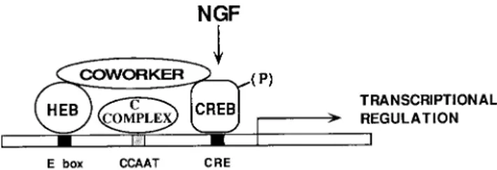
present in neuronal and glial cells and particular populations of lymphocytes (9). Repressors bound to E boxes may contrib-ute significantly to the tissue specificity of vgf, for instance, by ensuring that the action of transcriptional activators is limited to neuronal cells. We find particularly interesting, in this re-gard, the association of this E protein with a CRE-bound factor. A diagram illustrating the possible role of protein-protein interactions between factors bound to different vgf
promoter elements (the E box and CRE) is shown in Fig. 9. It is postulated that the transcriptional activity of the vgf pro-moter relies on a multiprotein complex involving an HEB protein and factors bound to CRE, such as CREB/ATF; co-regulators such as CBP and p300, which associate with CREB as well as with bHLH factors (11, 42), may mediate the inter-action. This complex could have a dual role in controlling gene expression. Gain- and loss-of-function experiments will help to establish the role of these interactions in the proper execution of a neural differentiation program.
ACKNOWLEDGMENTS
We are indebted to Harvey Eisen (Fred Hutchinson Cancer Re-search Center, Seattle, Wash.) for hospitality and critical suggestions. We thank E. Blackwood for the baboon library; D. Littman for anti-HEB serum; D. Mercanti for NGF; E. Beccari, L. Soucek, and A. Sacco for discussions; R. Gargamelli for photographs; and the Fonda-zione Pasteur-Istituto Cenci Bolognetti for radioisotopes. We acknowl-edge the technical assistance of N. Rizzo.
This work was supported by grants from C.N.R. (P.F. Ingegneria Genetica, P.F. ACRO, and P. Bilaterale), AIRC, and Telethon. G. Di Rocco, N. Canu, and B. Illi were supported by AIRC, Fondazione Villa Rusconi, and Telethon fellowships, respectively.
G. Di Rocco and M. Pennuto contributed equally to this work.
REFERENCES
1. Benezra, R., R. L. Davis, D. Lockshon, D., D. L. Turner, and H. Weintraub. 1990. The protein Id: a negative regulator of helix-loop-helix DNA binding proteins. Cell 61:45–69.
2. Blackwell, T. K., and H. Weintraub. 1990. Differences and similarities in DNA-binding preferences of MyoD and E2A protein complexes revealed by binding site selection. Science 250:1104–1110.
3. Bonni, A., D. D. Ginty, H. Dudek, and M. E. Greenberg. 1995. Serine 133-phosphorylated CREB induces transcription via a cooperative mecha-nism that may confer specificity to neurotrophin signals. Mol. Cell. Neurol.
6:168–183.
4. Bonven, B. J., A. D. Nielsen, P. L. Norby, F. S. Pedersen, and P. Jorgensen. 1995. E-box variants direct formation of distinct complexes with the basic helix-loop-helix protein ALF-1. J. Mol. Biol. 249:564–575.
5. Bradshaw, R. A., T. Blundell, R. Lapatto, N. McDonald, and J.
Murray-Rust.1993. Nerve growth factor revisited. Trends Biochem. Sci. 18:48–52. 6. Bunz, F., R. Kobayashi, and B. Stillman. 1993. cDNAs encoding the large
subunit of human replication factor C. Proc. Natl. Acad. Sci. USA 90:11014– 11018.
7. Cabrera, C., and M. Alonso. 1991. Transcriptional activation by het-erodimers of the achaete-scute and daughterless gene products of Drosoph-ila. EMBO J. 10:2965–2973.
8. Campos-Ortega, J. A., and Y. N. Yan. 1991. Genetic and molecular base of neurogenesis in Drosophila melanogaster. Annu. Rev. Neurosci. 14:399–420. 9. Chiaramello, A., K. Neuman, K. Palm, M. Metsis, and T. Neuman. 1995. FIG. 9. Model of vgf promoter regulation. E, CCAAT, and CRE sites par-ticipate in vgf transcriptional regulation. E and CRE elements cooperatively bind a multiprotein complex involved in the control of tissue specificity and of acti-vation by neurotrophins. This complex prevents transcription in nonneuronal cells or in the absence of neurotrophin signalling. Incoming signals from neuro-trophin receptors, travelling via a p21Ras pathway, cause phosphorylation of at least a CRE binding protein (CREB) and activate transcription. The E-CRE complex is formed by an E protein, one of the HEB gene products; by a CRE binding protein; and by a coworker, an adapter molecule that mediates the interaction of the first two proteins. Proteins like p300/CBP, which bind CREB and bHLH factors, might provide the adapter function. Interactions between the E-CRE complex and the complex bound to the CCAAT element (C complex, of unknown nature) may provide a finer tuning of vgf transcription.
on October 3, 2016 by ISTITUTO di NEUROBIOLOGIA e MEDICINA MOLECOLARE
http://mcb.asm.org/
Helix-loop-helix transcription factors mediate activation and repression of the p75LNGFR gene. Mol. Cell. Biol. 15:6034–6044.
10. D’Arcangelo, G., R. Habas, S. Wang, S. Halegoua, and S. R. J. Salton. 1996. Activation of codependent transcription factors is required for transcrip-tional induction of the vgf gene by nerve growth factor and Ras. Mol. Cell. Biol. 16:4621–4631.
11. Eckner, R., T. Yao, E. Oldread, and D. M. Livingston. 1996. Interaction and functional collaboration of p300/CBP and bHLH proteins in muscle and B-cell differentiation. Genes Dev. 10:2478–2490.
12. Einarson, M. B., and M. V. Chao. 1995. Regulation of Id1 and its association with basic helix-loop-helix proteins during nerve growth factor-induced dif-ferentiation of PC12 cells. Mol. Cell. Biol. 15:4175–4183.
13. Ephrussi, A., G. M. Church, S. Tonegawa, and W. Gilbert. 1985. B lineage-specific interactions of an immunoglobulin enhancer with cellular factors in
vivo. Science 227:134–140.
14. Ferri, G. L., A. Levi, and R. Possenti. 1992. A novel neuroendocrine gene product: selective VGF8a gene expression and immuno-localisation of the
VGF protein in endocrine and neuronal populations. Mol. Brain Res. 13:
139–143.
15. Fuse, N., S. Hirose, and S. Hayashi. 1994. Diploidy of Drosophila imaginal cells is maintained by a transcriptional repressor encoded by escargot. Genes Dev. 8:2270–2281.
16. Genetta, T., D. Ruezinsky, and T. Kadesch. 1994. Displacement of an E-box-binding repressor by basic helix-loop-helix proteins: implications for B-cell specificity of the immunoglobulin heavy-chain enhancer. Mol. Cell. Biol.
14:6153–6163.
17. Ginty, D. D., A. Bonni, and M. E. Greenberg. 1994. Nerve growth factor activates a Ras-dependent protein kinase that stimulates c-fos transcription via phosphorylation of CREB. Cell 77:713–725.
18. Guillemot, F., L. Lo, J. Johnson, A. Auerbach, D. Anderson, and A. Joyner. 1993. Mammalian achaete-scute homolog 1 is required for the early devel-opment of olfactory and autonomic neurons. Cell 75:463–476.
19. Hawley, R. J., R. J. Scheibe, and J. A. Wagner. 1992. NGF induces the expression of the VGF gene through a cAMP response element. J. Neurosci.
12:2573–2581.
20. Henthorn, P., M. Kiledjian, and T. Kadesh. 1990. Two distinct transcription factors that bind the immunoglobulin enhancermE5/kE2 motif. Science
247:467–470.
21. Hu, J. S., E. N. Olson, and R. E. Kingston. 1992. HEB, a helix-loop-helix protein related to E2A and ITF2 that can modulate the DNA-binding ability of myogenic regulatory factors. Mol. Cell. Biol. 12:1031–1042.
22. Idzerda, R. L., W. G. Carter, C. Nottemburg, E. A. Wayner, W. M. Gallatin,
and T. St. John.1989. Isolation and DNA sequence of a cDNA clone encoding a lymphocyte adhesion receptor for high endothelium. Proc. Natl. Acad. Sci. USA 86:4659–4663.
23. Johnson, J. E., S. J. Birren, and D. Anderson. 1990. Two rat homologues of Drosophila achaete-scute specifically expressed in neuronal precursors. Na-ture 346:858–861.
24. Kadesh, T. 1992. Helix-loop-helix proteins in the regulation of immunoglob-ulin gene transcription. Immunol. Today 13:31–36.
25. Klein, E. S., D. M. Simmons, L. W. Swanson, and M. G. Rosenfeld. 1993. Tissue-specific RNA splicing generates an ankyrin-like domain that affects the dimerization and DNA-binding properties of a bHLH protein. Genes Dev. 7:55–71.
26. Lee, J., S. Hollenberg, L. Snider, D. Turner, N. Lipnick, and H. Weintraub. 1995. Conversion of Xenopus ectoderm into neurons by NeuroD, a basic helix-loop-helix protein. Science 268:836–844.
27. Levi, A., J. D. Eldridge, and B. M. Paterson. 1985. Molecular cloning of a gene sequence regulated by nerve growth factor. Science 229:393–395. 28. Levi Montalcini, R. 1987. The nerve growth factor 35 years later. Science
237:1154–1162.
29. Littlewood, T., and G. Evan. 1994. Transcription factors 2: helix-loop-helix. Protein Profile 1:639–709.
30. Lombardo, A., S. A. Rabacchi, F. Cremisi, T. Pizzorusso, M. C. Cenni, R.
Possenti, G. Barsacchi, and L. Maffei.1995. A developmentally regulated NGF-induced gene, VGF, is expressed in geniculocortical afferents during synaptogenesis. Neuroscience 65:997–1008.
31. Murre, C., et al. 1994. Structure and function of helix-loop-helix proteins. Biochim. Biophys. Acta 1218:129–135.
32. Murre, C., P. S. McCaw, and D. Baltimore. 1989. A new DNA binding and dimerization motif in immunoglobulin enhancer binding, daughterless, MyoD and myc proteins. Cell 56:777–783.
33. Neuman, T., A. Keen, E. Knapik, D. Shine, M. Ross, H. O. Nornes, and M. X.
Zuber.1993. ME1 and GE1: basic helix-loop-helix transcription factors ex-pressed at high levels in the developing nervous system and in morphoge-netically active regions. Eur. J. Neurosci. 5:311–318.
34. Neumann, J. R., C. A. Morency, and K. O. Russian. 1987. A novel rapid assay for chloramphenicol acetyltransferase gene expression. BioTechniques 5: 444–447.
35. Nielsen, A. D., N. Pallisgaard, F. S. Pedersen, and P. Jorgensen. 1992. Murine helix-loop-helix transcriptional activator proteins binding to the E-box motif of the Akv murine leukemia virus enhancer identified by cDNA cloning. Mol. Cell. Biol. 12:3449–3459.
36. Piccioli, P., A. Di Luzio, A. Surani, J. Donnerer, R. Amman, R. Schuligoi,
and A. Cattaneo.1995. Neuroantibodies: ectopic expression of a recombi-nant SP antibody in the central nervous system of transgenic mice. Neuron
15:373–384.
37. Poli, V., F. P. Mancini, and R. Cortese. 1990. Il-6DBP, a nuclear protein involved in interleukin-6 signal transduction, defines a new family of leucine zipper proteins related to C/EBP. Cell 63:643–653.
38. Possenti, R., G. Di Rocco, S. Nasi, and A. Levi. 1992. Regulatory elements in the promoter region of vgf, a nerve growth factor-inducible gene. Proc. Natl. Acad. Sci. USA 89:3815–3819.
39. Possenti, R., J. D. Eldridge, B. M. Paterson, A. Grasso, and A. Levi. 1989. A protein induced by NGF in PC12 cells is stored in secretory vesicles and released through the regulated pathway. EMBO J. 8:2217–2223. 40. Ruezinsky, D., D. Beckmann, and T. Kadesch. 1991. Modulation of the IgH
enhancer’s cell type specificity through a genetic switch. Genes Dev. 5:29–37. 41. Salton, S. R. J., D. J. Fischberg, and K. W. Dong. 1991. Structure of the gene encoding VGF, a nervous system-specific mRNA that is rapidly and selec-tively induced by nerve growth factor in PC12 cells. Mol. Cell. Biol. 11:2335– 2349.
42. Sassone-Corsi, P. 1995. Transcription factors responsive to cAMP. Annu. Rev. Cell. Dev. Biol. 11:355–377.
43. Sawada, S., and D. R. Littman. 1993. A heterodimer of HEB and an E12-related protein interacts with the CD4 enhancer and regulates its activity in T-cell lines. Mol. Cell. Biol. 13:5620–5628.
44. Schreiber, E., P. Matthias, M. M. Muller, and W. Shaffner. 1989. Rapid detection of octamer binding proteins with “mini-extracts” prepared from a small number of cells. Nucleic Acids Res. 17:6419.
45. Singh, H., R. G. Clerc, and J. Le Bowitz. 1989. Molecular cloning of se-quence-specific DNA binding proteins using recognition site probes. Bio-Techniques 7:252–261.
46. Therrien, M., and J. Drouin. 1993. Cell-specific helix-loop-helix factor re-quired for pituitary conserved domains in a subset of genes rere-quired for neurogenesis and their homology to myc. Cell 50:415–424.
47. Trani, E., T. Ciotti, A. M. Rinaldi, N. Canu, G. L. Ferri, A. Levi, and R.
Possenti.1995. Tissue-specific processing of the neuroendocrine protein VGF. J. Neurochem. 65:2441–2449.
48. Umek, R. M., A. D. Friedman, and S. L. McKnight. 1991. CCAAT-enhancer binding protein: a component of a differentiation switch. Science 251:288– 292.
49. van den Pol, A. N., C. Decavel, A. Levi, and B. M. Paterson. 1989. Hypotha-lamic expression of a novel gene product, VGF: immunocytochemical anal-ysis. J. Neurosci. 9:4122–4137.
50. Weintraub, H., R. Davis, D. Lockshon, and A. Lassar. 1990. MyoD binds cooperatively to two sites in a target enhancer sequence: occupancy of two sites is required for activation. Proc. Natl. Acad. Sci. USA 87:5623–5627. 51. Zhang, Y., J. Babin, A. L. Feldhaus, H. Singh, P. A. Sharp, and M. Bina.
1991. HTF4: a new human helix-loop-helix protein. Nucleic Acids Res.
19:4555.
52. Zhuang, Y., P. Soriano, and H. Weintraub. 1994. The helix-loop-helix gene E2A is required for B cell formation. Cell 79:875–884.