Autore:
Nafiseh Mirzajani ______________
Relatori:
Prof. Francesco d’Errico ______________
Prof. Giorgio Curzio ______________ Dr. Riccardo Ciolini ______________
Characterization of the Mixed Radiation Fields for
BNCT
Anno 2013
UNIVERSITÀ DI PISA
Scuola di Dottorato in Ingegneria “Leonardo da Vinci”
Corso di Dottorato di Ricerca in
SICUREZZA NUCLEARE E INDUSTRIALE
Acknowledgements
I would like to express best gratitude and most sincere thanks to my tutors Prof. Francesco d’Errico, Prof. Giorgio Curzio and Dr. Riccardo Ciolini for their encouragement, supervision and profound enthusiasm, discussion, patience for reading and their comment on this thesis.
I am also grateful to Prof. Walter Ambrosini for his guidance and helpful advice during my work.
I wish to thank Dr. Angela Di Fulvio, Dr. Dahmane Mazed, Chiara Romei, Daniele Del Serra, Aldo Del Gratta and Fabio Pazzagli for collaboration and advice during this project.
I would like to acknowledge the kind, care and support of all staff of departments of Civil and Industrial Engineering (DICI), University of Pisa.
I must sincerely thank my parents, my lovely sister, my family and all of my friends in Pisa whose support and encouragement have helped me throughout my studies.
Ringraziamenti
Vorrei esprimere la mia gratitudine e i più sinceri ringraziamenti ai miei tutori Prof. Francesco D'Errico, Prof. Giorgio Curzio e il Dott. Riccardo Ciolini per
l'incoraggiamento, la supervisione, il profondo entusiasmo, le discussioni e nonché la pazienza per la lettura e i loro preziosi commenti su questa tesi.
Sono anche grato al Prof. Walter Ambrosini per la sua guida e consigli utili provveduti durante lo svolgimento di questo lavoro.
Desidero ringraziare il Dott. Angela Di Fulvio, Dr. Dahmane Mazed, Chiara Romei, Daniele Del Serra, Aldo Del Gratta e Fabio Pazzagli per la collaborazione e la consulenza nel corso di questo progetto.
Vorrei riconoscere anche in genere, la cura e il sostegno di tutto il personale del Dipartimento di Ingegneria Civile e Industriale (DICI), Università di Pisa.
Devo sinceramente ringraziare i miei genitori, mia bellissima sorella, la mia famiglia e tutti i miei amici a Pisa per il sostegno e l'incoraggiamento e l'aiuto provveduto durante i miei studi.
Index
INTRODUCTION ... 5
1. BORON NEUTRON CAPTURE THERAPY ... 9
1.1 The fundamental of BNCT ... 11
1.2 The BNCT history and present status ... 14
1.3 Application of neutron sources for BNCT ... 20
1.3.1 Nuclear reactors ... 21
1.3.2 Accelerator-based neutron sources (ABNS) ... 24
2. NEUTRON SPECTROMETRY APPLICATION, DETECTORS AND
TECHNIQUE ... 42
2.1 Neutron spectrometry ... 42
2.2 History ... 42
2.3 Current techniques ... 43
2.3.1 Recoil nuclei spectrometers ... 43
2.3.2 Nuclear reaction as neutron spectrometer ... 44
2.3.3 Time-of-flight (TOF) method ... 44
2.3.4 Threshold detectors spectrometers ... 44
2.3.5 Bonner sphere spectrometer (BSS) ... 45
2.3.5.1 Overview ... 45
2.3.5.2 BSS response and efficiency ... 48
2.3.5.3 The principle of BSS operation ... 49
2.3.5.4 Thermal neutron detector ... 51
2.3.5.5 Active neutron detectors ... 52
2.3.5.6 The 6LiI(Eu) scintillator ... 53
2.3.5.7 The 10BF3 filled proportional counter ... 54
2.3.5.8 The 3He filled proportional counter ... 55
2.3.5.9 Passive neutron detectors ... 57
2.3.5.10 Activation foils ... 58
2.3.5.11 Thermoluminescent dosimeters (TLDs) ... 59
2.3.5.12 Superheated drop or Bubble detector ... 62
2.3.5.13 Track detector ... 64
3. MONTE CARLO CALCULATION OF THE ENERGY RESPONSE
FUNCTIONS OF BSS-UNIPI
...73
3.1 Overview of the Monte Carlo code ... 73
3.1.1 Historical development ... 73
3.1.2 Monte Carlo N-Particle eXtended (MCNPX) ... 74
3.2 Calculation of energy response functions of BSS-UNIPI based on 6LiI(Eu) 75 3.2.1 MCNPX model ... 75
3.2.2 The response matrix of BSS-UNIPI based on 6LiI(Eu) ... 78
3.2.3 BSS-UNIPI response to 241Am-Be neutron source ... 85
3.3. Investigation of the activation-based BSS-UNIPI set (3 and 10 in.) response to monoenergetic neutrons ... 90
4. UNFOLDING PROCEDURES ... 96
4.2 Neutron spectrum unfolding code: MAXED ... 97
4.3 Neutron spectrum unfolding code: FRUIT ... 99
5. EXPERIMENTAL CHARACTERIZATION OF BSS- UNIPI ... 103
5.1 General characteristics of BSS-UNIPI ... 103
5.2 Discrimination of background and gamma ... 106
5.3 Calibration in neutron radiation field ... 106
5.4 Evaluation of the fluence response ... 108
5.5 Unfolding data ... 109
5.6 Analysis of the application of the shadow cone technique for the determination of neutron spectrum with BSS ... 111
5.6.1 Overview ... 111
5.6.2 The shadow cone method ... 111
5.6.3 Experimental measurements ... 113
5.6.4 Results and discussion ... 114
5.6.5 Conclusions ... 120
6. ASSESSMENT OF THE ANGULAR NEUTRON ENERGY SPECTRUM
OF AN ACCELERATOR-BASED BNCT FACILITY (TRASCO-BNCT
PROJECT) BY BONNER SPHERE SPECTROMETER ... 122
6.1 Overview ... 122
6.2 The primary experimental measurements ... 123
6.2.1 Unfolding procedures ... 124
6.3 Application of the shadow cone technique with BSS-UNIPI for the determination of the angular neutron energy spectrum of INFN-LNL SPES-BNCT facility ... 126
6.3.1 Experimental measurements ... 126
6.3.2 Unfolding procedures ... 129
6.3.3 Results and discussion ... 130
6.3.4 Conclusions ... 134
7. SUMMARY, CONCLUSIONS AND PROSPECTS ... 137
APPENDEX A- INPUT SIMULATION FILE OF MCNP ... 139
Introduction
Mixed radiation fields are normally encountered in the treatment of tumors with ionizing radiations in Boron Neutron Capture Therapy (BNCT). BNCT is a binary treatment modality for a variety of tumors, mainly high-grade primary brain tumors (glioblastoma multiforme), skin malignant melanoma, hepatic metastases and other cancers. Several hundred patients have been treated in the United States of America, Europe, Argentina and Japan. Survival times are comparable with those obtained with standard radiation therapy but improved patients' quality of life have been reported as a result of several of these BNCT treatments (Stupp, 2005; Menendez, 2009; Skold, 2010; Barth, 2012).
The BNCT facilities use research reactors or accelerator-based neutron beams with a broad epithermal energy spectrum, ranging from few eV up to about 30 keV, which also includes fast neutrons and gamma rays as contaminants. Before delivering this type of therapy, it is necessary to obtain detailed information on the neutron energy spectrum of the BNCT facility and evaluate the neutron dose delivered to the tumor and also to the adjacent healthy tissues of the patient, because the effectiveness of neutrons beams varies greatly depending on their energy. Thus, one of the major challenges of BNCT is to characterize the neutron beam in terms of intensity and energy spectrum.
The main aim of the present research is the definition of a suitable methodology for a complete neutron characterization of mixed field BNCT facilities, but the developed method applies generally also to other type of mixed fields, as found in the LINAC irradiation rooms. These neutron fields are complex to characterize because normally their energy extends from thermal (about 10-8 MeV) up to tens of MeV. Different neutron spectrometric techniques are available, which differs from what concerns their limitation and specific energy response, but only multi sphere systems (Bonner sphere Spectrometers, BSS) were found to have a wide energy range of application with an isotropic response (Alevra, 2003; Thomas, 2002). This type of neutron spectrometer consists of a set of moderating Bonner spheres with different diameters, typically made of polyethylene with a neutron thermal detector (passive or active) positioned at the center of each Bonner sphere. The accuracy of the resulting spectral neutron fluence is related to the application of an unfolding procedure to the experimental readings of the BSS and the availability of accurate spectrometer response matrix. Very precise response matrixes have been calculated solving the neutron transport equation or using Monte Carlo methods (Mares, 1994; Vega-Carrillo, 2008). The unfolding process is one of the most complicated parts of the BSS-based spectrometry. Most of the commonly employed unfolding codes rely on mathematical convergence algorithms, thus requiring a guess spectrum as similar as possible to the one to be determined (Matzke, 1994). The FRUIT (Bedogni, 2007) and MAXED (Reginatto, 2002) unfolding code were selected as a proper tool to obtain the neutron energy spectrum from the experimental data.
Neutron scattering contribution in the neutron energy spectrum profile is generally present due to the interactions of neutrons with accelerator room structures (walls, air and the ancillary experimental apparatus). To evaluate this contribution, the shadow cone technique (Eisenhauer, 1985; ISO, 2000; IAEA, 2000) was applied with BSS for the measurement of neutron spectra in the considered mixed radiation fields. The main item of this technique was counting the scattered neutrons contribution in the considered neutron installation. For this purpose, three shadow cones were realized and positioned between the source and BSS. Experiments were performed to select the optimal source to shadow cone distance by using an unfolding code to determine the neutron spectrum. The difference between the unfolded spectra obtained by using the shadow cones and the ISO reference spectra was considered as a parameter to select the optimum condition in the shadow cone technique application.
BSS and shadow cone method were applied for the investigation of neutron spectrum for an under construction BNCT facility. In particular, in this work, experimental activities were performed at INFN Legnaro National Laboratories (LNL) (Padua, Italy) aimed at angular-dependence neutron energy spectra measurements produced by the 9Be (p,xn) reaction, under 5 MeV proton beam. Such a work was performed in the framework of the INFN TRASCO-BNCT project. In this work, a BSS based on 6LiI(Eu) scintillator, named from now on BSS-UNIPI, coupled with the shadow cone technique was applied. The response function matrix for the set of BSS-UNIPI Bonner spheres and bare 6LiI(Eu) detector was calculated by MCNPX code (Pelowitz, 2008) for an extended neutron incidence energy range from 1 meV to 100 MeV. To obtain the neutron spectrum from measurements, the response function determined for each Bonner sphere was used and both the FRUIT and MAXED unfolding codes were applied to obtain the final spectra. The results are in agreement. This thesis is organized as follows: Chapter 1 is an overview of physical bases, history and present status of BNCT, with a description of the applications of different available neutron sources for this type of therapy.
Chapter 2 introduces neutron spectrometry detectors and technique with special attention dedicated to BSS, with the more common active or passive detectors. Moreover, BSS based on 6LiI(Eu), which are selected for measuring the neutron spectrum in BNCT facility in this work, are described.
Chapter 3 describes a general review of Monte Carlo methods, Monte Carlo calculation response function and matrix of BSS-UNIPI based on 6LiI (Eu), the response function of BSS-UNIPI based on activation foils.
Chapter 4 gives a general review of the unfolding method and the description of the selected unfolding codes, FRUIT, version 5, and MAXED.
Chapter 5 describes the experimental calibration of BSS and the set-up of the shadow cone technique.
Chapter 6 is devoted to the application of BSS-UNIPI for the determination of the angular neutron energy spectrum produced by the 9Be (p,xn) reaction, under 5 MeV proton beam, to be applied for an accelerator-based BNCT facility at INFN-LNL.
Chapter 7 reports the summary, conclusions and prospects.
References
Alevra, A.V and Thomas, D. J. (2003). Neutron spectrometry in mixed fields: multisphere spectrometers. Radiation Protection Dosimetry, 107 (1–3), pp. 37-72. Barth, R.F., et al. (2012). Current status of boron neutron capture therapy of high grade gliomas and recurrent head and neck cancer. Radiation Oncology, 7, 146, pp. 8-21.
Bedogni, R., et al. (2007). FRUIT: an operational tool for multisphere neutron spectrometry in workplaces. Nuclear Instruments and Methods A, 580, pp. 1301– 1309.
Ceballos, C., et al. (2009). The BSA modeling for the accelerator-based BNCT facility at INFN LNL for treating shallow skin melanoma. Applied Radiation and Isotopes, 67 (7-8), pp. S274-S277.
Eisenhauer, C.M., et al. (1985). Calibration techniques for neutron personal dosimetry. Radiation Protection Dosimetry, 10, pp. 43–57.
IAEA, Safety Reports Series No. 16, 2000.
ISO 8529-2, 2000. Reference neutron radiations—part 2: calibration fundamentals of radiation protection devices related to the basic quantities characterizing the radiation field, International Organization for Standardization, Geneva, Switzerland.nt with other authors.
Mares, V and Schraube, H. (1994). Evaluation of the response matrix of a Bonner sphere spectrometer with Lil detector from thermal energy to 100 MeV. Nuclear Instrumental Method A, 337, pp. 461-473.
Matzke, M. (1994). Unfolding Methods, Report D38116, Physikalisch-Technische Bundesanstalt, Germany.
Menendez, P.R., et al. (2009). BNCT for skin melanoma in extremities: Updated Argentine clinical results. Applied Radiation and Isotopes, 67(S), pp. S50–S53. Pelowitz, D.B. (2008). MCNPX User's Manual, 2.6.0 ed., LA-CP-07-1473.
Pisent, A., et al. (2006). Progress on the accelerator based SPES-BNCT project at INFN Legnaro. Journal of Physics: Conference Series, 41, pp. 391-399.
Prete, G., et al. (2008). Selective production exotic Species, Chapter XII – SPES applied Science, BNCT and LENOS. Technical design report. INFN-LNL-224, pp. 181-214.
Reginatto, M., et al. (2002). Spectrum unfolding, sensitivity analysis and propagation of uncertainties with the maximum entropy deconvolution code MAXED. Nuclear Instruments and Methods A, 476, pp. 242-246.
Skold, K., et al. (2010). Boron neutron capture therapy for newly diagnosed glioblastoma multiforme: An assessment of clinical potential. British Journal of Radiology, 83(991), pp. 596-603.
Stupp, R.M., et al. (2005). Radiotherapy plus concomitant and adjuvant temozolomide for glioblastoma. The New England Journal of Medicine, 352(10), pp. 987-996.
Thomas, D.J., et al. (2002). Bonner sphere spectrometers—a critical review. Nuclear Instruments and Methods in Physics Research, A 476, pp. 12-20.
Vega-Carrillo, H.R., et al. (2008). Calculation of Response matrix of a BSS with 6LiI scintillator. Revista Mexicana de Física S, pp. 54-57.
1. Boron Neutron Capture Therapy
Boron neutron capture therapy (BNCT) is one of the experimental types of radiotherapy treatments cancers. BNCT has started to show its ability to treat types of cancers. It has traditionally been used to treat the malignant brain tumour called glioblasoma multiforme (GBM) and then melanomas since the mid-1980s. There are some reports about successful trial BNCT about especially head and neck tumors, but it hasn't become a conventional therapy yet. Thus, BNCT is still in the trial stage now (Barth, 2012; Kankaanranta, 2011; 2012; Miyatake, 2012).
In recent years, BNCT also has been used to treat recurring of primary or recurrent cancers such as liver, lungs, thyroid and skin cancers (Emiliano, 2012; Masunaga, 2012; Pisarev, 2012; Gonzalez, 2004).
Fig. 1.1 shows a patient with a recurrent mucoepidermoid carcinoma neck cancer after 22 months of the first BNCT session treatment. Three treatment sessions caused not only a beneficial reduction in tumor size with BNCT, but also resolution of a cutaneous ulcer and reepithelization were recovered from normal skin. These results clearly demonstrate that BNCT is a highly tumor-selective treatment modality, and the patient lived for 7 years more before die.
Figure 1.1: In the left a patient with a recurrent mucoepidermoid carcinoma cancer of the
BNCT has some advantages and disadvantages in comparison to external photon radiotherapy. The advantages included are: first, BNCT has the ability to selectively deliver a high radiation dose to the tumor with a much lower dose to surrounding normal tissues. This is an important feature that makes BNCT particularly attractive for salvage therapy of patients who have been treated to tolerance with chemotherapy and radiotherapy; second, it has the potential to more effectively target multicentric deposits of tumor than is possible with stereotactic radiosurgery of primary and metastatic brain tumors. Third, although it may be only palliative, it can produce striking clinical responses, as evidenced by the experience of several groups treating patients with recurrent, therapeutically refractory head and neck cancer (Diaz, 2003; Busse, 2003; Barth, 2012); finally, median survival times of patients (GBM) have been increased who were treated by BNCT combining with conventional fractionated photon radiotherapy (BNCT-XRT) and temozolomide in comparison with using BNCT alone (Kawabata, 2011; Yamamoto, 2011; Nakai, 2011).
However, there are few disadvantages of using BNCT. There are only few of neutron beams which has quality required for BNCT (Catharine, 2005): nuclear reactors, charged-particle accelerators, compact neutron generators and hospital radiotherapy facilities (Photo Neutron Source (PHONES)) for BNCT that can produce the required low-energy neutron beam for BNCT (Burian, 2006; Prest, 2010; Autrerinen, 2001; 2005; 2012). Moreover BNCT facilities of the radiation are required to support medical facilities. While there are many research reactors around the world, only a few of them were modified to develop BNCT. Then, there are not yet available boron compounds with a sufficiently high ratio of boron delivery tumor to healthy tissue, to ensure that healthy tissues will not be affected by BNCT treatment.
A third disadvantage is due to the additional undesirable dose components which will be produced as an unavoidable side-effect, the most significant of these being the gamma ray and fast neutron contamination components which come from the neutron source. Also a supplementary gamma dose is generated via neutron capture in hydrogen of the tissues during patient irradiation. These doses, which are not directed at the tumor cells, place a limit on the total dose which can be delivered to the region of the body.
Although the research to the concept of BNCT is going on about seventy years, all the current work is still under investigation. The major challenges for the BNCT research include improving the beam quality of incident neutrons, developing new boron delivery agents, devising better methods to accurately calculate dose, finding alternative sources of neutrons, and creating beams with epithermal neutrons (he energy range 1 eV-10 keV), that are capable of reaching deep seated tumors without the need for surgery.
This chapter describes the fundamental of BNCT, an overview of the BNCT history and present status, and then neutron sources which were used or under development for BNCT.
1.1 The fundamental of BNCT
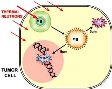
BNCT is based on thermal neutrons interaction with 10B (~20 g/g weight or ~109 atoms/cell), injected to the patient’s tumor, the reaction produces two high LET1
particles: 7Li isotope (6 m) and 4He () (9 m) particle and their ranges are comparable to a cell diameter (10 m) as (Figs. 1.3 and 1.4). Nuclear reactions take place inside a tumor cell in the following (Walker, 1998):
10
B +1n (0.025 eV) 11B* 7Li * (0.84 MeV)+ (1.47 MeV) (94 %)
7
Li* 7Li + (0.48 MeV)
10
B +1n (0.025 eV) 11B* 7Li (1.01 MeV) + (1.78 MeV) (6 %)
The cross section of the 10B(n,)7Li reaction is very high (about 3837 barns) at thermal energy, the Q-value is 2.790 MeV. When the 10B nucleus captures a thermal neutron it becomes 11B nucleus in the excited state for a very short time (~ 10-12 s). Then the excited boron (11B*) decays by producing an alpha particle, and an excited-state lithium (7Li*) with a probability of 94%. In the sequence, 7Li* decays to its fundamental level with a gamma ray emission (0.478 MeV) (Chadwick, 2006) (Fig. 1.2).
Figure 1.2: Energy level diagram for 11B* showing its decay scheme (Chadwick, 2006).
Figure 1.3: Schematic method of BNCT like the one performed by Hatanaka in Japan
(Takayanagi, 2012).
Figure 1.4: The process of thermal neutrons interaction with 10B inside the tumor cell which are producing two particles (7Li and 4He()).
When the reaction takes place inside a cell, the two ionizing particles leave the nucleus of tumor cells and can cause non repairable damages to the DNA, as double strand breaks. Because of their short ranges and high LET, the lethal damage is largely restricted to the tumor cells (Fig. 1.4) (Soyland, 2000; Pouget, 2001; Hall, 2000; Soloway, 1998). The energy of the neutrons which should be used depends on the depth of the tumour in BNCT. Thermal neutrons (0.025 eV) were used to irradiate shallow tumors, while epithermal neutrons (1 eV - 10 keV) were applied for treating deep tumors because they penetrate farther into the body before being slowed down to thermal energies.
Thermal neutrons have little effect on normal cells, since the capture cross-sections of the major tissue elements 16O, 12C, 1H, and 14N are only 1.9 × 10⁻⁴ b, 3.4 × 10⁻³ b, 3.32 × 10⁻¹ b and 7.5 × 10⁻² b, respectively (Chadwick, 2006).This is one advantage for BNCT because the surrounding healthy cells would be spared from radiation damages coming from the capture reactions occurring in tumor cells. By fluence of 1012 neutrons/cm2 and 109 boron atoms per cell would be enough to produce 2 to 3 neutron captures. Thus, the cell would be damaged. The proper borate compound such as (L)-4-dihydroxy-borylphenylalanine, referred to as boronophenylalanine (BPA), and Na2B12H11SH, known as sodium borocaptate
(BSH), have been used in clinical trials (Barth, 2012). These two compounds represent two different approaches to delivering boron to the tumours. BSH relies on passive diffusion from the blood into brain tumours. Brain tumors disrupt the blood brain barrier (BBB), and as a result BSH is able to diffuse into cancerous cells but not into healthy areas of the brain where the blood brain barrier is still intact.
In recent years, the development of novel improved boron carriers for BNCT has focused on nanoparticles (Steen Paterson, 2008; Mandal, 2011), porphyrin type (Shinji, 2011), nucleosides (Byun, 2006) and amino acids (Kabalka, 2003; Semioshkin, 2007) which are still used in the BNCT research programs for their ability to concentrate the boron atoms preferably in tumor cells.
Whilst the most of attention world-wide has focused on 10B as the neutron capture isotope in BNCT but there are other possibilities to use isotopes (11B, 12C, 1H, 14N,
23
Na, 35Cl, 235U, 153Gd and 157Gd). Gadolinium-157 (157Gd) has the highest neutron cross section (248,000 barns) for thermal neutrons among the stable nuclides and produces the neutron capture reaction (n,) as follows (nth is a thermal neutron):
157
Gd + nth (0.025 eV) →
158Gd* → 158
Gd + + 7.94 MeV
Thus, 157Gd have been proposed as Gadolinium Neutron Capture Therapy (GdNCT) for brain cancer in the 1980’s. The released energy of 157
Gd interaction with neutron thermal is more than 90% (~ 7.5 MeV) deposited by prompt gamma rays that reduce the local energy deposition. The emitted gamma rays partially interact with tumors but mainly diffuse in the body damaging healthy tissues. Also different Gd nuclear reactions and the generated Auger electrons in particular, cause a high local energy deposition, which results in a tumour cell inactivation. DNA single and double strand breaks from Auger electrons emitted during 157Gd(n,) events (Cerullo, 2009; Goorley, 2004).
Some research was done on In vivo experiments and toxicity studies for this new compound in GdNCT (De Stasio, 2001; 2005; Cerullo, 2009; Crossley, 2010). There are very few studies demonstrating the efficacy of GdNCT in experimental animal tumor models (Tokumitsu, 2000). Gadolinium such as Gd-DTPA currently is widely used to enhance images in MRI2 (Geninatti-Crich, 2011). Nowadays, it is commonly assumed the use of gadolinium instead of boron in neutron capture therapy and it is under investigation in worldwide.
Recently an investigation was done about using components H2TCP and H2DCP
as carboranyl porphyrins instead of boronophenylalanine. The result showed that it can be used as delivery agents for BNCT of an experimental brain tumor. In addition they have enhanced cellular uptake and improved therapeutic efficacy (Kawabata, 2011). However BNCT needs to develop new delivery agents in future. In following section an overview of BNCT history and present status will be described.
1.2 The BNCT history and present status
Two years after the discovery of neutrons in 1932 by Chadwick, the first research about the interaction of thermal neutron with boron which produces charged particle (7Li and 4He) was done at Cambridge University (Goldhaber, 1986). BNCT was first proposed by a biophysicist, Locher, of the Franklin Institute in Pennsylvania in 1936. He suggested treating tumors with thermal neutron irradiation and 10B which has a high reaction cross section (Locher, 1936). Then, the first radiobiological studies by using the 10B(n,)7Li reaction was performed at the University of Illinois in 1938 (Kruger, 1940). During the years 1951 to 1961, the first clinical application of BNCT was done for a group of malignant glioma patients at Brookhaven National Laboratory (BNL) in USA. At the same time, other clinical application of BNCT was done for 18 patients (brain cancer) at Massachusetts Institute of Technology (MIT) in USA. BNL and MIT used the thermal neutron beams extracted from the reactors of the two laboratories and applied a borate compound named Borax (10B-enriched ) for trials. These trials were not successful because of the thermal neutrons penetrated insufficiently into the deep tumors, damaging the scalp and the 10B carrier accumulated inadequately in the tumor cells. In additional, an induced tumor was found at depth following doses that exceeded the tolerance of normal surface tissues. Thus, all clinical trials in the USA had been stopped in 1961 (Farr, 1954; Soloway, 1967; Slatkin, 1991).
In 1960, Hatanaka confirmed that BNCT has advantages for the patient’s treatment of brain cancers in comparison between BNCT and conventional chemo-immuno – radiotherapy in Japan (Hatanaka, 1986). After that he applied new borate compounds BSH that were able to deliver a higher boron concentration inside the tumor cells for avoiding thermal neutron penetration problem. His trial had three processes: First, the tumor was removed as much as possible by surgery. Second,
BSH was slowly injected into the residual tumor that was able to deliver a higher boron concentration inside the tumor cells. Third, half a day later, the residual tumor was irradiated by a thermal neutron beam at the Hitachi Training Reactor (HTR) (Hatanaka, 1973; 1991; Hawthorne, 2003).
In the 1980s, BPA was utilized for intravenous administration in cutaneous and intracerebral melanoma, combined still with the thermal neutron beams in Japan. The application of BNCT for treating brain tumors gave better results, even if the mean survival was no longer than the one obtained with conventional radiotherapy. Nevertheless the high number of patients treated (120 patients in Japan) pushed the research to go on and improve the clinical results during the years 1960 to 1990. (Mishima, 1989 ; Hatanaka, 1994; Nakagawa, 1997).
In 1980, a preliminary clinical BNCT was started on glioblastoma multiforme and supported by the BIOMED I program of the European commission. In this program the BNCT facility was the high flux reactor (HFR) for treating patients at Petten in the Netherlands. At the time, a European team of experts worked in collaboration of German radiotherapist of the University of Essen (Germany) (Gabel, 1990; Moss, 1990; Sauerwein, 1997).
In 1984 a great multidisciplinary program of research started on application of BNCT to treatment of cancer at the University of Pavia in Italy, with the following goals: 1) to define the conditions under which the neutron irradiation could obtain a therapeutic result on an explanted organ bearing areas of neoplastic populations; 2) to verify the possibility to achieve these conditions in the liver; 3) to apply the BNCT procedure both in cell cultures and in the experimental animal (Zonta, 2006). Previously, the trials which were done in USA and Japan applying thermal neutron beams in the treatment by BNCT; however, the next trials have shifted, USA using of higher energy epithermal neutron beams, while in Japan, both epithermal and thermal-epithermal neutron beams are being used in BNCT facilities (Savolainen, 2012).
During 1994 to1999, a new clinical trial started at the BNL, USA: it was the first time that the higher energy epithermal neutrons using for the treatment of 53 GBM patients. The method allowed reaching a better penetration, to spare the skin and to deliver a higher BNCT dose in the tumor. While the neutron was passing the layers of tissue, it is thermalized in the first layers of tissue (skin, scalp). Moreover, a new generation of borate compounds BPA-f (borophenylalanine-fructose complex) was used, which was able to carry the boron atoms inside the cells penetrating their membrane (Coderre, 1997; Chanana, 1997; Diaz, 2003). In 1996 to 1999, at MIT a trial started for the treatment of 20 GBM patients with orally taken BPA and using epithermal neutron beams and 2 years later another trial was done for intra-cerebral melanoma or lioblastoma in the same laboratory (Madoc-Jones, 1999; Busse, 2003).
In 1996, the first configuration of an epithermal neutron beam for BNCT was constructed at the Finnish research reactor FiR1 in Otaniemi, Espoo (Finland). After the final configuration, FiR1 was used for radiobiological studies and patient
treatments in 1997. Two years later, over hundred patient irradiations have been performed (Auterinen, 2001).
During 1997 to 2002, the first clinical trial of BNCT was started at HFR in Petten (Netherlands) by the collaboration EORTC3 (BNCT group). 26 GBM patients have been treated by combining BSH and epithermal neutron beams, in collaboration with at the Academic Hospital of the Free University (AZVU) in Amsterdam (NL) and the Department of Radiotherapy of the University of Essen (Germany) within the BIOMED II Program (Sauerwein, 1999). The aims of this study were to investigate the systemic toxicity of i.v. administration of BSH, the maximum tolerated radiation dose and the dose limiting toxicity of BNCT. However, dose limiting toxicities were observed in this study (Hideghéty, 1999; Sauerwein 2002; Capala, 2003; Vos, 2005; Nievaart, 2007).
In 1997, the Japanese clinical BNCT group started to use BPA and epithermal neutrons for brain cancer treatments in Japan (Imahori, 1998).
In Finland, from 1999 to 2001, trials started with BPA and epithermal neutrons for 18 GBM patients at Helsinki University Central Hospital and VTT (Technical Research Center). In additional, the patients had surgery before BNCT (Joensuu, 2003).
From 1999 to 2005, preliminary study of clinical trial BNCT for high grade gliomas was done that was carried out using BPA and an epithermal neutron beam at Studsvik (Sweden) (Capala, 2003). Next investigation was done for increasing the infusion time boron concentrations in invading tumor cells in glioma bearing rats (Smith, 2001). Then, the higher dose and longer infusion time of the BPA were well tolerated by 29 patients and the result showed improved survival data. Using a 6-hour infusion time and a higher dose of BPA it could represent a significant step forward in BNCT of brain tumors, especially if combined with a photon boost. Moreover, all the patients had surgery before BNCT. BNCT facility was closed at Studsvik (Sweden) in 2006 (Sköld, 2010; Hopewell, 2011).
During 2000 to 2002, the first BNCT trails was performed in nine GBM patients with using BSH and the epithermal neutron facility of LVR-154 reactor at NRI Rez Reactor in Czech Republic (Burian, 2002).
From 2001 until 2003, two liver cancer patients were successfully treated with the TAOrMINA5 method by BNCT in Pavia (Italy). In the method, the patient had surgery and the liver removed from the body. Then, BPA was injected into the liver. Later then, the liver transported to a reactor for irradiation with thermal neutrons in the thermal column of the TRIGA Pavia research reactor. After irradiation, the liver was autotransplanted to the patient. Both patients were treated successfully with the TAOrMINA method. This success showed that BNCT could be a beneficial
3
option for a large number of patients suffering from liver cancer by using the TAOrMINA method (Pinelli, 2002; Zonta, 2006).
In 2001, the firstly application of BNCT was done to treat patients with cancers of the head and neck (HN) region who had failed all other therapies at the Kyoto University Research Reactor Institute (KURRI) in Japan (Kato, 2004). During 2002 to 2004 an additional investigation for BNCT was done in Japan about the possibility of applying BNCT to treat liver tumors. This research was done by using normal liver cells in mice. BNCT with the use of boron-lipiodol was evaluated to have the potential to treat VX2 liver tumor (Lin, 2002; Suzuki, 2004).
In 2001, in Argentina, BNCT was demonstrated in treating oral cancers. In the research, because of oral cancers could be easily exposed to the neutron beams, the oral cancer of hamsters was carried out and human oral mucosa tumors were transplanted in the hamsters. BPA was proven to be an effective boron-carrying agent for oral cancer in Argentina (Kreimann, 2001). Another investigation was done for using BNCT on treating skin melanoma in 2003. A phase I/II clinical trial was made using BPA and a mixed thermal-epithermal neutron beam at a RA-3 nuclear reactor in Bariloche (Argentina) (Gonzales, 2004; Menéndez, 2009). In 2002, the first animal trail started for investigating treatment of lung cancer in the mice by using BNCT at the Ohio State University in USA. In the research, Na3B20H17NH3 was selected as indirect boron-10 delivery agent for the treatment of
murine lung carcinoma in mice by using BNCT. The results showed that folate receptor tumor targeting was not significantly enhance overall tumor localization but may improve boron delivery at the cellular and subcellular levels (Pan, 2002). In 2003, the WIDEST6 project is studying the feasibility of the in situ BNCT treatment of lung metastases from colon adenocarcinoma. As the indispensable condition for the treatment implementation, the selective 10B uptake of the tumour cells with respect to the normal parenchima was investigated in a small animal model of lung metastases induced in BD-IX rats and treated with the 10B carrier BPA-fructose (Bortolussi, 2011).
In 2004, a research group of MIT and Harward (USA) by collaboration with Oxford (UK) started to demonstrate the treatment of lung cancer using rats which were irradiated with X-rays, thermal neutrons, or thermal neutrons in the presence of p-boronophenylalanine (BPA). The result indicated a positive breathing rate response. It was defined as a 20% increase in breathing rate using thermal neutron and BPA at any time during the period of 40–80 days after irradiation (Kiger, 2004). In 2004, the Argentinian research groupstudiedBPA-BNCT which induced control of experimental squamous cell carcinomas (SCC) of the hamster cheek pouch mucosa with no damage to normal tissue. They explored the feasibility and safety of treating spontaneous head and neck tumors, with particular focus on SCC, of
6
terminal feline patients with a low dose BPA-BNCT applying the thermal beam of the RA-1 Reactor within a preclinical context in Argentina (Rao, 2004).
In 2004 and 2005, the second trial clinical of BNCT was performed for treating malignant melanoma using the boron carrier BPA (EORTC protocol 11011) at HF in Petten (Netherlands) by the collaboration with the University of Essen (Germany) (Wittig, 2006).
In 2006, the first investigation was carried out on the liver tumor with BNCT at Kyoto University Research Reactor (KUR) in Japan: a patient multiple hepatocellular carcinomas (HCCs) was treated with BNCT without surgical procedure, using a newly developed boron delivery system: intra-arterial administration of the boron compound with a vessel-embolizing agent, lipiodol, in the hepatic arteria. The first patient has been treated successfully by BNCT (Suzuki, 2007).
At the same time, Suzuki demonstrated the feasibility of BNCT for lung cancer, malignant pleural mesothelioma (MPM), for 3 patients from a viewpoint of dose distribution analysis using Simulation Environment for Radiotherapy Applications (SERA). In this study, the 10B concentrations were assumed in the tumor and normal lung from data observed in clinical trials. The maximum, mean, and minimum doses to the tumors and the normal lung were assessed for each plan. The results were obtained from the analysis of the dose-distribution, indicated that BNCT has the possibility to be a promising treatment for MPM patients who are inoperable because of age and other medical illnesses (Suzuki, 2006 ).
In 2006, interest some BNCT researchers increasedto treat lung cancer (MPM) by BNCT in Italy.
In 2006, It was the first study using a rat model for exploring post-irradiation damage to the healthy lung after the irradiation of whole-thorax in Italy by BNCT. The investigations were planned for three kinds of experiments using rat by BNCT. Firstly, the rat colon carcinoma cells which incubation in BPA was followed by in vitro thermal neutron radiation. The half of the post-irradiation boron was to assist the biological effectiveness of BNCT. Secondly, it was in vitro post-irradiation boron rich cells injected through the inferior vena cava of rat to stimulate lung metastases and determine the survival endpoint. Thirdly, there were injections through the inferior vena cava of the rat to determine the histological characterization of metastatic tumor (Bortolussi, 2006).
In 2006, the preliminary treatment of liver cancer studied by BNCT and using the autotransplant method. The projects have been created to modify the thermal column of TRIGA reactors and to accomplish irradiation facilities around epitermal beams at the HFR Petten, Netherlands (Nievaart, 2006).
In Japan, during 2004 to 2008, BPA and BSH were applied simultaneously with epithermal neutrons in brain tumor and also HN cancer BNCT (Kato, 2004;
pancreatic, prostate osteosarcoma by Monte Carlo calculation. The result showed that BNCT has the potential to be applied for treating those cancers under certain conditions in Japan (Matsumoto, 2007). In 2008, the first study was performed for treating of lung cancer patients a malignant pleural mesothelioma (MPM) using both BPA and BSH with an epithermal neutron beam. The same study for other malignant short spindle cell tumor with BPA by BNCT in Japan (Suzuki, 2008). During 2003 to 2006, a BNCT trial was performed using BPA and epidermal neutron at the FiR 1 BNCT facility in Finland for patients who had recurred inoperable head and neck cancer. Most patients responded despite prior conventional radiation therapy in history, and most responses lasted for several months. Four (33%) patients were alive without cancer recurrence after a follow-up time exceeding 1 year, suggesting that some patients treated with BNCT may achieve a durable treatment response (Kankaanranta, 2007).
In 2009, BNCT was combined with conventional fractionated photon radiotherapy in primary treatment of brain tumors In Japan (Matsumura, 2009).
In 2009 and 2011, the other Italian research group had some investigation about the application of BNCT for treating lung cancer. The research was done about the possibility to apply boron neutron capture therapy (BNCT) for lung tumors. In the study, some rats are considered to be irradiated in the thermal column of the TRIGA reactor of the University of Pavia. Before the irradiation, lung metastases were induced in BDIX rats, which subsequently were infused with boronophenylalanine (BPA). During the irradiation, the rats were positioned in a box designed to shield the whole animal except the thorax area (Baking, 2009; Protti, 2009; Bortolussi, 2011). In the other research, small animal treatment planning and in vivo BNCT was performed at the Pavia reactor. The Monte Carlo treatment plan optimizations and validations for several animal models are presented together with some very preliminary in vivo tests to prove the suitability of the irradiation chamber of the Pavia reactor in 2011 (Protti, 2011).
During 2007 to 2012, BNCT was applied as the last salvage therapy for heavily pretreated HN patients with recurrent cancer and also was successfully applied as the first-line treatment of a large inoperable HN tumor in combination with intensity-modulated chemoradiotherapy in Finland (Kankaanranta, 2011; 2012).
During 2009 to 2011, the research of BNCT project was focused on the treatment of liver tumors applying BNCT to auto-transplanted livers at the University of Mainz. Consequently, a clinical trial was performed in four patients, suffering from liver metastases of colorectal carcinoma. Additionally, in vitro experiments have been initiated to investigate radiobiological effects of radiation generated during BNCT. For the in vitro experiments, as well as for the treatment of liver cancer, a reliable dosimetry system is necessary. At the same time, the other preclinical study was carried out for investigation the uptake of the neutron capture pharmaceutical (10B) under conditions identical to the proposed therapy for 15 patients at the Department of Transplantation Surgery of the University Mainz. Moreover, all experiments were done with human hepatoma cell lines to expand the research on the treatment for liver cancer in the BNCT. The other study investigated the
dosimetric feasibility BNCT of explanted livers in the thermal column of the research reactor in Mainz by Monte Carlo method. The simulation results showed that dosimetry should guarantee effective BNCT treatment of the organ to be better shielded from all gamma radiation (Hampel, 2009; Blaickner, 2012).
In 2011, Japan research group demonstrated patients (GBMs) which had surgery before treatment by BNCT with XRT conventional fractionated photon radiotherapy (BNCT-XRT) and temozolomide in combination with BSH. The result showed that they were treated successfully by the method and the median survival times of patients increased in comparison to the method that just uses BNCT (Kawabata, 2011; Yamamoto, 2011; Nakai, 2011).
In 2010-2011, ten patients with recurrent, late stage cancer of the head and neck region have been treated at the National Tsing Hua University THOR reactor in Taiwan using BPA (Wang, 2011).
In 2012, a Japanese research group evaluated the effects of employing a (10) B-carrier and manipulating intratumour hypoxia on local tumour response and lung metastatic potential in BNCT by measuring the response of intratumour quiescent (Q) cells. The result showed that BPA-BNCT, especially in combination with nicotinamide treatment, and the potential to reduce the number of metastases in lung more than BSH-BNCT (Masunaga, 2012).
A recent report about BNCT research activities and clinical trials was published in the proceedings of the 15th International Congress on Neutron Capture Therapy, held at Tsukaba, (Japan) in September 2012. A detailed description of the current status of BNCT of high grade gliomas and recurrent head and neck cancers has been reported by Barth in 2012 (Barth, 2012).
Today some countries such as USA, Argentina, Japan, England, Australia, Italy, Germany, Sweden, Netherlands, Slovakia, Czech Republic, Russia, and Taiwan developed wide BNCT research activities and clinical trials. Moreover, many of Institutes with the objective to develop neutron beams for medical applications in the world wide. Nowadays, current or recently completed clinical trials have been carried out in Japan, Filand, Argentina and Taiwan. In the hope that progress BNCT could be success in the treatment of many kinds of tumors in the future. BNCT should be on the development of new drugs, which would ensure a higher boron concentration ratio between tumour and healthy tissue. In additional, the possibility to irradiate larger targets which mean shorter irradiation times and, with a high dose delivery to the tumour and an almost total sparing of neutron sources for BNCT.
1.3 Application of neutron sources for BNCT
The required neutron beams for BNCT facilities must to provide and are difficult to achieve in practice. One of the major problems for the BNCT technique is to obtain
a suitable beam of neutrons in terms of intensity and energy spectrum. The characteristics of the neutron beam are given below:
1. The thermal neutron flux must have high intensity at the tumor cell for reducing irradiation time and the epithermal neutron flux usually is about 109 neutron cm-2sec-1;
2. The optimum neutron beam energy for treating deep-seated tumors with BNCT is from the 4 eV to 40 keV energy spectrum peaking at 10 keV.
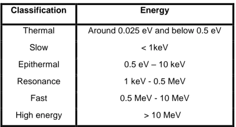
Neutrons were produced by neutron sources which are classified according to its energy as follows in Table 1.1.
Table 1.1: Classification of neutron in term of the kinetic energy.
Classification Energy
Thermal Around 0.025 eV and below 0.5 eV
Slow < 1keV
Epithermal 0.5 eV – 10 keV
Resonance 1 keV - 0.5 MeV
Fast 0.5 MeV - 10 MeV
High energy > 10 MeV
Thermal neutron and epithermal neutron can be very effective for the treatment of tumors in BNCT (Daquino, 2008; IAEA, 2001; Chadwick, 2006).
Neutron sources for BNCT currently have been limited to specially modified nuclear reactors, which are available in Japan, United States, European countries (Finland, Netherlands, Germany, Czech Republic), Argentina and Taiwan. Accelerators producing epithermal neutron beams also could be used for BNCT and these are being developed in several countries. It is anticipated that the first Japanese accelerator will be available for therapeutic use in 2013.
1.3.1 Nuclear reactors
Reactors are the most suitable type for neutron source because of it provides a suitable energy neutron spectrum with high intensity thermal neutron flux. Neutrons are generated by the fission reaction occurring in the core, mean energy is 1.98 MeV, in a nuclear reactor. It requires suitable moderator in order to obtain the epithermal neutron. From the beginning of BNCT history, the nuclear experimental reactors have been used as the only neutron sources to provide correct energy spectrum and adequate thermal neutron flux. Today, BNCT move from using
thermal neutron beam to the use of a more energetic, epithermal neutron beam. In principle epithermal neutrons have the capability to penetrate deep into the tissue by reducing the skin exposure and thermalizing at the same time. Thus, epithermal neutrons are useful for BNCT treatments of different tumor types. In addition epithermal thermal neutrons can be easily moderated further to be less penetrating by using a tissue-equivalent (TE) material on the patient’s skin (Seppälä, 2004). A number of reactors, with a very good neutron beam quality have been developed and applied clinically for BNCT, are shown in Table 1.2 (Hatanaka, 1973; Storr,
1992; Auterinen, 2001; Burian, 2002; Pinelli, 2002; Capala, 2003; Kato, 2004; Wittig, 2006; Nigg, 2006; Sauerwein, 2009; Wang, 2011; Gustavo, 2012).
Table 1.2 : Reactor- based neutron source for BNCT. USA
The Brookhaven Graphite Research Reactor (BGRR) at Brookhaven National Laboratory
The Massachusetts Institute of Technology Research Reactor (MITR)
Europe
The TRIGA II in Mainz, Germany The TAPIRO reactor in Rome, Italy
The TRIGA in Pavia, Italy JRC-HFR reactor at Petten, Netherlands
The Finnish TRIGA reactor, FiR 1, as clinical reactor in Helsinki, Finland LVR reactor, Czech Republic
The R2-0 Reactor, Studsvik Medical in Nyköping, Sweden Light Water Reactor (LWR) in Budapest, Hungary The WWR-M Kyiv Research Reactor (KRR) in Kyiv, Ukraine
The research reactor IRT-2000 in Sofia, Bulgaria The MARIA reactor in Świerk, Poland
Japan
The Hitachi Training Reactor (HTR)
Kyoto University Research Reactor (KURR) in Kumatori JRR4 at the Atomic Energy Research Institute (JAERI)
The Musashi Institute of Technology Reactor (MuITR)
Elsewhere
The High Flux Australian Reactor (HIFAR) at Lucas Heights (Australia) RA-6, RA-3, RA-1 CNEA reactor at Bariloche (Argentina) The National Tsing Hua University THOR reactor (Taiwan)
In following, two facilities, FiR 1 (Finland) and MITR (USA) which can be applied as a epithermal neutron source for BNCT, will be described.
In order to generate epithermal neutrons, the moderator block consisting of Al+AlF3
(Fluental™) was developed and produced in Technical Research Centre of Finland (VTT). The Fluental™ neutron moderator was installed into the space of the thermal column at FiR 1. The epithermal neutrons are produced from the irradiation the fast fission neutrons by The Fluental™ neutron moderator at 250 kW power. The epithermal neutron fluence has been obtained about 1.1 ×10⁹ n/cm2sec. The undesired fast neutron dose per epithermal fluence is 2 Gy/1013 cm-2 and the corresponding gamma contamination 0.5 Gy/1013 cm-2.
The epithermal neutron fluence was suitable for BNCT purposes. A 14 cm diameter collimator has been installed for directing the neutrons into the tumor area (Auterinen, 2001, 2005 and 2012) (Fig. 1.5).
Figure 1.5: Diagram of requirements for BNCT treatment at FiR 1 nuclear research reactor in Otaniemi, Finland (Auterinen, 2001).
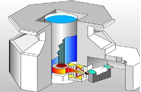
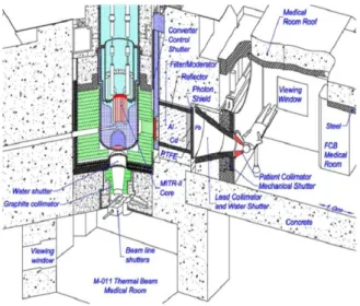
The fission converter based epithermal neutron irradiation (FCB) facility is placed in the experimental hall of the MITR and operates in parallel with other user applications. The FCB contains an array of 11 MITR-II fuel elements cooled by forced convection of heavy water coolant. The converter power is 120 kW at 6 MW reactor power. A shielded horizontal beam line contains an aluminum and Teflon filter-moderator to tailor the neutron energy spectrum into the desired epithermal
energy range. A patient collimator defines the beam aperture and extends into the shielded medical room to provide circular apertures ranging from 16 to 8 cm in diameter. The in-air epithermal flux is 6.2 × 10⁹ n/cm2s at the patient position with the 12 cm collimator (Fig. 1.6) (Barth, 2005; 2007; 2012).
The measured specific absorbed doses were constant for all field sizes and below the inherent background of 2.8 × 10⁻¹² GyWcm2/n produced by epithermal neutrons in tissue. The dose distributions were achieved by using the FCB approach and the theoretical optimum for BNCT. This facility is useful for clinical studies for superficial cancers and small animal studies (Harling, 2002; Riley, 2003). However, the application of nuclear reactors is limited and most of them are not located near the hospital. Thus, it would be necessary to have hospital-based neutron sources (Barth, 2007). Finally, an “in-hospital” compact nuclear reactor, which will be used exclusively for BNCT, has been designed and built in Beijing, China (Yiguo, 2010). But the new idea of the small reactors is not attractive for BNCT due to its low acceptability of such structure in a hospital environment and the high investment cost. Hence, the potential utility of accelerators provides an attractive idea in a real hospital environment. Nowadays, a number of groups around the world are involving their research toward into this topic.
Figure 1.6: Diagram of the BNCT facility at the Massachusetts Institute of Technology Reactor (MITR) in USA (Barth, 2012).
1.3.2 Accelerator-based neutron sources (ABNS)
Accelerator speed up the charge particles (protons, heluim-4, deuterons, tritons and electrons) with defined energy and let them interact to suitable target such as
the neutrons are produced from nuclear reactions. Some reactions used for generating neutrons by accelerator are listed in Table 1.3.
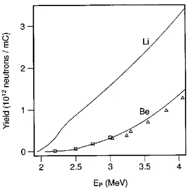
As can be seen in Table 1.2, there are different target materials and different incident particles but generally attention has been focused on low energy proton accelerators with the choice of either lithium or beryllium as the target material. In an investigation, total neutron yields have been calculated based on cross sections which are obtained from the thick target lithium bombarded by proton beam (Listen, 1975; Randers-Pehrson, 1998). Campbell with collaborators has measured direct total neutron yields from thick beryllium targets bombarded by proton beam (Campbell, 1977). The both results are shown in Fig. 1.7. In Fig. 1.7 the neutrons yields are high and, additionally, the kinetics are such that the secondary neutron spectrum has relatively low energy. Because of these /advantages, early designs for accelerator based BNCT systems focused on lithium targets (Randers-Pehrson, 1998).
Table 1.3 Characteristics of the charged particle, targets and nuclear reactions which are
considered for ABNS-BNCT.
Reaction Bombarding energy (MeV) Average neutron energy (MeV) Maximum neutron energy (MeV) Neutron production rate (nmA-1s-1) 7 Li(p,n)7Be 2.5 0.55 0.79 9.11011 9 Be(p,n)9B 4.0 1.06 2.12 1.01012 9 Be(p,n)10B 1.5 2.01 5.08 3.31011 13 C(d,n)14N 1.5 1.08 6.77 1.91011 2 H(d,n)3He 0.15 2.5 4.7 4.7108 3 H(d,n)3He 0.15 14.1 14.1 5.01010
Figure 1.7: The total neutron yields after proton bombardment of thick lithium and beryllium
targets as a function of incident proton energy.
Moreover, fusion reaction (D-T and D-D) can be used for generating neutron and are also considered as a neutron source. However, according to bombarding engineers, type of the charged particle and targets, the accelerator will be capable to generating the currents neutron needed to deliver therapy for BNCT in reasonable times.
Accelerators can generally be classified to linear (electrostatic quadrupole (ESQ) and radio frequency quadrupole (RFQ), Tandem cascade accelerator (TCA) and recirculating (cyclotron). Mostly these types of accelerator are suitable for producing neutron as ABNS which are applied for BNCT research and clinical trials (Green, 1998; Thomas, 2003; Culbertson, 2004; Kreiner, 2007; Pisent, 2006; Forton, 2009; Mitsumoto, 2010; Aleynik, 2011; Tanaka, 2009).
Theoretically RFQ accelerators have the capacity to be used as a neutron source in BNCT (Pisent, A. 2006).
Some of ABNS developed and applied clinically for BNCT are shown in Table 1.4 (Green, 1998; Thomas, 2003; Culbertson, 2004; Yonai, 2004; Kreiner, 2007; Pisent, 2006; Forton, 2009; Chiojdeanu, 2009; Sauerwein, 2009; Mitsumoto, 2010; Aleynik, 2011).
Table 1.4: ABNS for BNCT. USA
The proton-cyclotron-based accelerator facility, Washington in Seattle
The proton linear accelerator facility at Fermi National Accelerator Laboratory in Illinois The deuteron cyclotron facilities at Harper Hospital in Detroit
Europe
The proton-RFQ under investigated in Legnaro, Italy The proton-cyclotron-based in Louvain-La-Neuve, Belgium
The proton-Tandem in Bucharest-Mãgurele, Romania Dynamitron proton accelerator facility in Birmingham, England The proton-cyclotron-based facility in Bratislava, Slovak Republic
Japan
The proton-cyclotron-based at Sumitomo Heavy Industries in Sumitomo The proton-cyclotron-based at Tohoku University in Sendai
Elsewhere
The proton Tandem-Electro- Static-quardapol (ESQ) facility in Buenos Aires, Argentina
The proton Tandem in Novosibirsk, Russia
Two ABNS-BNCT facilities (the dynamitron proton accelerator facility of the University of Birmingham in Birmingham, England, and the proton-RFQ under investigate at the National Institute of Nuclear physics (INFN) in Legnaro (Italy) will be described.
The dynamitron proton accelerator facility of the University of Birmingham was the first clinical accelerator-based BNCT facility in the world. The accelerator had proton currents (1 mA) by bombarding a 7Li target via 7Li(p,n)7Be reaction with high energy protons (2.8 MV) generated thermal neutron flux 1.371012 ns-1. The facility used the Fluental TM as moderator to obtain the neutron beam in appropriate therapy energies as well as epitermal neutron. The proton beam with a Fluental TM, graphite reflector, lead filter, lithium target and shield (delimiter) Li-polyethylene was shown in Fig. 1.8 (Green, 1998; Culbertson, 2004).
Figure 1.8: University of Birmingham accelerator-based neutron source system.
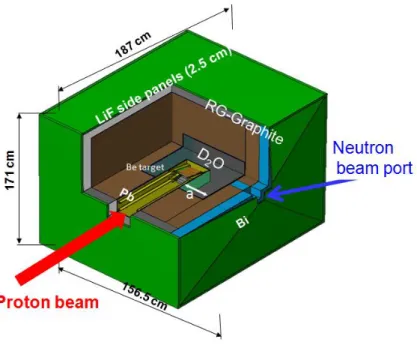
The RFQ is a unique facility in worldwide at INFN Legnaro (Italy). It's able to deliver 30 nA with 5 MeV proton beam which is useable in research project SPES-BNCT. The beryllium target (9Be) is bombarded by 30 nA proton via 9Be(p,xn)9B reaction, generating neutrons which are moderated via heavy water and graphite. Consequently the neutron flux of thermal neutrons is obtained about 2.5109/(scm2) which are suitable for therapy (Figs. 1.9 and 1.10).
Figure 1.10: Schematic layout of neutron source at irradiation facility INFN-LNL.
The LNL-BNCT facility is foreseen to explore the treatment of extended skin melanoma with such a therapeutic modality and, at the next stage, hepatic metastases in the explanted liver (Agosteo, 2001; Prete, 2008; Ceballos, 2009). The main items of the research program are being mainly focused on the neutron irradiation facility design, involve quality neutron beam, the development of a new boron carrier and a new, on-line, biological dose monitoring in both tumor and healthy tissues (Pisent, 2006).
The new application of neutron source for BNCT is using of process photoneutron production by a linear electron accelerator.
When medical electron accelerator (LINACs) works with acceleration potentials above 10 MV, they could generate secondary neutrons via photon-neutron (, n) and electron-neutron reaction (e, en) giant dipole resonance reactions of incident photons and electrons, respectively, with all the heavy materials present inside the gantry and along the beam line.
During the mid-1990s, the Idaho National Laboratory and Idaho State University has been involved in the first idea about the photoneutron production process driven by an electron accelerator which could be applied for BNCT in futures. In this research, relativistic electron beams impinge upon heavily-shielded tungsten targets located at the outer radius of a small cylindrical tank of circulating heavy water (D2O). A fraction of the energy of the electrons is converted in the tungsten
subsequently generated by photodisintegration of deuterons in the D2O within the
tank are directed to the patient through a suitable beam tailoring system. Initial proof-of-principal tests using a low-current benchtop prototype of the epithermal photoneutron source concept for BNCT were conducted (Nigg, 1997 and 2006). The results of these experiments demonstrated that on the basis of neutronic performance, the proposed photoneutron device could offer as an approach for the production of epithermal neutrons in BNCT.
Another research was done as PhoNeS (PhotoNeutron Source) project at Trieste University with collaboration Turino University in Italy. In this study, the photo-reaction model assumed which can be applied as a neutron source. Thus, a photoconverter has been designed. In order to increase the photoneutron production inside the moderating materials, the photoconverter materials have been selected: graphite blocks for the external moderator, lead target, moderators in polyethylene, PMMA box, 5 cm thick volumes filled with heavy water (D2O 99%),
and irradiation cavity. Finally, the photoconverter placed at off-axis points of photon beam with 18 MV at medical electron accelerator (Varian Clinac 210). As consequently, thermal and/or epithermal neutron beam with neutron fluence rate about 1.06108 cm-2s-1 has been obtained and could be used for BNCT treatment of shallow tumors (as melanoma or limb sarcoma) (Fig. 1.11) (Giannini, 2006).
Accelerator-based neutron sources (ABNS) have potential advantages over reactor-based neutron sources for clinical applications. First, ABNS can be easily turned off when the neutron field is not required during therapy. But the fast neutrons are not stopped via a critical assembly of fissile material, what means that licensing and regulations associated with maintaining the neutron source are substantially simplified. Second, the variety of neutron producing reactions that are accessible to accelerators, allows a number of neutron energy source spectra to be produced. Consequently, for some accelerator types, epithermal neutron beams can be produced the neutron flux energy spectrum can be tailored to the spatial characteristics of a particular patient’s tumor. Third, the capital expenses of an accelerator-based BNCT system will be substantially lower than those associated with the installation of a reactor system in or near a hospital. And finally, accelerators have been prominent features of radiotherapy departments in hospitals for years; clinicians have a longstanding and comfortable experience with such devices for patient irradiation. It is likely that accelerator hardware for BNCT irradiations could be sited within an existing radiotherapy room with the addition of extra shielding.
References
Aihara, T., et al. (2006). First clinical case of boron neutron capture therapy for head and neck malignancies using 18F-BPA PET. Head Neck, 28, pp. 850-5. Agosteo, S., et al. (2001). Advances in the INFN-Legnaro BNCT Project for Skin Melanoma. In Proceedings of International Physical and Clinical Workshop on BNCT Candiolo (Torino), February 7, 2001.
Aleynik, V., et al. (2011). BINP accelerator based epithermal neutron source. Applied Radiation and Isotopes, 69, pp. 1635-1638.
Auterinen, I., et al. (2001). Metamorphosis of a 35 years old Triga reactor into a modern BNCT facility. In: Frontiers in Neutron Capture Therapy, Plenum Press, New York, 1, 267-275.
Autrerinen, I., et al. (2005). Fir 1 Reactor in service for boron neutron capture therapy (BNCT) and isotope production. STI/PUB/1212. IAEA, pp. 313-324. Autrerinen, I. (2012). Introducing the BNCT option in a national health care system- the Finnish experience. In proceedings of 15 international congress on neutron capture therapy, 10-14 September, Tsukuba, Japan.
Bakeine, G.J., et al. (2009). Feasibility study on the utilization of boron neutron capture therapy (BNCT) in a rat model of diffuse lung metastases, Applied Radiation and Isotopes, 67, pp. S332-S335.
Barth, R.F., et al. (2005). Boron Neutron Capture Therapy of Cancer: Current Status and Future Prospects. Clinical Cancer Research, pp. 3987-4002
Barth, R.F., et al. (2007). Boron neutron capture therapy for the treatment of glioblastomas and extracranial tumors: As effective, more effective or less effective than photon irradiation? Radiation Oncology, 82, pp. 119-122.
Barth, R.F., et al. (2012). Current status of boron neutron capture therapy of high grade gliomas and recurrent head and neck cancer. Radiation Oncology, 7, 146, pp. 8-21.
Blaickner, M., et al. (2012 ). Dosimetric feasibility study for an extracorporeal BNCT application on liver metastases at the TRIGA Mainz. Journal of Applied Radiation and Isotopes, 70, pp. 139-143.
Bortolussi, S., et al. (2011). Boron uptake measurements in a rat model for Boron Neutron Capture Therapy of lung tumours. Applied Radiation and Isotopes, 69, pp. 394-398.
Byun, Y., et al. (2006). Anti-Cancer Agents in Medicinal Chemistry, 6, pp. 127-144. Burian, J., et al. (2006). Physics for BNCT. Journal of Physics Conference Series, 41, pp.174-186.
Burian, J., et al. (2002). Report on the first patient group of the phase I BNCT trial at the LVR-15 reactor. In proceedings of Conference Research and Development in Neutron Capture Therapy, Bologna, pp. 1107-1112.
Busse, P.M., et al. (2003). A critical examination of the results from the Harvard-MIT NCT program phase I clinical trial of neutron capture therapy for intracranial disease. Neuro-Oncology, 62, pp. 111-121.
Campbell, J., et al. (1977). Absolute neutron yield measurements for protons on Li, Cu, Co, and Be from threshold to 3 MeV. In Proceedings of the 4th Conference on the Scientific and Industrial Applications of Small Accelerators, IEEE, New York, pp. 517-521.
Capala, J., et al. (2003). Boron neutron capture therapy for glioblastoma multiforme: clinical studies in Sweden. Journal of Neuro-Oncology, 62(1-2), pp. 135-144.
Catharine, M., et al. (2005). Boron neutron capture therapy for glioblastoma multiforme. Journal of Pharmacy World & Science, 27, pp. 92-95.
Ceballos, C., et al. (2009). The BSA modeling for the accelerator-based BNCT facility at INFN LNL for treating shallow skin melanoma. Applied Radiation and Isotopes, 67 (7-8), pp. S274-S277.
Cerullo, N., et al. (2009). Progress in the use of gadolinium for NCT. Applied Radiation and Isotopes, 67, pp. 157-160.