A
A
l
l
m
m
a
a
M
M
a
a
t
t
e
e
r
r
S
S
t
t
u
u
d
d
i
i
o
o
r
r
u
u
m
m
–
–
U
U
n
n
i
i
v
v
e
e
r
r
s
s
i
i
t
t
à
à
d
d
i
i
B
B
o
o
l
l
o
o
g
g
n
n
a
a
DOTTORATO DI RICERCA IN
Oncologia Ematologia Patologia
Ciclo XXXI
Settore Concorsuale: 06/A4
Settore Scientifico Disciplinare: MED/08
TITOLO TESI
GENETIC PROFILES OF FIELD CANCERIZATION
AND INTRATUMORAL HETEROGENEITY IN ORAL
SQUAMOUS CELL CARCINOMA
Presentata da:
Dott. Gabusi Andrea
Coordinatore Dottorato
Supervisore
Prof. Pier Luigi Lollini
Prof.ssa Maria Pia Foschini
CONTENTS:
HEAD AND NECK SQUAMOUS CELL CARCINOMA AND ORAL SQUAMOUS CELL CARCINOMA…3
Definition………...3
Epidemiology……… 4
Risk factors………...7
TNM CLASSIFICATION……… 10
TNM classification to define lip and oral cavity squamous cell carcinoma……… 11
ORAL SQUAMOUS CELL CARCINOMA AND THE THEORY OF FIELD CANCERIZATION………….. 13
Introduction……….……….……….……... 13
Slaughter’s principles and first evidences……….……….………. 13
Biomolecular evidences of Slaughter’s field cancerization….……….……….14
Multiple Oral Lesions: Different models for field cancerization and the problem of clonality……….. 16
THE CONCEPT OF INTRATUMORAL HETEROGENEITY………... 21
Introduction……….……….……….……….……….. 21
Clonal evolution theory……….……….……….……… 21
Cancer Stem Cells theory……….……….……….……….. 22
EXPERIMENTAL SECTION……….……….……….……….. 24
AIMS OF THE PHD PROJECT(ABSTRACT).……….……….………... 24
PROJECT 1: INVESTIGATION OVER IMMUNOHISTOCHEMICAL EXPRESSION OF KI67 IN DISTANT MUCOSAE AS A PROGNOSTIC FACTOR IN ORAL SQUAMOUS CELL CARCINOMA…… 25
Aims……….……….……….……….……….. 25
Methods……….……….……….……….……… 25
Results……….……….……….……….……….. 29
Discussion……….……….……….……….……… 33
PROJECT 2: INVESTIGATION OVER PROGNOSTIC VALUE OF mtDNA PHYLOGENETIC ANALYSIS AS A TOOL FOR CLONAL DIAGNOSIS OF SECONDARY EVENTS IN ORAL SQUAMOUS CELL CARCINOMA……….. 36
Aims……….……….……….……….……….. 36
Methods……….……….……….……….……… 36
Results……….……….……….……….……….. 40
Discussion……… 44
PROJECT 3: STUDY OF INTRATUMOR HETEROGENEITY IN RECURRENT-METASTATIC ORAL SQUAMOUS CELL CARCINOMA BY MEANS OF MULTI REGION NEXT GENERATION SEQUENCING AND mtDNA ANALYSIS………... 46
Aims……….……….……….……….……….. 46
Methods……….……….……….……….……… 46
Results……….……….……….……….……….. 49
Discussion……….……….……….……….……… 58
PROJECT 4: INVESTIGATION OVER INTRATUMOR HETEROGENEITY AS A PROGNOSTIC FACTOR IN PREDICTING DISEASE RELAPSE IN ORAL SQUAMOUS CELL CARCINOMA…………. 61
Aims……….……….……….……….……….. 61
Methods……….……….……….……….……… 61
Results……….……….……….……….……….. 65
Discussion……….……….……….……….……… 74
Supplementary material Project 4……….……….……….………. 79
Summary……… 84
References……….……….……….……….………. 88
1.HEAD AND NECK SQUAMOUS CELL CARCINOMA AND ORAL SQUAMOUS CELL CARCINOMA
1.1. Definition
The term head and neck cancer (HNC) defines a group of malignant cancers that arise in different anatomical districts in the region of the head and neck including oral cavity, pharynx and larynx.
However different definitions have been proposed.
For example the National Institute of Health, and more precisely, the National Cancer Institute reports that HNC correspond to “Cancer that arises in the head or neck region (in the nasal cavity, sinuses, lips, mouth, salivary glands, throat, or larynx).(1) Differently, UK’s National Health service describes HNC as a miscellaneous group of cancers from 30 different organs or tissues in the head and neck region including “eye cancer, nasal and paranasal sinus cancer (cancers in the nasal cavity and in the sinuses around the nose),nasopharyngeal cancer (the area that connects the back of the nose to the back of the mouth), mouth and oropharyngeal cancer (cancers of the tongue, the gums, cheeks, lip and floor and roof of the mouth), larynx or laryngeal cancer (cancer of the voice box) and oesophageal cancer (cancer of the food pipe or gullet).”(2)
Almost 90% of HNC are squamous cell carcinomas deriving from epithelial cells of mucosal upper digestive tract.
Head and Neck Squamous Cell Carcinoma (HNSCCs) are strongly associated with tobacco exposure, alcohol, areca nut, low vitamins intake.
Recently, also oncoviruses infection has been recognized as a risk factor for HNSCC development and in particular HPV 16,18, EBV, HHV-8.(3)
HNSCC usually adopts an aggressive clinical behaviour due to its ability to invade rapidly adjacent tissue and cervical lymph nodes.(4)
Moreover, HNSCC local destructive action is linked to high risk of subsequent local relapses or distant lymph node metastasis which comprises several demolitive surgeries.
The tendency of multiple aggressive local or distant relapses is biologically explained by the theory of “field cancerization “. This theory was first proposed by Slaughter in 1953 and defines a genetically altered mucosal filed extended far beyond the border of the primary tumour, in which several squamous cell carcinomas may arise regardless surgical excision.(5)
Treatment modalities include surgical excision of the neoplasia, associated to radiotherapy or adjuvant chemotherapy in advanced stages.
Early diagnosis proved to impact positively overall survival and treatment efficacy. Nevertheless, despite clinical efforts in improving early diagnosis the survival rate hasn’t significantly improve in the last decade.(6)
Therapy for HNSCC includes also the use of Cetuximab, a monoclonal antibody against Epidermal Growth Factor Receptor (EGFR) approved by the FDA (Food and Drug Administration).(7)
1.2. Epidemiology:
Incidence of HNSCC worldwide varies significantly depending on the anatomical site
included in the statistics. On average, HNSCC classifies as the 6th more common cancer
accounting for 4,3 % of the tumours, 599.637 new cases and 224.834 new deaths each year. (Figure)
However, if squamous cell carcinoma of the upper 2/3 of the oesophagus was also included in the HNSCC group, incidence would rise sharply, classifying as the third more frequent carcinoma in men worldwide.
As far as oral squamous cell carcinoma ( OSCC) is concerned data estimates that 300000 cases new cases per year and the mortality rate reaches levels around 145 000 cases per year in Europe.(8)
However, higher incidence levels occur in Asia and in particular in Melanesia and India. Widespread use of areca nut and betel leaves in these regions explains the results.
FIG.1 The Graph shows the value of incidence (blue) and mortality(red) of Head and Neck Cancer worldwide. Left horizontal bars shows values for male population while right horizontal bars the values for women. Source: https://gco.iarc.fr/
Unfortunately, high incidence levels of OSCC are followed by similar data on mortality. In Taiwan, for example, OSCC embodies the first cause of death among men aged 25-44 years old. Low access to health services is a significant co-factor of high mortality in the region.(9)
HNSCC incidence is greater in men. Lifestyle plays an important role in these findings. In fact, despite men are traditionally more exposed to well known risk factors for oral squamous cell carcinoma such as tobacco smoke or alcohol consumption, incidence of OSCC in women is increasing as effect of similar exposure to risk factors in both sexes. Additionally, deficit in estrogenic hormones has also been advocated to play a role in OSCC tumorigenesis. (10)
OSCC tends to affect men in their sixth decade of life. Epidemiologic studies confirm worse trends in South Asia ( fifth decade of life ) with respect to North America where mean age of developing an OSCC stands above the threshold of seventh and eighth decade of life.(11)
However, recent data shows that the number of young patients affected by OSCC is increasing. So far, No scientific evidence seems to support that HPV is responsible for the increase of OSCC in young patients.
Yet, HPV related OSCC show better responsiveness to therapies and better survival rates. (12)
1.3. Risk Factors
Tobacco smoke and alcohol abuse are major risk factors in OSCC development. Nearly 80% of OSCCs seem to be related by the action of these two factors. Accordingly, suspension of tobacco or alcohol exposure would reduce approximately the incidence of OSCC by 80%.(13)
Additional risk factors include viral agents ( i.e. HPV) chronic oral trauma, UV exposure and immunodepression.
Tobacco:
The risk of developing OSCC is 5-9 times greater in smokers with respect to non-smokers. OSCC risk is dose-dependent. (14)The risk of developing OSCC doubles if more of 20 cigarettes/day are smoked. Additionally, in patients who do not quit smoking after diagnosis, the risk of developing a second neoplasia after resective surgery is up to six times higher the risk of patients who quit.(15)
Cancerogenetic action of tobacco smoke is mainly due to the presence of mutagenic compounds that can be found in products of combustion. More than 70 carcinogenetic compounds have been identified.
These include : polycyclic aromatic hydrocarbons ( benzopyrene, anthracene) nitrosamines of tobacco ( N-nitrosonornicotine, N-nitrosodimethylamine ) aromatic amines (2-toluidine ), aldehydes ( formaldeyde) metals and organic compounds.(16)
Direct termical irritation of tobacco smoke on mucosae seems to play an important role as co-factor.(17) In fact, reverse smoking, or the habit of smoking a cigarette with the lit end inside the mouth which is widespread in Andhra Pradesh and in Phylipinnes,correlates with higher risk of malignant transformation.(17)
Tobacco exposure other than smoking is also related to OSCC development. For example, in India and South East Asia, the diffusion of chewing betel leaves in association with tobacco, areca nut and other irritating compounds not only induces the onset of submucous fibrosis, a pre-malignant lesion at high risk of cancer, but also reflects greater rates of OSCC incidence.(18)
Alcohol
Recent data indicate that daily alcohol consumption exceeding 10g g/day cause negative effects on general health. Epidemiological data partially correlates an increase in HNSCC in young adults in UK with heavy alcohol drinkers.(19)
Carcinogenetic effect of alcohol seems to synergic to tobacco smoking. In fact, the risk of OSCC in both heavy smokers and heavy drinkers appears to be 13 fold greater the risk of tobacco or alcohol considered independently.(20)
Similar to tobacco smoking carcinogenetic action, also alcohol action seems to be dose dependant in increasing OSCC development . Alcohol abuse in the post-operative interval resulted in a significant reduction in survival rates, not necessarily related to disease
relapses.(21)
Experimental studies proved that ethanol acts as a mutagen as well as can act as solvent for other mutagens. Direct oncogenetic action seems to be related to acetaldehyde, one of the primary metabolites of alcohol and a powerful mutagen. (22,23)
Fungal and viral infection
Yeats and viruses have been investigated for years as potential triggers or co-factors of malignant transformation in OSCC.(24)
In particular Candida spp. is able to produce powerful mutagens such as N-Nitroso
bezilmetilamine which seems to play a pivotal role in cancer development. Candida spp. is also associated with premalignant lesions at moderate- high risk of evolving into OSCC such a hyperplastic chronic candidiasis.(25)
However, neither Candida associated premalignant lesions nor the presence of Candida spp in samples from OSCC are able to differentiate between Candida direct carcinogenesis
and Candida super-infection of premalignant/malignant lesions. (26)Consequently, role of Candida spp. in oral carcinogenesis are yet to be fully understood.
HPV infection has been recently related to HNSCC development, and in particular
genotypes 16 and HPV 18. HPV has preferential tropism in the pharynx where lymphoid tissue is abundant, and invasion of the mucosal barrier is easier. (26)
In the oral cavity HPV associated OSCC tend to arise posteriorly, at the base of the tongue and in proximity to palatine tonsils while are far less frequent in the anterior part of the oral cavity.(27)
Oncogenetic effect of HPV is mediated by HPV associated oncoproteins such as E6 and E7 which can interfere with in many important pathways such as TP53 inducing tumoral
degeneration.(28)
UV radiations and immunodepression
Long-time exposure to UV is a well-known risk factor for the development of OSCC of the lower lip.
In the last years awareness of risk factors among general population has improved incidence rates for lip OSCC, whose prognosis is less aggressive than other OSCCs.(29) Immunodepression has a deep negative impact on tumorigenesis. As far as OSCC is concerned, bone marrow transplant and related immunosuppressant regimes expose the patient to a 6-10 folds higher risk of developing OSCC.(30)
Similarly, HIV+ patients suffering from AIDS show and augmented risk of developing OSCC with respect to general population.
In these patients immune system is compromised and tumours can escape immune surveillance more easily with more descriptive clinical effects. (31)
2. TNM CLASSIFICATION:
TNM classification systems is based on the clinical extension of the disease and includes three parameters: dimension of primary tumour (T), nodal involvement (N), presence of distant metastasis (M).
Clinical classification (cTNM) is based on data obtained before surgical treatment. After tumour resection, cTNM is normally compared to histological classification ( pTNM). In HNSCC T parameter differs depending on the site of primary tumour. N classification of cervical nodes, on the contrary, is almost universal and only nodal involvement of
nasopharynx has an independent classification.(32)
TNM CLASSIFICATION TO DEFINE LIP AND ORAL CAVITY SQUAMOUS CELL CARCINOMA
TAB1. The Table shows the criteria of classification for T category according to TNM classification. Source: AJCC 8th Edition
TAB2. The Table shows the criteria of classification for N category according to TNM classification. Source: AJCC 8th Edition
FIG.2 (left) The mesasure of depth of invasion is assessed by dropping a “plumb line” from the horizon (level of basement membrane relative to the closest intact squamous mucosa)
FIG.3 (right) Difference between “depth of invasion” ( blue bar) and tumour thickness (white bar)
3. OSCC AND THE THEORY OF FIELD CANCERIZATION: 3.1 Introduction:
The term “ field cancerization” was introduced by Slaughter and colleagues in 1953 (5) to explain the appearance of multiple oral squamous cell carcinomas (OSCC)in the same patient. This model for head and neck cancer is still actual nowadays.
3.2 Slaughter’s principles and first evidences.
Analysing a population of 783 patients with OSCC Slaughter et al.(5) recorded in a publication titled “field cancerization in oral stratified squamous epithelium. Clinical
implications of multicentric origin” that 1) oral cancers usually had the tendency of spreading more easily in laterality than in depth 2) that the mucosa surrounding the neoplasia
frequently harboured clinical or morphological atypia. 3) that OSCC may consist of multiple independent foci that eventually may converge 4) OSCC may develop multifocally in distant areas presenting preneoplatic features 5) the persistence of altered epithelium after surgical resection may induce the formation of new carcinomas.
A similar apparently independent multifocality, they acknowledged, was “well above the statistical possibility of chance occurrence, therefore they concluded that multiple OSCC should be the effect of a “field cancerization,” in which an “area of epithelium has been preconditioned by an as-yet-unknown carcinogenic agent. Such a carcinogenic influence if operative long enough in time and intense enough in exposure produces an irreversible change in cells and cell groups in the given area, so that change of the process toward cancer becomes inevitable. “
Nowadays we know that not only was Slaughter’s intuition correct but also that it deeply helped our understanding of the natural history of OSCC. At that time, however, molecular techniques could hardly be performed hence it should not be surprising that a work citing the theory of field cancerization appeared no sooner than 16 years after Slaughter’s publication.
In 1969 Roth et al (33) studying the healing performances of UV-induced damaged cells acknowledged that epithelium from patients affected by oral and upper aero digestive tract squamous cells carcinoma showed a reduced repairing ability. They reported that a similar deficit could be the result of a damage at DNA level predisposing for further tumour
development.
Albeit Slaughter’s theory had gained some clinical evidence it still lacked proper scientific basis and it could not gain widespread popularity.
In 1982, Incze et al (34) observed, at electron microscope level, biopsies from normal appearing epithelium in patients with squamous cell carcinoma of the upper aero digestive tract and they reported that morphologic abnormalities were consistent with the concept that carcinogenesis is a multistep process of sequential neoplastic development extending over a long period of time.(Fig.1)
Only two years later, in 1984, Strong et al (35) observed that Field cancerization could be demonstrated by supravital staining with toluidine blue or by electron microscopic study of random biopsies taken from apparently normal mucosa.
Both authors not only confirmed Slaughter’s statements with clinical and morphological means but also identified tobacco and alcohol exposure as the “yet-unknown carcinogenic agent” able to condition mucosal behaviour towards cancer development.
3.3 Biomolecular evidences of Slaughter’s field cancerization
In 1996 Califano et al(36), in response to the lack of knowledge surrounding genetic progression of head and neck squamous cell carcinoma and genetic basis for field
cancerization, published a PCR based analysis of loss of heterozygosis (LOH) at selective genes putatively involved in head and neck carcinogenesis. They studied a population of eighty-seven lesions of the head and neck, including pre-invasive lesions and benign lesions associated with carcinogen exposure. Observing the level of accumulation of gene losses at different degrees of pre-neoplastic lesions they found that it was possible to
identify a model that could explain the progression of a mutated epithelium towards squamous cell carcinoma. However, despite some mutations were more likely to occur at specific stages or pre-malignancy, they suggested that accumulation rather than the order of genetic events led mutated cells in the progression towards cancer.
Genes studied by Califano and colleagues(36) included the 9p21 locus corresponding to an area of genetic loss common to many solid tumours containing p16, a cyclin-dependent kinase inhibitor involved in cell cycle regulation. Region 11q13 that includes the bcl-1/int-2 locus, an amplicon carrying the proto-oncogene cyclin D1, the p53 gene locus located at 17p13, the 3p21 locus and 13q21 locus that contains an area with frequent LOH near the retinoblastoma locus(36).
In particular, 3p and 17p losses were more frequent in mucosa undergoing dysplastic modification while 11q and 13 q losses could be found in epithelium preceding malignant transformation.
Califano’s results were confirmed by Patridge et al. in 1997 (37) and Lydiatt(38,39) in 1998 where LOH was found in dysplastic tissue surrounding tumours and histologically normal mucosa respectively.
Genetic approach paved the way to a large number of studies with different molecular techniques that tried to disclose hidden mechanisms of oral carcinogenesis with the aim at discovering clinically relevant biomarkers to be used in clinical practice.
As a consequence, the extension of the field and the related premalignant lesion whence the field could have been arisen, became a major concern.
Noteworthy, Ai H. (39) in 1999 detected by FISH chromosome aneuploidy in mucosa
distant from the carcinoma. Interestingly, 9/10 tested patients were smokers with respect to only one non-smoker patient with aneuploidy. Molecular tests can be of difficult application in daily practice, therefore several authors tested immunohistochemical markers useful to evidence areas od filed cancerization.
Van Oijen e Slootweg in 2000 (40)demonstrated that immunostaining of P53 and EGFR was abnormal in apparently normal mucosa of smokers when compared to non-smokers subjects. (Fig.2)
Tabor et al. (41)demonstrated that the presence of Ki67 positive mature keratinocytes , corresponded to areas of LOH. This observation has been validated by Montebugnoli et al(42–44)who demonstrated that the immunohistochemical analysis of ki67 in oral mucosa located in the cheek opposite to the OSCC can act as prognostic biomarker as, when overexpressed, had an impact on the aggressiveness of the primary tumour.
On the other hand, studies speculated on how far the field could extend. Interestingly
Griffioen GH, et al.(45) In 2015 reported that patients successfully treated for head and neck cancer died of primary lung cancers suggesting that the field could extend not only through the entire upper aero digestive tract, but also in the deep respiratory system.(Fig.3)
3.4 Multiple Oral Lesions: Different models for field cancerization and the problem of clonality
The theory of field cancerization derives from the effort of explaining the increased occurrence of local secondary tumours in oral cavity and upper aero digestive tract.
However, the exact mechanisms through which this phenomenon occurs are still matters of debate and different theories has been formulated so far.
In 1999 Garcia et al (46)described the presence of cluster of cells usually positive for TP53 in the normal mucosa of patients with head and neck squamous cell carcinoma. He called these clusters “patches”.
Assuming a monoclonal origin for oral squamous carcinoma, he hypothesized that these clusters of genetically altered cells could clonally expand as a consequence of proliferative advantages over non mutated cells generating greater area of mutated mucosa, better defined as “field”. Hence, according to Califano’s model of carcinogenesis(47), the
accumulation in the field of further mutations could lead to the development of independent neoplastic events.
On the other hand, an alternative theory reviewed by van Oijen et al. in 2000(40), reported that the occurrence of multiple pre(neoplastic) events could also result from a migration of mutated cells that, acting as micro metastasis, could generate different tumours at different local sites.
Apart from field cancerization theoretical models, the distinction between a local recurrence (LR) a metastasis or a second primary tumour (SPT) always represented a critical clinical issue able to deeply influence therapy and prognosis.
Before genetic approach, Second Primary Tumours (SPT) were usually defined by many researchers using Warren and Gates(48) criteria developed in 1932. This method was based on clinical and morphological parameters. In particular, SPTs were defined as two neoplastic lesions showing definitive and distinct pictures of malignancies after excluding that one could be the metastasis of the other. However, in case of two lesions both in the same anatomical area, the minimal distance to exclude a local recurrence was controversial as some researchers accepted 2 cm while others 1.5 cm.
Furthermore, second events may chronologically develop synchronously or
methachronously depending on whether the interval between the two carcinomas is lower or higher than six months respectively. Similarly, to spatial criteria, also time of occurrence could easily be confounding in the diagnosis of a second event.
As aforementioned criteria were easily matter of debate and scarcely took into account the theory of field cancerization, in 2002 Braakhuis (49), proposed a modification based on genetic profiles.
Evidence deriving from many works on LOH and TP53 mutations led Braakhuis to identify four types of second events: Local recurrence, metastasis, second primary tumours and second field tumours.
In particular, common molecular profiles between the primary tumours and the second lesions should be interpreted as consistent with local recurrence or metastasis depending if the second event developed in adjacent or distant site respectively. On the other hand, partially different genetic profile between the two lesions, should be suggestive of a secondary field tumour that, arising from a preconditioned field but followed different carcinogenetic pathways. Second Primary Tumours, being genetically and clinically independent should instead show different genetic profiles.
Further studies(50–53) confirmed the utility of Braakhius modified criteria and disclosed new genetic methodologies to perform clonal analysis of multiple squamous cell carcinomas. Remarkably, the high frequency rate of mtDNA mutations in tumors, especially those found in the D-loop region, a non-coding region, along with numerous mitochondrial genomes present in a single cell, has made mtDNA a reliable marker for clonality assays from microdissected paraffin-embedded tissue samples. (54,55)
As a result, mtDNA analysis not only demonstrated higher diagnostic sensibility when
compared to clinical and temporal criteria, but also resulted more informative with respect to TP53 analysis, reflecting the ability of some squamous cell carcinoma to follow carcinogenic pathways different from TP53 mutations.
In addition, mtDNA analysis resulted highly useful in disclosing possible mechanisms behind the cancerization of skin graft after surgery such as the spread of the clonal cell population to the cutaneous flap stimulated by cytokines produced by the grafted skin. In fact, as reported by Foschini et al,(50) in all three studied cases, the neoplastic lesions arising in the skin graft showed a clonal relationship with the previous OSCC and, on the basis of the results obtained by mtDNA analysis, could be considered as a recurrence of the primary OSCC.
FIG.3:Apparently normal oral mucosa can present small areas of altered epithelial keratinocytes
FIG.4: Immunohistochemical staining for TP53 in a restricted “patch” of oral epithelium supports the theory according to which the progression of a field of genetically altered kerationcytes precedes and promotes oral carcinogenesis
FIG 5: Smokers can present small areas of dysplasia in bronchial epithelium
4. THE CONCEPT OF INTRATUMORAL HETEROGENEITY: Introduction
The idea of heterogeneity among tumours and within tumours is not new. The observation that microscopic morphology may not be uniform throughout the entire extension of a primary tumor suggested that differences may exist among tumours cells.
Slaughter et al, studying morphology of OSCC recorded that OSCC may consist of multiple independent foci that eventually may converge. (5)
In addition, the hypothesis of tumoral heterogeneity was supported by clinical data even before bimolecular investigations, as tumours with similar morphology but deeply different behaviour and prognosis.
However, only in recent years, thank to improved sequencing technologies it was possible to study tumor heterogeneity at a deeper level.
Tumours should thus be regarded as complex biological entities. Heterogeneity seems to involve clinical, phenotypic, genetic and epigenetic variables as disclosed by recent studies of molecular biology applied to tumours.
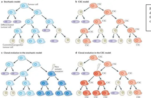
Two theories have been proposed to explain intratumour heterogeneity: clonal evolution models and cancer stem cells models (CSC).(56,57)
FIG 6: According to field cancerization theory three different types of second events can develop after primary tumour resection: Local Recurrence, Second Primary tumour and second field tumour. Source: https://www.tandfonline.com/doi/pdf/10.1586/1473715 9.2016.1126512
Both theories assume that tumours are initiated by single cells that acquire molecular aberrations that confer on them proliferative advantages and escape from programmed death and believe that microenvironment plays an important role in tumor progression. Nevertheless, the two theories also display important differences.
4.1 Clonal evolution theory:
In 1976 Nowell proposed a theory of clonal evolution assuming that a single cell could give rise to a tumor through a continued accumulation of genetic mutations.(58)
Genetic instability could then result in progressive formation of clonal populations contributing to intra-tumor heterogeneity.
Two types of clonal evolution have been described : linear and branched evolution. In linear evolution mutations are sequential and fitter clones tend to replace their
predecessors. The grade of heterogeneity in linear evolution is low and can be observed only when new clones has only partially replaced the old ones.
Linear evolution has been observed in acute myeloid leukaemia and multiple myeloma. In the branched evolution theory, different subclones coexist and evolve as the branches of a tree.
Early mutations, being shared by all subclones, can be seen as the trunk of the tree and reflect the genetic profile of the founder cell. During the evolution, genetic instability induces the formation of new subclones who acquire new genetic features.
Clonal evolution, due to its branching evolution, reflects higher grade of heterogeneity. However, it is important to mention that proliferation and expansion of a given subclone is the result of selective pressures that follow Darwinian rules.
4.2 Cancer stem cell theory:
This theory claims that only a small amount of cells with great self renewal ability features has the potential to promote tumor progression. These cells are cancer-stem-cells (CSCs) When CSCs lose their “stemness” and differentiate into a non CSC phenotype they give rise to subclones with individual genetic profile contributing to intratumor heterogeneity.
The CSC theory was firs demonstrated in hematopoietic tumours and later in solid tumours.
A set of membrane surface markers permits to identify CSCs ( i.e CD44+ /CD 24 low).(59)
Experimental studies demonstrate that cell with CSCs profiles can be isolated and if injected in xenografts are able to induce tumor formation and progression.
CSCs divide asymmetrically, resulting in a self-renewal CSC and a non.CSC. Non CSCs represent the majority of tumor mass but contribute less to tumor proliferation.(60)
Interestingly, according to CSC model, tumor cells show cellular plasticity. In fact, not only CSC may differentiate into non.CSC but also a non-CSC may reversibly switch to a CSC particular conditions.(61)
Tumor biology is thus regulated not only through a hierarchic organization of cells ( with and without self-renewal ability) but also by a homeostatic equilibrium between CSCs and non CSCs.(62)
Cancer stem cell model has gained increasing attention in recent years and studies have tried to study CSC markers in both OSCC and adjacent mucosa. The goal was to integrate our understanding of OSCC biology with evidence that markers associated with a stem-cell like behaviour.
Noteworthy, Simple et et al have proposed an intriguing theoretical model for field cancerization driven by genetically mutated stem-cells. In particular, according to that model, field cancerization is initiated by the carcinogen assault leading to genetic mutations (p53/p16) in a stem cell residing in normal epithelium. This cancer stem cell will proliferate and initially form a patch), which ultimately spreads to form a field. Histologically, at this
stage the cells remain in dysplastic or a premalignant stage. After getting a subsequent hit (RB), one of the cells in the field will form the primary tumor. The tumor will also host the increase in expression of different CSC-specific genes and other downstream markers of HNSCC tumorigenesis.
The progression of the field occurs either by the monoclonal or the polyclonal mode of cancerization. The CSCs of the field can also migrate (CD44h/ALDH1A1h) laterally to spreadthe field or get implanted at a new site and form a genetically similar tumor at a later stage signifying the monoclonal mode of field cancerization. On the other hand, multiple hits to the normal stem cells in the epithelium will lead to the development of independent
clones (polyclonal mode). (63)
Nevetheless, as pointed out by Gonzales-Moles et al, results are divergent depending on which marker of CSC is used. Hence, the search for specific markers to identify these cells in routine laboratory workup are strongly encouraged.(64)
FIG 7 Stochastic model and CSC model describe different patterns of tumour evolution but both include the formation of genetic heterogeneity among tumour cells.Source: www.nature.com/articles/nrc3597
EXPERIMENTAL SECTION:
1. AIMS OF THE PHD PROJECT (ABSTRACT):
Worldwide high mortality rate of Oral squamous cell carcinoma (OSCC) gives rise to a considerable global public health burden (65). Despite the currently available therapeutic strategies, comprising the surgical excision of malignant tissue and a combination of
radiotherapy and chemotherapy, the 5-year survival rate is still poor (66). The high mortality rate is usually attributed to late diagnosis, but some cases of OSCC surgically treated at an early stage still present with aggressive behaviour and disease progression (67,68) The aggressive behaviour of OSCC has been related to the “field cancerization concept” as the mucosa surrounding the primary mass is characterized by genetically altered epithelial cells that can escape clinical and histological examination and that may be responsible for cancer progression(69).
Recently, also the existence of a heterogeneous population of cells within the tumour mass (intratumor heterogeneity) has been linked to tumour aggressiveness and resistance to therapy.
Therefore, current research efforts focus on the discovery of new therapeutic strategies to determine the risks of OSCC occurrence, progression, and metastatic spread, and thereby to reduce mortality rates. Aim of the present PhD project was to investigate field
PROJECT 1: INVESTIGATION OVER IMMUNOHISTOCHEMICAL EXPRESSION OF KI67 IN DISTANT MUCOSAE AS A PROGNOSTIC FACTOR IN ORAL SQUAMOUS CELL CARCINOMA (PUBBLISHED AS: Ki67 Overexpression in mucosa distant from oral carcinoma: A poor prognostic factor in patients with long-term follow-up, Journal of Cranio-Maxillofacial Surgery, Volume 44, Issue 9, September 2016, Pages 1430-1435)
AIMS :
Genetically altered cells may escape macroscopic or histopathological examination and may require sophisticated biomolecular approaches to investigate genetic abnormalities of the epithelial field surrounding the tumor.(41,70–72). Immunohistochemistry is a simple, low-cost procedure that is frequently used to improve histological diagnosis. The Ki67 index is a good predictor of the presence of genetically altered cells in oral mucosa (41,73–76) and a good surrogate of sophisticated analyses, i.e., loss of heterozygosity (LOH)(41) when applied to the “non-neoplastic” mucosa surrounding a primary OSCC.
Aim of this project was to investigate whether Ki67 in distant mucosa from a long-term follow-up data from a cohort of patients treated for OSCC is associated with a poor
prognosis in terms of locoregional control (LRC) of disease (appearance of local recurrence, second primary tumor, and lymph node metastasis) and disease-specific survival (DSS).
METHODS
Patients and procedures
The studied population consisted of 55 patients with a histological diagnosis of OSCC referred to our Department with a minimum of 12 months follow-up. The diagnosis and surgical treatment of OSCC were performed at the Department of Biomedical and Neuromotor Sciences University of Bologna, Sections of Oral Sciences, Anatomic Pathology at Bellaria Hospital and the Maxillofacial Surgery Unit, Sant'Orsola Hospital.
Patient assessment at presentation, before surgery, included examination and diagnostic imaging (head and neck computed tomography [CT] scan and/or magnetic resonance imaging [MRI]). Patient treatment consisted of a composite resection, including excision of the primary tumor with ipsilateral or bilateral neck dissection. Selective supraomohyoid neck dissection with lymph node evaluation on frozen sections was performed simultaneously with tumor resection in all cases diagnosed as cN0. If a lymph node metastasis was
disclosed on frozen section, the neck dissection was extended to levels IV and V. Radical or modified radical neck dissection was performed for stage cNþ patients. During surgery, a specimen of macroscopically non-neoplastic mucosa was removed from the cheek opposite the primary OSCC for immunohistochemical evaluation of Ki67.
The work was approved by the ethical committee of the University of Bologna and all patients gave their written informed consent (CH-MAX-HNC Markers code
037/2008/O/Tess). The cohort of the present study included some of the patients analysed in previous studies(42,77)
Adjuvant therapy was administered according to the National Comprehensive Cancer Network guidelines(78) in locally advanced T3 and T4 lesions, lesions with high-grade histology or positive or close to the margins of surgical resection, and in cases with an N-stage higher than N1.
Follow-up was performed every 2 weeks for the first 2 months after surgery and then monthly during the first year after surgery, every 3 months during the second year after surgery, and finally every 6 months. A CT scan or MRI was requested every 6 months during the first 3 years after surgery and then once a year. Clinicopathological information obtained from each patient included: age; sex; tumor location; tumor stage, according to the TNM classification of the International Union Against Cancer(79); CT and clinical
examination before surgical management to identify all patients with clinically positive cervical lymph node metastasis (LNM) using criteria defined in(80); histological grade,
defined according to(65); status of surgical margins assessed at the closest point to the
surgical resection margin and classified in four categoriesaccording to the guidelines of the
Royal College of Pathologists in the United Kingdom(81) as follows:
▪ cleared no evidence of microscopic carcinoma or presence of epithelial precursor lesions (EPLs) within 5 mm of the margin
▪ closed histological evidence of carcinoma between 5 and 1 mm of the margin but not at the margin
▪ involved when neoplastic cells appeared on the inked margin (cases with involved margin were excluded from the present study)
▪ EPL of moderate-to-severe dysplasia (high grade squamous intra-epithelial lesions according to the Ljubljana classification) or in situ carcinoma (82) but not invasive carcinoma within 5 mm of the margin.
Ki-67 expression was evaluated in each patient from both the biopsy samples obtained within the tumor mass and the clinically non-neoplastic mucosa located in the cheek opposite the primary OSCC. No distant site showed any sign of premalignant
transformation.
All areas in the opposite cheek chosen for the biopsy sample were free of any trauma or clinically visible abnormal condition. All tissues were fixed in 10% formalin and embedded in paraffin according to routine practice. Serial sections were cut from each block and stained with hematoxylin and eosin for histologic evaluation and immunohistochemical analysis using the anti-Ki-67 monoclonal antibody (clone MIB 1, diluted 1:200 Dako, Glostrup, Denmark). The processing was performed in an automatic stainer
(Autostainer, Ventana Medical Systems, Tucson, AZ, USA). Counting the percentage of positive nuclei in 400 consecutive epithelial cells from selected areas yielded a
semiquantitative evaluation of the immunohistochemical results. In addition, the presence of mature
keratinocytes positive for Ki67 was recorded. Cut-off values of Ki67-positive cells of 42% in OSCC and 20% in non-neoplastic mucosa served to separate cases with “high” and “low” proliferative indexes(42,77) . These cut-off values in normal tissue were taken as reference according to previous works and were based on literature validated data. All slides were evaluated by a pathologist who was unaware of the clinical and follow-up information. The mucosal biopsy obtained from the cheek opposite to the OSCC was evaluated for the presence of oral potentially malignant lesions, which were reviewed and graded according to the recently proposed Ljubljana system(82) The disease-free survival endpoints were
defined as the durationbetween treatment completion and the diagnosis of recurrence,
lymph node or distant metastasis, death, or the last follow-up visit.
Statistical analysis
The outcomes of interest of the present study were disease specific survival (DSS) and locoregional control (LRC). DSS was defined as the time from diagnosis of the primary tumor to death from OSCC; LRC was defined as time from OSCC diagnosis to appearance of local recurrence (LR), second primary tumor (SPT), or LNM. The patient's age, sex, primary localization, T score, clinical staging, lymph node at appearance, tumor stage, histological grade, status of surgical margins, perineural and vascular invasion, Ki67 within tumor mass, and Ki67 from distant mucosa were analysed for their relationship to DSS and LRC. The survival rate was estimated using the Kaplan Meier method. Statistical
significance was evaluated using the log-rank test. For those variables found to be statistically significant by univariate analysis, the Cox proportional hazards method with forward selection was used for further evaluation by multivariate survival analysis. Time was defined as the period between treatment and the target event (DSS or LRC) or last follow-up. P values <0.05 were considered to be statistically significant in all analyses.
RESULTS
Descriptive analysis
A total of 55 cases met the inclusion criteria. Of the 55 patients, 32 were male and 23 were female, with a median age of 61.9 ± 16.1
years. Index tumor locations were the following: in 24 of 55 patients, the tongue; in 9, the floor of mouth; in 9, the cheek; in 1, the soft palate; in 11, the gingival and hard palate; and in 1, the lower lip. In all, 46 patients were treated with surgery alone, and 9 patients were treated with adjuvant radiotherapy. All disease-free patients had a minimum of 12 months' follow-up (mean 53.7 ± 32.4 months; range 12e110 months). During the follow-up, 23 of 55 patients (41.8%) experienced a second locoregional neoplastic manifestation: 6 of 55 (10.9%) developed an LR, 12 (21.8%) developed an SPT, and 11 (20%) an LNM. Six of 55 patients presented with multiple second locoregional neoplastic manifestations. One of 55 patients (1.8%) developed a distant metastasis. Eleven of 55 patients (20%) died of OSCC; all presented with a locoregional neoplastic manifestation before death. The features of the
study population in relation to DSS and LRC are summarized in Table3.
Ki-67 values in the OSCC
The Ki-67 values in the OSCC tumor mass ranged from 8% to 90% with a mean Ki67 value of 46.6 ± 22.3; 17 of 50 patients showed “low” Ki67 values and 33 showed “high” Ki67 values in the tumor mass (>42%).
Mucosal biopsies performed on the cheek opposite the OSCC:
None of the biopsies presented features of squamous intraepithelial lesions (82). The Ki-67 values in the clinically and histologically “non-neoplastic” mucosa distant from the primary OSCC ranged from 2% to 41% with a mean Ki67 value of 18.69 ± 9.1. Low Ki67 values (<20%) were observed in the 37 of 55 patients with “low” Ki67 values. Eleven patients with “low Ki67 values” (29.7%) presented with a second locoregional neoplastic event, of whom
4 died of disease after a time interval ranging from 11 to 52 months (mean 25.5 ± 18.2 months). High Ki67 values (>20%) and the presence of Ki67-positive mature keratinocytes were observed in 18 of 55 patients. Twelve patients (66.7%) with “high” Ki67 values (>20%) presented with a second locoregional neoplastic manifestation, of whom 7 died of disease after a time interval ranging from 1 to 66 months (mean 14.6 ± 23.1 months). Results of Kaplan Meier analysis showed that “high” Ki67values resulted in a variable significant
association with worse LRC (χ2=10.6; p < .05). “High” Ki67 values also resulted in a variable
significant association with worse DSS (χ2=6.5; p < .05) (Figs. 8 and 9). Histological grade
also resulted in a variable significant relation with worse LRC (χ2= 6.02; p < .05) and DSS
(χ2=16.8; p < .01); 3 of 5 patients (60%) with a poorly differentiated OSCC showed a second
locoregional neoplastic manifestation, compared with 15 of 31 patients (48.4%) with a moderately differentiated OSCC and 5 of 19 (26.3%) with a diagnosis of well-differentiated OSCC.
Three of 5 patients (60%) with a poorly differentiated OSCC died during follow-up (mean 5.6 ± 3.05 months; range 3e9 months), compared with 8 of 31 patients (48.4%) with a
moderately differentiated OSCC (mean 23.37 ± 23.26 months; range 1e56 months).
All patients with a diagnosis of well-differentiated OSCC survived during the follow-up period of the present study.
Finally, Kaplan Meier analysis showed that LNM at presentation was related to lower
survival in terms of LRC (χ2=3.8;p ¼ 0.04): 8 of 14 patients (57.1%) with LNM presented
with a locoregional manifestation during follow-up, in comparison to 15 of 41 patients
(36.6%) with N0 at presentation. N status was also related to worse DSS (χ2= 4.3; p ¼
0.03): 5 of 14 patients (35.7%) with a positive N value died during follow-up, in comparison to 6 of 41 patients (14.6%) with a negative N value at presentation.
Predictivity of LRC by multivariate analysis using the Cox proportional hazards model showed that “high” Ki67 values in clinically and histologically normal distant mucosa were
the most powerful prognostic factor (Table 4). Predictivity of DSS by multivariate analysis using the Cox proportional hazards model showed that histological grade was the most powerful prognostic factor (Table 5).
Considering the group of 31 T1-2N0 OSCCs, univariate analysis demonstrated that the only
variable statistically related to a worse LRC (χ2 =9.5; p < .01) and DSS ( χ2= 5.51; p < .05)
was the presence of “high” Ki67 values in non-neoplastic distant mucosa : 7 of 11 patients (63.6%) with “high” Ki67 values (>20%) showed the appearance of a second locoregional event, in comparison to 4 of 20 (20%) patients with “low” Ki67 values (<20%). All 3 patients in the T1-2N0 group who died during follow-up showed “high” Ki67 values (>20%) (Figs. 10 and 11).
TAB.3 Univariate analysis for potential prognostic variables related to locoregional control and disease-specific survival. Entries in boldface and asterisk indicate statistically significant p values.
FIG.8 Kaplan-Meier for disease free survival rate by Ki67 expression in the “normal” mucosa from the cheek opposite the OSCC. Significantly locoregional control( LCR p<0.01) was found for patients with high (>20%) Ki67 scores.
FIG.9 Kaplan-Meier for disease free survival rate by Ki67 expression in the “normal” mucosa from the cheek opposite the OSCC. Significantly worse Disease Specific Survival ( DSS p<0.01) was found for patients with high (>20%) Ki67 scores.
TAB4.Statistically significant variables by multivariate analysis for predicting locoregional control
FIG.10 Kaplan-Meier for disease free survival rate by Ki67 expression in the “normal” mucosa from the cheek opposite the OSCC. Significantly locoregional control( LCR p<0.01) was found for T1-2N0 patients with high (>20%) Ki67 scores.
FIG.11 Kaplan-Meier for disease free survival rate by Ki67 expression in the “normal” mucosa from the cheek opposite the OSCC. Significantly worse Disease Specific Survival ( DSS p<0.01) was found for T1-2N0 patients with high (>20%) Ki67 scores.
DISCUSSION
The poor survival of OSCC patients has traditionally been ascribed to the high rate of local recurrence (LR), second primary tumours (SPTs), and deaths due to comorbidity. Despite recent progress, accurate prediction of prognosis and choice of appropriate treatment has been hampered by the fact that even patients with a small tumor may have a fatal outcome (83,84)
In 1953, Slaughter et al. proposed the “field cancerization” concept as a pathogenic
pathway for the development of multiple primary OSCCs arising in different areas of the oral cavity, frequently associated with pre-neoplastic lesions. They postulated that OSCCs are often surrounded by genetically altered cells, and that SPTs can develop within this field as a result of independent events affecting multiple cells after continuous exposure to
carcinogenic agents(5). Recent studies based on molecular techniques supported this hypothesis(69,74). Two previous studies proposed the analysis of Ki67 expression in areas distant from the original tumor as a prognostic marker, particularly in the clinically and histologically nonneoplastic mucosa located on the cheek opposite the primary OSCC (42,77).
TAB5.Statistically significant variables by multivariate analysis for predicting Disease Specidfic Survival
The Ki67 labelling index is a simple, low-cost procedure frequently used to assess cell epithelial turnover in the oral mucosa. Its role as a prognostic marker has been analyzed in several OSCC cohorts showing a good correlation between Ki67 expression in tumor mass
and histological grading of OSCC(83,85–87). The authorsof two studies proposed
overexpression of Ki67 in the tumor mass as an independent prognostic marker(88,89) , although other studies did not find this relationship (90,91).
In an article published in 2010, Gonzales-Moles et al. showed a good relationship between high Ki67 expression in non-tumor epithelium associated with OSCC and the risk of multiple tumours. They identified a significant difference in Ki67 expression in distant (>1 cm) and close (<1 cm) epithelium associated with OSCC in patients who experienced multiple tumours compared to controls and patients with single tumours(73).
The results of both studies conducted in 2009 and 2011 showed that the Ki67 mean value in the oral mucosa distant from the primary mass was significantly higher than that in controls, and 20% of OSCC patients had an abnormally high cell turnover in the clinically and histologically non-neoplastic oral mucosa from the cheek opposite the primary tumor. Moreover, those data disclosed a relationship between abnormally proliferating areas and primary tumor aggressiveness in terms of locoregional recurrence.
However, the limit of both studies was the short follow-up of patients, ranging from 1 to 49 months, with approximately one third of patients followed up for less than 9 months. Instead, the OSCC cohort in the present study has a longer follow-up (mean 53.7 ± 32.4 months; range 12e110 months), and the role of Ki67 expression in clinically and histologically normal distant mucosa was analyzed in relation not only to LRC but also to disease-specific
survival (DSS).
The results showed a higher mean Ki67 value in patients who experienced a second
neoplastic event and in patients who died of disease complications. High KI67 expression in distant mucosa together with tumor differentiation and LNM at presentation were all
predictive variables for a worse prognosis in terms of LRC and DSS, but Ki67 expression was the most powerful independent prognostic factor related to LRC. Similar results were obtained considering only the group of T1-T2N0 OSCCs, emphasizing that Ki67 expression in distant mucosa is related to LRC and DSS even among early-stage OSCCs.
These results confirm the findings of studies with shorter follow-ups and suggest that Ki67 in clinically and histologically “non-neoplastic” mucosa distant from the primary tumor could be a promising biomarker to better understand the biological nature of OSCC, including its aggressiveness and long-term survival rate.
The long-term prognosis and choice of the most appropriate treatment in OSCC patients are routinely based on clinical and histological staging systems, such as histological grading or LNM(65,92,93)
The present study confirmed the clinical value of these parameters as reliable prognostic markers, but they seem to lose their efficacy in predicting LRC and survival in early stage tumors, whereas Ki67 expression remains a reliable predictive marker also in this group of patients. The use of Ki67 in distant mucosa may be included in the list of clinical
pathological biomarkers to be screened preoperatively, in surgical decision making, and as a good prognostic indicator of a more intensive surveillance during follow up.
In conclusion, although our data must be considered with caution due to the relatively small size of the cohort, the results of the present population study with a long-term follow-up period confirm the value of Ki67 expression in distant mucosa as a prognostic marker for OSCC patients.
PROJECT 2 :INVESTIGATION OVER PROGNOSTIC VALUE OF mtDNA
PHYLOGENETIC ANALYSIS AS A TOOL FOR CLONAL DIAGNOSIS OF SECONDARY EVENTS IN ORAL SQUAMOUS CELL CARCINOMA( PUBBLISHED AS Clonal analysis as a prognostic factor in multiple oral squamous cell carcinoma.Oral Oncol. 2017 Apr;67:131-137)
AIMS :
A novel classification based on molecular methods to assess clonality defines three types of secondary oral squamous cell carcinoma (OSCC): second primary tumour (SPT)
independent from the index tumour, local recurrence (LR), clonally related to the primary tumour, and second field tumour (SFT), derived from the same genetically altered mucosal field as the primary tumour. (49)The present study applied mtDNA analysis in a group of patients experiencing a second loco-regional neoplastic manifestation.
The purpose was to differentiate secondary tumours into LRs, SPTs and SFTs and evaluate the prognostic impact in terms of survival rate.
METHODS :
The study population comprised 23 consecutive patients who experienced a second
neoplastic loco-regional manifestation after complete surgical resection of a primary OSCC. The cohort included some patients analysed in previous reports (52,53), but a minimum follow-up of 24 months after the appearance of the second tumour was required for
enrolment. The study was approved by the institutional ethical committee (mtDNA01, code 020/2013/U/Tess), and informed consent was obtained from all patients. Twenty (86.9%) second neoplastic manifestations were OSCCs limited to the oral cavity whereas the remaining 3 (13.05%) presented a delayed lymph node metastasis (LNM) as a second event. Patients with LNM were enrolled only when the metastasis appeared 6 months or more after primary OSCC surgery. A surgical margin involved in the primary OSCC was considered an exclusion criterion.
All 23 patients were treated at the Maxillofacial Surgery Unit of Bellaria Hospital, and at the Oral and Maxillofacial Surgery Unit of S. Orsola–Malpighi University Hospital during the period 2002– 2011. They all underwent surgical resection of OSCC in accordance with standard treatment practice(65). Surgery consisted of composite resections, including excision of the primary tumour with ipsilateral or bilateral neck dissection. Microvascular reconstruction was performed for patients with locally advanced stages. Post-operative radiation therapy was performed when indicated, depending on the tumour stage, surgical margins, node involvement, and extra-nodal spread, according to currently accepted criteria(78). Tissue samples of the primary tumour and second neoplastic manifestation were sent for histological analysis to the Sections of Anatomic Pathology of the University of Bologna at Bellaria Hospital and S. Orsola–Malpighi University Hospital. A sample of
clinically healthy oral mucosa located on the cheek contralateral to the OSCC was collected from the study population during surgical resection of the index lesion and second tumours. Cells from clinically healthy mucosa were selected and served as control reference DNA for mtDNA analysis and/or to evaluate a potentially altered genetic field distant from the
neoplastic lesion. All tissues were formalin-fixed and routinely paraffin-embedded (FFPE). From each block, sections stained with haematoxylin and eosin were obtained for routine diagnosis. Histological diagnoses were made following the criteria proposed in the World Health Organization Blue Book(94). The mucosal biopsy obtained from healthy mucosa was always evaluated histologically to exclude any oral potentially malignant lesions according to the Ljubljana system(82).
Microdissection and DNA extraction
Ten micrometer-thick sections were carefully microdissected for DNA extraction by means of the laser-assisted SLlcut Microtest (MMI GmbH, distributed by Nikon,
populations of tumour cells. Cells from normal mucosa were also selected for mtDNA analysis. DNA was purified using the Quick ExtractTM FFPE DNA extraction kit (Epicentre, Madison, WI, USA) following the manufacturer’s instructions.
Mitochondrial DNA sequencing and analysis DNA was sequenced for mtDNA D-loop region and for TP53 by 454 platform (GSJunior, Roche, Branford, CT, USA). In brief, mtDNA D-loop sequence analysis was performed by amplifying four segments, covering the whole region from position 15,995 to position 700, according to Anderson et al.(95) as described in the human mitochondrial database (NC_012920 gi:251831106, MITOMAP: a Human
Mitochondrial Genome Database. Center for Molecular Medicine, Emory University, Atlanta, GA, USA, http://www.mitomap.org). Primers were designed using primer3 (http://www-genome.wi.mit.edu/cgi-bin/primer/primer3_www.cgi) and their sequences have been reported(52,53). To generate amplicons for the 454 NGS library, fusion primers were
designed to contain specific mtDNA primers, the A and B sequencing adapters and the key sequence required for 454 NGS, and one of 14 different10 bp multiplex identifier (MID) barcodes, according to the manufacturer’s GS FLX standard sequencing method. PCR reactions were performed in a 20 ll volume containing 5 pmol of each forward and reverse primer using Phusion II HotStart High Fidelity DNA Polymerase, following the instructions of the supplier (ThermoScientific, Pittsburg, PA, USA). PCR products were purified using the AmpPure kit (Agencourt, Beverly, MA, USA). The DNA sequence was analysed
bidirectionally by the GSJunior sequencer (Roche, Branford, CT, USA) following the supplier’s recommendation with a threshold of at least 5% mutant reads using Amplicon Variant Analyzer software 2.7. Phylogenetic and cluster analyses were conducted using MEGA software version 5.2 (http://www.megasoftware.net) using the NJ method and Kimura-2 parameter with a Gamma model that corrects for multiple hits taking into account transitional and
transversional substitution rates and differences in site substitution. Every NJ tree was tested for standard error by the bootstrap method as previously described(52,53).
Statistical analysis
Disease-specific survival was the outcome of interest in the present study. Patient age, gender, type of second neoplastic event (local event or LNM), localization of secondary OSCC, presence of a phylogenetic relationship between the index OSCC and the
secondary tumour, genetic diagnosis of a second neoplastic event following the Braakhuis classification (LR, SPT or SFT) (49)were analysed for their relationship with outcome. Survival rate was estimated using the Kaplan-Meier method. Statistical significance was evaluated using the log rank test. Time was defined as the period between the appearance of the second neoplastic lesion and death of disease, or last follow-up visit. The statistical analysis also evaluated the potential influence of outcome variables related to index OSCC (T score of index OSCC, N score of index OSCC, tumour differentiation of index OSCC, localization of index OSCC). For this second analysis, time was defined as the period between the appearance of the primary neoplastic lesion and death of disease, or last follow-up visit. P values < 0.05 were considered statistically significant in all analyses.
RESULTS:
Study population:
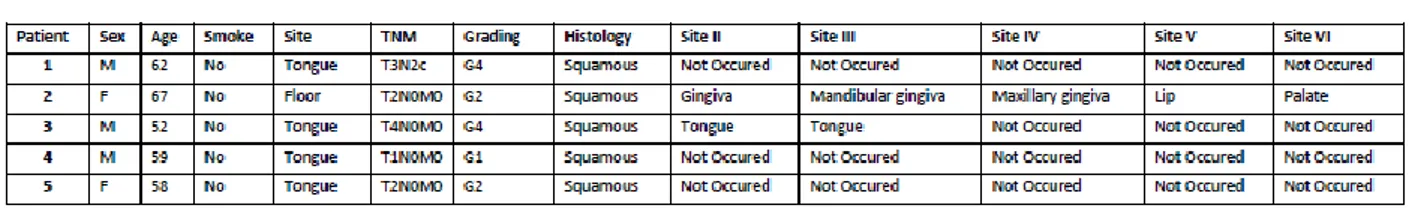
The study population comprised 16 women and 7 men aged 26–89 years with a mean age at second neoplastic event presentation of 63.65 ± 17.69.
Genetic classification of secondary OSCCs in LR, SPT and SFT Secondary tumours were genetically categorized as LR, SPT and SFT following the Braakhuis et al. classification(49) on the basis of the phylogenetic relationship between primary and secondary OSCC, and between the clinically and histologically normal mucosa distant from the index OSCC and
the mucosa distant from the secondary OSCC. Based on mtDNA results, cases were classified as
follows:
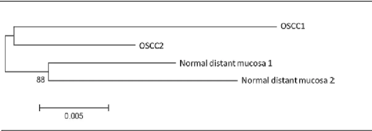
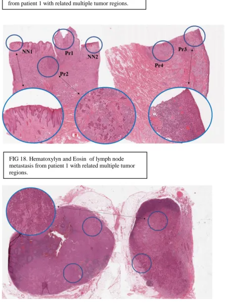
LR: when second manifestations were phylogenetically related to the index OSCC, and normal mucosa samples clustered together and were not genetically related to the index tumour or the recurrence. In our series seven second neoplastic events (30.4%) were phylogenetically related to the index OSCC. In all 7 cases the normal mucosa samples clustered together and were not genetically related to the index tumour or the recurrence (Fig. 12). On histology the LRs were not associated with epithelial precursor lesions (Fig. 15a).
SPT: when second manifestations were phylogenetically independent to the index OSCC, and normal mucosa samples clustered together and were not genetically related to the index or secondary OSCC. Four cases were classified as SPT as they presented a clonal relationship between the normal mucosa located distant from the tumour, sampled at the time of the primary
OSCC and the time of the second manifestation, whereas the two tumours were not clonally related, suggesting the occurrence of two genetically distinct neoplastic events (Fig. 13); SFT: when second manifestations were phylogenetically independent to the index OSCC and normal mucosa samples did not cluster together but may be genetically related to the index tumour or the recurrence. In such cases the genetic distance in normal mucosa suggested presence an altered genetic field. Twelve patients showed no genetic
relationship between the normal distant mucosa at the time of the primary OSCC and the normal distant mucosa at the time of the second manifestation, while both neoplastic events showed a clonal relation with the respective apparently normal mucosa. These features suggested a wide altered mucosal field, leading to a diagnosis of
On histology SPT (Fig. 15b) and SFT (Fig. 15c) were associated with areas of high grade squamous intraepithelial lesions (HG SIL)(82,94). In SFT cases the areas of HG-SIL were multiple.
The clinical and molecular profile of the second neoplastic event and related index OSCC are summarized in Table 6.
Results from Kaplan-Meier statistics
The log-rank test showed that the only independent prognostic factor related to a better survival rate (p < 0.05) was an altered mucosal field in non-clonal patients classifying the second neoplastic
manifestation as SFT; only 2/12 (16.6%) SFT events failed, compared to 5/7 LRs (71.4%) and 3/4 SPTs (75%) (Fig. 16). Results from Kaplan-Meier statistics were summarized in Table 7.
FIG 12 Phylogenetic tree of a case interpreted as local recurrence (LR). The secondary tumour (OSCC2) resulted phylogenetically related to the index tumour (OSCC1) and the
respective normal distant mucosa was phylogenetically related.
FIG 13 Phylogenetic tree of a case interpreted as second primary tumour (SPT). The secondary tumour (OSCC2) resulted phylogenetically distant from the index tumour(OSCC1) and the respective normal distant mucosa was phylogenetically related.
FIG 14 Phylogenetic tree of a case interpreted as second field tumour (SFT). The secondary tumour (OSCC2) resulted phylogenetically distant from the index tumour (OSCC1) and the respective normal distant mucosa did not show a phylogenetic relationship.
Fig.15 Histology: (a) LR is characterized by the presence of neoplastic cells (arrow) in the muscular wall, not related with the epithelium. (b) SPT is associated with HG-SIL.
The interface between non neoplastic oral epithelium and HG-SIL is indicated by the arrow. (c) SFT: the present case is a microinvasive OSCC (star). The surrounding mucosa
presents an area of HG-SIL (empty arrow); normally looking oral epithelium (black arrow) is interposed between microinvasive OSCC and HG-SIL.
Fig.16 Kaplan–Meier estimate for disease-free survival rate by genetic diagnosis of a second neoplastic event following the Braakhuis classification (LR, SPT or SFT). An altered mucosal field in non-clonal patients was the only prognostic factor related to a significantly better survival rate (p < 0.05). Indeed only 2/12 (16.6%) SFTs failed compared to 5/7 LRs (71.4%) and 3/4 SPTs (75%).
DISCUSSION:
mtDNA (D-loop) sequence analysis followed by NJ is a useful molecular method to assess tumour clonality [13–16,18–20]. In previous studies it was evaluated the reliability of mtDNA analysis in establishing the clonal relationship between paired neoplastic lesions in OSCC in comparison with the Hong classification based on clinical and histological criteria. Total agreement between mtDNA analysis and the Hong classification was found in 19/25 cases (76%). Specifically, complete agreement was achieved when mtDNA was compared with histopathological criteria, while discrepancies arose only in 6 cases in which the Hong classification was based only on the spatial or temporal distance of the second lesion(52). Subsequently it was evaluated the relationship between primary OSCC and lymph node metastasis in a series of patients with synchronous and metachronous metastases,
comparing mtDNA results with those obtained by another clonality test, i.e. TP53 sequence analysis. The results of TP53 and mtDNA analysis were consistent, showing that all neck metastases clonally related to the index tumour also shared similar mutations in the same
TP53 gene regions(53). Establishing a clonal relationship between a second neoplastic
lesion and the index tumour is not simply a problem of classification but yields new insights into the patient’s tumour biology and can influence the prognosis and treatment of the second lesion.
TAB 7. Univariate analysis for Potential Prognostic Variables related to disease specific survival. Entry in boldface and with asterisk indicate statistically significant P values
Few studies to date have analysed the difference in prognosis of patients with LR or SPT and SFT. Gonzalez Garcia et al. reported a lower survival rate in LR patients compared with patients with SPT(96) whereas Renmeno et al. found no differences in survival(97).
However, the two studies used different clinical and histological criteria to differentiate LR from SPT: Gonzalez Garcia et al. used the Hong classification(98) whereas Renmeno et al. used Warren and Gates’ criteria(48). To the best of our knowledge, this is the first study to analyse the prognosis of patients with multiple OSCCs following a classification based on molecular clonality assessment. The results revealed that the majority of second OSCC should be considered SFT, i.e. tumours phylogenetically independent from the primary and originating from a genetically altered mucosa. The present series found 12/23 SFT, 7/23 LR and 4/23 SPT. These results confirm the field cancerization theory that the mucosa
surrounding the primary OSCC mass is characterized by genetically altered epithelial cells that can escape clinical and histologic examination and might be responsible for cancer progression. This concept is further supported by the presence of multiple HG-SIL areas observed in cases of SFTs. The presence of a genetically altered filed surrounding OSCC, originally proposed by Slaughter et al.(5), has been widely confirmed by biomolecular approaches during the last two decades(99). Recently, Dasgupta et al. identified mitochondrial DNA (mtDNA) mutations in histologically negative resection margins of OSCC(100), confirming that even clinically and histologically radical surgical excision can lead to genetically altered mucosa capable of further neoplastic transformation. Kaplan Meier analysis of our results showed that these second manifestations had a better prognosis (16% failures) than LRs (71% failures) or SPTs (75% failures). The worse
prognosis of LRs with respect to SFTs in terms of survival is in accordance with the clinical data of Gonzalez Garcia et al.(96), while the higher failure rate of LRs is probably due to the difficulty of obtaining a radical excision of primary tumours when it is hard to identify