UNIVERSITÀ POLITECNICA DELLE MARCHE
Scuola di Dottorato di Ricerca della Facoltà di Medicina e Chirurgia
Corso di Dottorato in Salute dell’Uomo
Role of microRNAs in inflammaging and age related diseases
Dottoranda:
Docente tutor:
Angelica Giuliani
Prof.ssa Maria Rita Rippo
XXX Ciclo
Index
Summary ... 1
1. Introduction ... 5
1.1 Cellular senescence contributes to onset and maintenance of inflammaging ... 5
1.2 The hallmarks of cellular senescence... 7
1.2.1 Senescence markers: What defines a senescent cell? ...8
1.2.2 Senescence-associated secretory phenotype (SASP) ... 10
1.3 Mitochondria in aging cells and their role in inflammaging ... 15
1.3.1 Electron transport chain and mitochondrial ROS production ... 15
1.3.2 Mitochondria fuel Inflammaging through activation of receptors of the innate immune system 17 1.3.3 Mitochondria proteins: the potential role of Bcl-2 family members in regulating apoptosis and autophagy in aging cells ... 18
1.4 Epigenetic biomarkers of cellular senescence and aging ... 19
1.5 MicroRNAs ... 20
1.5.1 miRNA Biogenesis and Function ... 21
1.5.2 Inflamma-miRs ... 23
1.5.3 Cellular Senescence Associated (SA)-miRs ... 25
1.5.4 MitomiRs ... 27
1.6 A miRs subset belonging to inflamma-miRs, SA-miRs, and mitomiRs ... 28
1.7 Circulating microRNAs ... 31
3. Results and discussion ... 37
3.1 MitomiR-34a, -146a and -181a/Bcl-2 axis in senescence endothelial cells affects mitochondrial function and autophagy ... 37
3.1.1 Human senescent endothelial cells show altered mitochondrial activity and morphology ... 37
3.1.2 MiR-34a, -146a and -181a are up-regulated in senescent endothelial cells and modulate Bcl-2 expression ... 40
3.1.3 Overexpression of Mito-miR-146a, -34a and -181a induces mPTP opening, apoptosis and caspase-1 activation in young endothelial cells ... 42
3.1.4 Human senescent endothelial cells are more resistant to apoptosis than younger cells ... 44
3.1.5 Senescent endothelial cells show increased accumulation of autophagosomes and miR-34a, -146a and -181a modulate LC3 ... 45
3.1.6 Discussion ... 46
3.2 Anti-TNF-α treatment modulates SASP and SASP-related microRNAs in endothelial and circulating angiogenic cells... 49
3.2.1 Anti-TNF-α treatment of THP-1 cells ... 49
3.2.2 Anti-TNF-α treatment of HUVECs ... 51
3.2.3 Adalimumab treatment of sHUVECs affects their secretome ability to promote tumor cells migration ... 56
3.2.4 Effects of TNF-α inhibition in the secretome of senescent HUVECs on mammospheres assay 56 3.2.5 Effects of TNF-α inhibition on CACs from psoriatic patients ... 58
3.2.6 Discussion ... 59
3.3 MiR-21-5p and miR-126a-3p levels in plasma and circulating angiogenic cells: relationship with type 2 diabetes complications... 61
3.3.1 Discussion ... 67
4. Conclusion ... 70
5. Methods ... 73
HUVEC and THP-1 culture ... 73
Cell viability assay ... 74
Mitochondria isolation ... 75
Cell transfection ... 76
Cytokine production... 76
Psoriasis patients ... 76
CAC isolation and RNA extraction ... 77
Cultures and generation of MCF7 mammospheres (MS) ... 78
RNA isolation ... 78
Quantitative RT-PCR of mature miRNAs ... 78
Quantitative RT-PCR of mRNA ... 79
Protein extraction and immunoblotting ... 79
Flow cytometer analysis ... 80
TEM analysis ... 82
Patients ... 83
Laboratory assays ... 84
Statistical analysis ... 84
Summary
The elderly population is growing in the world due to increased life expectancy combined with declining birth rates. As people reach advanced age, they often face years of disability marked by multiple chronic diseases, poor mobility, and loss of independence. This burden of disability has caused many to worry about the impact of an aging population on social and economic stability. A huge challenge for modern biomedical research is to shorten as much as possible this period of frailty and disability to improve health span (Tchkonia et al., 2013) and to achieve “healthy aging” (Olivieri et al., 2017).
“Aging” is not a static but rather a dynamic phenotype that changes over time. Continuous interaction between individuals’ genetic makeup and environmental factors results in a spectrum of states that range from healthy aging to age-related disease. A pervasive feature of aging is a chronic and systemic pro-inflammatory status, termed “Inflammaging”. The source of this age-related systemic chronic inflammation was mainly attributed to the progressive activation of immune cells over time (Franceschi et al., 2000) even if today a variety of non-immune aging cells have been targeted as pro-inflammatory. Regardless of the cell types involved, there are growing epidemiological evidences that a state of bland inflammation is associated with and predicts several age-related diseases (ARDs), including
type 2 diabetes mellitus (T2DM) and its complications (Prattichizzo et al., 2016). Therefore, aging is the main risk factor for several pathological conditions.
Cellular senescence is historically associated with aging. However recently, the role of senescent cells in tissue remodelling during many physiological processes, including placental biology, embryonic patterning, wound healing, and tissue stress responses caused by cancer therapy has been suggested and it’s getting a lot of interest (Malaquin et al., 2016). Besides growth arrest, a significant feature of senescent cells is their ability to modify their immediate microenvironment using a senescence-associated (SA) secretome, commonly termed the SA secretory phenotype (SASP) (Coppè et al., 2008). This pro-inflammatory status of senescent cells could be a fundamental additional contributor to systemic inflammation. The identification of pathways that control age related inflammation across multiple systems is therefore important in order to understand whether treatments that modulate inflammaging may be beneficial in old people. Potential strategies for mitigating the deleterious effects of senescent cells include interfering with pathways that lead to senescence-associated growth arrest, eliminating senescent cells, and interfering with the adverse effects of senescent cells by targeting the SASP (Tchkonia et al., 2013).
Mitochondria dysfunction is deeply involved in the aging process and with the pathogenesis of major ARDs. Hallmarks of mitochondrial dysfunction include decreased efficacy of the respiratory chain and ATP production, decreased mitochondrial membrane potential, damaged cristae, swollen organelles, increased oxidative stress, and decreased mitochondrial DNA (mt-DNA) copy number. In addition to energy production, mitochondria play a central role in apoptosis, buffering calcium release, retrograde signalling to the nuclear genome, producing reactive oxygen species (ROS), participating in steroid synthesis, signalling to the
immune system, as well as controlling the cell cycle and cell growth (Coppotelli and Ross, 2016). Furthermore, mitochondria can promote inflammation releasing pro-inflammatory molecules and inducing inflammasome activation.
The identification of the pathways that control age related inflammation across multiple systems is therefore important because their modulation by specific treatments may be beneficial in old people. Recently, microRNAs (miRNAs or miRs) were largely investigated. MicroRNAs are small single-strand non-coding RNAs involved in gene expression that are increasingly recognized as epigenetic regulators of gene expression, modulating virtually all cellular processes, including senescence. MiRs can silence the mRNA target in most of cases by binding its 3’-untranslated region (UTR) in the cytoplasm, but they can also exert post-transcriptional control when bound to a region outside of the 3’UTR specifically within the 5’UTR and coding regions of the mRNA target. Moreover, in some cases, they activate transcription of a specific gene or stabilize the mRNA. A single miRNA has the ability to regulate multiple targets and, in turn, a single mRNA can be targeted by several miRs (Breving and Esquela-Kerscher, 2010). Over the years, a set of miRNAs with a well-recognized role in inflammaging, organismal aging, and development of ARDs has been defined (Olivieri et al., 2013; Olivieri et al., 2015); examples are miR-146a-5p, -21, -126a-3p. Interestingly, recent studies suggest that microRNAs can be found associated or inside mitochondria (mitomiRs) affecting their function, and that subset of these are SA-microRNAs (Bandiera et al., 2011). These miRs may play an important role in cellular senescence.
Interestingly, miRNAs can be released and also detected outside the cells. Recent evidence has confirmed that some features of aging are reflected in the profile of circulating miRNAs
and their shuttles (Olivieri et al., 2017). For these reasons, miRNAs are considered to be attractive candidates as diagnostic, prognostic and predictive biomarkers (Iorio and Croce, 2012), especially in ARDs such as T2DM (de Lucia C et al., 2017; Prattichizzo et al., 2015). The ARDs may well be characterised by molecule signatures whose identification would take us a little closer to discovering the biomarkers of health deterioration during aging. MiRNAs seem good candidates in this context.
1. Introduction
1.1 Cellular senescence contributes to onset and maintenance of
inflammaging
Aging is an inevitable outcome of life, characterized by a gradual decline in tissue and organ function and increased risk of disease and death (López-Otín C et al., 2013). It is a complex phenomenon that results from environmental, stochastic, genetic, and epigenetic events in different cell types and tissues and their interactions all the lifelong (Franceschi and Campisi, 2014). Human aging is characterized by a chronic, low-grade inflammation and this phenomenon has been termed “inflammaging”. Inflammaging is a systemic condition in the absence of overt infection (“sterile” inflammation) and it represents a highly significant risk factor for both morbidity and mortality in the elderly people, as most if not all age-related diseases share an inflammatory pathogenesis (Franceschi, 2000).
Acute inflammation can be beneficial: transient immune response is necessary to counteract harmful conditions such as traumatic tissue injury or an invading pathogen. However, acute inflammatory responses may be defective during aging. Many of the features of acute inflammation continue as the inflammation becomes chronic, but this one is usually of low
grade and persistent, resulting in responses that lead to tissue degeneration (Franceschi and Campisi 2014; Freund et al., 2011).
The number of senescent cells increases in multiple tissues with chronological aging, and senescent cells are present at sites of age-related pathology (Herbig et al., 2006; Lawless et al., 2010; Krishnamurthy et al., 2006; Jeyapalan et al., 2007). Senescent cells are usually cleared in normal tissues by the immune system (Kang et al., 2011). However, in aged person immunosenescence, characterized by the loss of function of immune system, compromises the clearance of senescent cells and exacerbates inflammation (Franceschi et al., 2000; Shaw et al., 2010).
Because the number of senescent cells increases with aging, it has been widely assumed that senescence contributes to aging. This claim is mainly sustained by data on animal models showing that periodic clearance of senescent cells, using genetic systems or drugs, is accompanied by a mean lifespan and healthspan extension, coupled with a reduced inflammatory gene expression in multiple tissues, including kidney and the heart (Baker et al, 2011). Moreover, the accumulation of senescent cells has recently been suggested to be a crucial factor contributing to systemic inflammation. Mechanistically, senescent cells likely fuel chronic inflammation because of acquisition a specific phenotype named “senescence-associated secretory phenotype” (SASP), characterized by the enhanced secretion of pro-inflammatory molecules and mediators (Tchkonia et al., 2013). Thus, senescent cells burden can modify the tissue microenvironment in a SASP-mediated manner and these spots are prominent sites of many age-related pathologies. Clearly, preventing development of the SASP or ameliorating its effects can mitigate the deleterious effect of senescent cells.
1.2 The hallmarks of cellular senescence
Cellular senescence can be defined as a stable arrest of the cell cycle coupled to continued viability, metabolic activity and stereotyped phenotypic changes (Campisi and d’Adda di Fagagna, 2007; Collado et al., 2007; Kuilman et al., 2010). More than 50 years ago, Hayflick and Moorhead observed that fibroblasts from healthy human donors had a limited ability to proliferate in culture. The non-dividing cells remained viable for many weeks, but failed to duplicate despite the presence of space and nutrients in the medium. When these cells became irreversibly arrested, they reached the so-called “Hayflick limit” (Hayflick and Moorhead, 1961). Today, a similar irreversible cell cycle arrest, now designated as “cellular senescence” can be triggered by a myriad of stimuli, including activation of oncogenes, telomere erosion or damage, mitochondrial deterioration, oxidative stress, and excessive DNA damage (Campisi and d’Adda di Fagagna, 2007; Collado et al., 2007; Kuilman et al., 2010; Baker and Sedivy 2013).
Telomeres are regions of repeat sequences, at the end of each linear chromosome, bound by multiple telomeric proteins. In mammalian cells, telomere DNA contains double-stranded tandem repeats of TTAGGG followed by terminal 3′ G-rich single-stranded overhangs. Telomere DNA probably adopt the T-loop structure, where the telomere end folds back on itself and the 3′ G strand overhang invades into the double-stranded DNA (the so-called D-loop). Functional telomeric structure prevents the degradation or fusion of chromosome ends, and thus is essential for maintaining the integrity and stability of eukaryotic genomes (Lu W et al., 2013). Due to the intrinsic inability of the replication machinery to copy the ends of linear molecules, telomeric DNA is subject to attrition during mitosis and the replication process can go on until a critical threshold of telomere length is reached.
Telomeric DNA is also highly prone to oxidative damage, and the increase in oxidative stress induces its shortening (Rhee et al., 2011; von Zglinicki, 2000). For these reasons, telomeres shortening is a widely used indicator of replicative senescence and cumulative genomic damage in somatic cells (Allsopp RC and Harley CB, 1995).
Dysfunctional telomeres, and non telomeric-DNA damage, trigger a classical DNA damage response (DDR), which imply the recruitment and activation of an apical DDR kinase like ATM (ataxia-telangiectasia mutated), to the damage site (d'Adda di Fagagna et al., 2003). In addition, compounds (e.g. histone deacetylase inhibitors), which relax chromatin without physically damaging DNA, activate the DDR proteins (Bakkenist and Kastan, 2003). The DDR is an evolutionarily conserved signaling cascade, which directs cell fate toward cell cycle progression, senescence or apoptosis. Both damage- and telomere-initiated senescence depend strongly on p53 and are usually accompanied by expression of p21 (Campisi and d’Adda di Fagagna, 2007). In addition to p53, replicative senescence is linked to p16INK4A (a cyclin-dependent kinase inhibitor). P16INK4A is encoded, together with ARF, by the INK4a/ARF locus. Indeed, activation of both the p53 and p16INK4A–RB pathways is essential for induction of senescence in a variety of human cell types (Kuilman et al., 2010).
1.2.1 Senescence markers: What defines a senescent cell?
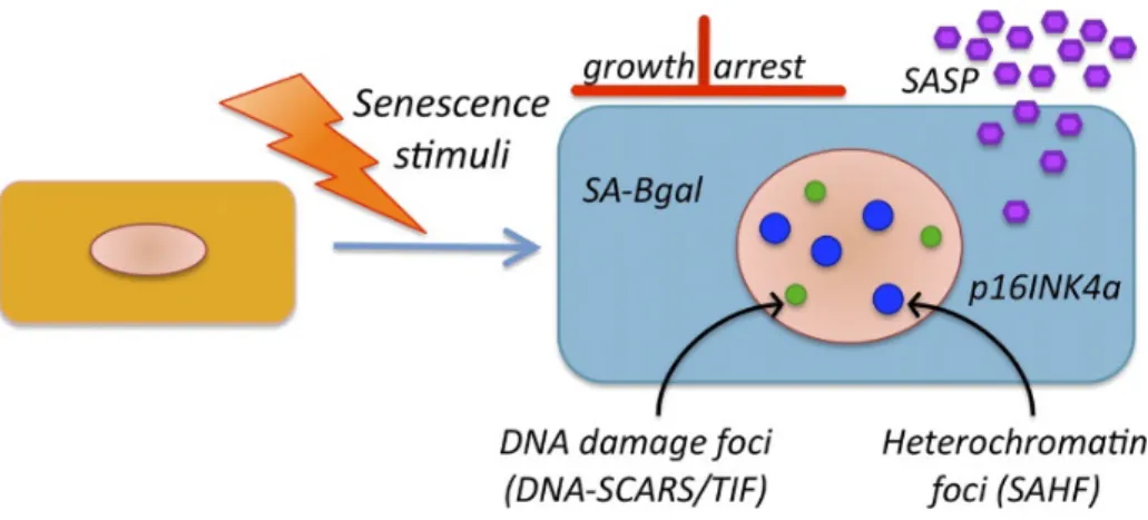
Senescent cells display several characteristics (Figure 1) which, together, can partially let us define the senescent state:
i. permanent growth arrest state, which cannot be reversed by known physiological stimuli;
ii. morphological changes, such as increased cell size and distinctive enlarged flat morphology;
iii. increased activity of SA–β-galactosidase (β-gal) (Dimri et al., 1995), which partly reflects the expansion of the lysosomal compartment, giving rise to an increase in β-gal activity that can be measured also at suboptimal pH 6 (Kurz et al., 2000); iv. activation, by most senescent cells, of p16INK4a (Alcorta et al., 1996);
v. appearance of persistent nuclear foci DNA segments with chromatin alterations reinforcing senescence (DNA-SCARS) and accumulation of SA heterochromatic foci (SAHF) (Salminen et al., 2012). These foci contain activated DDR proteins, including phospho-ATM and phosphorylated ATM/ataxia telangiectasia and Rad3 related (ATR) substrates (d’Adda di Fagagna et al., 2003). This leads to the local phosphorylation of multiple ATM substrates in the chromatin surrounding the DNA lesion, usually including γH2AX (a phosphorylated form of the histone variant H2AX);
Figure 1. Hallmarks of senescent cells. Markers of senescent cells include an essentially irreversible growth
arrest; expression of SA-β-gal and p16INK4a; robust secretion of numerous factors (SASP); and nuclear foci containing DDR proteins (DNA-SCARS/TIF) or heterochromatin (SAHF) (Rodier and Campisi, 2011).
vi. secretion of a plethora of proinflammatory factors, including growth factors, proteases, cytokines and chemokines, which have potent autocrine and paracrine activities (Coppé et al., 2008).
vii. altered susceptibility to apoptosis and un-efficient autophagy.
1.2.2 Senescence-associated secretory phenotype (SASP)
Cells undergoing senescence display dramatic alterations in their secretome, acquiring a typical secretory phenotype termed SASP (Coppé et al., 2008; Rodier et al., 2009). The SASP consists of a plethora of factors that includes cytokines (i.e. interleukin (IL)-1α/β, IL6, IL8), growth factors (i.e. EGF, FGF, VEGF), proteases (i.e. metalloproteinase MMP1, -2, and -3) and other non-soluble extracellular matrix proteins (i.e. collagens, fibronectin, laminin). Although a core of pro-inflammatory factors is a feature of all senescent cells, the specific composition of the SASP depends on the cell type and senescence inducer (Coppé et al., 2010). The SASP is a temporally regulated program. In culture, cells develop a full SASP five days later the senescence induction. Not all SASP factors start to be secreted at the same time (Coppé et al., 2008). Indeed, most of them are released in small amounts until several days after the induction of genotoxic stress. Accordingly, recent studies have shown that a certain number of secreted factors can be expressed much earlier than others, and can influence the subsequent expression of other SASP factors (Malaquin et al., 2016). In particular, pro-inflammatory cytokines IL-1α and IL-1β play critical role in the early phase of the SASP. Indeed, IL-1α acts as upstream regulator of IL-6/IL-8 cytokine network (Orjalo et al., 2009). Of note, during senescence, IL-1α and IL-1β are both secreted at low levels compared to IL-6 and IL-8 (Bhaumik et al., 2009; Coppe et al., 2008), which can act in an autocrine manner to reinforce the senescence growth arrest (Acosta et al., 2008). IL-1β
originates from its precursor pro-IL1β and it is activated to its mature secreted form after a proteolytic cleavage mediated by a component of the inflammasome complex, the caspase-1 (Galliher-Beckley et al., 20caspase-13). IL-caspase-1α acts intracellularly or as a cell surface-bound protein. Beyond the early phase of the SASP, IL-1α has been implicated in the upregulation of microRNA-146a/b, which are responsible for the inhibition of interleukin-1 receptor-associated kinase 1 (IRAK-1) and the resulting reduction in IL-6 in senescent fibroblasts naturally expressing high levels of IL-6 in their mature SASP. This mechanism suggests the possibility that a negative feedback loop may restrict the activity of the late mature SASP to prevent pervasive chronic inflammation, and that this loop may be established or alternatively controlled via microRNAs after the appearance of the mature late SASP signature (Bhaumik et al., 2009).
The SASP program is triggered by a variety of genotoxic senescence-inducing stress (e.g. telomere shortening or damage, cytotoxic drugs, radiation, oncogene activation, oxidative stress) (Coppé et al., 2008). DNA damage activates DDR, which allows for the recruitment and activation of ATM and subsequent DDR signaling cascade. Importantly, when DNA lesions are irreparable and a DDR signal is persistent, cells activate the senescence program and consequently all SA phenotypes, including SASP. Persistent DNA damage signaling is required for maintaining senescence-associated inflammatory cytokine secretion (Rodier et al., 2009). However, when a senescent-like phenotype is triggered in cells that overexpress cell-cycle inhibitors, such as p16 or p21, cells undergo a growth arrest with many characteristics of senescent cells, but fail to activate DDR and do not produce SASP (Coppé et al., 2010). Notably, although the DDR is important for both the SASP and SA growth arrest, the molecular regulation of these two processes is quite different, as p53 plays no role in establishing and maintaining the SASP despite its crucial function in regulating the SA
growth arrest (Coppé et al., 2008). Passos et al. proposed that a persistent DDR triggers continued production of reactive oxygen species (ROS), forming a dynamic feedback loop that actively maintains a deep senescent state (Passos et al., 2010).
However, canonical DDR signaling seems not be sufficient for the SASP. Another important pathway activated in response to cell stress is the mitogen-activated protein kinase p38 (p38MAPK). P38MAPK phosphorylation is important for the establishment of the senescence growth arrest due to its ability to activate both the p53 and pRb/p16 pathways. Freund and colleagues showed that p38MAPK activity is necessary and sufficient for the development of a SASP in cells induced to senesce by direct DNA damage or oncogenic RAS. In these contexts, p38MAPK is not activated quickly, but rather with delayed kinetics characteristic of the SASP (Freund et al., 2011).
These different signaling pathways (DDR, p38MAPK and IL-1 pathway) converge toward the activation of the nuclear transcription factor-κB (NF-κB), necessary for the expression of genes encoding SASP factors (Salminen et al., 2012) and CCA AT/enhancer–binding protein beta (C/EPBβ) (Acosta et al., 2008). Interestingly, these transcription factors were previously known to be involved in the regulation of cellular stress and inflammatory signals by promoting the expression of many cytokines (Pahl, 1999). Recently, another transcription factor - GATA4 - has also been identified as a senescence and SASP regulator via indirect activation of NF-κB. Indeed, GATA4 is degraded by p62-mediated selective autophagy, except during senescence, where its activation depends on ATM action (Kang et al., 2015). Interestingly, mTOR-inhibitor rapamycin, which has long been known to extend lifespan and health span in mice (Harrison et al., 2009) controls SASP protein secretion by enhancing IL-1α translation (Laberge et al., 2015). Recently, sensing of cytoplasmic chromatin by the
cGAS/STING pathway has been proposed as a trigger for SASP induction (Dou et al., 2017; Glück et al., 2017).
A recent work by Wiley et al. has shown that mitochondrial dysfunctions cause cell senescence and a distinct secretory phenotype termed MiDAS (mitochondrial dysfunction-associated senescence). Senescent cells with mitochondrial dysfunction do not secrete inflammatory components typical of the SASP, such as IL-6, IL-8 or IL-1β. The authors show that MiDAS was not caused by accumulation of nuclear DNA damage or oxidative stress but rather by a decreased ratio of NAD+/NADH and the resulting activation of AMP-activated protein kinase (AMPK), which in turn activates p53 (Wiley et al., 2015; Gallage and Gil, 2015).
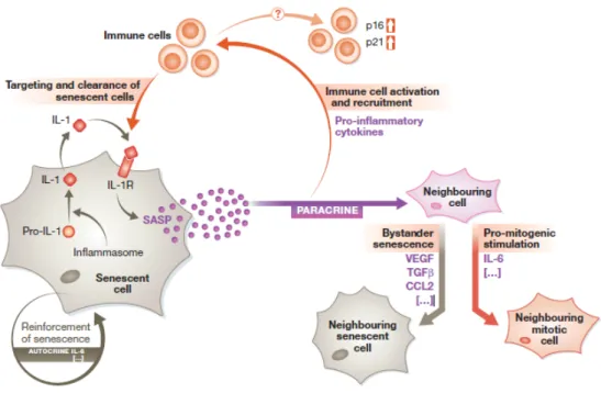
SASP can have a beneficial role in a variety of processes modifying the microenvironment, triggering immune surveillance and enforcing senescent cell cycle arrest in an autocrine and paracrine manners (Rodier and Campisi, 2011). In the physiological setting, senescent cells play an important beneficial role in embryonic development and in the organization of tissue repair responses that promote wound healing (Demaria et al., 2014; Muñoz-Espín et al., 2013; Storer et al., 2013). Moreover, SASP can play critical role in removing the transient senescent cells inducing immune-mediated clearance and modulating the differentiation of neighboring cells (Parrinello et al., 2005; Krizhanovsky et al., 2008). In fact, it has been suggested that senescent cells can also promote cellular plasticity and tissue regeneration (Ritschka et al., 2017). In the context of cancer, SASP also contributes to the clearance of damaged senescent tumor cells by enhancing both innate and adaptive immunity (Xue et al., 2007; Kang et al., 2011).
In contrast, the effects of the SASP could be deleterious: it can transmit cellular senescence on nearby healthy cells, thus representing a non-cell-autonomous mechanism (Acosta et al.,
2013). This paracrine effect could potentially explain persistent chronic inflammation, also known as inflammaging that contributes to multiple age-related phenotypes (Franceschi and Campisi, 2014). In the case of cancer microenvironment, SASP of senescent stromal fibroblasts can sustain tumor growth and invasion and can even promote long-term cancer therapy resistance (Krtolica et al., 2001). Thus, we can conclude that the ultimate outcome of SASP and in general of senescence within a tissue seems to be highly dependent on the context (Figure 2).
Figure 2. Cell autonomous and non-cell autonomous effects of cellular senescence. Stress stimuli can trigger
normal mitotic cells to go into senescence. This involves inflammasome-mediated activation of IL-1 signaling, which initiates the SASP response. The SASP acts cell autonomously (autocrine) to reinforce the senescent phenotype via cytokines such as IL-6. The SASP also acts non-cell autonomously (paracrine) to influence the cells in the surrounding environment. Paradoxically, the SASP can also exert pro-mitogenic stimulation of neighboring cells via cytokines like IL-6, which appear to play dual roles depending on the context. Furthermore, the SASP can act on the immune system via pro-inflammatory cytokines, leading to immune cell recruitment and subsequent targeting and clearance of senescent cells. Alternatively, the SASP can trigger upregulation of p16 and p21 levels on neighboring immune cells, the functional consequences of which are not yet so clear (Tasdemir and Lowe, 2013).
1.3 Mitochondria in aging cells and their role in inflammaging
Mitochondria play an essential role in energy generation, cell signaling, differentiation, death and obviously in the aging process. Any given mitochondrion is not a discrete, autonomous organelle. In fact, the identity of an individual mitochondrion is short-lived, because it will fuse with a neighboring mitochondrion in the near future. Therefore, the entire mitochondrial population is in constant flux, driven by continual fusion and division of mitochondria (Chan, 2006).
Recently, Correia–Melo et al. have shown that mitochondria are required for the pro-oxidant and pro-inflammatory phenotypes of senescent cells. A DDR pathway converging on mTORC1 phosphorylation promoted peroxisome proliferator-activated receptor gamma coactivator 1-beta (PGC-1β) dependent mitochondrial biogenesis, contributing to a ROS-mediated activation of the DDR and cell cycle arrest. Of note, the reduction in mitochondrial content in vivo, by either mTORC1 inhibition or PGC-1β deletion, prevented senescence in the aging mouse liver of the senescent phenotype (Correia-Melo et al., 2016).
1.3.1 Electron transport chain and mitochondrial ROS production
Mitochondria are the principal functional players of energy metabolism, as they provide most of the cellular energy through oxidative phosphorylation (OXPHOS). OXPHOS operates through five complexes, embedded in the inner mitochondrial membrane, that altogether represent the electron transport chain (ETC). The ETC is assembled from the genes of both the nuclear DNA and the mitochondrial DNA (mtDNA). The mtDNA is a circular, double-strand molecule, which encodes 13 polypeptides essential for OXPHOS, 2 rRNAs and 22 tRNAs for mitochondrial protein synthesis. Mammalian cells can have
hundreds to thousands of mitochondria, and each mitochondrion contains several mtDNA genomes. In physiological condition, during aerobic respiration, a variable percentage of electrons leaks from the ETC, particularly from complexes I and III, prematurely reduce oxygen and generate ROS (Bratic and Trifunovic, 2010). This process is exacerbated in senescent cells and leads to an over-production of ROS with concomitant ATP production decrease (Zwerschke et al., 2003). Low levels of ROS may improve systemic defense mechanisms by inducing an adaptive response, acting as signaling molecules. This concept is termed “mitohormesis”. On the contrary, high levels of ROS have been implicated into cellular damage and telomeres shortening hence promoting aging process (Ristow and Schmeisser, 2014; von Zglinicki et al., 2002; Parrinello et al., 2003). In the 50s, Harman with his Free Radical Theory of Aging (FRTA) theorized increased formation of ROS to be a major cause of aging (Harman, 1956). Because of mitochondria are the dominant source of cellular ROS, he extended his initial FRTA theory to the Mitochondrial Free Radical Theory of Aging (MFRTA) (Harman, 1972). One of the current opinions is that the relationship between ROS levels and aging is not always linear and oxidative stress is not
per se the major determinant of aging. Indeed, ROS-lowering interventions did not give
expected results (Sesso et al., 2012; Rautiainen et al., 2012) and as a matter of fact, as opposed to severe mitochondrial dysfunction, mild mitochondrial stress has an evolutionarily conserved anti-aging effect. However, excessive or uncontrolled free radical production can induce an inflammatory response, and free radicals are themselves inflammation effectors (Naik and Dixit, 2011). Thus, in aging cells, mitochondria-derived ROS and oxidative stress should be considered for their role as pro-inflammatory triggers rather than damaging molecules that progressively disrupt cell components and cellular homeostasis alone. Accordingly, ROS can partly mediate the senescence “bystander effect”,
i.e. the transition of the senescent phenotype from a senescent cell (SC) to neighbor cells (Nelson et al., 2017).
Even if ROS production is the best-known mitochondria biochemical change, other important phenomena occur which alter mitochondrial function. One of this concerns mitochondrial genome mutation: a general age-unrelated rule is the higher mitochondrial genome mutation rate than the nuclear genome (Trifunovic, 2006); Greaves and colleagues have shown that during aging selected pathogenic mutations increase and clonally expand but not the frequency of mutation (Greaves et al., 2014). Of note, mtDNA mutation accumulation in mouse tissues is influenced by the nuclear genetic background and correlates to neither cellular ROS content nor tissue senescence. MtDNA presents a number of genetic variants, called haplogroups. These haplogroups contain a functional, single-base polymorphism (SNPs) and can thus confer to their mitochondria a differential efficiency in the oxidative metabolism. Interestingly, they were associated with lifespan and longevity (Salvioli et al., 2006).
1.3.2 Mitochondria fuel Inflammaging through activation of receptors of the innate immune system
Mitochondria play a major role in inflammaging via not only ROS but also releasing several molecules inducing the inflammasome activation. This is partly due to the mitochondrial molecules conserved similarities to bacteria, as phylogenetically bacterial symbionts of early eukaryotic cells: mtDNA, N-formyl peptides and cardiolipin, a lipid present in the inner mitochondrial membrane, can act as damage-associated molecular pattern (DAMP)s activating pattern recognition receptors (PRRs), a large family of proteins that include
Toll-like receptors (TLRs) and NOD-Toll-like receptors (NLRs) (Franceschi et al., 2017). In particular, oxidized mtDNA and cardiolipin, released into the cytosol, could be recognized by Nlrp3 inflammasome, leading to IL-1β release (Shimada et al., 2012; Iyer et al., 2013). MtDNA is also recognized by TLR9, which senses DNA of bacterial and viral origin (Lamphier et al., 2006), inducing activation of NF-kB (Zhang et al., 2014). Further, mtDNA can even activate the cytosolic DNA sensor cyclic GMP-AMP Synthase (cGAS)/Stimulator of Interferon Genes (STING) pathway thus triggering expression of SASP factors (de Galarreta and Lujambio, 2017; Yuan et al., 2017).
1.3.3 Mitochondria proteins: the potential role of Bcl-2 family members in regulating apoptosis and autophagy in aging cells
Aging cell show an altered susceptibility to apoptosis and a reduction of autophagy efficiency. Consequently, cells accumulate dysfunctional organelles, which in turn contribute to the aging cell phenotype. Among the master regulators of both processes, the Bcl-2 family members should be considered. These proteins can be associated to the outer mitochondrial membranes. They are functionally described as either pro-apoptotic (e.g. BAX and BAK), which promote the mitochondrial outer membrane permeabilization (MOMP) or anti-apoptotic (e.g. Bcl-2, Bcl-xL and Mcl-1), which preserve outer mitochondrial membrane (OMM) integrity by directly inhibiting the pro-apoptotic proteins of the family. The balance of BCL-2 members and the regulation of their interactions dictate survival or commitment to apoptosis (Chipuk et al., 2010).
During programmed cell death MOMP allows soluble proteins (e.g. cytochrome c and the apoptosis initiating factor AIF) sequestered in the mitochondrial intermembrane space (IMS)
to diffuse into the cytosol. Cytochrome c release is a key event leading to activation of initiator caspase-9, cysteine proteases necessary for cell death, which then activates executioner caspases-3 and -7 (Chan, 2006).
In addition Bcl-2, Bcl-xL and Mcl-1 negatively regulates autophagy through the interaction with Beclin-1, which has a central role in this process (Liang et al., 1998).
Macroautophagy (or simpler autophagy) is a self-degradative mechanism that involves the engulfment of cytoplasm material and intracellular organelles within a double-layered membrane structure that then fuses with lysosomes (referred to as an autolysosome). Autophagy is a basic mechanism for cellular homeostasis that is important for balancing sources of energy in response to nutrient stress, in removing misfolded or aggregated proteins, clearing damaged organelles - such as mitochondria, endoplasmic reticulum and peroxisomes - as well as eliminating intracellular pathogens (Glick et al., 2010). Apoptosis and autophagy are often regulated by similar pathway and engage common sub-cellular sites and organelles, leading to the suggestion that these two processes are mechanistically linked. As both processes are altered in aging cells the intriguing idea of the role of Bcl-2 family member in cellular dysfunction during aging has been suggested in my laboratory (Rippo et al, 2014) and by others (Uraoka M et al., 2011).
1.4 Epigenetic biomarkers of cellular senescence and aging
The term epigenetics includes several phenomena such as DNA methylation, histone tail modifications, and microRNAs mediated mechanisms, which are able to mould the chromatin structure and/or gene expression levels, without altering the primary DNA
sequence (Stoccoro et al., 2016). Epigenetic markers can be changed by environmental influences and can then be passed on to daughter cells or via the germ line to offspring. Epigenetic modifications of chromatin and DNA have been recognized as important permissive and suppressive factors in controlling the expressed genome via gene transcription. A variety of changes in epigenetic biomarkers have been found in ARDs (Ospelt, 2016). Global DNA methylation is often lost in repetitive sequences, while in several specific promoter regions hyper-methylation is found. DNA methylation is achieved by the action of DNA methyltransferases. In addition, noncoding RNA can be considered as epigenetic markers and changes in microRNAs levels were observed during senescence and in ARDs (Olivieri et al., 2013). Understanding their emerging role as regulators of inflammaging and cellular senescence will enhance our knowledge of the molecular mechanisms involved in physiological and pathological aging and will provide new diagnostic tools and treatment options for patients with the major ARDs.
1.5 MicroRNAs
MicroRNAs are versatile regulators of gene expression. Since their discovery, thousands of miRNAs have been identified in humans, as well as in other species. In the human genome alone more than 2580 have been estimated miRNA genes (miRBase Registry, release 21.0). Online sources for sequences, such as the miRbase database (www.mirbase.org), were also made available since then. Recent evidence in the last year suggests that miRNA-mediated activities are surprisingly complex and that an intricate network of factors stringently control miRNA processing and biological activities in both negative and positive ways (Breving and Esquela-Kerscher, 2010).
1.5.1 miRNA Biogenesis and Function
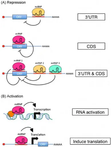
The microRNA biogenesis begins with genomic DNA transcription by RNA polymerase II (Pol II). RNA Pol II produces long pri-miRNAs from independent genomic transcription units or from the introns of protein-coding genes. Splicing is not required for production of miRNAs arising from introns. Furthermore, co-transcriptional processing of pri-miRNA into precursor miRNAs (pre-miRNAs) does not affect the splicing of the host pre-mRNA. In animals, the RNase III enzyme Drosha converts pri-miRNAs into pre-miRNAs, which are ~60 nucleotide stem–loop structures. Drosha acts as a component of a larger complex, the microprocessor, which includes a dsRNA-binding protein, named DGCR8 in mammals and Pasha in other animals. The nuclear transport receptor exportin 5 recognizes the ends and the stem of the pre-miRNA and exports the pre-miRNA from the nucleus to the cytoplasm via the nuclear pore. In the cytoplasm, the pre-microRNA is further processed by a second RNase III enzyme, Dicer, to double-stranded anti-parallel RNA (miRNA–miRNA*). This duplex is loaded into an Argonaute (AGO) protein. Subsequent maturation steps expel the miRNA*, producing a mature RNA-induced silencing complex (RISC). The RISC contains miRNA, an Argonaute protein and other protein factors. Finally, miRNA guides the RISC complex to complemental mRNA sequences to repress their expression (Bartel, 2004). The specificity of microRNAs is based on their mature sequence and is achieved through complementarity of target mRNAs with the “seed” region of the microRNA that is only six nucleotides in length. Typically, miRNA-binding sites of animal mRNAs reside in their 3’UTRs, resulting in down regulation of gene expression via translational repression and/or mRNA degradation (Ameres and Zamore, 2013). There is also increasing evidence that animal miRNAs can also exert post-transcriptional control when bound to sites located
outside of the 3’UTR and specifically within the 5’UTR and coding regions of the mRNA target (Duursma et al., 2008; Lytle et al., 2007; Tay et al., 2008). Moreover, accumulating evidences indicate that small RNAs bound to promoter regions in the nucleus can activate gene transcription, a phenomenon referred to as RNA activation (RNAa) (Janowski et al., 2007; Li et al., 2006) (Figure 3).
Figure 3: miRNAs can function to repress or activate target mRNA expression. (A) The canonical mode of miRNA-mediated repression occurs when a miRNA binds to mRNA regions of the 3’UTR, leading to down-regulation of target expression via translational repression and/or mRNA degradation. MiRNAs can also associate with binding sites located in the amino acid coding sequence (CDS) of mRNA transcripts to repress target expression (middle panel). Complex networks of distinct miRNAs can function together and bind to multiple miRNA complementary regions located both in the coding and non-coding regions of the mRNA target in order to fine-tune expression (bottom panel). (B) Recently, it has been shown that miRs can associate with incomplete complementarity to promoter elements of protein-coding genes and activate/enhance target transcription and protein expression via the phenomena RNA activation (RNAa) (top panel). Optimal promoter conditions (i.e., cofactors, chromatin structure) are apparently required for this activating effect to occur. MiRNAs can also induce protein translation by associating specifically with 5’UTR elements of mRNA targets (Breving and Esquela-Kerscher, 2010).
Of note, a single microRNA might control hundreds of distinct targets. These highly complex target networks pose a significant challenge to the mechanistic dissection of miRNA-mediated phenotypes. With such widespread control of gene regulation, miRNAs appear to have an influence on virtually every genetic pathway. Indeed, the growing number of miRNAs characterized to date reveals that they control a large range of essential biological processes related to cellular differentiation and growth, apoptosis, as well as aging, metabolic and immune responses.
1.5.2 Inflamma-miRs
The inflammatory response comprises complex biological reactions that require a fine-tuned integration between a range of immune system cell classes and an extensive network of biomolecules, which until recently had been thought to be largely cytokines. However, identification of the vast repertoire of miRNAs in the mammalian genome has completely revolutionized our understanding of most biological processes, including inflammation (Nilsen, 2007). Sensing of dangerous signals by the innate immune system involves a number of germline-encoded pattern recognition receptors (PRRs) that can detect both conserved pathogen- associated molecular profiles (PAMPs) expressed on microorganisms and altered endogenous ligands, mostly released by necrotic, senescent, and/or damaged cells (DAMPs). Among PRRs, toll-like receptors (TLRs) play a central role, since their engagement activates a potent pro-inflammatory pathway (Kawai and Akira, 2011). TLR signalling initiates from different adaptor proteins, such as myeloid differentiation factor 88 (MyD88) or TIR-domain-containing adapter protein-inducing interferon-β, which in turn activate several downstream pathways, leading to activation of NF-κB, mitogen-activated protein kinases (MAPKs), and members of the interferon regulatory factor family.
Fine-tuning of TLR signalling prevents generation of harmful and inappropriate inflammatory responses without lowering the surveillance for potentially dangerous signals. Deregulation of the whole network can have destructive effects and lead to tissue damage: this is a hallmark of chronic inflammation, which is often associated with ARDs development and progression (Olivieri et al., 2013a and b). A mounting body of evidence has been documenting a relatively small number of miRNAs that are involved in regulating inflammation: their prototypes are miR-155, miR-21, and miR-146a (Quinn and O’Neill, 2011), after referred to as inflamma-miRs. In physiological conditions, transcription of miR-155, miR-21, and miR-146a is at baseline levels; however, initiation of pro-inflammatory TLR signalling immediately results in strong co-induction of their expression through a mechanism that is largely NF-κB-dependent (Boldin and Baltimore, 2012). Although the importance of inflamma-miRs in innate immune response regulation is widely accepted, the molecular mechanism of their action has proved to be highly complex. Early investigations disclosed that miR-146a acts as a negative regulator of TLR signalling by targeting both tumour necrosis factor receptor-associated factor 6 (TRAF6) and IL-1receptor-associated kinase 1 (IRAK-1) (Taganov et al., 2006). MiR-21 was found to down-regulate the expression of IRAK and MyD88, as well as of programmed cell death protein 4 (PDCD4), switching the cell program from pro-inflammatory to anti-inflammatory, mainly as reflected by IL-10 production (Sheddy et al., 2010). Overall, these finding have generated a model where inflamma-miRs operate as a negative feedback loop to protect the organism against overwhelming inflammation. Subsequent research has disclosed that inflamma-miRs could play a dual function, inhibiting as well as inducing TLR signalling. This is the case of miR-155 and miR-21, which can function as an agonist of single-stranded RNA-binding TLRs and can therefore induce NF-κB activation and secretion of inflammatory molecules (Fabbri
et al., 2012). Although inflamma-miRs are co-induced during TLR signalling, several observations suggest that they do not act redundantly and simultaneously, but rather cooperate to control TLR signalling through functionally different performances. Such coordination of inflamma-miRs could go beyond suppression of TLR signalling and contribute to regulating the immunity/inflammation balance. Knockout mouse models have been instrumental in shedding light on the role of inflamma-miRs in inflammation and inflammation-related diseases. Transcriptome analysis of bic/miR-155-deficient CD4+ T cells in mice identified a wide spectrum of miR-155-regulated genes, including cytokines, chemokines, and transcription factors, suggesting that bic/miR-155 plays a key role in immune system homeostasis and function (Rodriguez et al., 2007). Knockout of miR-146a gene in C57BL/6 mice involves increased transcription of NF-κB-regulated genes (Zhao et al., 2011). These animals also develop myeloid sarcomas and lymphomas as well as chronic myeloproliferation in bone marrow. Genetic ablation of NF-κB p50 suppresses the myeloproliferation, demonstrating that NF-κB dysregulation is responsible for the myeloproliferative disease (Zhao et al., 2011). Details on the role of inflamma-miRs in controlling TLR signalling are just beginning to be explored, and further investigations are warranted to gain insights not only into their individual contribution to the homeostasis of the innate immune response, but also into the consequences of their deregulation in conditions characterized by chronic inflammation.
1.5.3 Cellular Senescence Associated (SA)-miRs
As mentioned above, several miRNAs have been shown to be involved in the regulation of pathways implicated in cellular senescence and exert important effects on cell cycle progression.
A particular aspect concerns p53, a critical regulator of cellular senescence. It enhances the post-transcriptional maturation of several miRNAs with growth-suppressive function (Suzuki et al., 2009; Liu et al., 2012; Munk et al., 2017) and it can be itself a target of microRNAs (Park et al., 2009; Inomata et al., 2009). P53 activation can be regulated by the NAD-dependent deacetylase Sirtuin-1 (SIRT-1), a gene that regulates cellular senescence and limits longevity, whose expression is under miR-34a. The miR-34 family regulates cell cycle progression, cellular senescence and apoptosis. One of the best-characterized SA-miR is just miR-34a. Expression of miR-34a leads to reduced SIRT1 levels, lead to an increase in p53 acetylation and activation. P53 in turn promotes the expression of the miRNA family miR-34; this creates a positive p53-activating feedback loop leading to increased p21 levels, thereby triggering senescence (Yamakuchi and Lowenstein, 2009).
Moreover, it has been demonstrated that miRs can also modulate p16 expression and telomere length (Lal et al., 2008; Benetti et al., 2008) and that miR-20a and the other miRs of the same cluster (miR-17-92 cluster) contribute to regulating cell cycle progression (Pickering et al., 2009; Cloonan et al., 2008; Ivanovska et al., 2008). Interestingly, ablation of miRNAs expression by knockdown of Dicer has been shown to result in the induction of premature senescence phenotype in embryonic fibroblast through activation of the p53 pathway (Mudhasani et al., 2008). Finally, several miRs that appear to be modulated during cellular senescence and aging are found in a range of ARDs, including cardiovascular diseases (CVD) and neurodegenerative diseases (Dimmeler and Nicotera, 2013).
1.5.4 MitomiRs
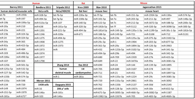
Recently, through different experimental approaches in different mammalian species, a number of studies allow identification of “signatures” of miRNAs located in the mitochondria (Table 1). These miRs were collectively called “mitomiRs”. Indeed, this subset includes both nuclear-encoded miRNAs that translocate into the mitochondria and target either mitochondrial or nuclear mRNA and a small number of mitochondrial-encoded microRNA (mit-mitomiRs). Early studies on the subject were mostly descriptive to detect the presence of miRNAs within the mitochondrion (Giuliani et al., 2017b). Kren and co-workers first detected 15 nuclear-encoded mitomiRs from adult rat livers that seemed to be involved in the modulation of genes associated with apoptosis, cell proliferation, and differentiation. They also postulated that given the central role that mitochondria play in apoptosis, they might serve as reservoirs of select miRNAs that may modulate these processes in a coordinate fashion (Kren et al., 2009). A subsequent study identified a pool of 20 miRs highly expressed in mitochondria of mice liver. Interestingly, mitochondria have a unique population of miRNAs, independent of the total cellular abundance of miRs: the miRs that were highly abundant in mitochondria were not those that were highly expressed in liver (Bian et al., 2010). For the first time, in 2011 Barrey and colleagues showed the presence of pre-miRs inside mitochondria, postulating that here some pre-miRNA sequences could be processed to mature miRNAs, which could be immediately active on the mitochondrial transcripts or exported in the cytosol in order to interfere with genomic mRNA (Barrey et al, 2010). Subsequently, other groups identified new mitomiRs from different tissue and cell types (Bandiera et al., 2011b; Mercer et al., 2011; Sripada et al., 2012; Dasgupta et al., 2015; Jagannathan et al., 2015). Overall these data indicate that
mitochondria have a discrete pool of mitomiRs and the association of miRs with mitochondria is species and cell type-specific (Geiger and Dalgaard 2017).
Table 1. List of miRs found within mitochondria (extracted from Giuliani et al, accepted for publication in
Mediators of Inflammation, 2017)
1.6 A miRs subset belonging to inflamma-miRs, SA-miRs, and mitomiRs
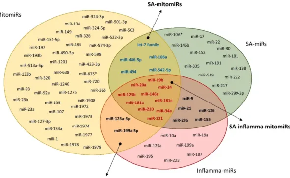
Since mitochondria play a key role in aging, it is reasonable to surmise that some mitomiRs are involved in the process, and that the most likely candidates should be found among the miRs involved both in aging and in inflammation (Rippo et al., 2014). Figure 4 shows a Venn diagram illustrating the intersections of the three different sets of miRNAs (inflamma-miRs, SA-miRs and mitomiRs). These three subtypes of miRs share a discrete pool of miRs (SA-inflamma-mitomiRs) that includes 10 microRNAs (19b, 20a, 24, miR-34a, miR-125b, miR-146a, miR-181a and miR-181c, miR-210, and miR-221).
Rat
Bandiera 2011 Sripada 2012 Kren 2009 Bian 2010 HeLa cells HeLa/HEK293 Rat liver mouse liver
let-7b miR-193b miR-328-5p let-7b-5p miR-130a-3p let-7f-5p let-7b miR-151-3p miR-1934-3p miR-130a miR-574-5p let-7g miR-197 miR-494-3p let-7g-5p miR-130b-3p miR-101-5p let-7a miR-203-3p miR-211-3p miR-497 miR-148a-3p miR-19b miR-199a-5p miR-513a-5p miR-107 miR-140-5p miR-122-5p let-7c miR-212-3p miR-3072-3p miR-188-5p miR-200c-3p miR-20a miR-210 miR-638 miR-181a-5p miR-320-3p miR-181b-5p let-7f miR-5112 miR-320-3p miR-3098-5p miR-300-3p miR-23a miR-221 miR-1201 miR-221-5p miR-494-3p miR-181d-5p miR-149-3p miR-135a-1-3p miR-1199-5p miR-30c-1-3p miR-181b-5p miR-23b miR-324-3p miR-1246 miR-320a miR-671 miR-188-5p miR-149-5p miR-721 miR-5108 miR-712 miR-5131 miR-24 miR-324-5p miR-1275 miR-494-3p miR-29a-3p miR-23b miR-125a-3p miR-375-3p miR-3102-5p miR-34a miR-365 miR-1908 miR-1275 miR-29c-3p miR-1 miR-1904 miR-203-3p miR-877-3p miR-92a miR-423-3p miR-1972 miR-1973 miR-361-5p miR-29a miR-1894-3p miR-126-3p miR-3963 miR-93 miR-484 miR-1973 miR- miR-432 miR-125b-5p miR-3102-5p miR-26a miR-341-3p miR-103 miR-486-5p miR-1974 miR-494-3p miR-29b miR-494 miR-23a miR-342-3p miR-106a miR-490-3p miR-1977 miR-680 miR-709 miR-1939 miR-27b miR-423-3p miR-107 miR-503 miR-1798 miR-689 miR-22 miR-3470a miR-99a miR-3081-5p miR-125b miR-501-3p Zhang 2014 Das 2012 miR-690 miR-24 miR-144 miR-139-3p miR-1895 miR-125a-5p miR-532-3p human rat miR-705 miR-680 miR-3107 miR-378 miR-720 miR-127-3p miR-542-5p skeletal muscle cardiomyocites miR-711 miR-21 miR-451 miR-27a miR-1897-5p miR-133b miR-574-3p miR-1 miR-181c miR-721 miR-133a-3p miR-1224 miR-29c miR-3085-3p miR-133a miR-598 Mercer 2011 miR-720 miR-133a-5p miR-2861 miR-30a miR-3092 miR-134 miR-720 143B cells Dasgupta 2015 miR-762 miR-133b miR-2137 miR-30d miR-2145 miR-149 miR-1974 miR-16 206 ρ° cells miR-805 miR-128-3p miR-1937c miR-30e miR-652-5p miR-151-5p miR-1979 miR-146a miR-181c miR-671-5p miR-3095-3p miR-466i-5p miR-3082-5p miR-1187 miR-181a miR-675* miR-103 mR-146a miR-1982-5p miR-1937b miR-705 miR-483-5p miR-466h-3p
human skeletal/muscular cells mouse heart
Human
Jagannathan 2015
Mouse
Figure 4. Venn diagram showing the mitomiRs involved in both aging and inflammation. Mitochondria-related
(mitomiRs), senescence-associated (miRs) and inflammation-related (inflamma-miRs) miR sets. SA-mitomiRs: miRs found both in the mitomiR and the SA-miR set; SA-inflamma-SA-mitomiRs: miRs found in all three sets (modified by Giuliani et al., 2017b).
In our laboratory, by using the bioinformatics tools Ingenuity Pathway Analysis (IPA) (www.ingenuity.com) the hypothesis that one role of SA-inflamma-mitomiRs may be the control mitochondria-associated protein expression was tested. They limited the research approach to human tissues and cell lines and to cytosolic targets observed experimentally, though not necessarily validated. This approach demonstrated that most SA-mitomiRs are involved in controlling the expression of at least one Bcl-2 family member (Rippo et al., 2014). Literature has also shown that several SA-mitomiRs e inflamma-mitomiRs can modulate Bcl-2 family members.
The above considerations suggest i) that in aging cells modulation of several mitomiRs should result in Bcl-2 deregulation and consequently alteration of Bcl-2-mediated processes
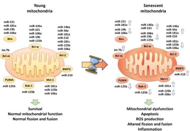
and ii) the hypothesis that SA-mitomiRs could influence mitochondrial function and, as a consequence, may promote ROS production and inflammation (summarized in Figure 5).
Figure 5. SA-mitomiRs and SA-inflamma-mitomiRs appear to be involved in controlling the expression of one or more Bcl-2 family member. In young HUVECs mitomiRs may control the expression of Bcl-2 family
members, resulting in their fine tuning and optimum function at the mitochondrial level. In senescent HUVECs, mitomiR deregulation may affect the balance of Bcl-2 family members, resulting in mitochondrial dysfunction (Giuliani et al., 2017b).
This hypothesis is supported by experimental evidence that 34a, 181a and miR-146a control Bcl-2 expression (Yang et al. 2014; Ouyang et al., 2012; Zhang F et al., 2015). In addition, IPA of “molecular and cellular functions” identified the main functions in which these SA-mitomiRs are involved. Most are among those affected by aging: cell cycle, cell growth and proliferation, survival, function, and maintenance. These data suggest that SA-inflamma-mitomiR deregulation, by altering these key functions, should be associated with
cell dysfunction and disease. Indeed, IPA of miR-146a, miR-34a, and miR-181a in relation to the pathway “diseases and disorders”, showed their association with several important age-associated diseases, including cancer, CVD, and neurological and metabolic disorders (Rippo et al. 2014). The authors proposed a model of how a mitomiRs subset (miR-181a, miR-34a and miR-146a) may control mitochondrial function/dysfunction and inflammation during cell aging via modulation of Bcl-2 family members.
1.7 Circulating microRNAs
MiRNAs are not confined within the cell. Accumulating evidence indicates that miRNAs are secreted in extracellular spaces in microvesicle-encapsulated form or are released in vesicle-free form and can enter into target cells. To move into extracellular circulatory biofluids, they take advantage of different systems:
1) miRNAs are bound to high-density lipoprotein (HDL) particles in a non-vesicle form,
2) they form complex with Ago2 proteins, 3) they are placed in exosomes,
4) they are encapsulated in micro-vesicles (MVs),
5) miRNAs accumulate in apoptotic bodies (Kumar and Reddy, 2016).
All miRNA transport strategies described to date allow communication between cells found in different organs. Exosomes may be the simplest and most robust way to realize a systemic miRNA-based signal network. However, little is known of how miRNA species are sorted into exosomes and which miRNA binding proteins are involved. HDL is another well-established circulating miR carrier, shuttling the potent gene regulators to distant tissues.
The report that miRNAs carried by HDL may be altered in disease states had further broadened our understanding of the complex effects that the lipoprotein can exert on target cells and tissues. The delivery of lipid-associated miRNAs to recipient cells is achieved by various routes, including endocytotic uptake, membrane fusion, and scavenger receptors. Circulating miRNA levels can be used as biomarkers for the major age-related diseases with important diagnostic and prognostic implications but their clinical relevance is still controversial (Cortez and Calin, 2009; D’Alessandra et al., 2010; Zampetaki et al., 2012; Olivieri et al., 2013c).
2. Aim of the work
Aging is one of the major risk factor for pathologies characterized by chronic inflammation status such as cardiovascular diseases, T2DM and neurodegenerative diseases – commonly termed ARDs. The understanding of mechanisms that regulates these inflammatory processes is one of the main target of the current scientific research to both prevent aging related pathologies and improve their outcome.
Non-coding nucleic acids, among which miRNAs represent one of the most studied category, contribute to these mechanisms by controlling and fine tuning protein expression. Since miRNAs expression can be regulated, they represent an intriguing opportunity as targets of new treatments; indeed, combinatorial therapies with conventional drugs and miRNA- or anti-miRNA-treatments are already in progress (van Beijnum, et al., 2017). Here three different aspects for which miRNAs are involved in cellular senescence, Inflammaging and ARDs were investigate by addressing three specific aims:
1. Assess whether age-related mitomiRs might play a direct role in controlling mitochondrial function by regulating mitochondrial protein expression.
Current research suggests that a large number of microRNAs are involved in the regulation of mitochondrial activities (mitomiRs) and some of them are modulated during senescence and inflammatory processes. Mitochondria have long been recognized for their canonical role in cellular respiration and oxidative phosphorylation. However, it is becoming increasingly evident that they represent key participants in sensing and integrating signal from the environment to trigger adaptive and compensatory responses in cells (Aon and Camara, 2015). Thus, acting as pleiotropic organelles, mitochondria might regulate aging and age-related diseases on several aspects and appear as indispensable for the pro-inflammatory and the pro-aging features of senescent cells (Correia-melo et al., 2015). Understanding the multitude of mechanisms that lead these organelles to be the “bad” players of aging could shed light on new potential druggable pathways. We focused on three mitomiRs (miR-34a, -146a and 181a) that can target Bcl-2 in different cellular systems and pathological context and that may be linked each other and to mitochondrial activity by an intriguing molecular network (Rippo et al., 2014). The aim of my PhD study was then to assess whether aging-related mitomiRs might play a direct role in controlling mitochondrial function by regulating mitochondrial protein expression. Their modulation could thus mediate the loss of mitochondrial canonical function in aging cells, inducing or contributing to the inflammatory response and to age-related diseases.
2. Evaluate whether and how prevention of TNF-α activity can restrain SASP.
Several miRNAs have been associated to the SASP phenotype. SASP attenuation, SASP factors modulation and selective senescent cells removal or killing are all potential strategies (senotherapies) for mitigating the deleterious effects of senescent cells. Highly promising results are coming from work on SASP suppressor and senolytic agents (Childs et al., 2015;
Chang et al., 2016). The second aim of this study was thus to evaluate whether and how adalimumab, a monoclonal antibody directed against tumor necrosis factor-α (TNF-α), a major SASP component, can prevent the SASP of endothelial cells in replicative senescence. The ability of adalimumab to restrain or delay SASP acquisition was tested by evaluating two of the senescence/SASP-associated miRNAs (miR-126-3p and miR-146a-5p) and the respective targets (Spred1 and Irak1).
3. Explore the potential of these a subset of circulating miRs as biomarkers of T2DM complications.
Increasing interests toward miRNA is justified by their role as reliable circulating markers of aging related disease. They have also the advantage to be easily accessible, rapidly analysable, highly stable and resistant so that their use for diagnostic and prognostic purpose could reduce the necessity of invasive procedures in patients introducing the concept of “liquid biopsy”. One of the ARDs, which arouses major interest for innovative diagnostic/prognostic biomarkers, is T2DM. T2DM is a chronic multi factorial metabolic disease, caused by environmental and genetic factors. A meta-analysis of randomised controlled trials has demonstrated that the benefit-risk ratio of intensive glucose lowering treatment, administered to prevent diabetic vascular complications, remains uncertain (Boussageon et al., 2011).This suggests that the parameters currently used to monitor T2DM progression do not adequately predict the probability of developing complications; therefore, innovative biomarkers are required to manage patients with T2DM and its severe complications. Circulating miR-126-3p levels have been associated with endothelial function (Wang et al., 2008) and circulating miR-21-5p with systemic inflammatory status (Olivieri et al., 2013a), two conditions that are closely related to glycaemia control. The last
aim of this work was therefore to explore the potential of these two miRs as biomarkers of T2DM complications.
3. Results and discussion
3.1 MitomiR-34a, -146a and -181a/Bcl-2 axis in senescence endothelial
cells affects mitochondrial function and autophagy
3.1.1 Human senescent endothelial cells show altered mitochondrial activity and morphology
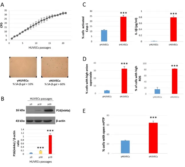
Senescent cells show altered phenotype and function. Among the subcellular compartments, mitochondria undergo profound morphological and biochemical changes reflecting their dysfunction. To characterize mitochondrial activity during senescence, we used replicative senescence of human primary endothelial cells (HUVECs) as a model. HUVECs were sub-cultured until complete growth arrested (Figure 6A, upper panel).
To confirm the acquisition of the senescent phenotype at later passages (Cell population doubling, CPD>25) β Gal staining was performed: senescent cells displayed a high SA-β Gal positivity (Figure 6A, bottom panels). In addition, p16(Ink4a), the prototypical protein characterizing senescent cells, showed a significant increase in both intermediate and senescent cells compared to younger cells (Figure 6B). We also evaluated the increase of Caspase-1 (Casp-1) activity and the production of its substrate IL-1β, a cytokine typically released by senescent cells and that characterizes the acquisition of SASP (Figure 6C).
Figure 6. Characterization of endothelial cells in replicative senescence. (A) Growth curve of three different
pool oh HUVECs - Y-axis: Cumulative Population Doubling (CPD); X-axis: cell passages from P1 to P21; Cells passages (upper panel) two representative photos of SA-β-Gal staining of young and senescent HUVECs. Cells with percentage of β-Gal <10% were considered young cells (yHUVECs), while those with % of SA-β-Gal > 60% were identified as senescent cells (sHUVECs). (B) P16(ink4a) protein level in young (P3), intermediate (P10) and senescent (P19) HUVECs. (C) Percentage of HUVECs with active Casp-1 and IL-1β concentration (pg/ml) in culture media. (D) Percentage of HUVECs with high levels of anion superoxide (MitoSOX) and ROS (DFCDA). (E) Percentage of HUVECs with open mPTP. *** p<0.001 intermediate or senescent cells vs young cells. Bar graphs indicate means±SD of three independent experiments.
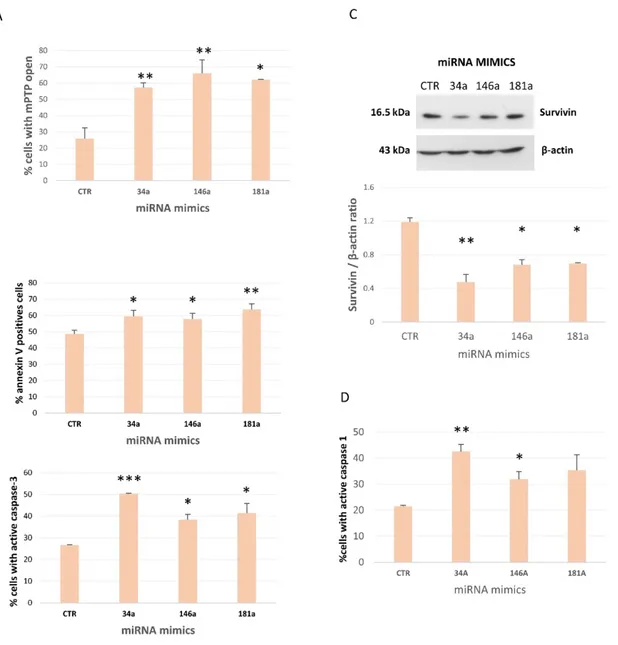
Furthermore, ROS and superoxide anion, markers of oxidative stress, increased in senescent (sHUVECs) compared to younger HUVECs (yHUVECs) (Figure 6D). As ROS in young cells are directly related to the opening of the permeability transition pore (mPTP) and both, in turn have a central role in in alterations of mitochondrial structure (Batandier et al., 2004),
we assayed for the first time this biochemical event. As shown in Figure 6E more than 60% of sHUVECs have got mPTP opened compared to 20% of yHUVECs. Altogether, these data support the assumption that in senescent cells mitochondria show altered biochemical functions.
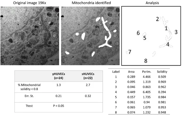
Finally, we evaluated mitochondrial morphology in young and senescent HUVECs in order to provide evidence on their dysfunction. Indeed, mitochondrial perimeter positively correlates with mitochondria about to undergo a fission event. Similarly, mitochondrial solidity (compact shape) positively correlates with mitochondria about to undergo a fusion event (Westrate et al., 2014). TEM analysis showed that sHUVECs have higher perimeter and lower solidity than younger cells (Figure 7). We also measured the area, a third morphological feature that positively correlates with mitochondrion propensity to fragment.
Figure 7. TEM analysis of mitochondrial morphology in endothelial cell. A representative image of
mitochondria in a senescent cell (upper panel). Mitochondrial Area, Perimeter and Solidity are measured (lower panel).
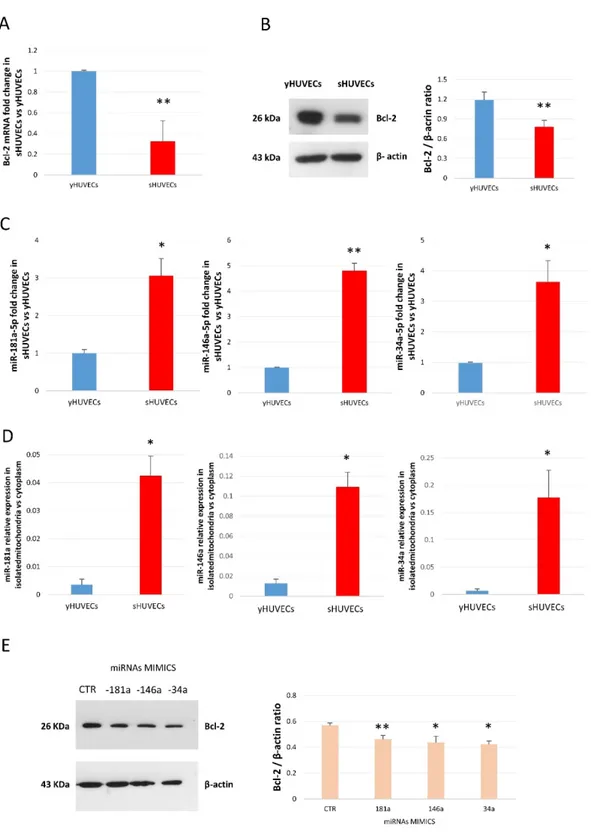
3.1.2 MiR-34a, -146a and -181a are up-regulated in senescent endothelial cells and modulate Bcl-2 expression
Bcl-2 family members are regulators of mitochondrial physiological functions; therefore, the expression of Bcl-2 protein, the master of the family, has been analyzed in both young and senescent cells. Bcl-2 showed a consistent down-regulation during senescence process (Figure 8 A and B). MiR-34a, miR-146a and miR-181a can modulate Bcl-2 and other Bcl-2 family members (Ouyang et al., 2012; Yang et al., 2014, Zhang et al., 2015), thus we hypothesized a role of these three miRs in Bcl-2 modulation during senescence (Rippo et al., 2014). Here we show that beside the already demonstrated up-regulation of miR-146a (Olivieri et al., 2013c), senescent HUVECs express higher miR-34a and miR-181a levels (Figure 8C) compared to younger cells. Then, to corroborate our hypothesis that these miRs could be defined mitomiRs in our experimental model, we have analyzed their expression in isolated mitochondria from young and senescent cells. All three miRs were found expressed within mitochondria and notably, their levels were increased in sHUVECs mitochondrial fraction compared with the cytoplasmic one (Figure 8D).
In order to demonstrate a relationship between miR-34a, miR-146a and miR-181a and Bcl-2, yHUVECs were singularly transfected with the corresponding miRNA mimic. After 24 hours, over-expression of all the three mature miRs resulted in Bcl-2 down-regulation: 34a induced the more relevant decrease (from 100% of control to 60%), followed by miR-146a (from 100% to 65%) and by miR-181a (from 100% to 80%) (Figure 8E).