UNIVERSITÀ DEGLI STUDI DI NAPOLI
“FEDERICO II”
PhD Thesis
“Analysis of differential gene expression in the aging
brain:
the case study of Col4a1, Col25a1, and Id3 and their
involvement in the adult neurogenesis of the
short-lived teleost Nothobranchius furzeri”
Coordinator Prof Giuseppe Cringoli Author Dr Adele Leggieri Tutor Prof Luigi Avallone
“Love is always patient and kind. It is never jealous. Love is never boastful or conceited. It is never rude or selfish. It does not take offense and is not resentful. Love take no pleasure in other people’s sins, but delights in the truth. It is always ready to excuse, to trust, to hope, and to endure
whatever comes" (St Paul to the Corinthians, 1th letter, 13,1-13)
Contents Acknowledgment...11 Abbreviations ...14 Figures ...18 Tables ...20 Abstract ...21 Introduction ...22 Bibliography ...25
1. N. furzeri: an extremely short-lived vertebrate...30
1.1. Phylogeny ...30
1.2. Sexual dimorphism, male color polymorphisms and sex determination ...31
1.3. Distribution and habitat ...33
1.4. Embryos development and life cycle ...36
1.5. Diet ...39
1.6. Housing ...40
1.7. Genetic structure ...43
1.8. N. furzeri in neurobiological and aging research ...44
1.9. Fish brain structure ...46
1.10. Fish brain versus mammalian brain ...48
1.11. N. furzeri brain structure ...52
2. Neurogenesis in vertebrates’ brain: focus on teleost fish ...64
2.1. Adult neurogenesis in vertebrates ...64
2.2. Adult neurogenesis in teleost fish ...68
2.3. Adult neurogenesis in the short-lived teleost Nothobranchius furzeri ...70
2.5. Neural cells: from progenitors to mature neurons ...75
2.6. Main neural cells markers ...76
2.6.1. Proliferating cell nuclear antigen (PCNA) ...76
2.6.2. S100 ...77
2.6.3. Doublecortex (DCX) ...78
2.6.4. HuC/D ...78
2.6.5. Tyrosine Hydroxylase (TH) ...78
3. Aim of the study ...87
3.1. Differentially expressed and positive selected genes in N. furzeri ...88
3.2. Collagen molecules ...88
3.2.1. Collagen type IV alpha 1 chain (Col4a1) ...90
3.2.2. Collagen type XXV alpha 1 chain (Col25a1)...92
3.3. Inhibitor of DNA binding 3 (Id3) ...93
4. Materials and Methods ...102
4.1. Protocols ...102
4.2. Animals and tissue preparation ...102
4.3. RNA isolation and cDNA synthesis ...103
4.4. Identification of Col4a1, Col25a1 and Id3 ...103
4.5. Quantitative Real Time PCR (qPCR) ...104
4.6. Riboprobes (mRNA) synthesis ...105
4.7. Probe validation with Dot Blot technique ...105
4.8. Fluorescence in situ hybridization (FISH) ...106
4.9. FISH and immunofluorescence (IF) double labeling ...108
5. Results ...113
5.1.1. Col4a1 ...113
5.1.2. Col25a1 ...114
5.1.3. Id3 ...116
5.2. Quantitative Real Time PCR ...117
5.3. Fluorescence In Situ Hybridization ...118
5.3.1. Col4a1 mRNA distribution ...119
5.3.2. Col25a1 mRNA distribution ...124
5.3.3. Id3 mRNA distribution ...129
5.4. Double labeling of fluorescence in situ hybridization and immunofluorescence ...136
5.4.1. Double staining for Col25a1, Col4a1, Id3 mRNAs with PCNA... ...136
5.4.2. Double staining for Col25a1, Col4a1, Id3 mRNAs with S100 ...137
5.4.3. Double staining for Col25a1, Col4a1, Id3 with DCX ...137
5.4.4. Double staining for Col25a1, Col4a1, Id3 mRNAs with HuC/HuD ...137
5.4.5. Double staining for Col25a1, Col4a1, Id3 mRNAs with TH ...138
5.5. Pearson correlation coefficient ...144
6. Discussion ...150
6.1. Col4a1, Col25a1 and Id3 are well conserved in course of evolution ...150
6.2. Age-related increase of Col4a1, Col25a1 and Id3 in the brain of N. furzeri ...151
6.3. Expression of Col4a, Col25a1 and Id3 mRNAs in the brain of young and old N. furzeri, and comparison with other vertebrates ...152
6.4. Col4a1, Col25a1 and Id3 are all expressed in proliferating brain
cells ...154
6.5. Col4a1, Col25a1 and Id3 neurogenic pattern in aged brain ...155
6.5.1. Forebrain ...156
6.5.2. Midbrain ...157
6.5.3. Hindbrain ...158
The last time I found myself, watching a white page and thinking about all those people who have been there for me was for my master thesis. At that time, I was hardly dreaming about doing a PhD, while now I am at its end. It has never been easy since the very first day, and it seemed to become harder during time, but I had many people by my side, helping and supporting me.
To my fiancé Giovanni, hardly to say in words how much I love him, how much I owe to all his encouragements, support, unconditional love, constant presence, and his desire in spending the rest of his life by my side. I would have been a different person without him.
I would thank my family. Thanks to my mother and my father, who raised me, helped me being who I am, and gave me the opportunity to study and follow my dreams. To my sister, who was always there supporting me, she wiped away my tears and helped me laugh even when life had been very tough. To my grandparents: I know how they are proud of me, and even if they could not be physically here with me, I can feel their constant presence. Thanks to the friends of a lifetime Lucky, Theodore and Buster, that made my days more sweet and delightful.
I am very grateful to my tutor, Professor Luigi Avallone, who gave me the opportunity to undertake this path and always had a great deal of confidence in me. I am also grateful to the Professors who “adopted” me in their working team, encouraging and supporting me during these three years: Professor Luciana Castaldo, who gave me infinite patience, and constant presence in the editing of this thesis; Professor Paolo de
Girolamo, who has always trusted my job and skills, who permitted me
profitable scientific discussions, which make my research more productive; Professor Carla Lucini, for her support and for sharing her knowledge and technical skills with me. I would express my gratitude to
Professor Livia D’Angelo, who introduced me to the model organism Nothobranchius furzeri, teaching me her knowledge and exploring with me
new ones; she has always been there for me, as mentor but, above all, as a friend; her encouragements and her confidence have always made me feel
sure of succeed during work and researches. I would like to thank also Professor Marina Paolucci, who has been the first person introducing me to the fascinating world of science and research and who first trusted in my skills.
I would like to thank also other member of the amazing team at Veterinary Medicine Department: Antonio Calamo, for sharing his graphic competences and for always answering at my numerous calls when I had troubles during imaging processing; Michele Sammarco, for being such a good mentor transmitting me his technical skills and making laboratory such a friendly place, together with Sabrina Alì. Thanks to the Professor
Lucianna Maruccio, a dear friend of an infinite humanity and love. I am
greatful to Dr Elena De Felice, with whom I confronted for her high technical skills, who has been always conforted me during the realization of this thesis, supporting me, and believing in me maybe even before really knowing me. I also express my gratitude to Professor Caterina
Squillacioti, Professor Lucia Francesca Menna, and Professor Alessandro Fioretti, for sharing their competences, and laboratory machinery, together
with their friendship and advises, giving me the opportunity to make my PhD experience more easy and productive.
I am also thankful to Professor Alessandro Cellerino and Dr Mario
Baumgart, who gave me the opportunity of being a guest PhD student at
FLI-Leibniz Institute on Aging of Jena and built with me the basis for the thesis project. I am also greateful to all members of Cellerino’s group, particularly to Sabine Matz for her constant kindness and sweetness, Dr
Alessia Montesano, and dr Cinzia Caterino for being a reference point and
a good companion during my staying in Jena.
Thanks to the amazing friends met during these years, Dr Antonio
Santaniello, Dr Federica Gilardi, Dr Laura Pacifico, Dr Giovanni Sgroi, Dr Francesco Buono, Dr Luca Borriello, dr Francesca Daniele, dr Sergio Catalano, dr Giovanni Cataldi, and Giovanni Palermo.
I am really grateful to my parents and brother in law, who have been my second family since almost ten years and have always had for me words of love and caring gestures.
Last, but not least, I would thank people who have constantly been by my side, everyday, in hard times more than better: my true friend Eleonora
Mazzarella, who I met at the very beginning of this journey and since then
has always been there in good times as in tough times, always remembering me that I will never be alone, because she will always be there for me; the constantly present Simone Botticelli and Francesca
Bosco, we have been through a lot together, but we are still here strongest
as ever and I am very lucky having two true friends like you; Federica
Ciullo, my cousin in law that has always been patient and caring with me,
mostly when I felt lost during my experience in Jena; my colleagues Imma
Prevenzano, and Sara Fioretti, I am very happy to have met such amazing
friends like them; Luigi Adinolfi, I can not forget how much you have done for me during my tough times in Naples; Jessica Raffaella Madera, a ten years old friend who never ask for something in turn of her true and deepest love.
Finally, thanks to all people that I forgot to mention, and have spent even only a second of their life helping mine being a little bit easier.
Abbreviations
A anterior thalamic nucleus aNCS adult neural stem cell
bHLH basic helix-loop-helix DNA-binding Cans ansulate commisure
Cant anterior commisure CC cerebellar crest CCe corpus of cerebellum Cmin minor commissure CNS central nervous system Col4a1 collagen type IV α 1 chain Col25a1 collagen type XXV α 1 chain CPN central pretectal nucleus Cpost posterior commissure
CRISPR clustered regularly interspaced short palindromic repeats DAO dorsal accessory optic nucleus
Dc1-4 central zone of dorsal telencephalon DCX doublecortin
Dd dorsal zone of dorsal telencephalon DEG differentially expressed gene
DIL inferior diffuse lobe of hypothalamus Dl lateral zone of dorsal telencephalon Dld dorso-lateral zone of dorsal telencephalon
Dll latero-lateral zone of dorsal telencephalon Dlv ventro-lateral zone of dorsal telencephalon Dm1-4 medial zone of dorsal telencephalon dph days post hatching
ecl external cellular layer EG granular eminentiae gc central griseum gl glomerular layer Ha habenular nucleus Hc caudal hypothalamus Hd dorsal hypothalamus HES hairy/enhancer of split Hl lateral hypothalamus
I intermediate thalamic nucleus icl internal cellular layer
Id inhibitor of DNA binding lfb lateral forebrain bundle llf lateral longitudinal fascicle LV nucleus of lateral valvula mfb medial forebrain bundle NE neuroepithelial cell
NEP neuroepithelial early progenitor cell NG glomerular nucleus
NRP nucleus of posterior recess NSC neural stem cell
nIII nerve III
NIII nucleus of nerve III (C)NS (central) nervous system OB olfactory bulb
ON optic nerve on over night OT optic tectum
PBS phosphate buffer saline
PCNA proliferating nuclear cell antigen PGc central preglomerular nucleus PGm medial preglomerular nucleus PGZ periventricular gray zone PM magnocellular preoptic nucleus
PPd dorsal periventricular pretectal nucleus PPp parvocellular portion of preoptic nucleus PPv ventral periventricular pretectal nucleus PVO paraventricular organ
RER rough endoplasmic reticulum RMS rostral migratory stream RT room temperature sf solitary fascicle
SGZ subgranular zone SOX SRY-related HMG-box SVZ subventricular zone
TALENs transcription activator-like effector nucleases TH tyrosine hydroxylase
Tl longitudinal tori
TNp posterior tuberal nucleus
TPp periventricular nucleus of posterior tuberculum TS1-4 layers of semicircular tori
ttb tecto bulbar tract Va valvula of cerebellum
VAO ventral accessory optic nucleus Vc central zone of ventral telencephalon Vd dorsal zone of ventral telencephalon Vl lateral zone of ventral telencephalon VL ventro-lateral thalamic nucleus Vm medial zone of ventral telencephalon VM ventro-medial thalamic nucleus
Vp posterior zone of ventral telencephalon
Vs supraccommissural zone of ventral telencephalon Vv vental zone of ventral telencephalon
Figures
Fig 1 | N. furzeri Phylogenetic position compared to the closest
Acanthopterygii and the Ostariophysan Danio rerio. Source: NFIN -
The Nothobranchius furzeri Information Network ...31
Fig 2 | N. furzeri, male and female. ...33
Fig 3 | Distribution of N. furzeri. ...35
Fig 4 | Small African N. furzeri pool. ...35
Fig 5 | N. furzeri annual life cycle. ...38
Fig 6 | Common crustacean used for N. furzeri diet known as Artemia salina. ...40
Fig 7 | Differences between fertilized (a) and unfertilized (b) N. furzeri egg. ...42
Fig 8 | Gross staging of N. furzeri eggs incubated in peat. ...42
Fig 9 | N. furzeri complete life cycle. ...46
Fig 10 | Fish brain in dorsal and lateral views.. ...48
Fig 11 | Model for eversion in teleost fish in contrast with evagination of telencephalic vesicles that occurs in other vertebrates. ...50
Fig 12 | Schematic representation of mammal (mouse) brain versus teleost (N. furzeri) brain. ...51
Fig 13 | Sagittal overview of N. furzeri brain. ...56
Fig 14 | Example of adult neurogenesis in rodents. ...67
Fig 15 | Neurogenic regions of the zebrafish brain. ...70
Fig 16 | Adult neurogenic niches distribution in the short-lived teleost N. furzeri, dorsal (a) and ventral (b) view. ...72
Fig 17 | Schematic depiction of N. furzeri adult major neurogenic niches 73 Fig 18 | Schematic PCNA+ cells drawing in zebrafish brain. ...77
Fig 19 | Collagen main structure. ...89
Fig 20 | Quantitative Real Time PCR for Col4a1, Col25a1 and Id3 upon aging (5 wph versus 27 wph). ...118
Fig 21 | Col4a1 evolutionary history. ...114
Fig 22 | Col25a1 evolutionary history. ...115
Fig 23 | Id3 evolutionary history. ...117
Fig 24 | Col4a1 mRNA expression in young N. furzeri brain. ...120
Fig 26| Col4a1 mRNA expression in old N. furzeri brain. ...122 Fig 27| Col4a1 mRNA expression in old N. furzeri brain. Hindbrain. ...123 Fig 28| Col25a1 mRNA expression in young N. furzeri brain. Transversal
cryosections, midbrain. ...125 Fig 29| Col25a1 mRNA expression in old N. furzeri brain. Transversal
cryosections, forebrain. ...126 Fig 30| Col25a1 mRNA expression in old N. furzeri brain. Transversal
cryosections, midbrain. ...127 Fig 31| Col25a1 mRNA expression in old N. furzeri brain. Transversal
cryosections, hindbrain. ...128 Fig 32| Id3 mRNA expression in young N. furzeri brain. Transversal
cryosections, whole brain. ...130 Fig 33| Id3 mRNA expression in old N. furzeri brain. Transversal
cryosections, forebrain. ...131 Fig 34| Id3 mRNA expression in old N. furzeri brain. Transversal
cryosections, midbrain. ...132 Fig 35| Id3 mRNA expression in old N. furzeri brain. Transversal
cryosections, hindbrain. ...133 Fig 36| Double labelling of FISH and IF with PCNA, in old N. furzeri
brain, transversal sections. ...139 Fig 37| Double labelling of FISH and IF with S100, in old N. furzeri brain,
transversal sections. ...140 Fig 38 | Double labelling of FISH and IF with DCX, in old N. furzeri brain, transversal sections. ...140 Fig 39| Double labelling of FISH and IF with HuC/D, in old N. furzeri
brain, transversal sections. ...141 Fig 40| Double labelling of FISH and IF with TH, in old N. furzeri brain,
transversal sections.. ...143 Fig41| PCC for DEGs quantitaive expression (Col4a1, Col25a1, Id3) in both young and old N. furzeri brain...147
Tables
Tab 1 | Schematic correspondence between N. furzeri and M. musculus forebrain divisions. ...52 Tab 2 | Serial dilution of mRNA probes in dilution buffer (DEPC H2O,
SSC 20x and formaldehyde 37%, 5:3:2) for Dot Blot validation. ...106 Tab 3 | Lists of primary antisera. ...110 Tab 4| Col4a1, Col25a1, and Id3 distribution in N. furzeri young and old
brain. ...135 Tab 5 | Pearson Correlation Coefficient for Col4a1 mRNA positive cells
expressing neural markers. Strong positive correlation is indicated by r > 0,7; moderate for 0,3 < r < 0,7; slight for 0 < r < 0,3. ...145 Tab 6 | Pearson Correlation Coefficient for Col25a1 mRNA positive cells
expressing neural markers. Strong positive correlation is indicated by r > 0,7; moderate for 0,3 < r < 0,7; slight for 0 < r < 0,3. ...145 Tab 7 | Pearson Correlation Coefficient for Id3 mRNA positive cells
expressing neural markers. Strong positive correlation is indicated by r > 0,7; moderate for 0,3 < r < 0,7; slight for 0 < r < 0,3. ...146
Abstract
Adult neurogenesis is a highly dynamic process, decreasing exponentially with aging in many vertebrate species. In mammalian brain, it is restricted to the subventricular and subgranular zones, while in teleost fish it occurs in approximately sixteen regions distributed along the entire rostro-caudal brain axis. Among teleost fish, Nothobranchius furzeri is a well-established model for aging studies, due to its very short lifespan together with anatomical and physiological aging features.
In vertebrates, Col4a1 is mainly located in brain ventricular areas, is involved in axonal growth and guidance, and participates in nerve repair;
Col25a1 is specifically expressed in neurons and encodes for a protein
named collagenous Alzheimer amyloid plaque component (CLAC), which binds to Alzheimer plaques reducing Aβ growth. Id3 is another gene required for embryonic neurogenesis and angiogenesis, and it persists in adult brain, where it regulates adult neural stem cells maintenance and function.
Quantitative and qualitative analysis of Col4a1, Col25a1 and Id3 was assessed in N. furzeri young (5 weeks post hatching (wph)) and old (27 wph) brains, showing all the three genes are upregulated during aging and are expressed in recognizable neurogenic areas. Furthermore, double labeling of fluorescence in situ hybridization (FISH), for genes' mRNAs, and immunofluorescence (IF), for PCNA, confirms that Col4a1, Col25a1 and Id3 are expressed in numerous PCNA containing cells, confirming that their expression in nerogenic areas is related to cells proliferation and / or differentiation. The phenotype of Col4a1, Col25a1 and Id3 expressing cells was assessed by FISH/IF double labeling with neural markers S100, DCX, and HuC/D. Our data confirm that, as for other vertebrates, also in
N. furzeri Col4a1, Col25a1 and Id3 are involved in adult neurogenesis.
Furthermore, they colocalize mainly with DCX and HuC/D expressing cells, suggesting that they are involved in proliferation and differentiation of neural cells more than radial glial ones. These data lay the foundations to further studies on the pathways regulating Col4a1, Col25a1 and Id3, to better understand their specific role in neural proliferation and differentiation pathways and identifying possible interactors.
Introduction
Collagen family comprises twenty-nine members, which mainly constitute extracellular matrices. However, few collagen molecules are expressed also in neuronal precursors, mature neurons, and glial cells (Sund et al., 2001; Hashimoto et al., 2002; Hubert et al., 2007; Seppanen et al., 2006). In the central nervous system (CNS), they are expressed in neurogenic areas where they regulate neural cells proliferation, differentiation and migration (Fox, 2008; Chernousov et al., 2006; Sertie et al., 2000). Members of the family are numbered from I to XXIX, even if collagen XXIX actually corresponds to a variant of collagen VI. They are all extracellular or transmembrane proteins, characterized by three α chains, which can assemble forming homo- or hetero-trimers (reviewed by Hubert et al., 2008). They are characterized by a triple helices structure, determined by a repetition of Gly-X-Y triplets in the primary sequence of each α chain, but sometimes triplets are interrupted by non-collagenous (NC) sequences. Homo- and hetero-trimers can assemble forming several superstructures that constitute seven different sub-families: fibrillary collagens (I, II, III, V, XI, XXIV, XXVII); fibril associated collagens with interrupted triple helices (FACITS) (IX, XII, XIV, XVI, XIX, XX, XXI, XXII and XXVI); collagens network forming (IV, VIII, X); collagen forming beaded filaments (VI); collagen forming anchoring fibrils (VII); collagens releasing anti-angiogenic NC fragment (endostatin) (XV, XVIII); transmembrane proteins (XIII, XVII, XXIII, XXV). For the collagen IV, six individual chains have been identified, α1 (IV) - α6 (IV), which assemble into three distinct protomers, α1.α1.α2 (IV), α3.α4.α5 (IV) and α5.α5.α6 (IV) (Boutaud et al., 2000; Hudson et al., 2003), but the major isoform is α1.α1.α2 (IV). In vertebrates, nervous system (NS), all isoforms are present in specific areas (Urabe et al., 2002). In details, chain α1 of collagen IV gene (Col4a1) is expressed in the pia mater and capillaries basement membranes (Hubert et al., 2008). More generally,
Col4 is involved in the development of the neuromuscular junction (Fox et
al., 2007), while mutations of Col4a1 are associated with intracerebral hemorrhages and porencephaly (Vahedi et al., 2007). Recently it was discovered that Col4a1 is located in ventricular brain regions, which are
described as one of the main neurogenic niche, where it is involved in axonal growth and guidance, and participates in nerve repair. These regions also express another collagen gene, collagen XXV (Col25). The gene encoding for the α1 chain of Col25 (Col25a1), has been discovered as the main component of Aβ plaques characterizing Alzheimer disease. In fact, Col25a1 encodes for a protein called collagenous Alzheimer amyloid plaque component (CLAC), which is supposed reducing Aβ growth. Furthermore, Col25a1 has been discovered as a novel essential factor that regulates the initial phase of intramuscular motor innervation, which is required for subsequent target-dependent motoneuron survival and neuromuscular junction formation during embryonic development (Tanaka et al., 2014).
Inhibitor of DNA (Id) binding proteins are a family comprising four members implicated in the control of cell-cycle progression and cells differentiation through preventing transcription factors from binding DNA (Lyden et al., 1999). Since Id lacks a basic helix-loop-helix DNA-binding (bHLH) domain, they negatively regulate the DNA binding capacities of bHLH proteins, which promote instead DNA transcription. Mice that lack different Id genes die during embryonic development (Norton, 2000). In vertebrates, all Id genes are expressed in endothelia outside of the brain, whereas only Id1 and Id3 are expressed in brain embryonic endothelial cells. In mice, all Id members are expressed during neurogenesis, particularly Id3 expression is observed in the roof and floor plate, and its signal become more intense along the ventricular zone in the third ventricle (Jen et al., 1996). The overall expression pattern of the Id genes during neurogenesis suggests that both Id1 and Id3 are expressed in undifferentiated neuroblasts cells (Jen et al., 1996).
Adult neurogenesis, also known as secondary neurogenesis, is a highly dynamic process that can be influenced by many factors and induced by many stimuli (Gould, 2007). It is an age-related process, decreasing exponentially with aging in many vertebrates' species (Kuhn et al., 1996; Pekcec et al., 2008; Knoth et al., 2010). In mammals, it occurs only into two neurogenic regions located in the subgranular zone (SGZ) of the dentate gyrus of the hippocampus and the subventricular zone (SVZ) of the lateral ventricles (Reynolds and Weiss, 1992; Eriksson et al., 1998; Gage,
2000). In contrast, teleost fish show a high rate of neurogenesis diffused in the entire brain (Zupanc 2001). These neural proliferating cells are mainly associated to the telencephalic region, mostly to the ventricular areas. However adult neurogenic niches are also present in preoptic and tectal areas, in the corpus of cerebellum (CCe) and in the medulla oblongata. Among teleost fish, N. furzeri represents a very well-established aging model due to its very short lifespan (about 4-6 months) maintained also in captivity. It is well known as African killifish, since it inhabits annual pools subject to desiccation during Monsoon seasons (reviewed by Cellerino et al., 2015). For this reason, N. furzeri eggs are subjected to diapause, a phenomenon consists in the arrest of the development, which restarts when pools refill with water during wet seasons. Adult neurogenesis studies in N. furzeri were conducted from Terzibasi Tozzini and co-workers (2012), demonstrating that its neurogenic areas are substantially similar to that of zebrafish. However, mechanisms regulating secondary neurogenesis in N. furzeri still remain unclear. We therefore decided to investigate the presence of three genes belonging to the above-mentioned families and already known to be involved in secondary neurogenesis in other vertebrate species: Col4a1, Col25a1, and Id3.
Bibliography
Boutaud A, Borza DB, Bondar O, Gunwar S, Netzer KO, Singh N,
Ninomiya Y, Sado Y, Noelken ME, Hudson BJ, 2000. Type IV collagen of the glomerular basement membrane: evidence that the chain specifity of network assembly is encoded by the noncollagenous NC1 domains. J Biol Chem, 275:30716-30724.
Cellerino A, Valenzano DR, Reichard M, 2015. From the bush to the bench: the annual Nothobranchius fishes as a new model system in biology. Biol Rev 91 (2): 511-533.
Chernousov M A, Rothblum K, Stahl RC, Evans A, Prentiss L, Carey DJ, 2006. Glypican-1 and alpha4 (V) collagen are required for Schwann cell myelination. J Neurosci 26, 508 – 517.
Eriksson PS, Perfilieva E, Björk-Eriksson T, Alborn AM, Nordborg C, Peterson DA, Gage FH, 1998. Nat Med 4, 1313-1317.
Fox MA, 2008. Novel roles for collagens in wiring the vertebrate nervous system. Curr Opin Cell Biol 20, 1 – 6.
Gage FH, 2000. Mammalian Neural Stem Cells. Sci 287 (5457): 1433-1438.
Gould E, 2007. How widespread is adult neurogenesis in mammals? Nat Rev Neuro 8, 481-488.
Hashimoto T, Wakabayashi T, Watanabe A, Kowa H, Hosoda R,
Nakamura A, Kanazawa I, Arai T, Takio K, Mann DM, Iwatsubo T, 2002. CLAC, a novel Alzheimer amyloid plaque component derived from a transmembrane precursor, CLAC-P/collagen type XXV. Embo J 21, 1524-1534.
Hubert T, Grimal S, Ratzinger S, Mechaly I, Grassel S, Fichard-Carroll A, 2007. Collagen XVI is a neural component of the developing and regenerating dorsal root ganglia extracellular matrix. Matrix Biol 26, 206 – 210.
Hubert T, Grimal S, Carroll P, Fichard-Carroll A, 2008. Collagens in the developing and diseased nervous system. Cell Mol Life Sci
66, 1223-1238.
Alport’s Syndrome, Goodpasture’s Syndrome, and type IV collagen. N Engl J Med, 348, 2543-2556.
Jen Y, Manova K, Benezra R, 1996. Expression patterns of Id1, Id2, and Id3 are highly related but distinct from that of Id4 during mouse embryogenesis. Dev Dyn 207, 235-252.
Knoth R, Singec I, Ditter M, Pantazis G, Capetian P, Meyer RP, Horvat V, Volk B, Kemperman G, 2010. Murine Features of Neurogenesis in the Human Hippocampus across the Lifespan from 0 to 100 Years. PLoS One 5(1): e8809.
Norton JD, 2000. ID helix-loop-helix proteins in cell growth, differentiation and tumorigenesis. J of Cell Sci 113:3897-3905-. Pekcec A, Fuest C, Mühlenhoff M, Gerardy‐ Schahn R, Potschka H,
2007. Targeting epileptogenesis‐ associated induction of neurogenesis by enzymatic depolysialylation of NCAM counteracts spatial learning dysfunction but fails to impact epilepsy development. J Neurochem 105, 389-400.
Reynolds BA, Weiss S, 1992. Generation of neurons and astrocytes from isolated cells of the adult mammalian central nervous system. Sci 255 (5052): 1707-1710.
Seppanen A, Autio-Harmainen H, Alafuzoff I, Sarkioja T, Veijola J, Hurskainen T, Bruckner-Tuderman L, Tasanen K, Majamaa K, 2006. Collagen XVII is expressed in human CNS neurons. Matrix Biol 25, 185 – 188.
Sertie AL. Sossi V, Camargo AA, Zatz M, Brahe C, Passos-Bueno MR, 2000. Collagen XVIII, containing an endogenous inhibitor of angiogenesis and tumor growth, plays a critical role in the maintenance of retinal structure and in neural tube closure (Knobloch syndrome). Hum Mol Genet 9, 2051 – 2058.
Sund M, Vaisanen T, Kaukinen S, Ilves M, Tu H, AutioHarmainen H, Rauvala H, Pihlajaniemi T, 2001. Distinct expression of type XIII collagen in neuronal structures and other tissues during mouse development. Matrix Biol 20, 215 – 231.
Terzibasi Tozzini E, Baumgart M, Battistoni G, Cellerino A, 2012.
Adult neurogenesis in the short-lived teleost Nothobranchius furzeri: localization of neurogenic niches, molecular characterization and effects of aging. Aging Cell 11, 241-251.
Urabe N, Naito I, Saito K, Yonezawa T, Sado Y, Yoshioka H, Kusachi S, Tsuji T, Ohtsuka A, Taguchi T, Murakami T, Ninomiya Y, 2002. Basement membrane type IV collagen molecules in the choroid plexus, pia mater and capillaries in the mouse brain. Arch Histol Cytol, 65133-143.
Zupanc JKH, 2001. Adult neurogenesis and neuronal regeneration in the central nervous system of teleost fish. Brain Behav Evol 58, 250-275.
Chapter 1
1. Nothobranchius furzeri: an extremely short-lived vertebrate
The genus Nothobranchius comprises about 50 species, mostly small freshwater teleost that inhabit temporary African water streams subject to annual desiccations. For this reason, Nothobranchius furzeri is also known as turquoise killifish ("kills" in Dutch means "stream"), while the name
Nothobranchius derives from the ancient Greek in which “nothos” means
"false" and “branchion” means "gill". In fact, they are provided with false gills opening in the operculum. The word “furzeri” derives from the discoverer Richard Furzer, who collected eggs in South-Eastern Africa over 40 years ago.
N. furzeri originates from the Gona Re Zhou National Park in Zimbabwe
(GRZ strain), while all recent population were collected far to the south, in Mozambique (MZM strain) (Reichard et al., 2009, 2014). Two colour morphs can be distinguished between males of the two strains. A yellow tail characterizes GRZ males, while red-tail chromatism is typical of the MZM strain. All species belonging to the genus express a stereotyped behavioral pattern in which the male drives the female toward the substrate in order to lay eggs in the muddy bottom.
1.1. Phylogeny
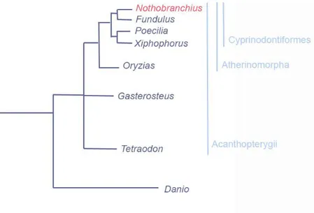
The teleost fish N. furzeri belongs to the order of Cyprinodontiformes, super-order Acanthopterygii, order Cyprinodontiformes, and comprises many other popular aquarium fish, such as other killifish and the so called live-bearing fish, comprising Anablepidae, Goodeidae and Poecilidae families. Molecular phylogenetic analyses have provided evidence that the order Cyprinodontiformes is a sister group and closest neighbours in a phylogenetic tree of Beloniformes [Fig 1]. N. furzeri phylogenetic position is very suitable for comparative genomics analysis and evolutionary studies, since its superorder Acanthopterygii comprehends also model genus for genomic and developmental studies, such as Fundulus, Poecilia,
the Japanese rice fish well known as Medaka (Setiamarga et al., 2008). N.
furzeri is also very close to the species Tetraodon nigroviridis, and the
stickleback Gasterosteus aculeatus.
Fig 1 | N. furzeri phylogenetic position compared to the closest Acanthopterygii and the Ostariophysan Danio rerio. Source: NFIN - The Nothobranchius furzeri Information Network
1.2. Sexual dimorphism, male color polymorphisms and sex determination
Nothobranchius are relatively small fish with a median size of 5 cm (ranging from 3 to 15 cm) with females smaller than males [
Fig 2]. All Nothobranchius are characterized by a marked sexual
fresh-water fish. Both sympatric and allopatric species occur in two or more colour forms. Females, on the other hand, are dull, their fins are translucent and body is pale brown, and it is very hard to distinguish between females of related species. N. furzeri females and females of other Nothobranchius species often possess iridescent scales. In some species, such as N. orthonotus, N. kadleci and N. melanospilus, female body may be pigmented with small dark dots. It is not clear yet whether this pigmentation arises as a by-product of male colouration (Sedlacek et al., 2014) or has any adaptive value. The bright male colouration is sexually selected and species-specific. In N. furzeri red and yellow morphs can be distinguished [
Fig 2] (Cellerino et al., 2016). A yellow caudal fin with a distal black band
characterizes the yellow morph. The red morph occurs in two forms: one with a distal black band and the other with a homogeneously red tail. Red and yellow reflectance may provide an advantage in different visual environments: the wavelength of red colour is too short to be reflected by turbid water and it is rapidly absorbed, whereas yellow wavelength is more visible (Litjens et al., 1999). This may have consequences for sexual selection via mate choice, as males with specific colouration may be more visible to females in particular light environments as the muddy ponds in which they live (Cellerino et al., 2016). The yellow sub marginal band with black marginal band is almost unique to N. furzeri. However, the red morph is very common in the genus Nothobranchius, typically co-existing with blue (rather than yellow) male morphs within a species.
Sex determination in N. furzeri is strictly genetic. Indeed, they possess a sex determination system XY based, like humans: the male is the heterogametic sex, XY, while female is the homogametic one, XX (Valenzano et al., 2009). Normally male/female ratio is 50%. However, in wild populations females typically dominate (Reichard et al., 2009). Given the equal adult sex ratio in the laboratory, high male mortality is the most plausible explanation for a female-biased sex ratio in the wild (Cellerino et al., 2016). In the wild predation on males is significantly higher than in females. Their vibrant colour, together with increased male mobility, mate searching, and their larger size, may further elevate the risk of predation (Tobler et al., 2007; Tobler et al., 2008). This situation is reported for
many taxa with high degrees of sexual dimorphism (Promislow et al., 1992), including many fish (Rosenthal et al., 2001). In addition, males compete aggressively for access to females (Polacik et al., 2011) and male competition for mates may contribute significantly to male mortality.
Fig 2 | N. furzeri, male and female. Yellow form, male (a); red form, male (b); adult female (c); and maturing male (d). Note opalescent hue on flanks and black pigmentation in the dorsal fin in d, temporary colours preceding the appearance of red, yellow and blue colours. Dorsal and anal fin are also relatively larger than in a female. Source: adapted from Polačik et al., 2016.
1.3. Distribution and habitat
Nothobranchius genus comprises four well-defined phylogenetic clades
that are almost exclusively allopatric (Dorn et al., 2014). The basal northern clade inhabits particularly dry regions in northern Kenya and Somalia. The southern clade is distributed south of the Zambezi River,
across a gradient from a humid coastal zone to dry habitats at higher altitudes. The inland clade is distributed in a high-altitude region between
Lake Victoria in Uganda and Kafue basin in Zambia and separated from the coastal clade (coastal basins in southern Kenya, Tanzania, and northern
clades occurred by vicariance events (Cellerino et al., 2016).
Nothobranchius pools are found in Savannah woodland, typically fed by
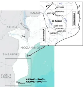
rainwater or in remnant pools within the channels of temporary streams (Reichard et al., 2009). Several Nothobranchius species may inhabit the same pool (Reichard et al., 2009). N. furzeri populations are distributed along an altitudinal gradient that ranges between 16 and 422 m above sea level, but most populations inhabit areas between 16 and 200 m above sea level (Reichard et al., 2009). Only two populations are known from higher altitudes: MZCS-323 (222 m) and GRZ (422 m) (Cellerino et al., 2016) [
Fig 3]. N. furzeri often inhabited shallow isolated pools separated from active stream channels and alluvial plains (Reichard et al., 2009). Vegetation is often present in the form of Nymphaea sp., grass vegetation and, occasionally, submerging vegetation (Reichard et al., 2009). N. furzeri typically occurred in close proximity to open water or at the interface between vegetation and open water. Many pools, however, lack any vegetation (Reichard et al., 2014). Vertisol soils on alluvial deposits are typical of N. Furzeri pools. Vertisol in fact provide suitable soil substrate for survival of dormant eggs during the dry season more than laterite soils. The substrate is soft and muddy and the water very turbid [Fig 4]. Water may be less turbid and even transparent when vegetation is abundant. Water conductivity (describing total dissolved solids, i.e. water hardness related to its mineral content) in N. furzeri habitats, varies widely, from 50 to 625 µS/cm (Cellerino et al., 2016).
Fig 3 | Distribution of N. furzeri. Broken lines indicates the range of each species (the extent of the range in Zimbabwe is not known). MZM and MZCS strain inhabit Mozambique pools. MZM comprises two sub strains, 0403 - 0410, while MZCS comprises five sub strains, 222 - 414 - 002 - 121 - 053. GRZ strain is located in the Gona Re Zhou National Park in Zimbabwe.
Fig 4 | Small African N. furzeri pool. The capacity of such a small pool is of about 80 fishes.
1.4. Embryos development and life cycle
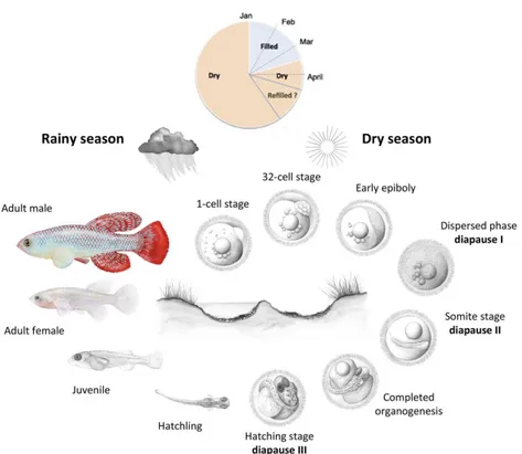
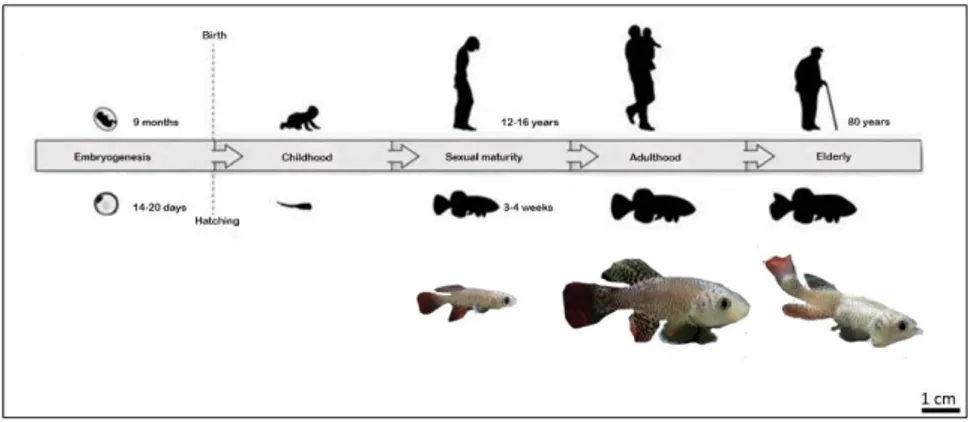
N. furzeri is an annual fish, with a median lifespan up to 12 months for the
MZM strain (long-lived phenotype) and a median life-span of just three months for the GRZ strain (short-lived phenotype) (Terzibasi et al., 2007). Eggs are spawned in the substrate and fish die when the pool evaporates. Embryos development is characterized by an early phase of embryonic development (cleavage) consisting in a very slow segmentation compared to other Cyprinodonts (Cellerino et al., 2016). Moreover, teleost cleavage lack of both G1 and G2 phases, so that cell proceed directly from the S to M phase (Graham and Morgan, 1966). Accordingly, all non-annual killifish species exhibit an average cell cycle length during cleavage of 20 – 45 min (Cellerino et al., 2016). By contrast, N. furzeri has a cleavage time of 75 min in the absence of a G1 phase (Cellerino et al., 2016). Annual life cycle is an ancient character state that has been lost several times during evolutionary history of African and Neotropical killifish clades (Murphy & Collier, 1997; Hrbek & Larson, 1999). Due to the short term of the rainy season, annual fish spend the majority of their life cycle in the dormancy state of diapause [Fig 5] (Cellerino et al., 2016). This mechanism is adopted from some species to survive unfavourable environmental conditions (extreme temperature, food availability, adverse climate such as drought). This latter is the case of N. furzeri going in diapause. In fact, as already mentioned above, South African pools are subject to periodically desiccation due to the alternation of the rainy seasons typically of the monsoon climate. The average duration of N.
furzeri habitats was only 75 days in one season (Terzibasi Tozzini et al.,
2013). This means embryos are expected to spend at least 10 months in diapause and they can survive in this state more than a year in captivity (Cellerino et al., 2016). Under laboratory conditions, it is possible to distinguish three different stages of diapause called diapause I, II and III (Wourms 1972). At the end of blastogenesis two cell types are produced: cells committed to forming the enveloping layer, which migrate as a uniform sheet, and dispersed macroblastomeres that migrate under this sheet (Cellerino et al., 2016). At the end of epiboly, there is a phase of
morphogenetic stasis with no evidence of a multicellular organizing centre and the macroblastomeres are under cell cycle arrest and migrate apparently randomly (Dolfi et al., 2014). This phase has fixed duration but may be prolonged under unfavourable conditions and represents diapause I (Cellerino et al., 2016). At the end of the dispersed phase, macroblastomeres re-aggregate to form the embryonic axis (Cellerino et al., 2016). In N. furzeri (at 26°C), epiboly is completed in 48 h and the dispersed phase lasts about 5 days, so that the embryonic axis appears only after approximately 1 week (Cellerino et al., 2016). After re-aggregation, the neural keel is formed and then somitogenesis and morphogenesis of the NS proceed until the embryo enters diapause II, in which embryogenesis is complete but organogenesis has not begun (Cellerino et al., 2016). At this stage, the number of somites is fixed, the heart is tubular and contractile, and the major divisions of the encephalon are present and so are the optic cups with the lens (Cellerino et al., 2016). The duration of diapause II is highly variable, from 2 days to up to 3 years. Temperature plays a major role in its regulation; embryos incubated at 28°C skip diapause and can hatch 12 days after fertilization. After diapause II, the embryo proceeds to complete development. South American annual killifish have an obligatory diapause III at the end of development (Berois et al., 2014), where the embryo is completely formed but metabolism is inhibited, while N. furzeri does not show diapause III under standard incubation conditions (Cellerino et al., 2016).
Fig 5 | N. furzeri annual life cycle. After flooding of the habitats (January - March), eggs hatch and the juveniles develop rapidly to reach sexual maturity. Eggs remain then diapause for about 10 months (April - December). Some embryos skip diapause and proceed through direct development and hatch if the habitat is wet. Source: adapted from Cellerino et al., 2015 and Platzer and Englert, 2016.
1.5. Diet
N. furzeri diet is mostly composed of aquatic invertebrates, both benthic
and pelagic (Polacik and Reichard, 2010). In addition, terrestrial insects were occasionally found in the guts (Polacik & Reichard, 2010). Their diet appears opportunistic and largely depends on prey availability (Polacik and Reichard, 2010). Food selectivity indices indicated that N. furzeri and N.
rachovii preferred to feed on small crustaceans (Cladocera, Copepoda, Ostracoda and Conchostraca) (Polacik & Reichard, 2010). Captive N. furzeri, as well as other species of the genus, readily consume live and
frozen dipteran larvae such as larvae of the genus Chironomus and
Chaoborus (Genade, 2005). Cannibalism has been observed in captivity in
juvenile fish, especially in N. orthonotus younger than 14 days, with large individual differences in body sizes (Cellerino et al., 2016). There is no record of cannibalism in the wild (Polacik & Reichard, 2010), but it is expected in juvenile fish whenever size differences exceed approximately 30% or more (Cellerino et al., 2016).
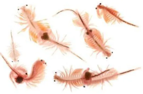
The most common crustacean often used in laboratory to provide live food for killifish is the Artemia salina [Fig 6]. Artemia eggs, also known as cysts, can be stored for long periods and hatched on demand in salt water. In laboratory conditions, cysts can be hatched in water and marine salt (1 liter of water with 8–10 g of salt). The amount of the eggs depends on the number of fish to feed; 3 g of eggs produce an amount of nauplii sufficient to feed at least 1000 freshly hatched N. furzeri. The hatching will occur in 18-36 hours. To collect fresh hatched nauplii, aeration has to be removed from the cone, then, after several minutes, shells and baby brine shrimps separate: baby brine shrimps are attracted from lights sources, so it can be helpful to have a lamp on the cone to let them move towards the light source. Shells will float to the surface and can be easily removed with a Pasteur pipette. Then it is important, before feeding them to fish, to rinse baby brine shrimps in a fine mesh net or sieve using clean fresh water.
Fig 6 | Common crustacean used for N. furzeri diet known as Artemia salina. Artemia is a genus of aquatic crustacean also known as brine shrimp.
1.6. Housing
After collection, killifish eggs can be incubated at 26°C in petri dishes containing boiled damp peat. To promote synchronously hatching, eggs can be pre-incubated in Yamamoto’s solution (17 mM NaCl, 2.7 mM KCl, 2.5 mM CaCl2, and 0.02 mM NaHCO3, pH 7.3) containing methylene blue
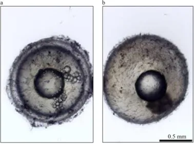
0.3%. Unfertilized eggs are characterized by a double-layered structure with a perivitelline space and remain clear [Fig 7 a], and should be removed by using a Pasteur pipette. Furthermore, if using methylene blue, they start decaying and acquire a bluish, rather opaque coloration [Fig 7 b]. Fertilized eggs will be translucent with no apparent structure inside [Fig 8 a], opaque with an advanced embryo [Fig 8 b, c], or white when dead [Fig 8 d] (Polacik et al., 20016). Ready-to-hatch embryos remain viable for only a limited time, as they are gradually consuming their energy reserves (Polacik et al., 20016), and the precise time interval depends on the incubation temperature (Polacik et al., 20016). The yolk sac begins diminishing ~60 d after the embryo reaches its pre-hatching stage (Polacik et al., 2016).
Eggs must be now moved with the peat (that maintains a slightly acidic environment, keeping waste nitrogenous substances in a safer, ammonium form) into a small aquarium (6 L for ~ 50 eggs). The peat layer on the bottom should be no deeper than 2 cm so that the freshly hatched fish can easily swim out and not get trapped in the peat (Polacik et al., 2016). Manipulation of fry, which are fragile, must be avoided, keeping them in their hatching aquarium with the peat for 3–4 dph; tank can be rearranged to a flat position ~24 h after hatching. Water temperature should be maintained at 27–28 °C with a small submersible aquarium heater and any aeration or filtration should be avoided. After 3-4 days, fry can be transferred into a larger aquarium without the peat (~4 L of water per fish). Water exchange can be provided by setting a recirculating system or by gradually exchanging of water adding mature system water to the tank with juveniles maintaining always the same water level (Polacik et al., 2016). When fish are sexually mature, at ~ 5 weeks post hatching (wph), they must be kept in a female biased ratio (1:3 ratio) to avoid lethal male– male interactions. In case, aggressive individual(s) should be removed from the aquarium.
Tanks have to be covered with lids and labeled with strain of fish contained, date of birth, number of fish and male/female ratio. Placing tanks with different strains adjacent to one another should be avoided. Colored labels are very helpful to indicate health status of fish and keep them monitored in order to their needs.
Young fish must be feed three times a day and adult fish, 5 days post hatching (dph)), two times per day, at regular intervals.
The tank must be provided with a spawning place, which could be a sieve in a bottle shaped glass jar (to prevent excessive spilling of the peat) filled with a 6 cm layer of wet autoclaved peat to protect eggs during long-term exposure to adult fish because N. furzeri tends to cannibalize their eggs. Before proceeding with eggs hatching, they should be bleached with a solution containing 0.1 mL of 5% sodium hypochlorite in 170 mL of system water: eggs are rinsed for 5 min in system water, placed in a second beaker of bleach solution for 5 min more, rinsed the with system water and placed into petri dishes.
Fig 7 | Differences between fertilized (a) and unfertilized (b) N. furzeri eggs. Fertilized eggs with typical double-layered structure. Unfertilized eggs lacking perivitelline space. Source: adapted from Polačik et al., 2016.
Fig 8 | Gross staging of N. furzeri eggs incubated in peat. (a–d) Diapaused N. furzeri egg lacking structures that are readily visible to the naked eye (a); advanced, post diapause II embryo with black-pigmented eyes (b); egg with a fully developed, ready-to-hatch embryo with gold-pigmented eye iris (c); and dead, decaying egg (d).
1.7. Genetic structure
Each Savannah pool consists of genetically homogenous populations of N.
furzeri and even adjacent pools containing genetically distinct populations
(Polacik et al., 2016). The same strong structure was found for three other species: N. orthonotus, N. kadleci and N. pienaari (Bartakova et al., 2013). Genetic differentiation between populations increases with geographical distance. The same two lineages co-occur in at least one population in another part of their contact zone and nuclear genetic markers (microsatellites) indicate no reproductive barrier (Bartakova et al., 2013). In the future, the use of genome-wide markers should reveal greater details of the population structure within and among populations.
At Fritz Lipmann Institute laboratories (Leibniz Institute for Aging, Jena, Germany), N. furzeri genome has been sequenced and annotated on NFIN genome browser (NFINgb) at http://nfingb.leibniz-fli.de/. NFINgb provides access to sequencing data of different short- and long-lived N.
furzeri strains: GRZ, MZM-0403, MZM-0410 and MZZW-0701
(Reichwald et al., 2015). It is also available a transcriptome browser, NFINtb (https://nfintb.leibniz-fli.de/nfintb/ ), in which are annotated 19,875 protein-coding genes plus comparisons data to Medaka, stickleback, tetraodon and zebrafish (Petzold et al., 2013).
1.3. Non-genetic and trans-genetic interventions
Species of the genus Nothobranchius are excellent model systems to test non-genetic interventions and their effects on ageing and survival (Cellerino et al., 2016). In particular, these fish offer a great opportunity to study the effects of water-soluble drugs and feeding on vertebrate survival and ageing on a large scale (Cellerino et al., 2016). Non-genetic manipulations, including changes in temperature, diet and drug administration on vertebrate survival and ageing-related phenotypes have recently been tested using several species of the genus Nothobranchius, (Cellerino et al., 2016). N. furzeri and N. rachovii ageing rate and overall survival in captivity can be modified by water temperature (Valenzano et al., 2006 b; Hsu & Chiu, 2009), with lower temperature increasing
lifespan, slowing down the appearance of ageing biomarkers, reducing adult body size and delaying the age-dependent decline in spontaneous activity (Cellerino et al, 2016). Dietary restriction by feeding every other day in inbred and wild-derived N. furzeri strains was effective in prolonging the survival of inbred strains and slowing down ageing but had a mixed effect in wild-derived strains, increasing early mortality while prolonging maximum lifespan (Terzibasi et al., 2009), similarly to results reported in mice (Weindruch, 1996). Different research groups have shown that in both N. furzeri and N. guentheri, the natural polyphenol resveratrol can slow down ageing and prolong lifespan (Valenzano et al., 2006 a; Yu & Li, 2012; Genade & Lang, 2013). Additionally, supplementation of melatonin into aged N. korthausae reduced age-dependent disruption of circadian rhythms (Lucas-Sanchez et al., 2013).
Due to its very short life cycle Nothobranchius fish are particularly suited for transgenic experiments. Transgenic N. furzeri lines are now available, with fish ubiquitously expressing green fluorescent protein (GFP) under several promoters (Valenzano et al., 2011; Hartmann & Englert, 2012; Allard, Kamei and Duan, 2013). Current work is aimed at further developing the transgenesis toolkit in this species and applying the recently developed transcription activator-like effector nucleases (TALENs) and systems (Zu et al., 2013; Sung et al., 2014) to introduce loss-of function mutations and perform site-specific genome editing (Cellerino et al., 2016). Since Nothobranchius is the shortest-lived vertebrate, transgenic mutants of its genus could be used to identify novel genes and genomic regions that modulate vertebrate survival and ageing. Nothobranchius transgenic studies could strongly promote its development as new powerful genetic models in several science fields, from developmental biology to ageing research.
1.8. N. furzeri in neurobiological and aging research
Vertebrates represent a model organism widely employed in laboratory research. In fact they are very suitable for biomedical and veterinary studies directed in identifieng and understanding mechanisms
characterizing human diseases. Among vertebrates, fish constitute the largest group and as early as 1978 the use of fish in research showed great potential (Woodhead, 1978) for the relatively low cost of maintaining large numbers of individuals for lifetime studies, well-developed genetics, in addition to the fact that they possess organs and system similar to ones of other vertebrates. The most common model so far employed in research is zebrafish and it is the best-characterised animal at genetic level (Gerhard et al., 2003). However, zebrafish does not provide a suitable model for aging studies (Yu et al., 2006) mainly because of the long lifespan in laboratory conditions (~ 5 years, Gerhard et al., 2003).
Many Cyprinodontiformes like Anablepidae, Goodeidae and Poecilidae families are often used in biomedical research and for ecology, evolutionary and genomic studies. In the recent years N. furzeri has been proposed as new non-mammalian model system in many fields, especially in aging research (Terzibasi et al., 2007). The African turquoise killifish possess in fact the shortest life span (4-6 months) among vertebrates that can be cultured in laboratory and it reproduces very fast (14-20 days between the spawn and the hatch) in captivity too [Fig 9]. More generally, teleost fish are very similar to mammals in many aspects. They have a telomeric like structure, an amazing regenerative capacity, human-like organs and systems (blood, spleen, bones), evident aging related characters (mitochondrial functional decline, neurodegeneration). Moreover, unlike other fish, N. furzeri possess an XY chromosomes sex determination system. Another interesting aspect is that their eggs can be stored dry at room temperature (RT) for months, even years, offering inexpensive methods of embryo storage (Cellerino et al., 2016). Fish organs are characterized by a continuous turnover and cellular proliferation and differentiation that change with aging. Even their skin undergoes continuously to age-dependent cellular turnover, changes in caudal fin colouration, and variation in gene expression.
Fig 9 | N. furzeri complete life cycle. From fertilization to hatching (usually between 14 and 20 days), animals are referred to as 'embryos'. Freshly hatched fish are referred to as 'fry' for the first few days and shortly after they are referred to as 'juvenile fish' (until the animals reach sexual maturity). At the age of 20-30 days post hatching (dph), the animals reach sexual maturity and are considered adults. The life cycle (from 'egg to egg') of the turquoise killifish is 5-7 weeks. Source: adapted from Harel et al., 2016.
1.9. Fish brain structure
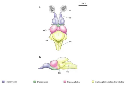
Fish brain is relatively small if compared to body size, except for some fish like mormyrid and sharks, which have a brain/body weight proportion like that of birds and marsupials (Helfman et al., 1997). The braincase may constrain brain size only in some of the smallest representatives of modern, perciform teleost. In most fish, however, the brain is considerably smaller than the space available and, in some cases, may occupy only about 6% of the brain cavity in an elasmobranch (Kruska, 1988). The excess space is commonly filled with lymphatic, fatty tissue (Kotrschal et al., 1998). Fish brain shows structures like mammals, and also their brain organization is overall similar, consisting of the three major regions of the forebrain, midbrain and hindbrain [Fig 10]. The telencephalon consists of paired cerebral hemispheres with olfactory bulbs attached to the rostral
hemispheres in most fish. The mesencephalon is capped by a pair of optic lobes (optic tectum), and the cerebellum arises from its rostral roof. Rostral, the spinal cord merges with the brain stem and tegmentum of mesencephalon and diencephalon (Kotrschal et al., 1998).
Forebrain performs functions associated with hormones and homeostasis. The pineal body lies just above the diencephalon. This structure detects light, maintains circadian rhythms, and controls color changes (Helfman et al., 1997). Forebrain olfactoy bulbs dimension depends of the development of smell sense (Kotrschal et al., 1998). Fish with larger olfactory bulbs are for example hagfish, catfish and sharks. Olfactory bulbs receive signals from one of two channels of the nose, the nostril, through the olfactory nerves (Helfman et al., 1997).
Midbrain dorsal region comprises the two optic lobes, which are very large in species that hunt by sight, such as rainbow trout and cichlids (Helfman et al., 1997).
Hindbrain division is particularly involved in swimming and balance (Helfman et al., 1997). It is divided in a rostro-dorsal area comprising the cerebellum, a rostro-ventral area comprising the pons and a caudal area comprising the medulla oblongata. Cerebellum is a single-lobed structure (Helfmann et al., 1997). Hagfish and lampreys have relatively small cerebellum, while the mormyrid cerebellum is massive and apparently involved in their electrical sense (Helfmann et al., 1997).
With few pathways descending from the brain, the motor system resides largely within the spinal cord except for several prominent brainstem reflexes (e.g. the Mauthner neuron system for escape (Kotrschal et al., 1998). Somatosensory information reaches the brain primarily via specialized cranial nerves, notably trigeminus (V), facialis (VII), vagus (X) and three lateral line nerves, two anterior and one posterior, rather than through ascending fiber systems of the spinal cord (Kotrschal et al., 1998).
Fig 10 | Fish brain in dorsal (a) and lateral views (b). Abbreviations: CC, crista cerebellaris; EG, eminentia granularae; OB, olfactory bulbs; oo, olfactory organ; OT, optic tectum. Lateral view does not include olfactory organs.
1.10. Fish brain versus mammalian brain
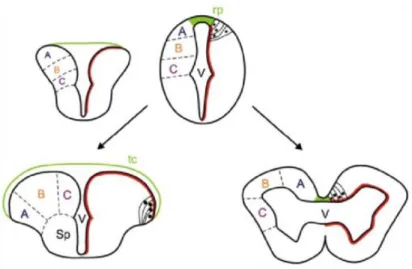
Both fish and mammalian brains are broadly structured similarly, but, generally, fish brain is quite small relative to their body mass (~ 1/15 the brain mass of a similarly size bird or mammal. They share similar areas and structures, the ability to direct visual attention at targets to achieve a goal, ignoring distracting stimuli that prevent to reach the goal (Gabay et al., 2013), the response to fear and pain. To make a comparison between teleost and mammals brain, it is remarkable to notice two different way of embryonic development: evagination and eversion [Fig 11]. The evagination process occurs in mammals, while the eversion process is typical of teleost fish. Consequently, there is a shift in the position of some telencephalic areas: teleost dorsal telencephalic areas correspond to the
mammalian pallial formation; ventral telencephalic areas to the subpallium; dorso-lateral telencephalon to the medial pallium or hippocampus; the everted part of dorso-medial telencephalon corresponds to the dorsal pallium (isocortex homologue); the posterior zone of dorsal telencephalic area would be the lateral pallium (the olfactory cortex homologue); the midline portion of the medial zone of dorsal telencephalic area would correspond to the homologue of the pallial amygdala. The pallial masses ventral to dorso-lateral would not be everted (Wullimann and Mueller, 2004) [
Fig 12, Tab 1]. However, exists also a partial eversion theory of teleostean telencephalon, contrasting the complete eversion theory (Nieuwenhuys, 2009). According to this latter theory, the dorso-lateral telencephalon is homologous to the medial pallial formation, the dorsal and ventro-central to the striatal formation, and the ventro-ventral and ventro-lateral to the septal formation. Generally, fish forebrain is bigger and much more primitive than mammalian one; it is mostly associated with olfaction, which is the reason why olfactory bulbs are larger and acute than those of mammals. On the other hand, mammals have a highly developed cerebral cortex. In fish, cerebellum, is involved in both swimming and balance maintenance and represents the biggest structure in fish brain, while in mammals it takes up only about 15% of volume.
Fig 11 | Model for eversion (left) in teleost fish in contrast with evagination of telencephalic vesicles that occurs in other vertebrates (right). In teleost fish, it is thought that the dorsal region of the telencephalic neural tube (pallium) folds over the ventral region (subpallium), stretching the dorsal roof-plate region of the neural tube (green) to form the tela choroidea. This would also relocate some ventricular cells to the dorsal telencephalic surface (red line) and cause medial to lateral rearrangements of pallial regions (compare the location of regions A, B, and C between everted and evaginated telencephalon). Abbreviations: rp, roof plate; Sp, subpallium; tc, tela choroidea; V, ventricle. Source: Folgueira et al., 2012.
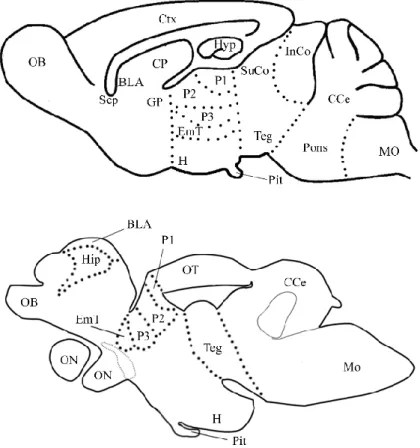
Fig 12 | Schematic representation of mammal (mouse) brain versus teleost (N. furzeri) brain. The massive difference is caused by the two different telencephalic development: eversion (teleost) and evagination (mammal). For this reason, mouse brain telencephalon shows two bilateral symmetric hemispheres surrounding by centrally located ventricles. In contrast, teleost telencephalon consists by two massive symmetric lobes covered by a dorsally located T-shaped ventricle. Teleost show a medial pallial region, homologous to the mammalian hippocampus (Hip), a dorsal pallial region, which corresponds to mammalian isocortex (Ctx), a ventral one corresponding to the mammalian amygdala (BLA), and a lateral pallial division corresponding to the piriform cortex (pirCtx). Abbreviations follow Muller et al., 2011.
Forebrain divisions
Nothobranchius furzeri Mus musculus
Dc1-2-3-4 Ctx Dld-Dlv Hyp Dm1-2-3-4 BLA Dp pirCtx Vd Str Vv Sep
Tab 1 | Schematic correspondence between N. furzeri and M. musculus forebrain divisions.
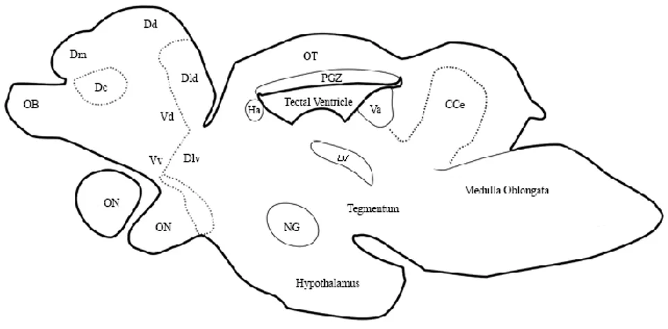
1.11. N. furzeri brain structure
The basic morphology of N. furzeri brain has been described in the N.
Furzeri brain atlas published in 2013 from D'Angelo. Anatomical structure
and abbreviations will follow atlas nomenclature. Its brain structure resembles that of close teleost (Setiamarga et al., 2008) i.e. Oryzias latipes (Ishikawa et al., 1999), Xiphophorus helleri (Anken & Rahmann, 1994),
Fundulus heteroclitus (Peter et al., 1975), with no significant
morphological differences between males and females. The gross morphology of N. furzeri CNS is characterized by: two small and elongate olfactory bulbs (OB), placed anterior-ventrally to the small telencephalic hemispheres; the diencephalon with the large paired inferior lobes of hypothalamus, bulgs out on the ventral surface of the brain; two pronounced lobes of the optic tectum, dorsal to diencephalon most rostrally, and the midbrain tegmentum most caudally; a prominent body of cerebellum; large medulla oblongata [