ARTICLE
OPEN
Decreased free
D
-aspartate levels are linked to enhanced
D
-aspartate oxidase activity in the dorsolateral prefrontal
cortex of schizophrenia patients
Tommaso Nuzzo1,2, Silvia Sacchi 3,4, Francesco Errico1,5, Simona Keller5,6, Orazio Palumbo 7, Ermanno Florio5,6, Daniela Punzo1,2, Francesco Napolitano1,5, Massimiliano Copetti8, Massimo Carella7, Lorenzo Chiariotti5,6, Alessandro Bertolino9,
Loredano Pollegioni3,4and Alessandro Usiello1,2
It is long acknowledged that the N-methylD-aspartate receptor co-agonist,D-serine, plays a crucial role in several N-methylD
-aspartate receptor-mediated physiological and pathological processes, including schizophrenia. BesidesD-serine, another freeD
-amino acid,D-aspartate, is involved in the activation of N-methylD-aspartate receptors acting as an agonist of this receptor subclass,
and is abundantly detected in the developing human brain. Based on the hypothesis of N-methylD-aspartate receptor
hypofunction in the pathophysiology of schizophrenia and considering the ability ofD-aspartate andD-serine to stimulate N-methyl D-aspartate receptor-dependent transmission, in the present work we assessed the concentration of these twoD-amino acids in the
post-mortem dorsolateral prefrontal cortex and hippocampus of patients with schizophrenia and healthy subjects. Moreover, in this cohort of post-mortem brain samples we investigated the spatiotemporal variations ofD-aspartate andD-serine. Consistent with
previous work, we found thatD-aspartate content was selectively decreased by around 30% in the dorsolateral prefrontal cortex,
but not in the hippocampus, of schizophrenia-affected patients, compared to healthy subjects. Interestingly, such selective reduction was associated to greater (around 25%) cortical activity of the enzyme responsible forD-aspartate catabolism,D-aspartate
oxidase. Conversely, no significant changes were found in the methylation state and transcription of DDO gene in patients with schizophrenia, compared to control individuals, as well as in the expression levels of serine racemase, the major enzyme responsible forD-serine biosynthesis, which also catalyzes aspartate racemization. These results reveal the potential involvement of
alteredD-aspartate metabolism in the dorsolateral prefrontal cortex as a factor contributing to dysfunctional N-methylD-aspartate
receptor-mediated transmission in schizophrenia.
npj Schizophrenia (2017) 3:16 ; doi:10.1038/s41537-017-0015-7
INTRODUCTION
D-serine (D-Ser) and D-aspartate (D-Asp) are the only two free D
-amino acids occurring at substantial levels in the mammalian brain.
D-Ser is present at high concentrations throughout prenatal and
postnatal life, especially in the forebrain regions1where N-methyl
D-aspartate receptors (NMDARs) are abundant. Together with
glycine, D-Ser activates the NMDAR co-agonist site and plays a
crucial role in NMDAR-mediated physiological and pathological processes, including those involved in schizophrenia (SCZ).2, 3 In this regard, abnormal D-Ser content and metabolism have been
observed in the cerebrospinal fluid and serum of subjects with SCZ.4–6 As in humans, several findings collected in preclinical models indicated an involvement of this D-amino acid in
SCZ-related phenotypes. For instance, reducedD-Ser levels due to the
ablation of the gene Serine racemase (Srr)7encoding the enzyme responsible for D-Ser biosynthesis, produce in mutant mice
NMDAR-related morphological and behavioral deficits reminiscent of those occurring in SCZ.3On the other hand,D-Ser
supplementa-tion in animal models was showed to be effective in improving a range of SCZ-related cognitive, sensorimotor, and social deficits.2,3 In contrast to D-Ser, D-Asp undergoes a strict developmental regulation since it occurs at high levels during embryonic and early postnatal phases, and markedly drops thereafter.1, 8 Remarkably, in the human prefrontal cortex (PFC) at 14 weeks of gestationD-Asp levels even exceed those of the corresponding L-form before the drastic postnatal decrease.1 The peculiar
temporal pattern ofD-Asp occurrence depends on the postnatal
onset ofD-aspartate oxidase (DDO or DASPO, EC 1.4.3.1) activity, a
peroxisomalflavoenzyme that selectively catabolisesD-Asp.9In the
mouse brain, the age-dependent increase in DDO enzymatic activity is paralleled by concomitant rise in Ddo mRNA levels that, in turn, is associated to progressive Ddo gene demethylation
Received: 30 November 2016 Revised: 10 February 2017 Accepted: 22 February 2017 1
Laboratory of Behavioral Neuroscience, Ceinge Biotecnologie Avanzate, 80145 Naples, Italy;2
Department of Environmental, Biological and Pharmaceutical Sciences and Technologies, Second University of Naples (SUN), 81100 Caserta, Italy;3
Department of Biotechnology and Life Sciences, University of Insubria, 21100 Varese, Italy;4
The Protein Factory Research Center, Politecnico di Milano and University of Insubria, 20133 Milan, Italy;5Department of Molecular Medicine and Medical Biotechnology, University of Naples “Federico II”, 80131 Naples, Italy;6
Endocrinology and Molecular Oncology Institute (I.E.O.S.), National Research Council (C.N.R.), 80131 Naples, Italy;7
Medical Genetics Unit, IRCCS Casa Sollievo della Sofferenza, 71013 San Giovanni Rotondo, Foggia, Italy;8Unit of Biostatistics, IRCCS Casa Sollievo della Sofferenza, 71013 San Giovanni Rotondo, Foggia, Italy and9
Department of Basic Medical Science, Neuroscience, and Sense Organs, University of Bari“Aldo Moro”, 70124 Bari, Italy Correspondence: Loredano Pollegioni ([email protected]) or Alessandro Usiello ([email protected]) Tommaso Nuzzo, Silvia Sacchi and Francesco Errico contributed equally to this work.
in the putative regulatory region surrounding the transcription start site.8
Several studies have so far indicated that D-Asp activates
NMDARs through its binding at the glutamate (Glu) site of this receptor.9,10Consistent with the knowledge that hypofunction of NMDARs elicits SCZ-like symptoms in humans and preclinical models,11,12it has been shown that higher cerebral levels ofD-Asp
in mice are protective against prepulse inhibition (PPI) deficits and abnormal circuits activation induced by the psychotomimetic drug phencyclidine (PCP). In addition, non-physiological D-Asp levels
improve cognition,13–15 enhance cortico-hippocampal connectiv-ity,16 and increase structural and functional synaptic plasticity in cortical and hippocampal pyramidal neurons.17 Translation of these preclinical studies to healthy humans indicated that the CC genotype of the DDO polymorphism rs3757351, predicting reduced DDO mRNA expression in the post-mortem PFC, is associated with prefrontal phenotypes relevant to SCZ, such as greater gray matter volume and enhanced activity during working memory tasks.17 Moreover, evidence obtained in human post-mortem samples have shown substantially decreased content ofD
-Asp in patients with SCZ, compared to non-psychiatric controls.18 Based on this previous finding, we used another collection of post-mortem dorsolateral prefrontal cortex (DLPFC) and hippo-campus samples of SCZ-affected patients and control individuals to investigate whether SCZ diagnosis affects the levels of D-Asp and its regulation by DDO by assessing DDO gene copy number, DDO gene methylation, DDO mRNA expression and DDO enzymatic activity. These brain samples were also analyzed for the content ofD-Ser and for the expression levels of SRR protein,
which is not only responsible forD-Ser production but has been
also recently proposed to contribute toD-Asp biosynthesis.19,20 RESULTS
Age-related distribution ofD-aspartate andD-serine in the brain of
patients with SCZ and healthy subjects
It has been previously reported that the content ofD-Asp in the
post-mortem PFC of healthy subjects drastically declines from prenatal to postnatal stage, and remains low during adulthood.1 However, there is no evidence relative to the temporal variations ofD-Asp content in the brain of SCZ-affected patients. Therefore,
we analyzed the changes inD-Asp levels across adult life in the
post-mortem DLPFC and hippocampus dissected from individuals with SCZ, compared to controls (representative images of these brain regions are shown in Fig.1a). In parallel toD-Asp, we also
assessed the age-span occurrence ofD-Ser.
We found that D-Asp levels do not significantly change along
adulthood in both the DLPFC and hippocampus of control subjects (DLPFC: r = 0.0368, p = 0.8733; hippocampus: r = −0.4924, p = 0.0620; Spearman correlation) and SCZ-affected patients (DLPFC: r = 0.1397, p = 0.5685; hippocampus: r = 0.2308, p = 0.4050) (Fig.1b, h), although there is a significant difference in the mean age of the two diagnosis groups (p < 0.01, Supplemen-tary Table1). Similarly, in both brain regions theL-Asp levels (Ctrl:
DLPFC, r = 0.0557, p = 0.8156; hippocampus, r =−0.0841, p = 0.7551; SCZ: DLPFC, r = 0.0053, p = 0.9829; hippocampus, r = 0.3488, p = 0.2016) and theD-Asp/total Asp ratio (Ctrl: DLPFC, r =
0.1153, p = 0.6285; hippocampus, r =−0.3133, p = 0.2481; SCZ: DLPFC, r = 0.1751, p = 0.4734; hippocampus, r = 0.1395, p = 0.6177) were unaffected by the age of the individuals, independently from their clinical diagnosis (Fig.1c, d, i, and j).
D- andL-Ser were concomitantly analyzed on the same samples.
The levels of both enantiomers remained constant during adulthood, in both the DLPFC and hippocampus of healthy individuals (D-Ser: DLPFC, r =−0.0685, p = 0.7742; hippocampus,
r =−0.2266, p = 0.3368;L-Ser: DLPFC, r = 0.2665, p = 0.2561;
hippo-campus, r =−0.1280, p = 0.5908) and SCZ patients (D-Ser: DLPFC,
r = 0.2966, p = 0.2042; hippocampus, r =−0.2597, p = 0.2689;L-Ser:
DLPFC, r = 0.2958, p = 0.2054; hippocampus, r =−0.0858, p = 0.7191) (Fig. 1e, f, k, and l). Accordingly, theD-Ser/total Ser
ratio did not change in the two brain regions during adult age in both clinical conditions (Ctrl: DLPFC, r =−0.1905, p = 0.4211; hippocampus, r =−0.1114, p = 0.6401; SCZ: DLPFC, r = −0.1046, p = 0.6607; hippocampus, r =−0.1626, p = 0.4934) (Fig.1g, m).
Overall, these data excluded significant age-dependent varia-tions of D-Asp and D-Ser levels in the DLPFC and hippocampus
associated to the diagnosis of SCZ.
Selective reduction ofD-aspartate levels in the DLPFC of patients
with SCZ
We evaluated whether the reduction (∼40%) in D-Asp levels
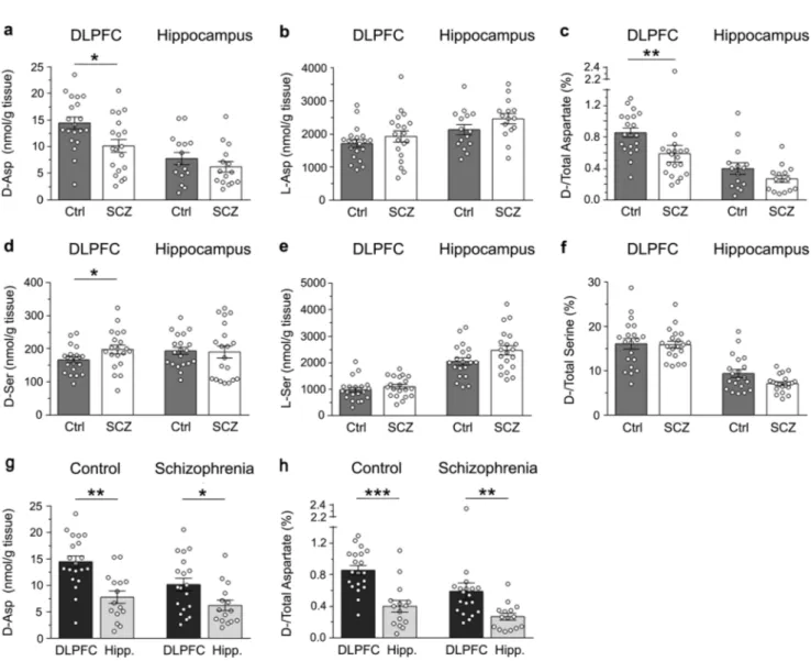
previously found in the post-mortem PFC of patients with SCZ (Institute of Psychiatry, King’s College London, UK)18 might be replicated in a new cohort of brain tissues from SCZ and control cases (Human Brain and Spinal Fluid Research Center, Los Angeles, CA, USA). In line with previous data, a significant (∼30%) reduction inD-Asp levels was detected in the DLPFC of SCZ-affected patients,
compared to healthy individuals (p < 0.05, Mann–Whitney test) (Fig. 2a and Supplementary Table 2). On the other hand, no difference inL-Asp levels was apparent between diagnoses (p > 0.05) (Fig.2b and Supplementary Table2). Accordingly, theD-Asp/ total Asp ratio was significantly lower in patients with SCZ, compared to controls (p < 0.01) (Fig. 2c and Supplementary Table 2). Differently from DLPFC, in the hippocampus we found no statistically significant change in D-Asp and L-Asp levels
between SCZ-affected patients and control subjects (p > 0.05 for both) (Fig.2a, b and Supplementary Table2), as well as in theD
-Asp/total Asp ratio (p > 0.05) (Fig.2c and Supplementary Table2). In addition, we found a mild increase inD-Ser content (∼15%) in
the DLPFC of SCZ patients, compared to control subjects (p < 0.05), while no difference was found in the hippocampus of SCZ-affected patients and healthy subjects (p > 0.05) (Fig.2d and Supplementary Table 2). However, in both brain regions and diagnostic groups analyzed, we detected comparableL-Ser levels andD-Ser/total Ser
ratio (p > 0.05) (Fig.2e, f and Supplementary Table2).
Finally, we assessed whether the diagnosis of SCZ could influence the regional differences of D-Asp levels occurring between the DLPFC and the hippocampus. We found that theD -Asp content was significantly higher in the DLPFC than in the hippocampus, in both healthy and SCZ-affected individuals (Ctrl: p < 0.01, SCZ: p < 0.05; Mann–Whitney test) (Fig. 2g and Supple-mentary Table 3). Similarly, the D-Asp/total Asp ratio was
significantly higher in the DLPFC than in the hippocampus, regardless of the diagnosis (Ctrl: p < 0.0001; SCZ: p < 0.05) (Fig.2h and Supplementary Table3).
DDO gene methylation and DDO mRNA expression in the DLPFC and hippocampus of patients with SCZ and healthy subjects We recently reported that the state of methylation of the regulatory region of the DDO gene spanning across the transcriptional start site controls the postnatal rise of DDO mRNA levels in the mouse brain thus predicting, in turn, the content ofD
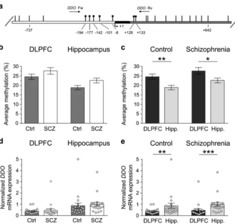
-Asp.8 Based on this finding, we evaluated whether epigenetic modification at the DDO locus was affected by the diagnosis of SCZ. To this aim, a bisulfite-seq analysis in the post-mortem DLPFC and hippocampus of SCZ-affected patients and healthy subjects was performed. We analyzed the average methylation state of the seven CpG sites (−194, −177, −142, −101, −8, +128 and +133) lying in the region of the human DDO surrounding the transcription start site spanning from −199 to +177 nucleotide positions (Fig.3a). No significant alteration in DDO gene methylation state was observed in the DLPFC of the two diagnostic groups (mean values: Ctrl, 24.5 ± 1.4%; SCZ, 27.6 ± 1.8%; p > 0.05, Mann–Whitney test) while a significant, moderate increase was detected in the AbnormalD-aspartate metabolism in schizophrenia
T Nuzzo et al 2
hippocampus of SCZ-affected patients, compared to healthy individuals (control, 18.8 ± 1.2%; SCZ, 22.6 ± 1.3%; p < 0.05) (Fig.3b).
Next, we analyzed the regional variations in the DDO gene methylation profile in both healthy and SCZ-affected subjects. The average methylation state of this gene was significantly higher in the DLPFC than in the hippocampus in both control individuals (mean values: DLPFC, 24.5 ± 1.4%; hippocampus, 18.8 ± 1.2%; p <
0.01) and in SCZ-affected patients (mean values: DLPFC, 27.6 ± 1.8%; hippocampus, 22.6 ± 1.3%; p < 0.05) (Fig.3c).
We then examined the DDO gene transcription in the post-mortem brain of patients with SCZ and relative controls. Interestingly, quantitative reverse transcription PCR (qRT-PCR) analysis did not reveal any significant difference in DDO mRNA levels between SCZ-affected and healthy individuals, in both the brain structures analyzed (mean values, DLPFC: Ctrl, 0.39 ± 0.05;
Fig. 1 Age-related changes inD-aspartate andD-serine levels between patients with schizophrenia and control subjects in the post-mortem
dorsolateral prefrontal cortex and hippocampus. a Representative images of the dorsolateral prefrontal cortex (DLPFC) and hippocampus dissected from post-mortem brains of healthy (control, Ctrl) and schizophrenia-affected individuals (SCZ). The dot plots represent the variations across lifespan in the content of free b, hD-aspartate and e, kD-serine, c, iL-aspartate and f, lL-serine, and d, g, j, mD-/total amino acids in the b–g dorsolateral prefrontal cortex (DLPFC) and h–m hippocampus of non-psychiatric individuals (DLPCF, n = 20; hippocampus, n = 15) and patients with schizophrenia (DLPCF, n= 19; hippocampus, n = 15). In each sample, all the amino acids were detected in a single run by HPLC and expressed as nmol/g of tissue, while the ratios are expressed as percentage (%)
Fig. 2 Changes inD-aspartate andD-serine levels between patients with schizophrenia and control subjects in the post-mortem dorsolateral
prefrontal cortex and hippocampus. aD-aspartate, bL-aspartate, dD-serine and eL-serine levels, cD-aspartate/total aspartate and fD-serine/ total serine are compared between non-psychiatric individuals (control, Ctrl) and patients with schizophrenia (SCZ) in the post-mortem dorsolateral prefrontal cortex (DLPFC) (Ctrl, n= 20; SCZ, n = 19) and hippocampus (Ctrl, n = 15; SCZ, n = 15). g, hD-aspartate levels andD/total aspartate ratio were also compared between the DLPFC and hippocampus (Hipp.) of non-psychiatric individuals (DLPCF, n= 20; hippocampus, n= 15) and patients with schizophrenia (DLPCF, n = 19; hippocampus, n = 15). In each sample, all the amino acids were detected in a single run by HPLC and expressed as nmol/g of tissue, while the ratios are expressed as percentage (%). *p< 0.05; **p < 0.01; ***p < 0.0001 (Mann–Whitney test). Dots represent the single subjects’ values while bars illustrate the means ± SEM
AbnormalD-aspartate metabolism in schizophrenia T Nuzzo et al 4
SCZ, 0.47 ± 0.14; p > 0.05; hippocampus: Ctrl, 0.87 ± 0.23; SCZ, 1.00 ± 0.18; p > 0.05) (Fig.3d).
On the other hand, consistently with an influence of DDO gene methylation on mRNA expression,8 we found reduced levels of DDO transcript in the DLPFC, compared to the hippocampus, independently of the clinical diagnosis (mean values, Ctrl: DLPFC, 0.39 ± 0.05; hippocampus, 0.87 ± 0.23; p < 0.01; SCZ: DLPFC, 0.47 ± 0.14; hippocampus, 1.00 ± 0.18; p < 0.0001) (Fig.3e). Interestingly, regional changes in the state of methylation and in the transcription of DDO gene found in the human brain seem to be conserved across species since we found similar epigenetic and gene transcription variations in the mouse ortholog Ddo gene (see Supplementary Fig.1).
DDO enzymatic activity in the DLPFC and hippocampus of patients with SCZ and healthy subjects
In order to link alteredD-Asp levels to abnormal DDO regulation,
the activity of the enzyme was evaluated in the post-mortem brain of SCZ-affected patients and healthy individuals. Interestingly, in line with reduced content of D-Asp found in the DLPFC of
SCZ-affected subjects (Fig.2a), in this brain region of patients we found increased DDO activity, compared to controls (mean values: Ctrl, 35.8 ± 4.3μU/mg protein; SCZ, 52.5 ± 4.4 μU/mg protein; p < 0.01,
Mann–Whitney test) (Fig. 4a). On the other side, no significant changes in the levels of DDO activity were observed in the hippocampus of patients, compared to healthy subjects (mean values: Ctrl, 27.7 ± 3.6μU/mg protein; SCZ, 30.0 ± 3.7 μU/mg protein; p > 0.05) (Fig.4a).
In spite of the different transcriptional regulation of DDO found between the DLPFC and hippocampus, the DDO enzymatic activity was not significantly changed between these two brain regions in control subjects (mean values: DLPFC, 35.8 ± 4.3μU/mg protein, hippocampus, 27.7 ± 3.6μU/mg protein; p > 0.05) while in SCZ-affected individuals it was significantly higher in the DLPFC than in the hippocampus (mean values: DLPFC, 52.5 ± 4.4μU/mg protein; hippocampus, 30.0 ± 3.7μU/mg protein; p < 0.0001) (Fig.4b).
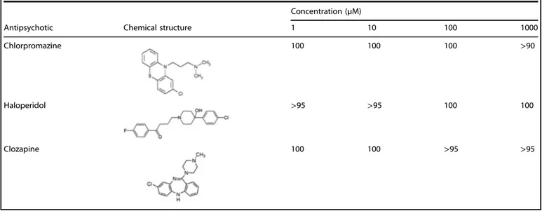
The enzymatic activity of human DDO (hDDO) is not affected by widely usedfirst- and second-generation antipsychotics Since the use of antipsychotic medications may represent a potential confounding factor for the analysis ofD-Asp levels in the
post-mortem SCZ brains, we tested in vitro whether some of the widely used antipsychotics may interfere with recombinant hDDO activity. To this aim, we tested twofirst-generation antipsychotics, haloperidol and chlorpromazine, and a second-generation
Fig. 3 DDO gene methylation and DDO mRNA expression in the dorsolateral prefrontal cortex (DLPFC) and hippocampus (Hipp.) of schizophrenia-affected patients (SCZ) and control (Ctrl) subjects. a Structure of the putative promoter of the human DDO gene. Arrow indicates the transcription start site (TSS,+1). White, black, and gray boxes represent, respectively, the putative regulatory upstream region, the exon 1 and thefirst inton. Position of primers used for bisulfite analysis is indicated by arrows at the top of the map (DdoFw, DdoRv). Vertical bars represent the relative positions of each CpG site. Black circles represent the CpG sites analyzed. b, c Average methylation of the seven CpG sites analyzed is compared b between the two diagnosis groups (DLPFC: Ctrl, n= 20; SCZ, n = 20−hippocampus: Ctrl, n = 20; SCZ, n = 19), or c between the two brain regions (Ctrl: DLPFC, n= 20; hippocampus, n = 20−SCZ: DLPFC, n = 20; hippocampus, n = 19). The percent (%) of methylation represents the average of the seven CpG sites contained in the analyzed region. d, e DDO gene transcription is compared d between the two diagnosis groups (DLPFC: Ctrl, n= 20; SCZ, n = 20−hippocampus: Ctrl, n = 20; SCZ, n = 20), and e between the two brain regions (Ctrl: DLPFC, n= 20; hippocampus, n = 20−SCZ: DLPFC, n = 20; hippocampus, n = 20). The DDO mRNA expression is normalized to the mean of two housekeeping genes and expressed as arbitrary units. *p< 0.05, **p < 0.01,***p < 0.0001 (Mann–Whitney test). Dots represent the single subjects’ values while bars illustrate the means ± SEM
antipsychotic, clozapine. Overall, the activity assays indicated that these compounds have no effect on hDDO activity, even when their concentration is in the low millimolar range (Table1). These results suggest that the exposure to common antipsychotic
drugs is unlikely to affect the catabolism ofD-Asp in the brain of
patients with SCZ.
Lack of difference in serine racemase protein expression in the DLPFC and hippocampus between patients with SCZ and healthy subjects
Recent findings indicated that the mammalian serine racemase (SRR), the primary enzyme responsible for brainD-Ser production,7
could be also involved in D-Asp biosynthesis.19, 20 Indeed, Srr
knockout mice display not only a strong D-Ser levels decrease
(~90%) in their brain,7but also a substantial reduction (50–65%) in
D-Asp amount in the frontal cortex and hippocampus,19,20where SRR is highly expressed.7Based on this knowledge, and consistent with the neurochemical alteration of D-Asp levels found in SCZ
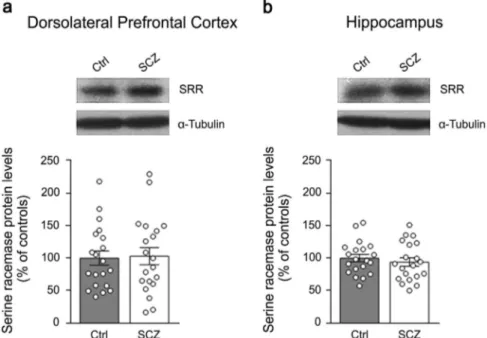
brain samples, we measured SRR protein levels in patients with SCZ and control subjects by western blotting approach. Our data revealed that the protein amount of SRR is comparable between SCZ-affected patients and healthy individuals, both in the DLPFC and hippocampus (p > 0.05, Mann–Whitney test) (Figs.5a, b). Such result highlights that the reported decreasedD-Asp concentration
found in the DLPFC of SCZ patients is not associated to alterations in SRR protein expression.
DISCUSSION
A large bulk of findings supports the influence of NMDAR hypofunction in the pathogenesis of SCZ.11, 12 Accordingly, complex dysfunctions in glutamatergic transmission have been reported at both presynaptic and postsynaptic level,11 including altered mRNA and protein expression of NMDAR subunits in the post-mortem SCZ brain.21,22Besides direct alterations in NMDARs, evidence achieved over the last 15 years suggested that altered metabolism of the co-agonist D-Ser may contribute to the
pathophysiological mechanisms leading to SCZ.2, 3 Accordingly, reduced D-Ser levels have been detected in the CSF4, 5 and serum,6,23,24but not in the post-mortem brain tissues of patients suffering from SCZ.4,25Furthermore, different clinical studies have demonstrated thatD-Ser supplementation could be beneficial for
SCZ symptoms.3,26 In line with previousfindings,4,25 herein we detected comparable or slightly higherD-Ser levels, respectively in
the post-mortem hippocampus and DLPFC of patients with SCZ, compared to healthy subjects. Interestingly, our data revealed that the levels of the enzyme responsible forD-Ser biosynthesis, SRR,7
Fig. 4 Enzymatic activity of humanD-aspartate oxidase (hDDO) in
the dorsolateral prefrontal cortex (DLPFC) and hippocampus (Hipp.) of patients with schizophrenia (SCZ) and control (Ctrl) subjects. DDO enzymatic activity is analyzed a between the two diagnosis groups (DLPFC: Ctrl, n= 20; SCZ, n = 20−hippocampus: Ctrl, n = 20; SCZ, n = 19), and b between the two brain regions (Ctrl: DLPFC, n= 20; hippocampus, n= 20−SCZ: DLPFC, n = 20; hippocampus, n = 20). The enzymatic activity of DDO is expressed as μU/mg of total proteins. **p< 0.01, ***p < 0.0001 (Mann–Whitney test). Dots repre-sent the single subjects’ values while bars illustrate the means ± SEM
Table 1. Inhibition assays showing the relative activity (%) of recombinant human DDO (hDDO) in the presence of different concentrations of antipsychotic drugs
Concentration (µM)
Antipsychotic Chemical structure 1 10 100 1000
Chlorpromazine 100 100 100 >90
Haloperidol >95 >95 100 100
Clozapine 100 100 >95 >95
AbnormalD-aspartate metabolism in schizophrenia T Nuzzo et al 6
do not significantly change between control and SCZ-affected individuals in both the brain regions analyzed. This result is in line with unaltered SRR immunoreactivity found in another cohort of SCZ DLPFC samples,27 although other studies indicate either increased or decreased SRR protein levels in this brain region or in the hippocampus of patients.4, 27, 28 Considering the acknowl-edged relevance of SRR as a risk gene to SCZ vulnerability,29future studies will be necessary to further investigate the SRR protein expression levels in the SCZ brain.
Differently from the co-agonist D-Ser, D-Asp, a D-amino acid
highly enriched in the embryonic brain, acts as an endogenous agonist of NMDARs by stimulating theL-Glu binding sites.9,10,30In
addition, through the activation of presynaptic NMDARs, D-Asp
also evokes the presynaptic release of cortical L-Glu in freely
moving mice analyzed by in vivo microdialysis.31RegardingD-Asp
occurrence, we confirmed in a new post-mortem brain bank our earlier results evidencing an approximate 40% D-Asp levels
reduction in the PFC of SCZ-affected patients.18 Indeed, herein we found a significant 30% decrease of thisD-amino acid content in the DLPFC of SCZ patients compared to control cases. Such a reduction is selective for the DLPFC since we detected comparable levels ofD-Asp in the hippocampus of SCZ-affected and healthy
individuals. In both the brain regions analyzed, our study revealed that the content ofD-Asp did not change over the age span of
subjects, regardless of their clinical diagnosis.
Interestingly, in the DLPFC of SCZ-affected patients the reduced
D-Asp content is linked to significantly increased (~25%) DDO
enzymatic activity. This observation highlights, for thefirst time, the existence of a dysfunctional metabolic process at the level of DDO activity in the SCZ brain. Notably, our in vitro enzymatic assays support a putative direct pathophysiological dysfunction of
D-Asp levels in SCZ since they exclude that the altered D-Asp
catabolism in the DLPFC of patients could depend on secondary effect associated to previous antipsychotic medication. In fact, some of the most commonly used typical (chlorpromazine and haloperidol) or atypical (clozapine) antipsychotic drugs were unable to affect the activity of the recombinant hDDO. However,
we cannot exclude that these drugs, rather than exerting a specific direct action on DDO, may influence D-Asp levels by cumulative effects on different neurotransmitter systems following a lifetime treatment. To our knowledge, this is thefirst report investigating the potential influence of antipsychotic drugs on DDO enzymatic activity; therefore, further investigations will be required to shed light on this important issue. Besides antipsychotic treatment, also the post-mortem delay (PMD)32,33and the age of donors represent potential confounding factors that could affect the tissue levels of amino acids. However, it is unlikely that the higher PMD observed in the brain samples of SCZ patients and their older age, compared to controls, may account for the reducedD-Asp levels
found in the DLPFC since no significant differences in D-Asp
concentration were detected in the hippocampus of the same subjects. Moreover,D-Asp levels did not correlate with age, PMD
and pH, thus reducing the potential confounding effect of these variables. Nevertheless, a larger sample size would be needed to replicate ourfindings.
The altered DDO enzymatic activity detected in this cohort of post-mortem SCZ brains is reminiscent of the increased activity of the D-Ser-degrading enzyme, DAAO, previously found in the
post-mortem brain of SCZ-affected subjects.34,35Altogether, these results suggest that an overall alteration of the enzymes controlling the catabolism of the two main neuroactiveD-amino
acids, i.e., DAAO and DDO, could contribute to NMDAR hypostimulation in SCZ. In agreement with an abnormal degradation of D-Ser in the SCZ brain, severalfindings
demon-strated a genetic association between this neuropsychiatric illness and the gene G72 (refs2,36), which encodes the protein pLG72 that interacts with DAAO and regulates its activity.37In addition to genetic evidence, altered pLG72 protein levels have been found in the plasma of patients with SCZ, compared to control subjects.38 Unlike DAAO, the existence of putative modulators of DDO has never been investigated so far. Therefore, in the light of the dysfunctional DDO activity found in the DLPFC of SCZ patients, future studies will be mandatory to assess the occurrence of regulatory molecules and cellular factors that could tune the
Fig. 5 Serine racemase protein levels in the post-mortem dorsolateral prefrontal cortex and hippocampus of patients with schizophrenia. The expression of serine racemase (SRR) was assessed by western blotting in the a dorsolateral prefrontal cortex and b hippocampus of schizophrenia-affected patients (SCZ) and control (Ctrl) individuals (n= 20 per brain region and diagnosis). The variations of SRR protein levels in patients are expressed as percentage (%) of the control group (healthy subjects).α-Tubulin protein was used to normalize for variations in loading and transfer. Representative blots comparing the two diagnostic groups are shown above the graphs. Dots represent the single subjects’ values while bars illustrate the means ± SEM
metabolic control played by DDO overD-Asp under physiological
and pathologic conditions. In this regard, the recent pioneering research of novel inhibitors specific for hDDO39well suits with the idea to reduce the DDO activity in SCZ brain in the attempt to increase the availability of D-Asp and potentiate, in turn, the
overall NMDAR-mediated transmission.
Besides the altered DDO enzymatic activity observed in the DLPFC of patients with SCZ, our work did not reveal significant variations in the methylation state of DDO transcriptional regulatory region and in the DDO mRNA levels between SCZ-affected and control individuals. We also assessed the potential existence of copy number variations in the DDO gene but the genetic analysis did not evidence alterations in both diagnosis groups (seeSupplementary Resultsand Supplementary Table 4). These data suggest that no gross molecular and genetic alterations occur in this cohort of SCZ cases until post-transcriptional regulatory level. However, the absence of reliable antibodies selective for DDO did not allow us to evaluate the absolute levels of this protein in the post-mortem human brains.
In addition to DDO, we also evaluated the potential contribu-tion of SRR to the reduccontribu-tion ofD-Asp levels observed in the DLPFC.
Indeed, recent data evidenced that suchD-Ser-producing enzyme
could be also involved inD-Asp biosynthesis.19,20 However, our
results excluded that the D-Asp reduction found in the DLPFC
could depend from altered SRR protein levels since our western blotting data revealed comparable amount of this protein between SCZ patients and healthy subjects.
In contrast to the decreasedD-Asp levels found in the DLPFC of
patients, no alteration in the hippocampus was observed, with the exception of an increase in DDO gene methylation, whose biological significance remains unclear. Remarkably, the selectivity of D-Asp reduction in the DLPFC and the reproducibility of this result in another post-mortem brain bank18could overall suggest a greater vulnerability toD-Asp metabolic dysfunctions in a brain are
acritically involved in the pathophysiology of SCZ. Indeed, several reports suggest the occurrence of functional and anatomical alterations in the DLPFC of SCZ-affected patients40that are related to aberrant NMDAR-dependent signaling,22and are likely respon-sible for the typical cognitive symptomatology of SCZ.41 In line with an involvement of the NMDAR agonist, D-Asp, in cortical
processes relevant to SCZ, we previously found that a polymorph-ism in the human DDO gene (rs3757351), associated to reduced DDO mRNA levels (and, therefore, potentially yielding higher D
-Asp levels), predicts greater in vivo prefrontal activity during working memory processing in healthy subjects.17As for humans, research in preclinical models has been recently indicating a link between D-Asp content and cortical phenotypes related to SCZ.
Indeed, we previously demonstrated that increased levels of this
D-amino acid in knockout mice for the Ddo gene (Ddo-/-) enhance
dendritic length and spine density in cortical pyramidal neurons,17 potentiate cortico-hippocampal connectivity, and attenuate the functional activation of cortical areas induced by PCP.16Moreover, oralD-Asp supplementation in mice improves cognitive and social
interaction impairments produced by spared nerve injury of the sciatic nerve.42 Altogether, the beneficial synaptic and cognitive effects ofD-Asp in preclinical models and its reduced
concentra-tion in the human SCZ post-mortem brain suggest that novel strategies, aimed at rising the cerebral availability of this endogenous NMDAR agonist, could be considered in future clinical studies in order to reverse the NMDAR dysfunctions observed in SCZ. The efficacy ofD-Ser as add-on pharmacotherapy
in the treatment of SCZ symptoms3encourages to pursue similar clinical approaches also withD-Asp.
Our research also investigated the regulation of endogenousD
-Asp levels in human DLPFC and hippocampus exerted by DDO, the only known enzyme involved in the catabolism of thisD-amino
acid.9 We confirmed in humans our previous evidence in the mouse brain about the pivotal control played by DDO gene
methylation on DDO gene transcription.8Indeed, our present data revealed that the regional changes in DDO gene methylation (higher in the DLPFC than in the hippocampus) are inversely related to the variations in DDO mRNA levels (lower in the DLPFC than in the hippocampus), regardless of the clinical diagnosis. However, we also found evidence that the regional diversity in DDO gene transcription does not help to explain the higherD-Asp
levels found in the DLPFC, compared to the hippocampus. Indeed, the DDO enzymatic activity detected in the DLPFC and hippocampus is comparable in control subjects or even higher in the DLPFC of SCZ patients. The comprehension of the metabolic mechanisms controlling the endogenous synthesis ofD-Asp in the
brain will be necessary to understand more clearly the present observations and the potential pathophysiological significance of
D-Asp dysregulation.
Differently from its low occurrence in adulthood,D-Asp reaches
the highest levels in the brain during the embryonic phase.1, 8 Although the biological significance ofD-Asp in prenatal phase is
still unknown, it is tempting to hypothesize that such abundance could imply a functional involvement for this D-amino acid in
glutamatergic neurotransmission since in this period NMDARs undergo a strict developmental regulation43 and critically affect fundamental cellular processes, including proliferation, migration, apoptosis, synaptogenesis, and differentiation.44 Moreover, it is noteworthy to mention that besides the mere direct effect produced by D-Asp on postsynaptic NMDARs, this D-amino acid
can also trigger the release of cortical L-Glu through the
stimulation of presynaptic NMDARs,31thus probably acting as an “amplifier” of glutamatergic system. Since the current working model of SCZ postulates that this disorder has a neurodevelop-mental origin linked to NMDAR hypofunction, we hypothesize that precocious alteration in D-Asp metabolism might significantly
impact on NMDAR activity in developmental phase, when genetic abnormalities and aversive environmental factors are known to confer greater susceptibility to develop SCZ symptoms at adulthood.45 Unfortunately, the present study cannot establish whether the altered corticalD-Asp metabolism found in the DLPFC
of SCZ patients has a neurodevelopmental onset or develops at later stages of life. In the light of our hypothesis, such dysfunction would acquire greater relevance if it already existed in the developing brain. The future generation of transgenic animal models with reduced prenatal levels ofD-Asp will be a strategic
tool for clarifying the neurobiological significance of thisD-amino
acid in the immature brain and, in turn, for better defining its potential implication in early NMDAR-dependent processes relevant to SCZ.
MATERIALS AND METHODS Human tissue collection
DLPFC and hippocampus samples from post-mortem brains of non-psychiatrically ill individuals (Ctrl) and SCZ patients (n = 20/diagnosis/brain region) were obtained from The Human Brain and Spinal Fluid Resource Center (Los Angeles Healthcare Center, Los Angeles, CA, USA). All tissue collection and processing was carried out under the regulations and licenses of the Human Tissue Authority and in accordance with the Human Tissue Act of 2004. Clinical diagnosis of SCZ was performed according to DSMIII-R criteria. Demographic characteristics of control and SCZ subjects are described in Supplementary Table1. Frozen tissues were pulverized in liquid nitrogen and stored at−80 °C for subsequent processings. All the following analyses were executed by experimenters blind to the clinical conditions of the subjects.
High-performance liquid chromatography analysis
Brain tissue samples were analyzed as previously reported46with minor modifications.8 Samples were homogenized in 1:20 (w/v) 0.2 M TCA, sonicated (three cycles, 10 s each) and centrifuged at 13,000 g for 20 min The precipitated protein pellets were stored at −80 °C for protein quantification, while the supernatants were neutralized with NaOH and AbnormalD-aspartate metabolism in schizophrenia
T Nuzzo et al 8
subjected to pre-column derivatization with o-phthaldialdehyde/N-acetyl-L-cysteine. Diastereoisomer derivatives were resolved on a Simmetry C8 5-μm reversed-phase column (Waters, 4.6 × 250 mm). Identification and quantification ofD-Ser,L-Ser,D-Asp, andL-Asp were based on retention times and peak areas, compared with those associated with external standards. The identity ofD-Asp,L-Asp, andD-Ser peaks was confirmed by adding known amount of external standards, and by the selective degradation catalyzed by the RgDAAO M213R variant,47StLASPO,48and wild-type RgDAAO,49respectively. The samples were added with 10μg of the enzymes, incubated at 30 °C for 60 min and then derivatized. The amino acids total amount detected in homogenates was normalized by gram of tissue.
DNA methylation analysis
Methylation status of the putative DDO promoter was assessed through a strategy based on the locus-specific amplification of bisulfite-treated genomic DNA, as described inSupplementary Methods.
RNA extraction and quantitative RT-PCR analysis
Total RNA extraction and qRT-PCR were performed as described inSupplementary Methods. DDO mRNA expression levels were normalized to the mean of two housekeeping genes:β-actin and cyclophilin (PPIA). The following primers were used for cDNA amplification: DDO-fw 5′-GGTGTTCATTTGGTATCAGGTTG-3′ and DDO-rev 5′-CTTTCGAAATCCCA-GAACCA-3′; β-actin-fw 5′-TCCTCCCTGGAGAAGAGCTA-3′ and β-actin-rev 5′-CGTGGATGCCACAGGACT-3′; PPIA-fw 5′-TTCATCTGCACTGCCAAGAC-3′ and PPIA-rev 5′-CACTTTGCCAAACACCACAT-3′. DDO mRNA expression was calculated using the relative quantification method (2−ΔΔCt). Enzymatic activity assay
DDO activity measurements in human post-mortem brains were performed by using the Amplex UltraRedfluorescent reagent (Invitrogen, Thermo-Fisher Scientific, Waltham, MA 0245, USA) adapting the procedure previously reported.37,50Detailed procedure is described inSupplementary Methods. DDO activity was normalized by the total protein content and expressed asμU/mg protein.
In vitro inhibition assays
The effect of antipsychotic drugs on hDDO activity was evaluated in vitro using a coupled enzyme assay and the Amplex UltraRed reagent (Life Technologies, Carlsbad, CA USA). Detailed procedure is described inSupplementary Methods.
Western blotting
Samples preparation and immunoblotting were performed as previously described.18 Frozen, powdered samples from post-mortem brains were sonicated in 1% SDS and boiled for 10 min Proteins were separated by sodium dodecyl sulfate polyacrylamide gel electrophoresis and electro-blotted onto polyvinylidene difluoride membranes (GE-Healthcare). Immunodetection was accomplished by using anti-serine racemase (1:500, Santa Cruz Biotechnology, Santa Cruz, CA, USA) and anti-α-tubulin (1:50,000, Sigma, St Louis, MO, USA) antibodies. Blots were then incubated in horseradish peroxidase-conjugated secondary antibodies. Immunoreac-tivity signals were detected by enhanced chemiluminescence (GE-Healthcare) and quantified by Quantity One software (Bio-Rad). Optical density values were normalized toα-tubulin for variations in loading and transfer.
Statistical analysis
Normal distribution assumption for continuous variables was checked by means of Q-Q plot and Shapiro–Wilks and Kolmogorov–Smirnov tests. We observed a non normal (or suspected non normal) distribution forD-/L-Asp and D-/L-Ser, both as absolute levels and as ratios, therefore, a non-parametric approach was used for all statistical analyses. Correlation between continuous variables was assessed using Spearman coefficient. Two-samples comparisons of continuous variables were performed using the Mann–Whitney U-test. A p-value < 0.05 was considered as statistically significant. All analyses were performed using SAS Release 9.4 (SAS Institute, Cary, NC, USA) and GraphPad (GraphPad Prism Software, Inc., La Jolla, CA, USA).
ACKNOWLEDGEMENTS
Tissue specimens were obtained from the Human Brain and Spinal Fluid Resource Center, VA West Los Angeles Healthcare Center, Los Angeles, CA 90073, which is sponsored by NINDS/NIMH, National Multiple Sclerosis Society, and Department of Veterans Affairs. We thank Livia D’Angelo and Clara Di Troia for careful reading of the manuscript and helpful suggestions, and Arianna De Rosa, Antonia Marcone and Vittorio Lucignano for their excellent technical support. F.E. was supported by 2015 NARSAD Young Investigator Grant from the Brain and Behavior Research Foundation (Grant no: 23968). S.S. and L.P. were supported from grants from Fondo di Ateneo per la Ricerca. L.C. was supported by Epigenomics Flagship Project (CNR).
AUTHOR CONTRIBUTIONS
T.N. and S.S. performed HPLC analyses; S.S. and L.P. set-up HPLC and enzymatic activity assays methods, interpreted data, and wrote the work; F.E. designed the work and wrote the manuscript; S.K. and E.F. performed DNA methylation studies under L.C. supervision; O.P. performed genetic studies under M.C. supervision; D.P. and F.N. performed quantitative real-time PCR analyses; M.Co. undertook the statistical analyses; A.B. helped to interpret results; A.U. and L.P. conceived the work and wrote the manuscript.
COMPETING INTEREST
The authors declare that they have no competing interests.
REFERENCES
1. Hashimoto, A. et al. Embryonic development and postnatal changes in free D-aspartate and D-serine in the human prefrontal cortex. J. Neurochem. 61, 348–351 (1993).
2. Labrie, V., Wong, A. H. & Roder, J. C. Contributions of the D-serine pathway to schizophrenia. Neuropharmacology 62, 1484–1503 (2012).
3. Balu, D. T. & Coyle, J. T. The NMDA receptor ‘glycine modulatory site’ in schizophrenia: D-serine, glycine, and beyond. Curr. Opin. Pharmacol. 20, 109–115 (2015).
4. Bendikov, I. et al. A CSF and postmortem brain study of D-serine metabolic parameters in schizophrenia. Schizophr. Res. 90, 41–51 (2007).
5. Hashimoto, K. et al. Reduced D-serine to total serine ratio in the cerebrospinal fluid of drug naive schizophrenic patients. Prog. Neuropsychopharmacol. Biol. Psychiatry 29, 767–769 (2005).
6. Hashimoto, K. et al. Decreased serum levels of D-serine in patients with schizophrenia: evidence in support of the N-methyl-D-aspartate receptor hypofunction hypothesis of schizophrenia. Arch. Gen. Psychiatry 60, 572–576 (2003).
7. Horio, M. et al. Levels of D-serine in the brain and peripheral organs of serine racemase (Srr) knock-out mice. Neurochem. Int. 59, 853–859 (2011).
8. Punzo, D. et al. Age-related changes in D-aspartate oxidase promoter methylation control extracellular D-aspartate levels and prevent precocious cell death during brain aging. J. Neurosci. 36, 3064–3078 (2016).
9. Errico, F., Napolitano, F., Nistico, R. & Usiello, A. New insights on the role of free D-aspartate in the mammalian brain. Amino Acids 43, 1861–1871 (2012). 10. Krashia, P. et al. Persistent elevation of D-Aspartate enhances NMDA
receptor-mediated responses in mouse substantia nigra pars compacta dopamine neurons. Neuropharmacology 103, 69–78 (2016).
11. Moghaddam, B. & Javitt, D. From revolution to evolution: the glutamate hypothesis of schizophrenia and its implication for treatment. Neuropsycho-pharmacology 37, 4–15 (2012).
12. Coyle, J. T. NMDA receptor and schizophrenia: a brief history. Schizophr. Bull. 38, 920–926 (2012).
13. Errico, F. et al. Increased D-aspartate brain content rescues hippocampal age-related synaptic plasticity deterioration of mice. Neurobiol. Aging 32, 2229–2243 (2011).
14. Errico, F. et al. Persistent increase of D-aspartate in D-aspartate oxidase mutant mice induces a precocious hippocampal age-dependent synaptic plasticity and spatial memory decay. Neurobiol. Aging 32, 2061–2074 (2011).
15. Errico, F. et al. D-aspartate prevents corticostriatal long-term depression and attenuates schizophrenia-like symptoms induced by amphetamine and MK-801. J. Neurosci. 28, 10404–10414 (2008).
16. Errico, F. et al. A role for D-aspartate oxidase in schizophrenia and in schizophrenia-related symptoms induced by phencyclidine in mice. Transl. Psy-chiatry 5, e512 (2015).
17. Errico, F. et al. Free D-aspartate regulates neuronal dendritic morphology, synaptic plasticity, gray matter volume and brain activity in mammals. Transl. Psychiatry 4, e417 (2014).
18. Errico, F. et al. Decreased levels of D-aspartate and NMDA in the prefrontal cortex and striatum of patients with schizophrenia. J. Psychiatr. Res. 47, 1432–1437 (2013).
19. Horio, M. et al. Decreased levels of free D-aspartic acid in the forebrain of serine racemase (Srr) knock-out mice. Neurochem. Int. 62, 843–847 (2013).
20. Ito, T. et al. Serine racemase is involved in d-aspartate biosynthesis. J. Biochem. 160, 345–353 (2016).
21. Kristiansen, L. V., Huerta, I., Beneyto, M. & Meador-Woodruff, J. H. NMDA receptors and schizophrenia. Curr. Opin. Pharmacol. 7, 48–55 (2007).
22. Weickert, C. S. et al. Molecular evidence of N-methyl-D-aspartate receptor hypofunction in schizophrenia. Mol. Psychiatry 18, 1185–1192 (2013). 23. Yamada, K. et al. Identification of multiple serine racemase (SRR) mRNA isoforms
and genetic analyses of SRR and DAO in schizophrenia and D-serine levels. Biol. Psychiatry 57, 1493–1503 (2005).
24. Calcia, M. A. et al. Plasma levels of D-serine in Brazilian individuals with schizophrenia. Schizophr. Res. 142, 83–87 (2012).
25. Kumashiro, S., Hashimoto, A. & Nishikawa, T. Free D-serine in post-mortem brains and spinal cords of individuals with and without neuropsychiatric diseases. Brain. Res. 681, 117–125 (1995).
26. Hashimoto, K., Malchow, B., Falkai, P. & Schmitt, A. Glutamate modulators as potential therapeutic drugs in schizophrenia and affective disorders. Eur. Arch. Psychiatry Clin. Neurosci. 263, 367–377 (2013).
27. Steffek, A. E., Haroutunian, V. & Meador-Woodruff, J. H. Serine racemase protein expression in cortex and hippocampus in schizophrenia. Neuroreport 17, 1181–1185 (2006).
28. Verrall, L. et al. d-Amino acid oxidase and serine racemase in human brain: normal distribution and altered expression in schizophrenia. Eur. J. Neurosci. 26, 1657–1669 (2007).
29. Schizophrenia Working Group of the Psychiatric Genomics C. Biological insights from 108 schizophrenia-associated genetic loci. Nature 511, 421–427 (2014).
30. Errico, F., Mothet, J. P. & Usiello, A. D-Aspartate: An endogenous NMDA receptor agonist enriched in the developing brain with potential involvement in schizophrenia. J. Pharm. Biomed. Anal. 116, 7–17 (2015).
31. Cristino, L. et al. d-Aspartate oxidase influences glutamatergic system homeostasis in mammalian brain. Neurobiol. Aging 36, 1890–1902 (2015). 32. Hashimoto, K., Sawa, A. & Iyo, M. Increased levels of glutamate in
brains from patients with mood disorders. Biol. Psychiatry 62, 1310–1316 (2007).
33. Perry, T. L., Hansen, S. & Gandham, S. S. Postmortem changes of amino compounds in human and rat brain. J. Neurochem. 36, 406–410 (1981). 34. Burnet, P. W. et al. D-amino acid oxidase activity and expression are increased in
schizophrenia. Mol. Psychiatry 13, 658–660 (2008).
35. Madeira, C., Freitas, M. E., Vargas-Lopes, C., Wolosker, H. & Panizzutti, R. Increased brain D-amino acid oxidase (DAAO) activity in schizophrenia. Schizophr. Res. 101, 76–83 (2008).
36. Sacchi, S., Binelli, G. & Pollegioni, L. G72 primate-specific gene: a still enigmatic element in psychiatric disorders. Cell. Mol. Life Sci. 73, 2029–2039 (2016).
37. Sacchi, S. et al. pLG72 modulates intracellular D-serine levels through its inter-action with D-amino acid oxidase: effect on schizophrenia susceptibility. J. Biol. Chem. 283, 22244–22256 (2008).
38. Lin, C. H. et al. Distinctively higher plasma G72 protein levels in patients with schizophrenia than in healthy individuals. Mol. Psychiatry 19, 636–637 (2014). 39. Katane, M. et al. Identification of novel D-aspartate oxidase inhibitors by in
silico screening and their functional and structural characterization in vitro. J. Med. Chem. 58, 7328–7340 (2015).
40. Weinberger, D. R. et al. Prefrontal neurons and the genetics of schizophrenia. Biol. Psychiatry 50, 825–844 (2001).
41. Gilmour, G. et al. NMDA receptors, cognition and schizophrenia--testing the validity of the NMDA receptor hypofunction hypothesis. Neuropharmacology 62, 1401–1412 (2012).
42. Palazzo, E. et al. D-Aspartate drinking solution alleviates pain and cognitive impairment in neuropathic mice. Amino Acids 48, 1553–1567 (2016).
43. Cull-Candy, S., Brickley, S. & Farrant, M. NMDA receptor subunits: diversity, development and disease. Curr. Opin. Neurobiol. 11, 327–335 (2001).
44. Jansson, L. C. & Akerman, K. E. The role of glutamate and its receptors in the proliferation, migration, differentiation and survival of neural progenitor cells. J. Neural. Transm. (Vienna) 121, 819–836 (2014).
45. Fatemi, S. H. & Folsom, T. D. The neurodevelopmental hypothesis of schizo-phrenia, revisited. Schizophr. Bull. 35, 528–548 (2009).
46. Topo, E. et al. Thyroid hormones and aspartic acid, aspartate oxidase, D-aspartate racemase, H2O2, and ROS in rats and mice. Chem. Biodivers. 7, 1467–1478 (2010).
47. Sacchi, S. et al. Engineering the substrate specificity of D-amino-acid oxidase. J. Biol. Chem. 277, 27510–27516 (2002).
48. Bifulco, D., Pollegioni, L., Tessaro, D., Servi, S. & Molla, G. A thermostable L-aspartate oxidase: a new tool for biotechnological applications. Appl. Microbiol. Biotechnol. 97, 7285–7295 (2013).
49. Molla, G. et al. Role of arginine 285 in the active site of Rhodotorula gracilis D-amino acid oxidase. A site-directed mutagenesis study. J. Biol. Chem. 275, 24715–24721 (2000).
50. Cappelletti, P., Campomenosi, P., Pollegioni, L. & Sacchi, S. The degradation (by distinct pathways) of human D-amino acid oxidase and its interacting partner pLG72--two key proteins in D-serine catabolism in the brain. FEBS J. 281, 708–723 (2014).
This work is licensed under a Creative Commons Attribution 4.0 International License. The images or other third party material in this article are included in the article’s Creative Commons license, unless indicated otherwise in the credit line; if the material is not included under the Creative Commons license, users will need to obtain permission from the license holder to reproduce the material. To view a copy of this license, visithttp://creativecommons.org/licenses/by/ 4.0/
© The Author(s) 2017
Supplementary Information accompanies the paper on the npj Schizophrenia website (doi:10.1038/s41537-017-0015-7). AbnormalD-aspartate metabolism in schizophrenia
T Nuzzo et al 10