OSAS
Transoral glossoepiglottopexy in the treatment of
adult obstructive sleep apnoea: a surgical approach
Glossoepiglottoplastica con approccio trans-orale nel trattamento delle apnee
ostruttive notturne nell’adulto
V. ROUSTAN1, M. BARBIERI1, F. INCANDELA1, F. MISSALE1, H. CAMERA1, F. BRAIDO2, R. MORA1, G. PERETTI1 1 Department of Otorhinolaryngology and Head and Neck Surgery, 2 Allergy and Respiratory Diseased Clinic DIMI,
University of Genoa, IRCCS AUO San Martino-IST, Genoa, Italy SUMMARY
The treatment of obstructive sleep apnoea syndrome (OSAS) is still a matter of debate; among the different therapeutic alternatives, both surgical and conservative, treatment with continuous positive airway pressure (CPAP) is considered the “gold standard”. The recent scien-tific literature reports that even if CPAP represents an effective solution for sleep apnoeas, 12% of patients do not benefit from its use. In most cases, primary collapse of the epiglottis is responsible for failure. We developed a surgical technique that provides a stable support to the epiglottis without influencing its function during swallowing while preserving laryngeal anatomy and physiology. The procedure we propose is based on that conceived by Monnier for children affected by laryngomalacia. We analysed a group of 20 patients who underwent glossoepiglottopexy between January 2015 and September 2016 and compared data (AHI, ODI, t90, ESS, EAT10, etc.) collected before and 6 months after surgery to demonstrate the safety and effectiveness of our glossoepiglottopexy (GEP). The results allow us to consider GEP as a valid choice to treat adults who suffer from sleep apnoeas.
KEY WORDS: OSAS • Epiglottis • Transoral laser microsurgery (TLM)
RIASSUNTO
I dati della letteratura scientifica internazionale stabiliscono che almeno il 10% della popolazione adulta sia affetto da Sindrome delle Apnee Ostruttive nel Sonno (OSAS). Nonostante le numerose proposte terapeutiche, chirurgiche e non, che si sono susseguite negli anni, la ventiloterapia con C-PAP rappresenta ad oggi il gold standard per il trattamento di questi pazienti. Esiste comunque una percentuale di pazienti, stimata attorno al 12%, che non trae beneficio dalla terapia ventilatoria o che, addirittura, peggiora in corso di C-PAP; il col-lasso primario dell’epiglottide, ovvero quello dipendente esclusivamente da un’eccessiva lassità della cartilagine, verso l’aditus laringeo rappresenta la causa più frequente di insuccesso in questi soggetti. Sulla base di queste osservazioni abbiamo deciso di sviluppare una tecnica chirugica volta unicamente alla stabilizzazione dell’epiglottide che fosse conservativa e potenzialmente reversibile. La procedura che proponiamo prende spunto dall’epiglottoplastica proposta da Monnier per i pazienti pediatrici affetti da laringomalacia. Lo studio comprende 20 pazienti sottoposti a glossoepiglottoplastica tra Gennaio 2015 e Settembre 2016; diversi parametri (tra i quali AHI, ODI, t90, ESS, EAT10 ed altri) sono stati confrontati tra quelli registrati nel per operatorio e dopo 6 mesi di follow-up per valutare l’efficacia dell’intervento chirurgico. I risultati evidenziano come la glossoepiglottopessi rappresenti una valida possibilità di trattamento delle epi-glottidi primarie responsabili della sindrome delle apnee ostruttive notturne nell’adulto.
PAROLE CHIAVE: OSAS • Epiglottide • Microchirurgia laser transorale (TLM)
Acta Otorhinolaryngol Ital 2018;38:38-44
Introduction
Obstructive sleep apnoea (OSA) is a disorder caused by repetitive collapse of the upper airway during sleep, re-sulting in either partial or complete airflow obstruction 1 2.
In the adult population, the prevalence of OSA is 22% in men and 17% in women 3. The morphology of
upper-airway structures plays a major role in the pathogenesis of OSA.
Treatment of OSA with continuous positive airway pres-sure (CPAP) is currently considered as the “gold stand-ard”. Despite its proven efficacy, a significant number of patients cannot tolerate the device and require therapeutic
alternatives such as surgery, oral appliances and/or posi-tional devices.
In the past, the prevalence of epiglottis collapse evaluated by clinical examination was estimated to be 12% in OSAS patients, although nowadays thanks to the introduction of drug-induced sedation endoscopy (DISE) it is possible to show that the prevalence of epiglottis collapse in deter-mining the airway obstruction is actually much higher 4-6,
and also allows for stratification of OSAS into different levels of obstruction. Nonetheless, the role of the epiglot-tis in airway collapse still remains a matter of debate in the medical community 1 7.
The laxity of the epiglottis is widely discussed in the pae-diatric literature as one of the possible presentations of congenital laryngomalacia; however, laryngomalacia in adults is a rare entity that has not been clearly described. Primary collapse of the epiglottis represents a challenging situation because CPAP treatment may aggravate airway obstruction by further pushing the epiglottis down into the laryngeal aditus 8, and up to now no standardised surgical
procedures have been described.
Herein we describe a new surgical procedure, obtained by modifying Monnier’s glossoepiglottopexy, used in malac-ic epiglottis in children 9, with the aim to resolve the
col-lapse of the epiglottis in adults without interfering with its fundamental functions.
Materials and methods
From January 2015 to September 2016, 20 patients (16 males and 4 females), aged between 35 and 65 years old (mean 49.2) affected by OSAS underwent glossoe-piglottopexy at the Department of Otorhinolaryngology and Head and Neck Surgery of the University of Genoa. All patients were submitted to multilevel surgery (nasal-pharyngopalatal-laryngeal) performed by a senior sur-geon. If tonsils were still in place a tonsillectomy was performed 10, sparing the palatoglossus and
palatopharyn-geus muscles and as much as possible the mucosal cov-ering both pillars 11. Septoplasty and turbinoplasty 12 was
performed if needed 13 .
All patients were evaluated in a preoperative setting and after 6 months after surgery by clinical, polysomnograph-ic and endoscoppolysomnograph-ic assessment. All data were extracted from a single database 14.
Inclusion criteria were: OSA confirmed by sleep study (with an apnoea hypopnoea index (AHI) ≥ 15 episodes/h, BMI < 35 kg/m2; primary epiglottic collapse evaluated by
drug-induced sedation endoscopy (DISE) and Laryngo-score < 6 15.
Major comorbidities, severe tongue base hypertrophy,
secondary epiglottis, cranio-facial malformations and la-ryngeal dysfunction were considered exclusion criteria. The study was approved by our institutional review board.
Clinical evaluation
Clinical evaluation included a complete head and neck ex-amination as well as body mass index (BMI) calculation. Moreover, all patients were evaluated by a comprehensive history that covered sleep habits and sleep disturbances. Ex-cessive daytime sleepiness was estimated by the Epworth sleepiness scale (ESS) 16, while swallowing by the Eating
Assessment Tool (EAT 10) 17 and the penetration aspiration
scale 18. EAT 10 is a self-administered, symptom-specific
outcome survey that was investigated before and one week after surgery, while the penetration aspiration scale is an endoscopic finding that expresses the accidental loss of food or liquid into the airway while eating or drinking. The possibility to have an adequate exposure of the larynx by a trans-oral approach was evaluated by the Laryngoscore 15.
Polysomnographic study
Polysomnographic study was performed with a cardiores-piratory monitor (Vital night, Vital aire, Milan Italy) to record the following variables simultaneously: nocturnal snoring sound, arterial oxygen saturation measured by finger oximetry, body position, nasal and mouth airflow, thoracic and abdominal respiratory movements recorded by inductive plethysmography and heart rate. The entity of sleep apnoea was evaluated by: AHI; oxygen desatu-ration index (ODI); T < 90% (average desatudesatu-ration and the percentage of total time with oxygen saturation level lower than 90%); mean oxygen saturation (SpO2 mean).
Endoscopic evaluation
Flexible video nasopharyngoscopy (V-NFL) was used for endoscopic evaluation. To perform this procedure, we used a transnasal flexible endoscopy using an ENF-V2 videoendoscope connected to an EvisExera II CLV-180B light source (Visera Elite OTV-S190, Olympus Medical Systems Corporation, Tokyo, Japan), integrated with high-definition television (HDTV). Upper airway and digestive tract morphology was examined. The Muller manoeuvre was performed in a sitting position to determine the extent of retropalatal (Muller RP) and retrolingual (Muller RL) obstruction; it was considered significant when the per-centage of collapse of the airway was at least 50%.
Drug-induced sedation endoscopy (DISE)
DISE was performed in a supine position without neck extension. The entire exam was recorded. No local anaes-thesia was used in the nasal cavity according to the
Eu-ropean position statement on DISE 19. Sleep was induced
with a bolus of 1.3 mg of midazolam in pre-anaesthesia and intravenous propofol in target controlled infusion (TCI) (1.1-1.6 μg/ml according Schnider’s model) 20. The
degree of obstruction was evaluated at the level of the na-sal cavity, nasopharynx, oropharynx, hypopharynx and larynx using the NOHL classification 21.
Surgical technique
During microlaryngoscopy, the patient lies supine in Boyce-Jackson’s position with the eyes protected by wet bandages and the superior teeth by a silicone device. The smallest efficient tube (Laser Shield®II Endotracheal
Tube, Medtronic Xomed, Jacksonville, FL USA) for trans oral-intubation was chosen. Exposure of the supraglot-tis and the base of the tongue was obtained by a Sataloff laryngoscope (Microfrance Sataloff Laryngoscopes 124, Medtronic ENT, Jacksonville FL USA) to achieve ad-equate exposure of both the base of the tongue and the entire valleculae and epiglottis. The position of the hyoid bone was marked on the skin as a landmark.
A CO2 laser (Ultrapulse Dualpro Laser CO2, Lumenis, Yokneam, Israel), set on ultrapulse mode, at 3 watts of de-livered power at a 400 mm working distance, was used to vaporize the mucosa overlying the valleculae and the base of the tongue and outlining two raw areas leaving a rim of 2-4 mm of healthy mucosa along the entire profile of the supra-hyoid epiglottis. The cicatricial retraction during the healing process determined an iatrogenic synechia attract-ing the epiglottis to the tongue base 14. The surgical field
was mostly bloodless thanks to the ability of the CO2 laser to coagulate vessels with a diameter less than 0.5 mm. For larger vessels, cauterising monopolar forceps was routinely adopted and surgical clips are also strongly recommended. In case of hypertrophy of the lingual tonsils, resection of the lymphatic tissue may be combined. A cottonoid, soaked in saline solution, was applied intraoperatively to remove char and clean the raw surface for detection of any residual intact mucosal island as a cause of possible retention cysts when the two surfaces adhere to each other.
To ensure complete sealing of the pexy, two Premilene®
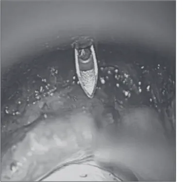
sutures number 1 (Premilene, Braun, Melsungen Germa-ny) embraced the hyoid bone guided by means of two 16 gauge catheters. The first suture was bent in the middle to form a loop and was inserted inside the first needle to exit from the tip. Later, the catheter was passed through the skin, in a supra-hyoid position, along the midline to avoid vascular injuries, until reaching the glosso-epiglottic val-lecula. This procedure was monitored under microscopic control (Fig. 1). The needle was then pushed through the epiglottis and retrieved out of the mouth.
The second suture was inserted into the second catheter and penetrated soft tissues beneath the hyoid (Figs. 2, 3). Finally, the surgeon inserted the proximal end of the wire
Fig. 1. The first needle penetrates the skin of the neck in a suprahyoid position. This needle contains a suture bent to form a loop. In MLSD the first surgeon controls that the needle penetrates the epiglottis in a correct posi-tion and he then retrieves the loop out of the mouth.
Fig. 2. The second suture was inserted into the second catheter and pen-etrated soft tissues beneath the hyoid.
within the loop while holding it with one hand (Fig. 4). The second surgeon retracts the distal part of the loop, with a firm movement, allowing the second suture to exit from the skin. Both wires are then fixed anteriorly to the larynx using a Silastic sheet, on the anterior neck, to protect the skin from local trauma. Before tying the suture, a small amount of
Tisseel® (Baxter, Westlake Village, CA USA) is spread in
the glosso-epiglottic vallecula using a Duplocath®
(Bax-ter, Westlake Village, CA USA) catheter.
Postoperative care
The patient was extubated at the end of the procedure. A clinical swallow evaluation was obtained on the first postoperative day. In case of normal scores, the patient was allowed to resume oral intake, including thin liquids, and was placed on a soft diet for 1 week. Intravenous an-tibiotics were administered for 24 hours. The patient was discharged after 48 hours of observation. Oral antibiotics and analgesics were recommended for one week.
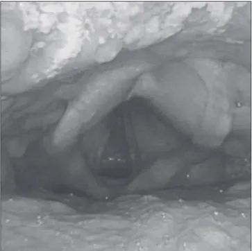
The stich was removed endoscopically after 4 weeks (Fig. 5); neither general or local anaesthesia is required. Flexible laryngoscopy was performed after 1 week, testing the penetration-aspiration scale, and was repeated 3 weeks postoperatively. All patients filled in the EAT 10. All sub-jects also underwent an additional polysomnography test-ing 6 months after the procedure, while postoperative views were recorded at the time of control polysomnography.
Statistical analysis
Statistical analysis was performed with IBM SPSS Statis-tics version 24.0. Data were collected on a paired sample basis and comparisons were done using a nonparametric Wilcoxon Signed Rank Test. The Wilcoxon Signed Rank
Fig. 3. Endoscopic view after the insertion of the second suture. Fig. 5. The suture is removed after 4 weeks under endoscopic control. The epiglottis results perfectly stabilised.
Fig. 4. The proximal end of the wire is inserted within the loop that is used as a guide to make the free suture exit the neck.
Test was used to test for statistically significant differenc-es between pre- and post-operative parameters. P < 0.05 was accepted as statistically significant.
Results
Twenty patients affected by OSAS with primary epiglottic collapse were included in this study. All patients (100%) underwent glossoepiglottopexy and pharyngoplasty, ac-cording to the palatal conformation: 10 (50%) Barbed Reposition Pharyngoplasty (BRP) 11, 6 (30%) Functional
Expansion Pharyngoplasty (FEP) 22, 4 (20%) Barbed
Ro-man Blinds Technique (BRB) 23 24. Tonsillectomy was
per-formed in 11 (55%) patients and septoplasty and turbino-plasty in 4 (20%) patients (Table I).
Transoral glossoepiglottopexy was associated with a sig-nificant decrease in ESS scores (p < 0.001) evaluated at 6 months after surgery (16.5 ± 4.3 vs. 3.1 ± 2.5).
Clinical swallow evaluation and EAT-10 score was nor-mal in all the patients; in particular, only one patient reported that extra effort was needed to swallow pills, with a total score of 2. All patients received a score of 1 on the penetration-aspiration scale in their first swal-lows.
Mean ± sd AHI values were 23.6 ± 6.5 and 5.2 ± 3.2 events/ hour, before and six months after surgery respectively, and this difference was statistically significant (p < 0.001). ODI
presented a significantly lower value at 6 months after sur-gery (19.3 ± 6.1 vs. 4.8 ± 2.7, p < 0.001). There was a sig-nificant decrease of T < 90% values, which decreased from 9.7 ± 3.4 to 1.4 ± 2.0 (p < 0.001). Mean oxygen saturation (mean SpO2) showed an increase at final evaluation vs. pretreatment levels 86.9 ± 2.3 vs. 94.7 ± 2.3 (p < 0.001) (Table I).
Discussion
A positive correlation between high BMI and OSA is well known in the literature; the pressure of the parapharyngeal fat pads and chronic collapse of the retroglottal airway dur-ing sleep create deformity of the epiglottis observed in pa-tients with OSA. Thus, the shape of the epiglottis reflects the degree of the chronic airway obstruction. In patients suffering from OSAS, primary collapse of the epiglottis is due to the high negative intrathoracic pressure generated during obstructive events, in contrast to that which charac-terises laryngomalacia.
However, even when the epiglottis represents a cause of ob-struction, its function in protecting the upper airway must be taken into account. The role played by the epiglottis in pre-venting inhalation of bolus is two-fold: on one hand, it offers a physical closure of the laryngeal aditus, while on the other its sensitive receptors (distributed on the laryngeal surface, ariepiglottic folds, arytenoids and posterior commissure)
Table I. Comparison of data collected before and 6 months after surgery.
Patients Surgery Questionnaires Polysomnography
Age Sex Pharyngoplasty Tonsillectomy Septoplasty and turbinoplasty ESS pre ESS post EAT10 pre EAT10 post PAS pre PAS post AHI pre AHI post ODI pre ODI post T90% pre T90% post mean SpO2 pre mean SpO2 post 50 M BRP Yes No 16 8 0 0 1 1 25 10 20 10 10 2 88 93 45 M FEP Yes No 11 3 0 0 1 1 17 5 10 5 5 0 92 92 63 M FEP No Yes 15 3 0 0 1 1 20 4 15 3 10 0 89 98 45 M BRP No No 20 6 0 0 1 1 30 8 30 8 20 8 86 95 51 M BRB No No 18 0 0 2 1 1 18 0 10 2 5 0 88 93 38 M BRB Yes No 24 0 0 0 1 1 38 10 25 5 10 3 86 92 42 M FEP No No 12 5 0 0 1 1 22 5 15 5 8 3 89 97 46 M BRP Yes No 10 2 0 0 1 1 28 8 19 7 10 0 86 95 50 M BRP Yes No 20 4 0 0 1 1 26 6 15 6 10 1 84 96
56 F FEP Yes Yes 20 5 0 0 1 1 40 10 35 8 15 2 83 93
53 F BRP No No 10 3 0 0 1 1 18 8 18 8 5 0 89 98 48 M BRB Yes No 11 0 0 0 1 1 22 1 20 1 12 0 86 97 35 M BRP Yes No 13 4 0 0 1 1 25 6 21 6 10 2 88 95 46 M BRP No No 15 0 0 0 1 1 17 1 15 2 8 0 89 95 49 M FEP No Yes 20 5 0 0 1 1 20 3 20 2 5 0 88 97 53 M BRB Yes No 20 0 0 0 1 1 18 2 16 2 5 0 87 95 65 M BRP Yes No 23 5 0 0 1 1 27 7 25 7 9 3 85 96 49 M BRP Yes No 20 3 0 0 1 1 22 5 20 2 10 0 86 94 55 F BRP No Yes 16 6 0 0 1 1 21 3 20 2 16 0 83 92 45 F FEP No No 15 0 0 0 1 1 18 2 16 5 11 3 85 90
stimulate the so-called “glottis closure reflex”. This reflex is coordinated by the vagal nerve, and more precisely by its superior laryngeal branch, which provides the abduc-tion of the vocal folds during deglutiabduc-tion thus avoiding the entrance of bolus in the airways. To maintain these two functions, two technical precautions must be taken into consideration: 1) it is necessary to leave a 3-4 mm rim of healthy cartilage and mucosa along the entire profile of the epiglottis to address food to the piriform sinuses; 2) the mucosa of the supraglottis (except the lingual aspect of the epiglottis) and the glottis should be preserved to allow the activation of reflexes.
Since the advent of DISE, the indications for the laryn-geal surgery for OSAS have significantly changed, due to the fact that the awake endoscopy with Muller manoeuvre alone underestimates the obstructive component at this level by 40% 25.
To date, surgical approaches in this region are techni-cally complex, significantly invasive and associated with a high rate of complications, such as bleeding, oedema, persistent dysphagia, dysgeusia, etc. 26. New technologies
(diathermy, CO2 laser and co-blation) have allowed the introduction of innovative surgical techniques aimed at modifying the epiglottis and negatively interfering with its sphincter function 27.
Conclusions
The surgical technique described herein is able to pre-serve laryngeal anatomy and physiology, and is safe and reversible in case of unexpected complications. Moreover, it provides stable support to the epiglottis without interfering with its function during swallowing and reinforces the wall of the airway by creating a type of landslide barrier for posterior falling of the tongue base.
References
1 Torre C, Camacho M, Liu SY, et al. Epiglottis collapse in adult obstructive sleep apnea: a systematic review. Laryngo-scope 2016;126:515-23.
2 Ma MA, Kumar R, Macey PM, el al. Epiglottis cross-sectional area and oropharyngeal airway length in male and female ob-structive sleep apnea patients. Nat Sci Sleep 2016;8:297-304.
3 Franklin KA, Lindberg E. Obstructive sleep apnea is a com-mon disorder in the population-a review on the epidemiology of sleep apnea. J Thorac Dis 2015;7:1311-22.
4 Cavaliere M, Russo F, Iemma M. Awake versus drug-induced sleep endoscopy: evaluation of airway obstruction in ob-structive sleep apnea/hypopnoea syndrome. Laryngoscope 2013;123:2315-8.
5 Fernández-Julián E, García-Pérez MÁ, García-Callejo J, et
al. Surgical planning after sleep versus awake techniques
in patients with obstructive sleep apnea. Laryngoscope 2014;124:1970-4.
6 Koutsourelakis I, Safiruddin F, Ravesloot M, et al. Surgery for obstructive sleep apnea: sleep endoscopy determinants of outcome. Laryngoscope 2012;122:2587-91.
7 Montevecchi F, Cammaroto G, Meccariello G, et al. Tran-soral robotic surgery (TORS): a new tool for high risk tracheostomy decannulation. Acta Otorhinolaryngol Ita 2017;37:46-50.
8 Dedhia RC, Rosen CA, Soose RJ. What is the role of the larynx in adult obstructive sleep apnea? Laryngoscope 2014;124:1029-34.
9 Monnier P. Pediatric airway surgery: management of laryn-gotracheal stenosis in infants and children. Berlin: Springer 2010.
10 Verse T, Kroker B, Pirsing W, et al. Tonsillectomy for treat-ment of obstructive sleep apnea in adults with tonsillar hy-pertrophy. Laryngoscope 2000;110:1556-9.
11 Vicini C, Hendawy E, Campanini A, et al. Barbed reposi-tion pharyngoplasty (BRP) for OSAHS: a feasibility, safety, efficacy and teachability pilot study. “We are on the giant’s shoulders”. Eur Arch Otorhinolaryngol 2015;272:3065-70.
12 Barbieri M, Salami A, Mora F, et al. High frequency surgery in the treatment of turbinate hypertrophy: 11’s years’ experi-ence. Acta Otorhinolaryngol Ita 2003;23:436-9.
13 Passali FM, Bellussi L, Mazzone S, et al. Predictive role of nasal functionality tests in the evaluation of patients before nocturnal polysomnografic recording. Acta Otorhinolaryn-gol Ita 2011;31:103-8.
14 Passali D, Caruso G, Arigliano LC, et al. Database appli-cation for patients with obstructive sleep apnoea syndrome. Acta Otorhinolaryngol Ita 2012;32:252-5.
15 Piazza C, Mangili S, Bon FD, et al. Preoperative clinical predictors of difficult laryngeal exposure for microlaryngos-copy: the Laryngoscore. Laryngoscope 2014;124:2561-7.
16 Rosenthal LD, Dolan DC. The Epworth Sleepiness Scale in the identification of obstructive sleep apnea. J Nerv Ment Dis 2008;196:429-31.
17 Belafsky PC, Mouadeb DA, Rees CJ, et al. Validity and re-liability of the Eating Assessment Tool (EAT-10). Ann Otol Rhinol Laryngol 2008;117:919-24.
18 Robbins J, Coyle J, Rosenbek J, et al. Differentiation of normal and abnormal airway protection during swallow-ing usswallow-ing the penetration-aspiration scale. Dysphagia 1999;14:228-32.
19 De Vito A, Carrasco Llatas M, Vanni A, et al. European po-sition paper on drug-induced sedation endoscopy (DISE).
Sleep Breath 2014;18:453-65.
20 Barbieri M, Del Piazzo A, Della Casa P, et al. La Sleep En-doscopy come criterio di valutazione pre-chirurgica nei pa-zienti affetti da turbe del sonno. Valsalva 2009 Vol LXXXV n 3/4.
21 Vicini C, De Vito A, Benazzo M, et al. The nose orophar-ynx hypopharorophar-ynx and larorophar-ynx (NOHL) classification: a new system of diagnostic standardized examination for OSAHS patients. Eur Arch Otorhinolaryngol 2012;269:1297-300.
22 Sorrenti G, Piccin O. Functional expansion pharyngoplasty in the treatment of obstructive sleep apnea. Laryngoscope 2013;123:2905-8.
23 Mantovani M, Rinaldi V, Torretta S, et al. Barbed Ro-man blind technique for the treatment ob obstructive sleep apnea: how we do it? Eur Arch Otorhinolaryngol 2016;273:517-23.
24 Salamanca F,Constantini F, Mantovani M, et al. Barbed
an-terior pharyngoplasty: an evolution of anan-terior palatoplasty.
Acta Otorhinolaryngol Ital 2014;34:434-8.
25 Friedman M, Tanyeri H, La Rosa M, et al. Clinical predictors of obstructive sleep apnea. Laryngoscope 1999;109:1901-7.
26 Bourolias C, Hajiioannou J, Sobol E, et al. Epiglottis reshap-ing usreshap-ing CO2 laser: a minimally invasive technique and its potent applications. Head Face Med 2008;4:15.
27 Lin HC, Friedman M, Chang HW, et al. Z-palatopharyngo-plasty combined with endoscopic coblator open tongue base resection for severe obstructive sleep apnea/hypopnea syn-drome. Otolaryngol Head Neck Surg 2014;150:1078-85.
Received: July 10, 2017 - Accepted: September 1, 2017
Address for correspondence: Valeria Roustan, Department of Otorhi-nolaryngology and Head and Neck Surgery, University of Genoa, San Martino Hospital, largo Rosanna Benzi 10, 16132 Genoa, Italy. E-mail: [email protected]