Head and neck
Pilot study on microvascular anastomosis:
performance and future educational prospects
Studio pilota sulle microanastomosi vascolari: risultati a confronto e prospettive
didattiche future
G. BERRETTI1, G. COLLETTI2, G. PARRINELLO1, A. IAVARONE1, P. VANNUCCHI1, A. DEGANELLO1
1 Department of Surgery and Translational Medicine, University of Florence, Florence, Italy; 2 Maxillofacial Surgery,
San Paolo Hospital, University of Milan, Italy SUMMARY
The introduction of microvascular free flaps has revolutionised modern reconstructive surgery. Unfortunately, access to training opportu-nities at standardised training courses is limited and expensive. We designed a pilot study on microvascular anastomoses with the aim of verifying if a short course, easily reproducible, could transmit microvascular skills to participants; if the chosen pre-test was predictive of final performance; and if age could influence the outcome. A total of 30 participants (10 students, 10 residents and 10 surgeons) without any previous microvascular experience were instructed and tested during a single 3 to 5 hour course. The two microanastomoses evaluated were the first ever performed by each participant. More than the half of the cohort was able to produce both patent microanastomoses in less than 2 hours; two-thirds of the attempted microanastomoses were patent. The pretest predicted decent scores from poor performances with a sensitivity of 61.5%, specificity of 100%, positive predictive value of 100% and negative predictive value of 40%. Students and residents obtained significantly higher scores than surgeons. Since our course model is short, cost-effective and highly reproducible, it could be in-troduced and implemented anywhere as an educational prospect for preselecting young residents showing talent and natural predisposition and having ambitions towards microvascular reconstructive surgery.
KEY WORDS: Microvascular anastomosis • Free flap • Microvascular training • Surgical skills • Education
RIASSUNTO
L’introduzione dei lembi liberi microvascolari ha rivoluzionato la moderna chirurgia ricostruttiva. Purtroppo, l’accesso a corsi specifi-ci e intensivi è attualmente limitato e costoso. Abbiamo organizzato uno studio pilota sulle microanastomosi vascolari con lo scopo di verificare: se un corso economico e facilmente riproducibile potesse trasmettere ai partecipanti delle abilità microchirurgiche di base; se il test preliminare scelto fosse predittivo dei risultati finali; e se l’età potesse essere un fattore in grado di influenzare la performance. Sono stati selezionati un numero complessivo di 30 partecipanti (10 studenti, 10 specializzandi e 10 chirurghi), senza nessuna precedente esperienza microchirurgica, ai quali è stato proposto un corso della durata di 3-5 ore. Le anastomosi conclusive, sottoposte a valutazione, sono state le prime realizzate da ciascun partecipante. Più della metà degli individui testati è stata in grado di confezionare entrambe le microanasromosi funzionanti e in un tempo inferiore alle 2 ore; nel complesso due terzi delle microanastomisi realizzate erano pervie. Il test preliminare è stato in grado di predire risultati finali buoni rispetto a risultati finali scarsi con una sensibilità del 61%, una specificità del 100%, un valore predittivo positivo pari al 100% ed un valore predittivo negativo del 40%. Studenti e specializzandi hanno ottenuto punteggi significativamente migliori rispetto ai chirurghi. Dato che il corso da noi proposto è breve, dai costi contenuti e facilmente ripro-ducibile, riteniamo possa essere facilmente implementato in altre strutture al fine di selezionare giovani specializzandi dotati di talento con una naturale predisposizione per la microchirurgia, e che dimostrino interesse ed ambizione nel campo della chirurgia ricostruttiva.
PAROLE CHIAVE: Anastomosi microvascolari • Lembi liberi • Esercitazioni microvascolari • Abilità chirurgiche • Apprendimento
Introduction
In the early years of the 20th century, Carrel codified the triangulation technique obtaining functioning vascular anastomosis 1-3 to investigate the feasibility of organ
trans-plantation.
Otolaryngologists were the first physicians to use micro-surgical techniques. In 1921, the microscope was used for the first time in ear surgery by a Swedish otologist, Carl Olof Nylen 3 4, the next year Gunnar Holmgren replaced
the first monocular microscope with a binocular one 4.
The application of the microscope to vascular anastomo-sis allowed the first successful microvascular transfer of a jejunal interposition flap in 1959 5.
The introduction of microvascular free flaps revolution-ised the surgical treatment of head and neck tumours and represents one of the major advancements in the manage-ment of these neoplasms. Microsurgery requires consider-able practice before any attempt is made at clinical appli-cation. Live animals represent the ideal training medium;
however, unlimited access to the animal laboratory is not always possible 6. In such situations, segments of fresh
human placenta or other surrogates such as pig coronaries or chicken wing vessels can provide adequate specimens for microsurgical practice.
At our tertiary referral academic hospital, to date, no specific microvascular training program is available for students, residents or surgeons, and no specific financial support is provided for those who intend to build up these skills attending national and/or international microvascu-lar courses. This situation is diffuse all over the country, and this aspect penalises even more youngest surgeons with access to limited economic resources, despite the fact that it seems that age in itself plays an important role in the abrupt of the learning process. In fact, a study comparing the results of microsurgical education after the completion of a comprehensive microsurgical course pro-gram between students and surgeons demonstrated that the students achieved higher scores, with a significant bet-ter performance in tissue handling 7.
We designed a pilot study on microvascular anastomoses with the aim of verifying if even a simple short course, easily reproducible, could transmit microvascular skills to participants; if the chosen pre-test was predictive of the final performance; and if age could influence the out-come.
Materials and methods
From April to September 2016, 30 voluntary participants were enrolled and tested in our pilot study.
Population
Inclusion criteria were: motivation and willingness to par-ticipate, complete lack of previous microvascular experi-ence, age in concordance with the professional position. The cohort was formed of 3 groups;
group 1 (G1): 10 students attending the sixth (last) year of medical school, all aged under 28 years (mean 24.8), with no surgical experience; 7 expressed ambition to a career in a surgical specialty;
group 2 (G2): 10 residents in surgical disciplines (5 in otolaryngology, 3 in general surgery, 1 in vascular sur-gery, and 1 in orthopaedic surgery), aged between 28 and 35 years (mean 29.8), usually attending macroscopic sur-gery as second operator, sometimes as first operator under supervision, but none with specific experience on micro-vascular anastomosis or microscopic ear surgery;
group 3 (G3): 10 surgeons (8 otolaryngologists, 1 vas-cular surgeon, and 1 maxillofacial surgeon) aged over 35 years (mean 51.5) usually performing macroscopic surgery as first operator. The 8 otolaryngologists were skilled in microscopic laryngeal surgery, but none had any experience in microvascular anastomosis or micro-scopic ear surgery.
Microanastomosis course
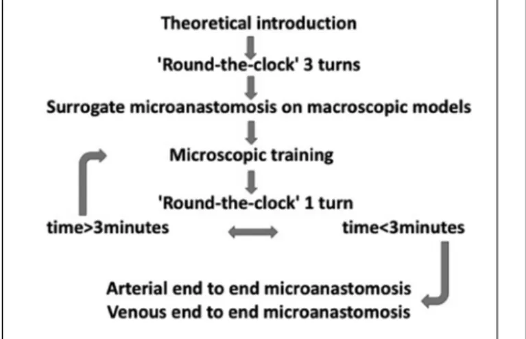
Each participant was instructed and then individually tested in one session that varied between 3 to 5 hours; a schematic representation of the structure of the course is displayed in Figure 1.
A brief theoretical introduction to experimental microsur-gery was provided: correct posture, use of the microscope, handling of microinstruments, end to end microsuture technique for artery and vein, exercises on a macroscopic model for microscopic arterial and venous microanasto-moses and tasks required.
A preliminary pre-test was initially administrated, namely the so-called ‘round-the-clock’ training model 8, which
proved to be an inexpensive and readily available valid tool to provide instant assessment of the individual micro-surgical predisposition (Fig. 2). Participants were asked to pass a 9/0 nylon microsuture needle through the needle eyes of nine sewing needles planted on a rigid support, each run had to be completed clockwise; the time of each run for 3 consecutive runs was recorded, without consid-ering steadiness or handling abilities.
Participants then familiarised with microsuture tech-niques for end-to-end arterial and venous anastomoses using a macroscopic model: the artery surrogate was a silicon tube, since the lumen is maintained; the vein surrogate was a finger glove, since lumen tend to col-lapse. When performing surrogate microanastomoses, the same handling rules and same techniques required for true microanastomoses had to be respected, with the only difference of working without the microscope and using a 5/0 suture. For the surrogate arterial microanas-tomosis, the triangulation technique with interrupted sutures was used; for the surrogate venous anastomosis, after placing the first two stay sutures at 2 o’clock and 10 o’clock, the superior 180° were sutured, and then the inferior 180°. During the surrogate microanastomoses the instructor surveyed the participant, pointing out and explaining all handling errors according to the
cal requirements previously explained in the theoretical introduction.
The next step was microscopic training on an animal mod-el; we used one half rooster per participant since it provided good quality vessels, very similar in diameter to those en-countered for microvascular head and neck reconstructions, with average diameter of 3 mm (range 2.5 mm - 3.5 mm). Participants were free to familiarise themselves with micro-sutures using the sciatic nerve or other tissues, but not ves-sels; no time restrictions were given for this training, while the instructor provided constant feedback on handling and errors, by pointing out: incorrect posture, incorrect use of the microscope, incorrect microinstrument handling, incor-rect tissue handling, incorincor-rect needle handling and needle damage, unwanted perforations, asymmetric knots place-ment, incorrect knots tying. All this followed the require-ments listed by the Northwestern Objective Microanasto-mosis Assessment Tool (NOMAT) 9.
After microscopic training, participants performed a fourth run at the ‘round-the-clock’ test, if the exercise was completed in less than 3 minutes (this cut-off was chosen considering four times the mean time recorded by the ex-perienced microsurgeon, AD) they could move forward to perform the microanastomoses, otherwise they had to re-turn to the microscopic training before re-attempting an-other ‘round-the-clock’ run until the threshold of 3 min-utes was satisfied. Times recorded on this fourth run at the ‘round-the-clock’ test were considered good when below 2 minutes, intermediate when between 2 and 3 minutes, and poor when above 3 minutes.
Microvascular end-to-end arterial anastomosis using 8/0 nylon interrupted sutures was performed first, then the
ve-nous end-to-end anastomosis was performed. Participants had one-hour to complete each microanastomosis or to complete at least the minimal-task. During the microanas-tomoses participants were surveyed, but no feedback on errors and no suggestions were provided by the instructor, who, instead, took note and counted all errors.
All macro-microsurgical instruments were propriety of the senior author (AD), all animal models were bought by the first author (GB) at her own expense; the macro-micro surgical suture materials were outdated and ready to be discarded.
Scoring system
Arterial and venous microanastomoses were separately evaluated. The minimal-task for the artery was considered to be the correct placement of the first 3 stay sutures and this was awarded with 50 points, the minimal-task for the vein was the correct suture of the superior 180°, for which 50 points were awarded. Zero points were scored in case of inability/incorrect placement of the first 3 stay sutures on the arterial microanastomosis, inability/incorrect suture of the superior venous wall, or in case of minimal-task result-ing in lumen obliteration. If the minimal-task was met, the participant could move forward to complete the microanas-tomosis as much as possible within the one-hour time limit. The time to complete each microanastomosis was re-corded, and microanastomoses were evaluated in terms of patency and continence, 100 points were awarded if the microanastomosis was patent and continent, 95 points if the microanastomosis was patent but leaking if one ex-tra stitch was needed and 90 points if two exex-tra stitches were needed. In case three or more stitches were required to restore continence, or if the microanastomosis was not patent, the score was 50 points considering only achieve-ment of the minimal-task. At the final score 2 points were deducted for each incorrect gesture performed during the microanastomosis.
Global (arterial + venous) scores above 160 were considered high, scores between 100 and 160 were considered interme-diate and scores below 100 were considered low.
Times recorded for the fourth run at the ‘round-the-clock’ test were correlated with the microanastomoses global scores.
Statistical analysis
Differences between groups were tested with ANOVA; the variables, all continuous, were expressed as mean val-ues and standard deviation, and were compared between two different groups with the t-Student test: p values less than 0.05 were considered statistically significant.
Results
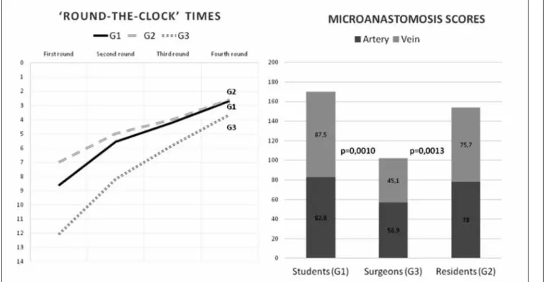
Times (minutes’ and seconds’’) of the first, second, third and fourth run at the ‘round-the-clock’ are shown in Ta-ble I; the analysis showed a significant difference for the
Fig. 2. The ‘round-the-clock’ test, each run had to be completed clockwise under microscopic magnification at 6X using 9/0 suture.
fourth round between G1 and G2 vs. G3 (p = 0.025). Mean arterial and venous microanastomoses scores are shown in Table II.
Comparison of the global (arterial + venous) perfor-mance between groups showed a significant difference between G1 vs. G3 (p = 0.0010) and between G2 vs. G3 (p = 0.0013), indicating a better performance for students and residents compared to surgeons, while no differences were seen between students and residents (p = 0.47), Figure 3.
As already stated, all participants had to complete each microanastomosis within a maximum time limit of 1 hour. The mean arterial and venous microanastomoses times were, respectively, 56 and 51 minutes for G1, 48 and 52 minutes for G2 and 52 and 52 minutes for G3.
The 3 minute cut-off time on the fourth run at the ‘round-the-clock’ test was able to predict good and intermediate microanastomoses global scores from poor performances with a sensitivity of 61.5% and a specificity of 100%; the positive predictive value was 100% and the negative pre-dictive value was 40%.
Discussion
The surgeon’s experience is a critical factor influencing the outcomes of microvascular reconstructions, and the suc-cess rate in experienced hands is usually above 95% 10-12.
However, a study on the first year of clinical experience of three microvascular head and neck surgeons yielded a success rate of 97.5%, showing that well-trained junior mi-crovascular surgeons can achieve survival rates comparable with those of experts 12.
Lascar et al. compared the patency rates obtained by resi-dents in plastic surgery with different microsurgical ex-perience, varying from residents of the first year with no microsurgical experience to residents of the sixth year with considerable experimental and clinical microsurgical experience. A proportional narrowing of outcome differ-ences among groups was seen with the increase in number of microanastomoses performed by the less experienced ones, until no outcome differences were recorded after 52 microanastomosis 13, demonstrating that constant intense
training is necessary to achieve high standards. In this view, a 3 to 5 hour course is usually considered much too short for formal microsurgery training. However, the principal
Table I. Results at ‘round-the-clock’ test
First run Second run Third run Fourth run G 1 8’38” 5’33” 4’11” 2’41”
G 2 7’ 4’59” 4’22” 2’35”
G 3 12’ 8’11” 5’5” 3’38”
Table II. Mean scores of arterial and venous microanastomoses.
ARTERY VEIN
G 1 82.8 (SD 21.66) 87.5 (SD 15.62) G 2 78 (SD 31.10) 75.7 (31.68) G 3 56.9 (SD 42.04) 45.1 (SD 38.45)
SD: standard deviation
philosophy of the study was not focused on offering inten-sive microsurgical education, but rather on implementing a highly accessible and inexpensive microsurgery facility. Our course was effective in transmitting a microvascular starting point, with the aim of identifying the youngest and most talented physicians for future advancements.
The results indicate that even a simple, rapid, highly repro-ducible and low-cost course can effectively transmit micro-vascular skills to participants. In fact, more than the half of the cohort (7 students, 7 residents and 3 surgeons) was able to produce, in less than 2 hours, both arterial and venous microanastomoses functioning. Furthermore, two-thirds (39 of 60) of the attempted microanastomoses were patent, and among these, 20 were also perfectly continent, while in 17 cases one extra stitch was needed to stop leakage, and in 2 cases 2 stitches were needed. The time limit of 1 hour per microanastomosis was set because from a clinical point of view two hours is an acceptable ischaemia time for most fascio-cutaneous and osteo-cutaneous free flaps.
These results are remarkable considering that none of the subjects possessed any microvascular experience, no one had attended other microvascular courses before and the two microanastomoses considered were the first ever per-formed by all participants. Our evaluation system is not validated, but we preferred to design a scoring system that was more focused on final microanastomosis evaluation rather than homogeneously evaluating the ongoing per-formance during the microanastomosis. We felt that the NOMAT system was more suitable for monitoring the evolution in building up the global microvascular experi-ence within more structured courses, rather than assessing the results of a single attempt in a mini-course.
The ‘round-the-clock’ test with a 3 minute cut-off time confirmed to be an effective exercise and a powerful pre-dictive tool with 100% specificity and positive prepre-dictive values, which means that it could be effectively used to preselect subjects who almost surely will perform well. In fact, none of the 16 subjects who completed the fourth run in less than 3 minutes had a poor microanastomosis global score: 13 had good performance (5 students, 6 residents, 2 surgeons) and 3 an intermediate performance (2 stu-dents and 1 resident).
Students and residents obtained significantly higher scores than surgeons. This seems to confirm that age is a real and crucial factor for acquisition of microvascular skills: 3 surgeons were unable to complete the minimal task and only 3 produced both functioning microanasto-moses. Therefore, our data indicate that age seems to be more determinant than surgical background experience for the acquisition of microvascular skills, and this is par-alleled by the finding that surgeons were more prone to errors than the younger participants.
For all participants, the arterial microanastomosis was per-formed first and the venous anastomosis was perper-formed subsequently. Venous microanastomosis is recognized to
be technically more difficult than an arterial one, mainly because the vessel lumen tend to collapse, and beginners are expected to perform better with less demanding du-ties; G1, instead, was the only group with venous scores higher than the arterial ones. This data is interesting: stu-dents demonstrated the ability to keep on learning and perfecting the technique as ’on the job’ training. When performing a venous anastomosis, students were already familiarised with the microscope and with microinstru-ments during the previous microanastomosis.
Conclusions
Our mini-course aroused substantial enthusiasm among all participants. In our country, surgery is unfortunately started late and practiced in autonomy mostly by aged surgeons after the achievement of permanent working po-sitions. Residents are often frustrated by the lack of suf-ficient hands-on experience during training, and our data confirm the importance of investing in young, talented and motivated surgeons, since they can produce excellent performances. Since this course model is short-timed, cost-effective and highly reproducible, it could be intro-duced and implemented anywhere as a future educational prospect for preselecting young residents showing talent and natural predisposition and having ambitions towards microvascular reconstructive surgery.
Acknowledgements
This paper was awarded with the 3rd SIO Price at the 104th
National Congress of the Italian Society for Otorhinolar-yngology and Head and Neck Surgery, Sorrento 2017.
References
1 Carrel A. The operative technique of vascular anastomoses and the transplantation of viscera. Lyon Med 1902;98:859-63.
2. Carrel A, Guthrie CC. Functions of a transplanted kidney.
Science 1905;22:473.
3 Carrel A, Guthrie CC. A new method for the homoplastic transplantation of the ovary. Science 1906;23:591.
4 Dohlman GF. Carl Olof Nylén and the birth of the otomicro-scope and microsurgery. Arch Otolaryngol 1969;90:813-7.
5 Seidenberg B, Rosenak SS, Hurwitt ES, et al. Immediate re-construction of the cervical esophagus by a revascularized isolated jejunal segment. Ann Surg 1959;149:162-71.
6 Goldstein M. Use of fresh human placenta for microsurgical training. J Microsurg 1979;1:70-1.
7 Mücke T, Borgmann A, Ritschl LM et al. Microvascular training of medical students and surgeons - a comparative prospective study. J Craniomaxillofac Surg 2013;41:e187-90.
8 Chan WY, Figus A, Ekwobi C et al. The ‘round-the-clock’ training model for assessment and warm up of
microsurgi-cal skills: a validation study. J Plast Reconstr Aesthet Surg 2010;63:1323-8.
9 Aoun SG, El Ahmadieh TY, El Tecle NE et al. A pilot study to assess the construct and face validity of the Northwestern Objective Microanastomosis Assessment Tool. J Neurosurg 2015;123:103-9.
10 Blackwell KE, Brown MT, Gonzalez D. Overcoming the learning curve in microvascular head and neck reconstruc-tion. Arch Otolaryngol Head Neck Surg 1997;123:1332-81.
11 Mathes SJ. Plastic Surgery. 2nd ed. Philadelphia: Elsevier;
2006. pp 507-538.
12 Khouri R. Avoiding free flap failure. Clin Plast Surg
1992;19:773-5.
13 Lascar I, Totir D, Cinca A et al. Training program and learn-ing curve in experimental microsurgery durlearn-ing the residency in plastic surgery. Microsurgery 2007;27:263-7.
Address for correspondence: Alberto Deganello, Unit of Otorhi-nolaryngology-Head and Neck Surgery, Department of Surgical Specialties, Radiological Sciences, and Public Health, University of Brescia, ASST Spedali Civili, piazzale Spedali Civili 1, 25123 Brescia, Italy. E-mail: [email protected]