ISSN: 1524-4628
Copyright © 2011 American Heart Association. All rights reserved. Print ISSN: 0039-2499. Online Stroke is published by the American Heart Association. 7272 Greenville Avenue, Dallas, TX 72514
DOI: 10.1161/STROKEAHA.110.592246
2011;42;17-21; originally published online Nov 24, 2010;
Stroke
Padovani and on behalf of the Italian Project on Stroke in Young Adults Investigators
Zini, Paolo Cerrato, Paolo Costa, Mauro Magoni, Licia Iacoviello, Alessandro
Rota, Maurizia Rasura, Massimo Del Sette, Alessia Giossi, Irene Volonghi, Andrea
Alessandro Adami, Maria Luisa DeLodovici, Elisabetta Del Zotto, Lidia Luciana
Gandolfo, Federica Casoni, Rossella Musolino, Rocco Salvatore Calabrò, Paolo Bovi,
Alessandro Pezzini, Mario Grassi, Corrado Lodigiani, Rosalba Patella, Carlo
Italian Project on Stroke in Young Adults
Predictors of Migraine Subtypes in Young Adults With Ischemic Stroke: The
http://stroke.ahajournals.org/cgi/content/full/42/1/17
located on the World Wide Web at:
The online version of this article, along with updated information and services, is
http://www.lww.com/reprints
Reprints: Information about reprints can be found online at
410-528-8550. E-mail:
Fax: Kluwer Health, 351 West Camden Street, Baltimore, MD 21202-2436. Phone: 410-528-4050. Permissions: Permissions & Rights Desk, Lippincott Williams & Wilkins, a division of Wolters
http://stroke.ahajournals.org/subscriptions/
Ischemic Stroke
The Italian Project on Stroke in Young Adults
Alessandro Pezzini, MD; Mario Grassi, PhD; Corrado Lodigiani, MD, PhD; Rosalba Patella, MD;
Carlo Gandolfo, MD; Federica Casoni, MD; Rossella Musolino, MD; Rocco Salvatore Calabro`, MD;
Paolo Bovi, MD; Alessandro Adami, MD; Maria Luisa DeLodovici, MD; Elisabetta Del Zotto, MD;
Lidia Luciana Rota, MD; Maurizia Rasura, MD; Massimo Del Sette, MD; Alessia Giossi, MD;
Irene Volonghi, MD; Andrea Zini, MD; Paolo Cerrato, MD; Paolo Costa, MD; Mauro Magoni, MD;
Licia Iacoviello, MD, PhD; Alessandro Padovani, MD, PhD; on behalf of the Italian Project on Stroke
in Young Adults Investigators
Background and Purpose—The mechanisms underlying the relationship between migraine and ischemic stroke remain uncertain. The aim of the present study was to investigate the predictive value of major cardiovascular risk factors, cardiac interatrial abnormalities, and additional biological markers on migraine subtypes in young adults with ischemic stroke. Methods—Ischemic stroke patients aged 45 years or younger were consecutively enrolled as part of the Italian Project on
Stroke in Young Adults. A comprehensive evaluation was performed including assessment of self-reported migraine and cardiovascular risk factors, interatrial right-to-left shunt, and genotyping to detect factor V Leiden and the G20210A mutation in the prothrombin gene.
Results—Nine hundred eighty-one patients (mean age, 36.0⫾7.6 years; 50.7% women) were included. The risk of migraine with aura increased with decreasing number of cardiovascular risk factors (OR, 0.50; 95% CI, 0.24 – 0.99 for 2 factors or more), increasing number of thrombophilic variants (OR, 2.21; 95% CI, 1.05– 4.68 for carriers of at least 1 of the 2), and the presence of right-to-left shunt (OR, 2.41; 95% CI, 1.37–3.45), as compared to patients without migraine. None of these factors had influence on the risk of migraine without aura.
Conclusions—In young adults with ischemic stroke, low cardiovascular risk profile, right-to-left shunt, and an underlying procoagulant state are predictors of migraine with aura. The biological effects of these factors should be considered in future studies aimed at investigating the mechanisms linking migraine to brain ischemia. (Stroke. 2011;42:17-21.)
Key Words: migraine 䡲 patent foramen ovale 䡲 stroke
A
lthough a large body of literature supports an association between migraine, especially migraine with aura (MA), and ischemic stroke,1the mechanisms underlying the relationbetween the 2 disorders remain to be elucidated. Several pathogenic processes have been advocated and, at least theoretically, might be operant synergistically. First, evidence indicating that MA is a risk factor for different ischemic
disorders, including cardiovascular death, stroke, myocardial infarction, angina, and claudication,2– 4has led to
hypothesiz-ing that MA may predispose to systemic atherosclerosis, which increases the risk for each of these vascular diseases through similar mechanisms. If this is the case, then a higher prevalence of risk factors known to be associated with cardiovascular disease among those with MA is expected.
Received June 2, 2010; accepted September 20, 2010.
From the Dipartimento di Scienze Mediche e Chirurgiche (A. Pezzini, E.D.Z., A.G., I.V., P.C., A. Padovani), Clinica Neurologica, Universita` degli Studi di Brescia, Brescia, Italia; Dipartimento di Scienze Sanitarie Applicate (M.G.), Sezione di Statistica Medica e Epidemiologia, Universita` di Pavia, Pavia, Italia; Centro Trombosi (C.L., L.L.R.), IRCCS Istituto Clinico Humanitas, Rozzano-Milano, Italia; Stroke Unit (R.P., M.R.), Azienda Ospedaliera Sant’Andrea, Roma, Italia; Dipartimento di Neuroscienze (C.G.), Oftalmologia e Genetica, Universita` di Genova, Genova, Italia; Stroke Unit (F.C., A.Z.), Clinica Neurologica, Nuovo Ospedale Civile “S. Agostino Estense,” AUSL Modena, Italia; Dipartimento di Neuroscienze (R.M.), Scienze Psichiatriche e Anestesiologiche Clinica Neurologica, Universita` di Messina, Messina, Italia; Istituto di Ricovero e Cura a Carattere Scientifico (R.S.C.), Centro Neurolesi Bonino-Pulejo, Messina, Italia; UO Neurologia (P.B.), Azienda Ospedaliera-Universitaria Borgo Trento, Verona, Italia; Stroke Center (A.A.), Dipartimento di Neurologia, Ospedale Sacro Cuore Negrar, Verona, Italia; Unita` di Neurologia (M.L.D.), Ospedale di Circolo, Universita` dell’Insubria, Varese, Italia; Unita` di Neurologia (M.D.S.), Ospedale S. Andrea, La Spezia, Italia; Dipartimento di Neuroscienze (P.C.), Stroke Unit, Universita` di Torino, Torino, Italia; Stroke Unit (M.M.), Neurologia Vascolare, Spedali Civili di Brescia, Brescia, Italia; Laboratori di Ricerca (L.I.), “RE ARTU,” Centro di Ricerca e Formazione ad Alta Tecnologia nelle Scienze Biomediche, “Giovanni Paolo II,” Universita` Cattolica del Sacro Cuore, Campobasso, Italia.
The online-only Data Supplement is available at http://stroke.ahajournals.org/cgi/content/full/STROKEAHA.110.592246/DC1.
Correspondence to Alessandro Pezzini, MD, Dipartimento di Scienze Mediche e Chirurgiche, Clinica Neurologica, Universita` degli Studi di Brescia, P.le Spedali Civili, 1, 25100 Brescia, Italia. E-mail [email protected]
© 2010 American Heart Association, Inc.
Stroke is available at http://stroke.ahajournals.org DOI: 10.1161/STROKEAHA.110.592246
Second, a number of investigators have reported an increased prevalence of patent foramen ovale, a potential risk factor for stroke among younger individuals, in patients with MA, prompting speculation that this cardiac interatrial abnormality may explain at least part of the ischemic risk in patients with MA.5Third, migraine might predispose to ischemic stroke by
inducing platelet-related hypercoagulability as a consequence of an underlying procoagulant state attributable to coexistent coagulation abnormalities and endothelial dysfunction.6,7
Be-sides the obvious implication in elucidating the biological mechanisms linking the 2 diseases, the identification of factors influencing such a relationship may help in stratifying subpopulations of those with migraines, enabling preventive strategies to be targeted at those subjects at higher risk for stroke. The Italian Project on Stroke in Young Adults (IPSYS) provides the opportunity to investigate these issues because of its large sample size, the homogeneous demo-graphic characteristics and clinical phenotype of the subjects included, and the standard diagnostic work-up. Therefore, in the present study we aimed at evaluating whether individual cardiovascular risk profile varies according to migraine sub-type, and whether specific risk factors clustering may be used as a predictive marker of migraine subtypes in a cohort of Italian ischemic stroke patients aged 45 years or younger.
Subjects and Methods
The study was approved by the local Ethics Committee. Informed consent was provided by all study participants. The IPSYS is a countrywide network of neurological centers with special interest in cerebral ischemia at a young age across Italy (see the online Appendix, available at http://stroke.ahajournals.org) aimed at recruiting patients with first-ever acute stroke who fulfill the following criteria: (1) age 18 to 45 years and 2) CT- or MRI-proven cerebral infarction in the setting of a hospital-based, multicentric, observational study. Centers are included in the network provided that the recruitment process of stroke cases takes place prospectively. Stroke was defined as a sudden loss of global or focal cerebral function that persisted for⬎24 hours with a probable vascular cause. Ischemic strokes attributable to sinus venous thrombosis, vasospasm after subarachnoid hemorrhage, cardiac surgery, occurring as an immediate consequence of trauma, and iatrogenic strokes were excluded. For the purpose of the present analysis, we screened data sets from patients consecutively admitted to 10 hospitals between January 2000 and July 2009.
Risk Factor Definition
The following risk factors for premature cerebral ischemia were retained: hypertension, diabetes mellitus, cigarette smoking, hyper-cholesterolemia, and oral contraceptive use. These variables were defined and dichotomized as follows: hypertension (systolic blood pressure ⱖ140 mm Hg and diastolic pressure ⱖ90 mm Hg in 2 separate measurements after the acute phase or use of antihyperten-sive drugs before recruitment), diabetes mellitus (history of diabetes, use of hypoglycemic agent or insulin, or fasting glucose ⱖ126 mg/dL), current smoking (including former smokers who had quit smoking for 6 months before the index event), hypercholesterolemia (cholesterol serum levelsⱖ220 mg/dL or use of cholesterol-lowering drugs), and oral contraceptive use (including former users who had quit using these medications for 1 month before the index event). Assessment of Migraine History
Personal history of headache was assessed in all patients by study physicians during a face-to-face interview in both acute phase and follow-up evaluations. History of migraine before stroke occurrence was considered for the present analysis. The diagnoses of migraine
without aura (MO) and MA were made according to the diagnostic criteria of the International Headache Society.8
Clinical and Laboratory Investigations
All patients underwent an etiologic work-up including complete blood cell count, biochemical profile, urinalysis, 12-lead ECG, chest roent-genography, Doppler ultrasonography with frequency spectral analysis and B-mode echotomography of the cervical arteries, transcranial Doppler ultrasonography, and CT and/or MR angiography to investigate extracranial and intracranial vessels. The performance of specialized coagulation testing (including prothrombin and activated partial throm-boplastin times, antiphospholipid antibodies, fibrinogen, protein C, protein S, activated protein C resistance, antithrombin III, and genotyp-ing to detect factor V Leiden and the G20210A mutation in the prothrombin gene) was left to the discretion of the investigator in charge of the patient and no obvious cause of infarct was detected. Transtho-racic and transesophageal echocardiography were performed to rule out cardiac sources of emboli. In particular, interatrial right-to-left shunt (RLS) was assessed in all patients with transesophageal echocardiogra-phy with a contrast study and Valsalva maneuver and/or transcranial Doppler sonography with intravenous injection of agitated saline. A RLS was considered present if any microbubble was seen in the left atrium within 3 cardiac cycles from maximum right atrial opacification on echocardiography.9Transcranial Doppler sonography with
intrave-nous injection of agitated saline was performed according to the Venice Consensus Conference.10Briefly, it consists of the injection of 9 mL of
saline solution and 1 mL air mixed with a 3-way stopcock by exchange of saline/air mixture between the syringes and injected as a bolus as a contrast-enhancing agent into the right cubital vein 5 seconds before the start of a 10-second Valsalva maneuver while recording the flow velocity of the middle cerebral artery, insonated through the temporal window on the right side at a depth of 50 to 60 mm, with a handheld probe. The appearance of transient spikes on the velocity spectral curve is considered positive for interatrial RLS. The method has an overall diagnostic accuracy comparable to that of transesophageal echocardi-ography with a contrast study and Valsalva maneuver.11Based on the
results of such investigations, patients were classified according to a classification based on the Trial of Org 10172 in Acute Stroke Treatment criteria, accommodated and validated for the cause of stroke in the young,12and divided into 5 etiologic categories: (1)
atheroscle-rotic vasculopathy; (2) nonatheroscleatheroscle-rotic vasculopathy; (3) small-vessel disease; (4) cardioembolism; and (5) other (cerebral infarction that did not meet the criteria for 1 of the categories outlined). Stroke was classified as “anterior circulation stroke” when involving the areas of the brain supplied by carotid arteries and their branches, and as “posterior circulation stroke” when involving the brain stem, the cerebellum, or territories supplied by the posterior cerebral arteries.
Statistical Analysis
An individual proatherosclerotic score (from 0 – 4) was computed based on the number of major cardiovascular risk factors (hyperten-sion, diabetes, smoking, and hypercholesterolemia). Similarly, a prothrombotic score (from 0 –2) was obtained for each subject based on the individual status of carrier of factor V Leiden and the G20210A mutations. The median value of each score (1 for proath-erosclerotic score and 0 for prothrombotic score) was used to get binary variables. Differences among the migraine groups (no mi-graine, MO, and MA) were examined with the2test, median test,
and ANOVA F test, when appropriate. Categorical (multinomial) logistic regression model was planned to examine the conditional effect of RLS, proatherosclerotic risk profile, and thrombophilic defects in the prediction of migraine subgroups, and adjusted for age and gender. Results are given as OR (as measures of migraine– covariate interaction effects on disease risk) with 95% CI. P⬍0.05 on 2-sided test was considered significant. Data were analyzed using the SPSS (version 16) package.
Results
The current study targets 1017 patients enrolled in the IPSYS registry. Among these, patients with missing migraine history
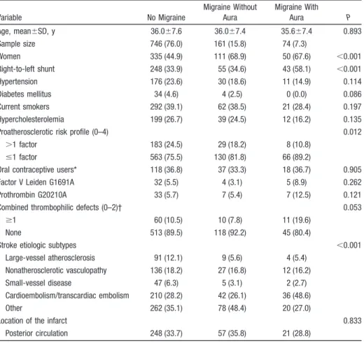
(n⫽36) were excluded, leaving a final sample size of 981 (mean age, 36.0⫾7.6 years; 50.7% women) for the analysis. Demo-graphic characteristics of the study population grouped accord-ing to migraine status and the prevalence of selected risk factors are presented in Table 1. Large-vessel atherosclerosis and small-vessel disease were the presumed cause of infarct in 112 (11.0%) cases and 55 (5.4%) cases, respectively, nonatheroscle-rotic vasculopathy in 179 (17.6%) cases, cardiac/transcardiac embolism in 301 (29.6%) cases, and other etiologies in the remaining 369 (36.3%) cases. Patients who did not have mi-graines were more often males, more often had hypertension, diabetes mellitus, and hypercholesterolemia, were more often smokers, and, overall, were more likely to have an unfavorable vascular risk profile compared to patients with migraine, espe-cially with the specific MA subtype. Conversely, RLS and individual and combined prothrombotic genotypes were more frequent in the subgroup of patients with MA than in the subgroup of patients with no history of migraine, and in the subgroup of patients with MO. There were no differences in the preva-lence of oral contraceptive users between females with or without personal history of migraine.
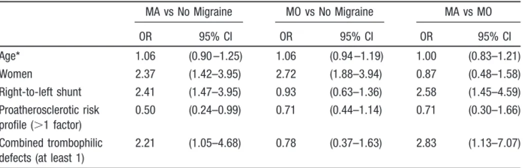
As summarized by the migraine– covariate interaction OR reported in Table 2, females had⬎2-fold increased risk for migraine (OR, 2.37; 95% CI, 1.42–3.95 for MA; OR, 2.72;
95% CI, 1.88 –3.94 for MO) as compared to the reference group of stroke patients without migraine. In addition, we observed a reduction in the risk of migraine with increasing number of the atherothrombotic risk factors. In particular, such a reduction (OR, 0.50; 95% CI, 0.24 – 0.99 for 2 factors or more) was significant only in the subgroup of patients with MA in comparison with the subgroup without migraines. A similar interactive effect of migraine with the 2 genetic thrombophilic variants was also observed: carriers of factor V Leiden mutation, the G20210A mutation in the prothrombin gene, or both the prothrombotic genotypes had ⬎2-fold increased risk for MA (OR, 2.21; 95% CI, 1.05– 4.68 as compared to stroke patients without migraine; OR, 2.83; 95% CI, 1.13–7.07 as compared to stroke patients with MO). Finally, the status of RLS carrier was associated with⬎2-fold increased risk of MA when compared to the other 2 sub-groups (OR, 2.41; 95% CI, 1.37–3.45 as compared to stroke patients without migraine; OR, 2.58; 95% CI, 1.45– 4.59 as compared to stroke patients with MO). None of these predis-posing factors had significant interaction with MO.
Discussion
The hypothesis that people with MA have a higher prevalence of traditional risk factors known to be associated with cardiovascular disease recently has been suggested to explain
Table 1. Demographics and Clinical Characteristics of the Study Group According to Migraine Status Variable No Migraine Migraine Without Aura Migraine With Aura P Age, mean⫾SD, y 36.0⫾7.6 36.0⫾7.4 35.6⫾7.4 0.893 Sample size 746 (76.0) 161 (15.8) 74 (7.3) Women 335 (44.9) 111 (68.9) 50 (67.6) ⬍0.001 Right-to-left shunt 248 (33.9) 55 (34.6) 43 (58.1) ⬍0.001 Hypertension 176 (23.6) 30 (18.6) 11 (14.9) 0.114 Diabetes mellitus 34 (4.6) 4 (2.5) 0 (0.0) 0.086 Current smokers 292 (39.1) 62 (38.5) 21 (28.4) 0.197 Hypercholesterolemia 199 (26.7) 39 (24.5) 12 (16.2) 0.135
Proatherosclerotic risk profile (0–4) 0.012
⬎1 factor 183 (24.5) 29 (18.2) 8 (10.8)
ⱕ1 factor 563 (75.5) 130 (81.8) 66 (89.2)
Oral contraceptive users* 118 (36.8) 37 (33.3) 18 (36.7) 0.905 Factor V Leiden G1691A 32 (5.5) 4 (3.1) 5 (8.9) 0.262 Prothrombin G20210A 33 (5.7) 7 (5.4) 7 (12.5) 0.121
Combined thrombophilic defects (0–2)† 0.053
ⱖ1 60 (10.5) 10 (7.8) 11 (19.6)
None 513 (89.5) 118 (92.2) 45 (80.4)
Stroke etiologic subtypes ⬍0.001
Large-vessel atherosclerosis 91 (12.1) 9 (5.6) 4 (5.4) Nonatherosclerotic vasculopathy 136 (18.2) 27 (16.8) 12 (16.2) Small-vessel disease 47 (6.3) 5 (3.1) 2 (2.7) Cardioembolism/transcardiac embolism 210 (28.2) 42 (26.1) 36 (48.6)
Other 262 (35.1) 78 (48.4) 20 (27.0)
Location of the infarct 0.833
Posterior circulation 248 (33.7) 57 (35.8) 21 (28.8) *For women only.
†259 genotypes missing.
the association between this migraine subtype and ischemic vascular disease, especially cerebral ischemia. If MA is associated with an unfavorable cardiovascular risk profile, then it would be likely that the increased propensity to ischemic disease observed in those with MA is the conse-quence of common comorbidities rather than of migraine-specific mechanisms. In line with this hypothesis, the population-based Genetic Epidemiology in Migraine study indicated that those with migraines, particularly those with aura, were more likely to smoke, have a parental history of myocardial infarction, an unfavorable cholesterol profile, elevated blood pressure, and high Framingham risk score, leading to speculation that MA may be a marker of progres-sive atherosclerosis.13Data from the population-based
Amer-ican Migraine Prevalence and Prevention study recently provided further support to the perception that those with migraines have a higher probability of having an unfavorable risk factor profile.14 Intriguingly, a number of large-scale
epidemiological analyses conducted over the past years have questioned these findings and suggested that the migraine– stroke association is particularly present in the absence of traditional cardiovascular risk factors,15,16 reinforcing the
assumption that biological mechanisms other than those atherosclerosis-mediated may link migraine to ischemic stroke. The results of the present study support this hypoth-esis and prompt speculation that migraine alone might be insufficient to increase the risk of ischemic stroke and additional, nonproatherosclerotic factors might be necessary, and that these factors might be of help in identifying the subgroup of those with migraines in which cerebral ischemia is more likely to occur. Actually, within a large cohort of young adults with ischemic stroke, we found that the coex-istence of migraine and cerebral infarcts is more frequent in those patients with a lower vascular risk factor profile, a higher prevalence of RLS, and underlying hypercoagulabil-ity. In particular, the fact that such a relation was limited to stroke patients with MA is an argument in favor of the idea that MA, RLS, and thrombophilic disorders are comorbid conditions acting together in a synergistic way and their effect is more pronounced and clinically apparent when not overcome by that of major cardiovascular risk factors. A number of data indirectly support this view. First, although controversial, there is evidence that MA may be associated
with patent foramen ovale.5Second, in line with our findings,
sparse reports indicate that the prevalence of migraine is higher among stroke patients with patent foramen ovale.17–19
Third, epidemiological evidence supports the assumption of a biological link between migraine and hypercoagulability,6as
well as its venous thromboembolic consequences.16Finally,
the reported relation among prothrombotic states, patent foramen ovale, and ischemic stroke20in a complex triangular
pattern indirectly strengthens this hypothesis.
Although such a biological mechanism seems likely, an alternative interpretation of our data is also plausible. Ac-cording to recent findings, vascular alterations may be the initial step of a cascade of events leading to neuronal dysfunction in those with migraines, and not the other way around.21,22 The emerging hypothesis is that brief cerebral
hypoxic-ischemic episodes induced by microembolization could trigger cortical spreading depolarization of neurons and, thus, a migraine attack, whereas prolonged occlusion of the same vessels might cause cerebral ischemia.23 In this
regard, stroke and MA should be considered different clinical phenotypes of a common process of focal cerebral hypoper-fusion. Lending support to this view, it might be that the coexistence of RLS and hypercoagulability we found in our stroke patients with MA increases the probability that small and clinically insignificant cerebral emboli shunted from the heart trigger spreading depolarization of neurons and cause MA in the same way they may induce major cerebral infarcts. Among other aspects, it is assumed that the duration is the main difference between spreading depolarization associated with MA and spreading depolarization associated with ischemic stroke.24-26If this were true, then MA should be conceptualized
as a marker of increased stroke risk, not as a stroke risk factor. The assumption of a pathogenic link between RLS and MA may also provide a potential interpretation for the weak association between migraine and myocardial infarction, as opposed to the strong association between migraine and ischemic stroke ob-served in numerous epidemiological studies.1
Several strengths of the present study should be noted, including the large number of participants, the homogeneous demographic characteristics and clinical phenotype of the cohort, the standardized diagnostic work-up and evaluation of risk factors, and the clinical diagnosis of migraine and its subtypes, which constitute the gold standard for a valid
Table 2. Migraine–Covariate Interaction OR of Age, Gender, Right-to-Left Shunt,
Proatherosclerotic Risk Profile, and Thrombophilic Defects According to Multinomial Logistic Regression Model MA vs No Migraine MO vs No Migraine MA vs MO OR 95% CI OR 95% CI OR 95% CI Age* 1.06 (0.90 –1.25) 1.06 (0.94 –1.19) 1.00 (0.83–1.21) Women 2.37 (1.42–3.95) 2.72 (1.88–3.94) 0.87 (0.48–1.58) Right-to-left shunt 2.41 (1.47–3.95) 0.93 (0.63–1.36) 2.58 (1.45–4.59) Proatherosclerotic risk profile (⬎1 factor) 0.50 (0.24–0.99) 0.71 (0.44–1.14) 0.71 (0.30–1.66) Combined trombophilic defects (at least 1)
2.21 (1.05–4.68) 0.78 (0.37–1.63) 2.83 (1.13–7.07)
MA indicates migraine with aura; MO, migraine without aura. *OR changes by 5-year units step.
diagnosis with respect to alternative methods adopted in other studies. Some limitations also should be considered. First, because of the retrospective migraine ascertainment in our stroke patients, a recall bias cannot be theoretically excluded. However, because patients were unaware of the hypothesis undergoing study, there is no reason why they should have reported migraine symptoms more frequently. Second, be-cause we did not assess migraine frequency and severity, as well as frequency of auras, in our cohort, we cannot evaluate whether the observed association differs according to specific migraine patterns. However, whether migraine frequency is a measure of migraine severity remains to be demonstrated. Third, because the transcranial Doppler technique prevents the assessment of atrial septal aneurysms, any further analyses comparing the prevalence of MA in stroke patients with RLS, atrial septal aneurysms, or both are hindered by the lack of precise data on the frequency of atrial septal aneurysms in our series. Furthermore, our protocol did not include any measure-ment, albeit semiquantitative, of the RLS diameter, an anatomic marker that may allow the identification of patients at high risk for embolism. Finally, although we adjusted for major potential confounders, residual confounding is possible given the obser-vational design of the study. The implications of these missing data are noteworthy, but it seems unlikely that they have altered the main findings of the present analysis.
Conclusion
In conclusion, the results of our study reinforce the prevailing idea that multiple factors contribute to migraine, especially MA, and stroke susceptibility. In particular, they indicate that in young adults with cerebral ischemia, MA, as opposed to MO, is related to a low cardiovascular risk profile and it is strongly associated with RLS and inherited prothrombotic disorders. Although it seems premature to conclude that approaches aimed at modifying these factors may modify the risk of ischemic stroke in those with migraines, their biological effects should be taken into account in future studies aimed at investigating the mechanisms linking migraine to brain ischemia.
Acknowledgments
The authors express their gratitude to all the doctors who assisted in the ascertainment and recruitment of patients in the IPSYS Centers, and to all the individuals who participated in the study.
Disclosures
None.
References
1. Schurks M, Rist PM, Bigal ME, Buring JE, Lipton RB, Kurth T. Migraine and cardiovascular disease: systematic review and meta-analysis. BMJ. 2009;339:b3914.
2. Kurth T, Gaziano JM, Cook NR, Logroscino G, Diener HC, Buring JE. Migraine and risk of cardiovascular disease in women. JAMA. 2006;296: 283–291.
3. Kurth T, Gaziano JM, Cook NR, Bubes V, Logroscino G, Diener HC, Buring JE. Migraine and risk of cardiovascular disease in men. Arch
Intern Med. 2007;167:795– 801.
4. Tietjen EG. Migraine and ischaemic heart disease and stroke: potential mechanisms and treatment implications. Cephalalgia. 2007;27:981–987. 5. Wammes-van der Heijden EA, Tijssen CC, Egberts AC. Right-to-left shunt and migraine: the strength of the relationship. Cephalalgia. 2006; 26:208 –213.
6. Tietjen GE. Migraine as a systemic vasculopathy. Cephalalgia. 2009;29: 989 –996.
7. Lee ST, Chu K, Jung KH, Kim DH, Kim EH, Choe VN, Kim JH, Im WS, Kang L, Park JE, Park HJ, Park HK, Song EC, Lee SK, Kim M, Roh JK. Decreased number and function of endothelial progenitor cells in patients with migraine. Neurology. 2008;70:1510 –1517.
8. Headache Classification Subcommittee of the International Headache Society. The international classification of headache disorders. Cephalalgia. 2004;24(Suppl 1):24 –36.
9. Webster MW, Chancellor AM, Smith HJ, Swift DL, Sharpe DN, Bass NM, Glasgow GL. Patent foramen ovale in young stroke patients. Lancet. 1988;2:11–12.
10. Jauss M, Zanette E. Detection of right-to-left shunt with ultrasound contrast agent and transcranial Doppler sonography. Cerebrovasc Dis. 2000;10:490 – 496.
11. Job FP, Ringelstein EB, Grafen Y, Flachskampf FA, Doherty C, Stockmanns A, Hanrath P. Comparison of transcranial contrast Doppler sonography and transesophageal contrast echocardiography for the detection of patent foramen ovale in young stroke patients. Am J Cardiol. 1994;74:381–384.
12. Johnson CJ, Kittner SJ, McCarter RJ, Sloan MA, Stern BJ, Buchholz D, Price TR. Interrater reliability of an etiologic classification of ischemic stroke. Stroke. 1995;26:46 –51.
13. Scher AI, Terwindt GM, Picavet HS, Verschuren WM, Ferrari MD, Launer LJ. Cardiovascular risk factors and migraine: the GEM population-based study. Neurology. 2005;64:614 – 620.
14. Bigal ME, Kurth T, Santanello N, Buse D, Golden W, Robbins M, Lipton RB. Migraine and cardiovascular disease. A population-based study Neurology. 2010;74:628 – 635.
15. Kurth T. Migraine and ischaemic vascular events. Cephalalgia. 2007;27: 967–975.
16. Schwaiger J, Kiechl S, Stockner H, Knoflach M, Werner P, Rungger G, Gasperi A, Willeit J. Burden of atherosclerosis and risk of venous throm-boembolism in patients with migraine. Neurology. 2008;71:937–943. 17. Lamy C, Giannesini C, Zuber M, Arquizan C, Meder JF, Trystram D,
Coste J, Mas JL. Clinical and imaging findings in cryptogenic stroke patients with and without patent foramen ovale: the PFO-ASA Study. Atrial Septal Aneurysm. Stroke. 2002;33:706 –711.
18. Sztajzel R, Genoud D, Roth S, Mermillod B, Le Floch-Rohr J. Patent foramen ovale, a possible cause of symptomatic migraine: a study of 74 patients with acute ischemic stroke. Cerebrovasc Dis. 2002;13:102–106. 19. Milhaud D, Bogousslavsky J, van Melle G, Liot P. Ischemic stroke and
active migraine. Neurology. 2001;57:1805–1811.
20. Pezzini A, Grassi M, Del Zotto E, Giossi A, Volonghi I, Costa P, Grau A, Magoni M, Padovani A, Lichy C. Do common prothrombotic mutations influence the risk of cerebral ischemia in patients with patent foramen ovale? Systematic review and meta-analysis. Thromb Haemost. 2009; 101:813– 817.
21. Dreier JP, Kleeberg J, Petzold G, Priller J, Windmu¨ller O, Orzechowski HD, Lindauer U, Heinemann U, Einha¨upl KM, Dirnagl U. Endothelin-1 potently induces Lea˜o’s cortical spreading depression in vivo in the rat: a model for an endothelial trigger of migrainous aura? Brain. 2002;125: 102–112.
22. Brennan KC, Beltra´n-Parrazal L, Lo´pez-Valde´s HE, Theriot J, Toga AW, Charles AC. Distinct vascular conduction with cortical spreading depression. J Neurophysiol. 2007;97:4143– 4151.
23. Nozari A, Dilekoz E, Sukhotinsky I, Stein T, Eikermann-Haerter K, Liu C, Wang Y, Frosch MP, Waeber C, Ayata C, Moskowitz MA. Micro-emboli may link spreading depression, migraine aura, and patent foramen ovale. Ann Neurol. 2010;67:221–229.
24. Oliveira-Ferreira AI, Milakara D, Alam M, Jorks D, Major S, Hartings JA, Lu¨ckl J, Martus P, Graf R, Dohmen C, Bohner G, Woitzik J, Dreier JP, COSBID study group. Experimental and preliminary clinical evidence of an ischemic zone with prolonged negative DC shifts surrounded by a normally perfused tissue belt with persistent electrocorticographic depression. J Cereb Blood Flow Metab. 2010;30:1504 –1519. 25. Dreier JP, Major S, Manning A, Woitzik J, Drenckhahn C, Steinbrink J,
Tolias C, Oliveira-Ferreira AI, Fabricius M, Hartings JA, Vajkoczy P, Lauritzen M, Dirnagl U, Bohner G, Strong AJ, COSBID study group. Cortical spreading ischaemia is a novel process involved in ischaemic damage in patients with aneurysmal subarachnoid haemorrhage. Brain. 2009;132:1866 –1881.
26. Dalkara T, Nozari A, Moskowitz MA. Migraine aura pathophysiology: the role of blood vessels and microembolisation. Lancet Neurol. 2010;9: 309 –317