Acute liver dysfunction after cardiac arrest
Enrica Iesu1☯‡, Federico Franchi1,2☯‡, Federica Zama Cavicchi1, Selene Pozzebon1,2, Vito Fontana1, Manuel Mendoza1,3, Leda Nobile1, Sabino Scolletta2, Jean-Louis Vincent1, Jacques Creteur1, Fabio Silvio Taccone1*1 Department of Intensive Care, Erasme Hospital, Universite´ Libre de Bruxelles, Brussels, Belgium, 2 Department of Medical Biotechnologies, Anesthesia and Intensive Care Unit, University of Siena, Siena, Italy, 3 Department of Intensive Care, Hospital Universitari de Tarragona Joan XXIII, Tarragona, Spain
☯These authors contributed equally to this work. ‡ These authors are co-first authors on this work.
Abstract
Few data are available regarding hypoxic hepatitis (HH) and acute liver failure (ALF) in patients resuscitated from cardiac arrest (CA). The aim of this study was to describe the occurrence of these complications and their association with outcome. All adult patients admitted to the Department of Intensive Care following CA were considered for inclusion in this retrospective study. Exclusion criteria were early death (<24 hours) or missing biological data. We retrieved data concerning CA characteristics and markers of liver function. ALF was defined as a bilirubin>1.2 mg/dL and an international normalized ratio�1.5. HH was defined as an aminotransferase level>1000 IU/L. Neurological outcome was assessed at 3 months and an unfavourable neurological outcome was defined as a Cerebral Performance Categories (CPC) score of 3–5. A total of 374 patients (age 62 [52–74] years; 242 male) were included. ALF developed in 208 patients (56%) and HH in 27 (7%); 24 patients devel-oped both conditions. Patients with HH had higher mortality (89% vs. 51% vs. 45%, respec-tively) and greater rates of unfavourable neurological outcome (93% vs. 60% vs. 59%, respectively) compared to those with ALF without HH (n = 184) and those without ALF or HH (n = 163; p = 0.03). Unwitnessed arrest, non-shockable initial rhythm, lack of bystander cardiopulmonary resuscitation, high adrenaline doses and the development of acute kidney injury were independent predictors of unfavourable neurological outcome; HH (OR: 16.276 [95% CIs: 2.625–81.345; p = 0.003), but not ALF, was also a significant risk-factor for unfa-vourable outcome. Although ALF occurs frequently after CA, HH is a rare complication. Only HH is significantly associated with poor neurological outcome in this setting.
Introduction
The outcome of patients after sudden cardiac arrest (CA) remains poor, in particular because a large proportion of these patients will eventually die after hospital admission because of exten-sive brain damage and cardiogenic shock [1–3]. Moreover, the ischemia-reperfusion injury that occurs after the return of spontaneous circulation (ROSC) contributes to a systemic a1111111111 a1111111111 a1111111111 a1111111111 a1111111111 OPEN ACCESS
Citation: Iesu E, Franchi F, Zama Cavicchi F, Pozzebon S, Fontana V, Mendoza M, et al. (2018) Acute liver dysfunction after cardiac arrest. PLoS ONE 13(11): e0206655.https://doi.org/10.1371/ journal.pone.0206655
Editor: Peter Rosenberger, University of Tu¨bingen, GERMANY
Received: November 28, 2017 Accepted: October 8, 2018 Published: November 5, 2018
Copyright:© 2018 Iesu et al. This is an open access article distributed under the terms of the
Creative Commons Attribution License, which permits unrestricted use, distribution, and reproduction in any medium, provided the original author and source are credited.
Data Availability Statement: The data set is available on Dryad via:https://doi.org/10.5061/ dryad.qv6fp83.
Funding: The author(s) received no specific funding for this work.
Competing interests: The authors have declared that no competing interests exist.
inflammatory response that has several similarities with sepsis [4] and may result in the devel-opment of multi-organ failure (MOF) [5], which further increases the likelihood of poor outcome.
Several studies have suggested that severe cardiovascular and respiratory failure may nega-tively impact on patient outcome after CA [5,6]. Nevertheless, there is a paucity of data on the prognostic value of extra-cerebral organ dysfunction after CA. In a recent meta-analysis, San-droni et al. showed that post-arrest acute kidney injury (AKI) had an early onset, occurred in more than 50% of CA patients and was significantly associated with increased mortality [7]. However, there are almost no data on liver dysfunction in this setting; in one recent study, Champigneulle et al. reported that hypoxic hepatitis (HH) occurred in 11% of patients resusci-tated after an out-of-hospital CA (OHCA) and, in multivariate analysis, was significantly asso-ciated with ICU mortality [8]. Hypoxic liver injury typically occurs in individuals with congestive heart failure and/or low cardiac output but is not always associated with altered liver function [9]. No studies have described the occurrence of acute liver failure (ALF), defined by severely altered coagulation and an increase in total bilirubin, in CA patients.
The aim of this study was, therefore, to describe the occurrence of ALF and HH among CA patients admitted to the intensive care unit (ICU) as well as their association with patient outcome.
Methods
Study population
This retrospective study was performed in the Department of Intensive Care at Erasme Hospital, Brussels (Belgium). The local Ethical Committee (Comite´ d’Ethique Hospitalo-Facultaire Erasme-ULB) approved the study (P2017/264), but waived the need for informed consent because of its retrospective nature. All comatose patients (Glasgow Coma Scale, GCS < 9) admitted after in-hospital CA (IHCA) or OHCA were included in a prospective institutional database (January 2007 to December 2015) and considered as eligible for the study. Exclusion criteria were missing data on liver function or death less than 24 hours after ICU admission, because the occurrence of liver dysfunction could not be fully evaluated in these patients.
Post-resuscitation care
Our standardized institutional protocol of post-resuscitation management has been exten-sively described elsewhere [10]. Briefly, all comatose CA patients were treated with targeted temperature management (TTM; target body temperature: 32–34˚C) for 24 hours. Deep seda-tion was obtained using midazolam and morphine, and cisatracurium was administered to control shivering. Rewarming (<0.5˚C/h) was achieved passively. Advanced hemodynamic monitoring was used (PiCCO, Pulsion, Munich, Germany) and cardiac function assessed by repeated trans-oesophageal and/or transthoracic echocardiography. Mean arterial pressure was maintained at >65–70 mmHg using volume resuscitation, dobutamine and/or noradrena-line, whenever needed. Intra-aortic balloon counterpulsation (IABP) or extracorporeal mem-brane oxygenation (ECMO) was also used in cases of severe cardiogenic shock. Ventilation was set to maintain normocapnia and SpO2>94%. Blood glucose was kept between 110 and
150 mg/dl using a continuous insulin infusion. Enteral nutrition was initiated during TTM and continued thereafter according to gastric tolerance. Neurological monitoring and prog-nostication of coma after CA were performed using a multimodal approach, as previously described [11].
Data collection
We collected data on demographics (including chronic use of any anticoagulant), pre-existing chronic diseases and cardiopulmonary resuscitation (CPR) (initial rhythm, bystander CPR, time to ROSC, total adrenaline dose) in all patients. Severity of disease was assessed using the Acute Physiology and Chronic Health Evaluation (APACHE) II score [12] and the Sequential Organ Failure Assessment (SOFA) score [13] on the day of admission. To assess liver function, we collected the results from blood samples for aspartate (AST) and alanine (ALT) transami-nases, lactate dehydrogenase (LDH, normal values < 200 IU/L), prothrombin time (PT, nor-mal values > 70%), international nornor-malized ratio (INR, nornor-mal values � 1.2), total bilirubin (normal values � 1.2 mg/dl) and fibrinogen (normal values > 150 mg/dl) on admission and then once daily until ICU discharge. Use of mechanical ventilation, continuous renal replace-ment therapy (CRRT) and vasoactive drugs was recorded, as was length of ICU stay. The development of infection during the ICU stay was noted.
Neurological evaluation was assessed at 3 months after CA using the cerebral performance categories score (CPC; 1 = no or mild neurological disability, 2 = moderate neurological dis-ability, 3 = severe neurological impairment, 4 = vegetative state, 5 = death) [13]. The CPC evaluation was performed prospectively during follow-up visits or by telephone interview with the general practitioner. Favourable neurological outcome was considered as a CPC 1–2; unfa-vourable neurological outcome as CPC 3–5 [14].
Definitions
The definition for ALF was based on previously published criteria, which included an INR �1.5, an elevated total bilirubin in the absence of chronic liver disease, and new onset enceph-alopathy (defined as a patient not obeying orders) [15]. In the presence of acute post-anoxic encephalopathy, we only considered the first two criteria for the definition of ALF.
Hypoxic hepatitis was defined as an increase in AST and/or ALT to more than 20 times the upper normal range (� 50 IU/L), i.e., > 1000 IU/L in the setting of acute cardiovascular failure after CA and in the absence of another cause of cell necrosis [16]. We also reported the time to the highest AST and/or ALT values after ICU admission.
AKI was defined according to AKIN criteria [17]. Shock was defined as the need for vaso-pressor agents for more than 6 hours. We specifically recorded the use of potentially hepato-toxic drugs/interventions (e.g., paracetamol,β-lactams, quinolones, isoniazid, azoles, metrodinazole, any chemotherapy, trimethoprim/ sulfamethoxazole [TMT/SMT] and anti-epileptic drugs).
Statistical analysis
Discrete variables are expressed as counts (percentage) and continuous variables as median (25th to 75th percentiles). The Kolmogorov-Smirnov test was used, and histograms and nor-mal-quantile plots were examined to verify the normality of distribution of continuous vari-ables. Demographics and clinical differences between groups (HH vs. HH; ALF vs. no-ALF; survivors vs. non-survivors; favourable neurological outcome vs. unfavourable outcome) were assessed using the chi-square test, Fisher’s exact test, Student’s t-test, or Mann–Whitney U-test, as appropriate. For the development of HH, and because the number of events was small, only variables with a p values < 0.05 in the univariate analysis were considered in the multivariable logistic regression and odds ratios (ORs) were estimated using a logistic regres-sion approach with penalized profile likelihood based confidence intervals for parameter esti-mates. For multivariable modelling, correlations between the predictors were checked because high correlations may induce large bias in the estimators of the regression coefficients. Two
multivariable logistic regression analyses with ALF or unfavourable neurological outcome as the dependent variables were performed in all patients; co-linearity between variables was excluded prior to modelling; only variables associated with a higher risk of ALF or unfavour-able neurological outcome (p <0.2) on a univariate basis were introduced in the multivariunfavour-able models. ORs with 95% confidence intervals (CI) were computed. A p value < 0.05 was consid-ered as statistically significant. Data were analyzed using IBM SPSS Statistics software, version 22.0 for Windows and R software, version 3.1.0 (CRAN project).
Results
From a total of 435 patients, 61 were excluded because of early death (n = 51) or the absence of data on liver transaminases, coagulation and/or total bilirubin (n = 10) and 374 patients were analysed (Table 1); 207 (55%) had had an out-of-hospital CA and 221 (59%) a non-shockable initial rhythm. Pre-existing liver disease was present in 17 (4%) patients; 12 of them had alco-holic liver cirrhosis (9 with Child-Pugh B and 3 with Child-Pugh C) and 5 had hepatitis B or C liver cirrhosis (all with Child-Pugh of 3). The ICU length of stay was 4 [2–9] days, 194 (52%) patients died and 227 (61%) patients had an unfavourable neurological outcome.
Acute liver failure and hypoxic hepatitis
ALF was observed in 208 patients (56%). The median time to ALF development was 3 [2–3] days. Patients with ALF had a longer time to return of spontaneous circulation (ROSC), were more likely to have had a non-shockable initial rhythm, received more adrenaline and more frequently received TTM than those who did not develop ALF; they were also more likely to receive vasopressor drugs, ECMO or CRRT during the ICU stay (Table 1). Patients with ALF received less frequently paracetamol (S1 Table), more often developed shock and HH, had higher blood lactate concentrations on admission and had lower lowest ScvO2/SvO2values.
Hospital mortality was significantly higher in patients with ALF than in those without (61% vs. 51%, p<0.05).
HH developed in 27 (7%) patients. Median AST and ALT on admission were higher in patients who developed HH than in those who did not, as were LDH and INR values (Table 1). The median time to HH development was 1 [0–1] days and the time to the highest AST/ALT values in these patients was 1 [0–1] days. Patients who developed HH had a longer time to ROSC, had more frequently had a non-shockable initial rhythm and received more adrenaline than those who did not develop HH; they were also more frequently treated with vasopressors, inotropic agents, ECMO and CRRT during the ICU stay (Table 1). Patients with HH received less frequently paracetamol,β-lactams or amiodarone (S1 Table) had higher lactate concentra-tions on admission, more frequently developed shock during the ICU stay, and had lower low-est ScvO2/SvO2values. ALF occurred more frequently in patients who developed HH than in
those who did not. ICU and hospital mortality rates and rates of unfavourable neurological outcome were significantly higher in patients with HH than in those without.
Patients who developed HH had higher mortality rates and rates of unfavourable neurologi-cal outcome compared to those with ALF without HH (n = 184) and to those without ALF or HH (n = 163; p = 0.03) (Fig 1).
Neurological outcome
Compared to those with favourable neurological outcome, patients with an unfavourable neu-rological outcome were older and less likely to have had a witnessed arrest, to have received bystander CPR and to have had an initial shockable rhythm; time to ROSC was longer and adrenaline dose higher in patients with an unfavourable neurological outcome (Table 2).
Table 1. Characteristics of the study population, according to the development of hypoxic hepatitis (HH) or acute liver failure (ALF). ALL (n = 374) HH (n = 27) NO-HH (n = 347) ALF (n = 208) NO-ALF (n = 166) DEMOGRAPHICS Age, years 62 [52–74] 59 [50–68] 62 [52–75] 61 [51–74] 62 [52–75] Weight, Kg 77 [67–85] 77 [65–85] 77 [68–85] 75 [65–85] 78 [68–88] Male, n (%) 142 (72) 17 (63) 253 (73) 152 (73) 118 (71)
ICU length of stay, days 4 [2–9] 3 [2–8] 4 [3–9] 5 [2–10] 4 [2–8]
APACHE II score 25 [20–8] 27 [22–9] 24 [20–29] 25 [20–29] 24 [20–28]
SOFA score 11 [9–14] 13 [10–15] 11 [9–14] 11 [9–14] 11 [9–13]
ARREST CHARACTERISTICS
Witnessed, n (%) 320 (86) 22 (81) 298 (86) 180 (86) 140 (84)
Bystander CPR, n (%) 254 (68) 18 (67) 236 (68) 147 (71) 107 (64)
Time to ROSC, min 15 [7–25] 21 [15–30] 15 [7–25]�� 15 [7–28] 14 [7–21]�
Adrenaline, mg 3 [2–5] 4 [3–6] 3 [1–5]� 4 [2–6] 3 [1-]� Out-of-hospital, n (%) 207 (55) 15 (56) 192 (55) 122 (59) 86 (52) TTM, n (%) 331 (89) 23 (85) 308 (89) 196 (94) 135 (81)� Non-cardiac cause, n (%) 153 (41) 13 (48) 140 (40) 93 (45) 60 (36) Non-shockable rhythm, n (%) 221 (59) 21 (78) 200 (58)� 136 (65) 85 (51)�� ICU mortality, n (%) 194 (52) 24 (89) 170 (49)�� 116 (56) 78 (47) Hospital mortality, n (%) 213 (57) 24 (89) 189 (54)�� 128 (61) 85 (51)�
Unfavourable neurological outcome at 3 months, n (%) 226 (61) 25 (93) 201 (58)�� 133 (64) 93 (56)
COMORBIDITIES
Chronic heart failure, n (%) 78 (22) 2 (7) 76 (22)� 54 (26) 24 (14)��
Hypertension, n (%) 159 (42) 10 (37) 149 (43) 85 (41) 74 (45)
Coronary artery disease, n (%) 146 (39) 7 (26) 139 (40) 83 (40) 63 (38)
Diabetes, n (%) 91 (24) 6 (22) 85 (24) 47 (23) 44 (26)
COPD/asthma, n (%) 63 (17) 6 (22) 57 (16) 35 (17) 28 (17)
Neurological disease, n (%) 54 (14) 2 (7) 52 (15) 25 (12) 29 (18)
Chronic renal failure, n (%) 62 (17) 6 (22) 56 (16) 29 (14) 33 (20)
Liver cirrhosis, n (%) 17 (4) 1 (4) 16 (5) 14 (7) 3 (2) HIV, n (%) 3 (1) 1 (4) 2 (1) 1 (1) 2 (1) Corticosteroids, n (%) Chronic anticoagulation, n (%) 85 (23) 65 (17) 12 (44) 3 (11) 73 (21)��62 (18) 55 (26) 40 (19) 30 (18) 25 (15) DURING ICU STAY
Infection, n (%) 241 (64) 18 (67) 223 (64) 137 (66) 104 (63) IABP, n (%) 24 (6) 2 (7) 22 (6) 17 (8) 7 (4) ECMO, n (%) 47 (13) 10 (37) 37 (11)�� 37 (18) 10 (6)�� Shock, n (%) 200 (53) 25 (93) 175 (50)�� 134 (64) 66 (40)�� Vasopressor therapy, n (%) 283 (76) 26 (96) 257 (74)�� 169 (81) 114 (69)� Inotropic agents, n (%) 201 (54) 25 (93) 176 (51)�� 132 (63) 69 (42) Mechanical ventilation, n (%) 374 (100) 27 (100) 347 (100) 206 (99) 163 (98)
At least one hepatotoxic drug, n (%) 254 (68) 10 (37) 244 (70)�� 142 (68) 112 (67)
CRRT, n (%) 61 (16) 14 (52) 47 (14)�� 47 (23) 14 (8)��
HH, n (%) 27 (7) 27 (100) 0 (0)�� 24 (11) 3 (2)��
ALF, n (%) 208 (56) 24 (89) 184 (53)�� 208 (100) 0 (0)��
AKI, n (%) 221 (59) 24 (89) 197 (57)�� 135 (65) 86 (52)��
Lowest platelet Count, /mm3 133 [79–187] 60 [39–120] 137 [83–189]�� 103 [56–168] 160 [113–215]�
Lowest ScvO2/SvO2, % 62 [56–66] 59 [54–65] 62 [57–66]� 61 [56–66] 63 [58–68]�
BLOOD SAMPLE ON ADMISSION
Lactate, mEq l-1 5.1 [4.1–7.7] 8.2 [5.6–12.7] 4.9 [4.0–7.2]�� 5.8 [4.4–9] 4.6 [3.9–6.0]��
ScvO2/SvO2, % 69 [64–74] 67 [60–74] 69 [64–74] 68 [63–74] 69 [64–74]
Patients with an unfavourable neurological outcome also had a higher lactate concentration and lower mean arterial pressure on admission and more frequently developed shock, and required vasopressors during the ICU stay than those with a favourable neurological outcome. HH occurred more frequently in patients with an unfavourable neurological outcome than in those with a favourable neurological outcome (25/226 vs. 2/148, p = 0.002).
Multivariable analyses
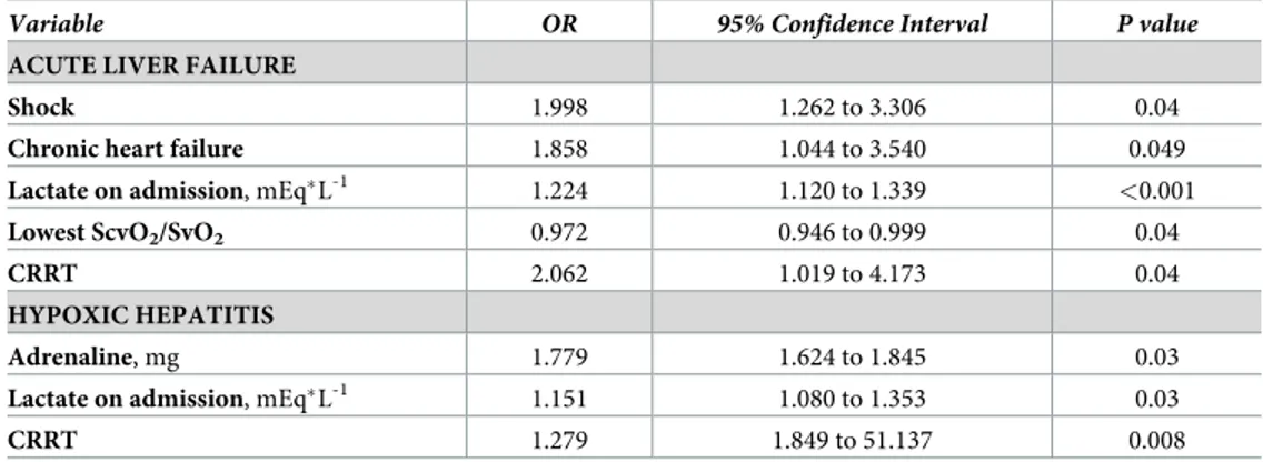
In multivariable logistic regression analysis, shock, chronic heart failure, high lactate concen-tration on admission, lowest venous (central or mixed) oxygen saturation, and use of CRRT during the ICU stay were independent predictors for development of ALF during the ICU stay
(Table 3). High doses of adrenaline during CPR, high lactate concentration on admission and
use of CRRT during the ICU stay were independent predictors for development of HH during the ICU stay (Table 3).
Unwitnessed arrest, non-shockable initial rhythm, lack of bystander CPR, high adrenaline doses and the development of AKI were all independent predictors of an unfavourable neuro-logical outcome (Table 4); HH, but not ALF, was also a significant risk factor for poor neuro-logical outcome. Table 1. (Continued) ALL (n = 374) HH (n = 27) NO-HH (n = 347) ALF (n = 208) NO-ALF (n = 166) AST, IU/L 95 [47–193] 704 [160–1559] 86 [44–168]�� 104 [51–235] 80 [41–164]� ALT, IU/L 68 [32–153] 394 [135–782] 62 [31–128]�� 69 [33–177] 64 [30–121]� LDH, IU/L 336 [240–488] 918 [476–2325] 326 [235–449]� 356 [244–525] 322 [236–425]� AP, IU/L 77 [58–106] 88 [68–153] 76 [58–104] 77 [58–114] 75 [59–102] GGT, IU/L 68 [42–103] 77 [38–95] 68 [43–103] 66 [41–103] 71 [53–103] Total bilirubin, mg dl-1 0.5 [0.3–0.9] 0.5 [0.4–1.1] 0.5 [0.3–0.9] 0.5 [0.3–0.8] 0.5 [0.3–0.9] APTT, sec 32 [27–44] 41 [29–71] 32 [27–43] 38 [30–60] 29 [25–34]� PT, % 65 [47–79] 46 [33–60] 67 [50–80]� 50 [38–69] 73 [64–88]� INR 1.26 [1.12–1.54] 1.64 [1.35–2.18] 1.25 [1.11–1.49]� 1.22 [1.11–1.53] 1.30 [1.17–1.56] Platelets, /mm3 201 [138–266] 169 [99–218] 202 [141–269] 169 [111–233] 220 [172–292]� Proteins, mg dl-1 5.7 [5–6.3] 5.4 [4.7–5.9] 5.7 [5–6.4] 6.0 [5.0–6.0] 6.0 [5.0–7.0] Glucose, mg dl-1 200 [155–289] 190 [143–237] 202 [158–291] 194 [147–294] 204 [163–282] pH 7.28 [7.21–7.38] 7.22 [7.125–7.31] 7.30 [7.22–7.38]�� 7.28 [7.17–7.38] 7.31 [7.23–7.38]� PaCO2, mmHg 37 [33–44] 34 [29–41] 38 [33–44]� 37 [32–43] 38 [34–45] PaO2, mmHg 111 [85–178] 113 [89–181] 111 [85–178] 119 [87–189] 106 [84–156] MAP, mmHg 86 [75–103] 78 [70–91] 87 [76–105]�� 83 [73–98] 94 [79–109]� Creatinine, mg dl-1 1.2[0.9–1.6] 1.3 [1.1–2.25] 1.2 [0.9–1.6] 1.2 [1.0–2.0] 1.1 [0.9–1.4] CRP, mg dl-1 40 [14–84] 45 [23–143] 37 [12–79] 44.5 [18.0–100.0] 32.5 [10–68.5]�
ICU = intensive care unit; CPR = cardiopulmonary resuscitation; ROSC = return of spontaneous circulation; TTM = targeted temperature management;
COPD = chronic pulmonary obstructive disease; HIV = human immunodeficiency; IABP = intra-aortic balloon counterpulsation; ECMO = extracorporeal membrane oxygenation; CRRT = continuous renal replacement therapy; HH = hypoxic hepatitis; ALF = acute liver failure; AKI = acute liver failure; ScvO2/SvO2 = central venous oxygen saturation or mixed venous oxygen saturation; AST = aspartate aminotransferase; ALT = alanine aminotransferase; LDH = lactate dehydrogenase; AP = alkaline phosphatase; GGT =γ-glutamine transferase; APTT = activated partial thromboplastin time; PT = prothrombin time; INR = international normalized ratio;
MAP = mean arterial pressure; CRP = C-reactive protein; APACHE = Acute Physiology and Chronic Health Evaluation; SOFA = Sequential Organ Failure Assessment. �p < 0.05
��p < 0.01 for HH vs. no-HH or ALF vs. no-ALF.
Discussion
In our study, HH and ALF were observed in 7% and 56%, respectively, of patients resuscitated after CA, HH patients more frequently had an unfavourable neurological outcome compared to those with ALF without HH or those without ALF or HH. High dose adrenaline during CPR, high lactate concentration on admission and the need for CRRT were independent pre-dictors of HH development. Development of HH, but not of ALF, was a significant risk factor for poor neurological outcome and ICU mortality.
Few studies have reported data on the development of liver dysfunction after CA; in one large database, liver failure, defined using only bilirubin levels, occurred in 10% of CA patients on ICU admission and in up to 20% during the ICU stay, with no differences between patients with favourable or unfavourable neurological outcome [18]. The occurrence of ALF in our study is quite high; however, some high bilirubin levels may have been due to hemolysis or rhabdomyolysis and increased INR levels could have been secondary to sepsis or previous anti-coagulant therapy, which may have resulted in an overestimation of the number of cases. In
Fig 1. Intensive care unit (ICU) mortality and unfavourable neurological outcome (UO) according to the occurrence of hypoxic hepatitis (HH) or acute liver failure (ALF).
Table 2. Characteristics of the study population according to hospital survival and neurological outcome.
SURVIVORS (n = 161)
NON-SURVIVORS (n = 213)
Favourable neurological outcome (n = 148)
Unfavourable neurological outcome (n = 226)
DEMOGRAPHICS
Age, years 57 [49–70] 66 [54–76]� � 58 [51–70] 66 [52–76]� �
Weight, Kgs 78 [70–85] 77 [65–85] 78 [70–85] 76 [65–85]
Male, n (%) 119 (74) 151 (71) 109 (74) 161 (71)
ICU length of stay, days 7 [4–13] 3 [2–7]� � 6 [3–11] 3 [2–8]� �
APACHE II score 23 [20–27] 26 [19–30]� 23 [20–27] 26 [20–30]�
SOFA score 10 [8–12] 13 [10–15]� � 10 [8–12] 12 [10–14]� �
ARREST CHARACTERISTICS
Witnessed, n (%) 148 (92) 172 (81)� � 136 (92) 184 (81)� �
Bystander CPR, n (%) 121 (75) 133 (62)� � 114 (77) 140 (62)� �
Time to ROSC, min 12 [5–20] 17 [10–25]� � 11.5 [5–20] 17 [10–25]� �
Adrenaline, mg 2 [1–4] 4 [2–6]� � 2 [1–4] 4 [2–6]� � Out-of-hospital, n (%) 91 (57) 116 (54) 82 (55) 125 (55) TTM, n (%) 138 (86) 193 (91) 126 (85) 205 (91) Non-cardiac cause, n (%) 54 (34) 99 (46)� 49 (33) 104 (46)� Non-shockable rhythm, n (%) 67 (42) 154 (72)� � 61 (41) 160 (71)� � ICU mortality, n (%) 0 (0) 194 (91)� � 0 (0) 194 (86)� � Hospital mortality, n (%) 0 (0) 213 (100)� � 0 (0) 213 (94)� �
Unfavourable neurological outcome at 3 months, n (%) 13 (8) 213 (100)� � 0 (0) 226 (100)� �
COMORBIDITIES
Chronic heart failure, n (%) 30 (19) 48 (23) 28 (19) 50 (22)
Hypertension, n (%) 73 (45) 86 (40) 65 (44) 94 (42)
Coronary artery disease, n (%) 59 (37) 87 (41) 55 (37) 91 (40)
Diabetes, n (%) 33 (20) 58 (27) 30 (20) 61 (27)
COPD/Asthma, n (%) 21 (13) 42 (20) 19 (13) 44 (19)
Neurological disease, n (%) 16 (10) 38 (18) 13 (9) 41 (18)�
Chronic renal failure, n (%) 23 (14) 39 (18) 21 (14) 41 (18)
Liver cirrhosis, n (%) 3 (2) 14 (7) 3 (2) 14 (6) HIV, n (%) 2 (1) 1 (0) 2 (1) 1 (0) Corticosteroids, n (%) Chronic anticoagulation, n (%) 27 (17) 27 (17) 58 (27)� �38 (18) 25 (17) 25 (17) 60 (27) 40 (18)
DURING ICU STAY
Infection, n (%) 110 (68) 131 (62) 98 (66) 143 (63) IABP, n (%) 7 (4) 17 (8) 6 (4) 18 (8) ECMO, n (%) 20 (12) 27 (13) 18 (12) 29 (13) Shock, n (%) 67 (42) 133 (62)� � 64 (43) 136 (60)� � Vasopressor therapy, n (%) 107 (66) 176 (83)� � 100 (68) 183 (81)� � Inotropic agents, n (%) 80 (50) 121 (57) 73 (49) 128 (57) Mechanical ventilation, n (%) 161 (100) 213 (100) 148 (100) 226 (100)
At least one hepatotoxic drug, n (%) 103 (64) 151 (71) 100 (68) 154 (68)
CRRT, n (%) 23 (14) 38 (18) 21 (14) 40 (18)
HH, n (%) 3 (2) 24 (11)� � 2 (1) 25 (11)� �
ALF, n (%) 80 (50) 128 (60) 75 (51) 13 (59)
AKI, n (%) 76 (47) 145 (68)� � 70 (47) 151 (67)� �
Lowest platelet count, /mm3 138 [94–181] 130 [70–188] 135 [94–180] 131 [70–190]
Lowest ScvO2/SvO2, % 61 [56–66] 63 [57–67] 61 [56–66] 62.65 [57–67]
BLOOD SAMPLE ON ADMISSION
Lactate, mEq l-1 4.6 [3.8–6.2] 5.5 [4.2–8.5]� � 4.6 [3.8–6.4] 5.3 [4.2–8.2]� �
ScvO2/SvO2, % 68 [64–73] 69 [64–75] 67 [63–73] 69 [64–76]
AST, IU/L 84 [40–209] 99 [51–182] 82 [38–206] 102 [52–186]
ALT, IU/L 68 [30–158] 66 [32–141] 66.5 [29–155] 69 [33–144]
two other studies, the incidence of HH ranged from 11% to 14% in patients with OHCA [8,
19]. CA is one of the main causes of HH in critically ill patients [20], and the prevalence of HH in heterogeneous critically ill populations is much lower, ranging from 1% to 12% [21,22], depending on the threshold of AST/ALT used to define HH. Interestingly, we did not observe a higher incidence of ALF or HH in IHCA patients, in whom the presence of a pre-existing pathological condition might have reduced the tolerance of the liver to an ischemic injury, compared to OHCA patients.
The total dose of adrenaline during resuscitation, lactate concentrations on admission and use of CRRT were independent variables associated with the occurrence of HH. Conversely, Oh et al. reported that no-flow time was the only predictor of HH [19], and Champigneulle et al. demonstrated that the delay from collapse to ROSC was the major predictor of HH [8]. However, the retrospective design of all these studies makes it difficult to accurately assess the time to CA and initiation of CPR [23]. In our study, we included patients with IHCA, which may have added an additional confounder regarding the role of resuscitation times on the occurrence of liver failure. Nevertheless, in our population, high lactate concentration on admission and the total amount of adrenaline received during resuscitation, which can be con-sidered as markers of the severity of the anoxic insult, better represented the extent of the ischemia/reperfusion injury on the liver than did the duration of resuscitation. In addition, experimental animal models of CA have reported that adrenaline may worsen the function of
Table 2. (Continued)
SURVIVORS (n = 161)
NON-SURVIVORS (n = 213)
Favourable neurological outcome (n = 148)
Unfavourable neurological outcome (n = 226) LDH, IU/L 308 [225– 461] 352 [250–498] 296 [222–429] 360 [251–507] AP, IU/L 69 [54–91] 84 [62–119] 68.5 [53–90] 83 [62–118] GGT, IU/L 60 [37–91] 77 [45–110] 59 [36–91] 77 [45–107] Total bilirubin, mg dl-1 0.5 [0.3–0.8] 0.6 [0.4–1] 0.5 [0.3–0.80] 0.5 [0.4–1] APTT, sec 32 [27–42] 33 [28–45] 32 [27–45] 33 [28–46] PT, % 69 [51–85] 62 [46–75] 69 [51–85] 62 [46–75] INR 1.2 [1.1–1.5] 1.3 [1.2–1.6] 1.2 [1.1–1.5] 1.3 [1.2–1.6] Platelets, /mm3 203 [147– 261] 193 [127–268] 203 [142–263] 197 [131–267] Proteins, mg dl-1 5.7 [5.1–6.3] 5.6 [5–6.3] 5.7 [5–6.2] 5.7 [5–6.4] Glucose, mg dl-1 194 [147– 274] 204 [160–293] 193 [145–276] 204 [166–293] pH 7.31 [7.23– 7.38] 7.29 [7.18–7.38] 7.3 [7.22–7.38] 7.29 [7.19–7.39] PaCO2, mmHg 39 [33–44] 37 [32–43] 38 [33–44] 37 [32–43.75] PaO2, mmHg 120 [87–175] 108 [84–182]� 120 [86–174] 109 [85–182]� MAP, mmHg 93 [80–110] 83 [72–100]� 93 [80–110] 83.5 [73–100]� Creatinine, mg dl-1 1.1 [0.9–1.5] 1.2 [0.9–1.7] 1.1 [0.9–1.5] 1.2 [0.9–1.7] CRP, mg dl-1 31 [10–74] 44 [19–94]� 31 [10–72] 44 [18–92]�
ICU = intensive care unit; CPR = cardiopulmonary resuscitation; ROSC = return of spontaneous circulation; TTM = targeted temperature management;
COPD = chronic pulmonary obstructive disease; HIV = human immunodeficiency; IABP = intra-aortic balloon counterpulsation; ECMO = extracorporeal membrane oxygenation; CRRT = continuous renal replacement therapy; HH = hypoxic hepatitis; ALF = acute liver failure; AKI = acute liver failure; ScvO2/SvO2 = central venous oxygen saturation or mixed venous oxygen saturation; AST = aspartate aminotransferase; ALT = alanine aminotransferase; LDH = lactate dehydrogenase; AP = alkaline phosphatase; GGT =γ-glutamine transferase; APTT = activated partial thromboplastin time; PT = prothtombin time; INR = international normalized ratio;
MAP = mean arterial pressure; CRP = C-reactive protein; APACHE = Acute Physiology and Chronic Health Evaluation; SOFA = Sequential Organ Failure Assessment. �p < 0.05
��p < 0.01 for survivors vs. non-survivors or favourable vs. unfavourable neurological outcome.
all organs, and in particular mesenteric perfusion, and its administration may lead to lower abdominal perfusion and higher lactate concentrations than other vasoconstrictor drugs [24,
25]. Finally, the use of CRRT could be considered as an index of patient severity or reflect the degree of the post-anoxic systemic inflammatory response, leading to MOF. Recently, Tujjar et al showed that 43% of patients resuscitated after CA developed AKI, and one third of these patients required CRRT; the total amount of adrenaline was independently associated with the development of AKI, because the adrenaline impaired abdominal perfusion [10].
The factors associated with development of ALF were shock, chronic heart failure, high lac-tate concentration on admission, low venous oxygen saturation and the use of CRRT.
Although shock is not one of the most common causes of ALF [26], significant alterations in liver function are observed in up to 20% of patients with severe cardiogenic shock treated with veno-arterial ECMO [27]. Patients with chronic heart failure are at high-risk of developing car-diogenic shock, which requires aggressive therapy, including IABP and assist devices [28]. Low values of venous oxygen saturation, together with high lactate concentrations, may sug-gest the presence of a severe low cardiac output status that would result in reduced hepatic blood flow and oxygenation, as well as hepatic congestion from venous outflow obstruction, which would finally lead to liver failure [29].
The mortality rate in our patients with HH was 89%, which is consistent with rates reported by other authors (75–86%) [8,19]. Our results showed that HH was also a significant predictor of poor neurologic outcome after CA, and previous studies have described an association of
Table 3. Logistic regression analysis for predictors of acute liver failure and hypoxic hepatitis after cardiac arrest.
Variable OR 95% Confidence Interval P value
ACUTE LIVER FAILURE
Shock 1.998 1.262 to 3.306 0.04
Chronic heart failure 1.858 1.044 to 3.540 0.049
Lactate on admission, mEq�L-1
1.224 1.120 to 1.339 <0.001
Lowest ScvO2/SvO2 0.972 0.946 to 0.999 0.04
CRRT 2.062 1.019 to 4.173 0.04
HYPOXIC HEPATITIS
Adrenaline, mg 1.779 1.624 to 1.845 0.03
Lactate on admission, mEq�L-1 1.151 1.080 to 1.353 0.03
CRRT 1.279 1.849 to 51.137 0.008
OR = odds ratio; CRRT = continuous renal replacement therapy; ScvO2/SvO2, central/mixed venous oxygen saturation.
https://doi.org/10.1371/journal.pone.0206655.t003
Table 4. Logistic regression analysis for predictors of unfavourable neurological outcome.
Variable OR 95% Confidence Interval P value
Witnessed CA 0.892 0.755 to 0.911 0.02 Bystander CPR 0.867 0.712 to 0.955 0.03 Non-shockable rhythm 2.923 1.528 to 5.592 0.001 Adrenaline, mg 1.179 1.024 to 1.357 0.02 AKI 2.360 1.228 to 4.536 0.01 HH 16.276 2.625 to 81.345 0.003
OR = odds ratio; AKI = acute kidney injury; CA = cardiac arrest; CPR = cardiopulmonary resuscitation; HH = hypoxic hepatitis.
HH with mortality in the same setting [8,19]. In two studies [5,18], hepatic failure, assessed by the hepatic SOFA score, was not associated with mortality; however, this score does not consider AST/ALT as a relevant marker of hepatic damage induced by anoxia. Our findings showing a role of HH and AKI as predictors of unfavourable neurological outcome support the need to better describe the occurrence of extra-cerebral organ dysfunction among patients admitted to the ICU after CA. These abnormalities may influence patient outcome and need to be recognized early and further studies performed to identify potential therapies.
Our study has some strength. We underlined the need to assess the development of HH after both OHCA and IHCA, as this may represent a significant predictor of poor outcome. We have also shown some important predictors of HH in this patients’ population, i.e. the total dose of adrenaline during resuscitation, high lactate concentrations and the use of CRRT. The presence of these factors, together with a prolonged duration of resuscitation [8,19], could identify those patients requiring a more accurate hemodynamic monitoring and/or the optimization of supporting therapies in order to minimize further liver injury. Also, we have included in our analysis the development of acute liver failure, which was not considered in previous studies [8,19]. Finally, we collected a larger amount of data, including biological find-ings, therapeutic data and the use of potentially hepatotoxic drugs, which provided a more accurate analysis of the variables independently associated with the occurrence of post-anoxic liver injury.
Our study has also several limitations. First, we only collected routine measures of liver function and injury, and liver function may have been better assessed using arterial ammo-nium levels or functional tests (e.g., indocyanine green clearance test or 13C- methacetin breath test). Also, additional tests of the biosynthetic capacity of the liver, including blood-clot-ting factors or cholinesterases activity, which are reduced in patients with liver dysfunction, were not routinely performed. Second, we analyzed a mixed population of patients with OHCA and IHCA, who may have different predisposing conditions for the development of ALF and HH; however, the location of arrest was not predictive of the development of HH or of unfavourable neurological outcome in multivariable analysis. Third, due to the design of the study, we cannot determine whether there is a causal relationship between and the develop-ment of HH and unfavourable neurological outcome, which may just reflect the intensity of the initial anoxic injury. Fourth, we did not routinely perform morphologic examinations of the liver (e.g., ultrasound); however, hepatic echography, although of interest to exclude other causes of ischemic liver injury (vascular clotting) and to identify signs of chronic liver disease, may have limited diagnostic/prognostic role in the setting of HH.
Conclusions
Hypoxic liver injury is rare after cardiac arrest but is an independent predictor of poor out-come. Data on the occurrence of extra-cerebral organ dysfunction should be collected in future studies dealing with prognostication and therapeutic interventions in this setting.
Supporting information
S1 Table. Presence of different hepatotoxic drugs, according to the occurrence of hypoxic hepatitis (HH) and acute liver failure (ALF) or according to patients’ outcome.
(DOCX)
Author Contributions
Data curation: Enrica Iesu, Federico Franchi, Federica Zama Cavicchi, Selene Pozzebon, Leda Nobile, Fabio Silvio Taccone.
Formal analysis: Leda Nobile, Sabino Scolletta. Funding acquisition: Fabio Silvio Taccone. Investigation: Enrica Iesu, Manuel Mendoza.
Methodology: Federico Franchi, Federica Zama Cavicchi, Selene Pozzebon, Vito Fontana, Leda Nobile, Fabio Silvio Taccone.
Project administration: Fabio Silvio Taccone. Resources: Manuel Mendoza.
Supervision: Sabino Scolletta, Jean-Louis Vincent, Jacques Creteur, Fabio Silvio Taccone. Validation: Vito Fontana, Manuel Mendoza, Fabio Silvio Taccone.
Writing – original draft: Enrica Iesu, Fabio Silvio Taccone.
Writing – review & editing: Enrica Iesu, Federico Franchi, Federica Zama Cavicchi, Selene Pozzebon, Vito Fontana, Sabino Scolletta, Jean-Louis Vincent, Jacques Creteur, Fabio Silvio Taccone.
References
1. Berdowski J, Berg RA, Tijssen JG, Koster RW. Global incidences of out-of-hospital cardiac arrest and survival rates: systematic review of 67 prospective studies. Resuscitation 2010; 81:1479–87.https:// doi.org/10.1016/j.resuscitation.2010.08.006PMID:20828914
2. Dragancea I, Rundgren M, Englund E, Friberg H, Cronberg T. The influence of induced hypothermia and delayed prognostication on the mode of death after cardiac arrest. Resuscitation 2013; 84:337–42. https://doi.org/10.1016/j.resuscitation.2012.09.015PMID:23000363
3. Lemiale V, Dumas F, Mongardon N, et al. Intensive care unit mortality after cardiac arrest: the relative contribution of shock and brain injury in a large cohort. Intensive Care Med 2013; 39:1972–80.https:// doi.org/10.1007/s00134-013-3043-4PMID:23942856
4. Nolan JP, Neumar RW, Adrie C, et al. Post-cardiac arrest syndrome: epidemiology, pathophysiology, treatment, and prognostication. A Scientific Statement from the International Liaison Committee on Resuscitation; the American Heart Association Emergency Cardiovascular Care Committee; the Coun-cil on Cardiovascular Surgery and Anesthesia; the CounCoun-cil on Cardiopulmonary, Perioperative, and Crit-ical Care; the Council on ClinCrit-ical Cardiology; the Council on Stroke. Resuscitation 2008; 79:350–79. https://doi.org/10.1016/j.resuscitation.2008.09.017PMID:18963350
5. Roberts BW, Kilgannon JH, Chansky ME, et al. Multiple organ dysfunction after return of spontaneous circulation in postcardiac arrest syndrome. Crit Care Med 2013; 41:1492–501.https://doi.org/10.1097/ CCM.0b013e31828a39e9PMID:23507719
6. Beitler JR, Ghafouri TB, Jinadasa SP, et al. Favorable neurocognitive outcome with low tidal volume ventilation after cardiac arrest. Am J Respir Crit Care Med 2017; 195:1198–1206https://doi.org/10. 1164/rccm.201609-1771OCPMID:28267376
7. Sandroni C, Dell’anna AM, Tujjar O, Geri G, Cariou A, Taccone FS. Acute kidney injury after cardiac arrest: a systematic review and meta-analysis of clinical studies. Minerva Anestesiol 2016; 82(9):989– 99. PMID:26957119
8. Champigneulle B, Geri G, Bougouin W, et al. Hypoxic hepatitis after out-of-hospital cardiac arrest: Inci-dence, determinants and prognosis. Resuscitation 2016; 103:60–5.https://doi.org/10.1016/j. resuscitation.2016.03.021PMID:27068401
9. Stravitz R, Kramer A, Davern T, et al. Intensive care of patients with acute liver failure: recommenda-tions of the U.S. Acute Liver Failure Study Group. Crit Care Med 2007; 35:2498–508.https://doi.org/10. 1097/01.CCM.0000287592.94554.5FPMID:17901832
10. Tujjar O, Mineo G, Dell’Anna A, et al. Acute kidney injury after cardiac arrest. Crit Care 2015; 19:169. https://doi.org/10.1186/s13054-015-0900-2PMID:25887258
11. Taccone FS, Cronberg T, Friberg H, et al. How to assess prognosis after cardiac arrest and therapeutic hypothermia. Crit Care 2014; 18:202.https://doi.org/10.1186/cc13696PMID:24417885
12. Knaus WA, Draper EA, Wagner DP, Zimmerman JE. APACHE II: a severity of disease classification system. Crit Care Med 1985; 13:818–29 PMID:3928249
13. Vincent JL, Moreno R, Takala J, Willatts S, De Mendonc a A, Bruining H, et al. The SOFA (Sepsis-related Organ Failure Assessment) score to describe organ dysfunction/failure. On behalf of the Work-ing Group on Sepsis-Related Prob- lems of the European Society of Intensive Care Medicine. Intensive Care Med 1996; 22:707–10. PMID:8844239
14. Jennett B, Bond M. Assessment of outcome after severe brain damage. Lancet 1975; 1(7905):480– 484. PMID:46957
15. McDowell Torres D, Stevens RD, Gurakar A. Acute liver failure: a management challenge for the prac-ticing gastroenterologist. Gastroenterol Hepatol 2010; 6:444–450.
16. Henrion J. Hypoxic hepatitis. Liver Int 2012; 32:1039–1052.https://doi.org/10.1111/j.1478-3231.2011. 02655.xPMID:22098491
17. Mehta RL, Kellum JA, Shah SV, et al. Acute Kidney Injury Network: report of an initiative to improve out-comes in acute kidney injury. Crit Care 2007; 11:R31.https://doi.org/10.1186/cc5713PMID:17331245 18. Nobile L, Taccone FS, Szakmany T, et al. The impact of extracerebral organ failure on outcome of
patients after cardiac arrest: an observational study from the ICON database. Crit Care 2016; 20:368. https://doi.org/10.1186/s13054-016-1528-6PMID:27839517
19. Oh SH, Kim HJ, Park KN, et al. Hypoxic hepatitis in survivors of out-of-hospital cardiac arrest. Am J Emerg Med 2015; 33:1166–70.https://doi.org/10.1016/j.ajem.2015.05.008PMID:26032661 20. Raurich JM, Llompart-Pou JA, Ferreruela M, et al. Hypoxic hepatitis in critically ill patients: incidence,
etiology and risk factors for mortality. J Anesth 2011; 25:50–6. https://doi.org/10.1007/s00540-010-1058-3PMID:21153035
21. Fuhrmann V, Kneidinger N, Herkner H, et al. Hypoxic hepatitis: underlying con-ditions and risk factors for mortality in critically ill patients. Intensive Care Med2009; 35:1397–1405.
22. Fuhrmann V, Kneidinger N, Herkner H, et al. Impact of hypoxic hepatitis onmortality in the intensive care unit. Intensive Care Med 2011; 37:1302–1310.https://doi.org/10.1007/s00134-011-2248-7PMID: 21647720
23. Jacobs I, Nadkarni V, Bahr J, et al. Cardiac arrest and cardiopulmonary resuscitation outcome reports: update and simplification of the Utstein templates for resuscitation registries: a statement for healthcare professionals from a task force of the International Liaison Committee on Resuscitation (American Heart Association, European Resuscitation Council, Australian Resuscitation Council, New Zealand Resuscitation Council, Heart and Stroke Foundation of Canada, InterAmerican Heart Foundation, Resuscitation Councils of Southern Africa). Circulation 2004; 110:3385–97.https://doi.org/10.1161/01. CIR.0000147236.85306.15PMID:15557386
24. Studer W, Wu X, Siegemund M, Seeberger M. Resuscitation from cardiac arrest with adrenaline/epi-nephrine or vasopressin: effects on intestinal mucosal tonometer pCO(2) during the post-resuscitation period in rats. Resuscitation 2002; 53:201–7. PMID:12009224
25. Perkins GD, Quinn T, Deakin CD, et al. Pre-hospital Assessment of the Role of Adrenaline: Measuring the Effectiveness of Drug administration In Cardiac arrest (PARAMEDIC-2): Trial protocol. Resuscita-tion 2016; 108:75–81.https://doi.org/10.1016/j.resuscitation.2016.08.029PMID:27650864
26. Hadem J, Tacke F, Bruns T, et al; Acute Liver Failure Study Group Germany. Etiologies and outcomes of acute liver failure in Germany. Clin Gastroenterol Hepatol 2012; 10:664–9.https://doi.org/10.1016/j. cgh.2012.02.016PMID:22373724
27. Blandino Ortiz A, Lamanna I, Antonucci E, et al. Altered liver function in patients undergoing veno-arte-rial extracorporeal membrane oxygenation (ECMO) therapy. Minerva Anestesiol 2017; 83(3):255–65. https://doi.org/10.23736/S0375-9393.16.11442-7PMID:27858410
28. Sintek MA, Gdowski M, Lindman BR, Nassif M, Lavine KJ, Novak E, Bach RG, Silvestry SC, Mann DL, Joseph SM. Intra-Aortic Balloon Counterpulsation in Patients With Chronic Heart Failure and Cardio-genic Shock: Clinical Response and Predictors of Stabilization. J Card Fail 2015; 21:868–76.https:// doi.org/10.1016/j.cardfail.2015.06.383PMID:26164215
29. Saner FH, Heuer M, Meyer M, et al. When the heart kills the liver: acute liver failure in congestive heart failure. Eur J Med Res 2009; 14:541–546.https://doi.org/10.1186/2047-783X-14-12-541PMID: 20149988