Contents lists available atScienceDirect
LWT - Food Science and Technology
journal homepage:www.elsevier.com/locate/lwtIn
fluence of thermal and electrical effects of ohmic heating on
C-phycocyanin properties and biocompounds recovery from Spirulina platensis
Pedro Ferreira-Santos
a,1, Rafaela Nunes
a,1, Filomena De Biasio
b, Giorgia Spigno
c,
Domenico Gorgoglione
d, José A. Teixeira
a, Cristina M.R. Rocha
a,∗aCEB - Centre of Biological Engineering, University of Minho, Campus de Gualtar, 4710-057, Braga, Portugal bOsun Solutions S.r.l, Località Galdo, 85044, Lauria, PZ, Italy
cDiSTAS - Department for Sustainable Food Process, Faculty of Agriculture, Università Cattolica del Sacro Cuore, Via Emilia Parmense, 84 -29122, Piacenza, Italy dEVRA S.r.l, Località Galdo Zona Industriale Lotto 20, 85044, Lauria, PZ, Italy
A R T I C L E I N F O
Keywords: Arthrospira platensis Moderate electricfields C-phycocyanin Biocompounds extraction Cell disruption
A B S T R A C T
Spirulina platensis is interesting for the food industry due to its overall composition and high content in C-phycocyanin. However, the sensitivity of C-phycocyanin makes its extraction a delicate process. The present study focuses on assessing the use of ohmic heating (OH) in the recovery of C-phycocyanin and other relevant compounds as an alternative method to freeze-thawing or conventional heating. Different ohmic and conven-tional heating treatments were applied both to purified C-phycocyanin and Spirulina powder. Evaluation of fluorescence and circular dichroism showed that moderated electric fields increased C-phycocyanin stability. This was confirmed in the extraction trials which revealed that OH assisted extraction at higher temperatures (44 °C), and shorter times (30 min) allowed significant higher extraction yield of C-phycocyanin (45 mg/gdw Spirulina), in comparison with conventional heating and freeze-thawing. OH allowed also up to 80% higher yields in phenolic compounds and carbohydrates.
1. Introduction
Spirulina (Arthrospira) platensis is a cyanobacterium that is arising attention in various industries and it is commonly used as food sup-plement due to its particular composition (e.g., proteins, lipids, carbo-hydrates, vitamins andfibers) and high antioxidant activity (Andrade et al., 2019;Papadaki, Kyriakopoulou, Tzovenis, & Krokida, 2017;Soni, Sudhakar, & Rana, 2017). Regarding Spirulina protein content, it is well known that these cyanobacteria have a high content in C-phycocyanin (PC), a high commercial value blue pigment used as a natural colorant. This blue natural colorant has been widely used in nutraceutical, cos-metic and pharmaceutical industries and represents more than 20% of Spirulina dry weight (Batista, Gouveia, Bandarra, Franco, & Raymundo, 2013; Martínez, Luengo, Saldaña, Álvarez, & Raso, 2017; Sharma, Goyal, & Sharma, 2016). PC is composed of two similar subunits of α-helix andβ-sheets and should be easily isolated as a protein complex because it is water-soluble (Kamble, Gaikar, Padalia, & Shinde, 2013; Léa;Vernès, Granvillain, Chemat, & Vian, 2015). On the other hand, though the protein fraction, and in particular C-phycocyanin, is the most explored fraction from Spirulina platensis, other compounds may
also be interesting and potentially have high added value, such as phenolics and carbohydrates. These compounds may have important bioactive properties, such as antioxidant, antimicrobial, anticancer, prebiotic, antiadherent, etc., which make these compounds interesting for applications in the food, health and pharmaceutical industries (Dai & Mumper, 2010; Ferreira-Santos, Genisheva, Pereira, Teixeira, & Rocha, 2019;Mena-García, Ruiz-Matute, Soria, & Sanz, 2019).
However, pigments, phenolic compounds, carbohydrates and pro-teins are presented in cytoplasm or internal organelles which makes difficult their extraction due to the resistance of the cell wall (Chen et al., 2020;Moraes, Sala, Cerveira, & Kalil, 2011;Zhu et al., 2007). Although various cell disintegration technologies have already been described (e.g., maceration, bead-beating and freeze-thawing), these are time-consuming and often require high solvent and/or energy in-puts which lead to an increase in the production costs and limiting their widespread application (Chittapun, Jonjaroen, Khumrangsee, & Charoenrat, 2020;Kamble et al., 2013;Kumar, Dhar, Pabbi, Kumar, & Walia, 2014). Moreover, the PC extraction without affecting its physico-chemical properties is also a challenge due to its instability to light and heat (low denaturation temperature, i.e. about 45 °C), which makes
https://doi.org/10.1016/j.lwt.2020.109491
Received 31 January 2020; Received in revised form 8 April 2020; Accepted 24 April 2020 ∗Corresponding author.
E-mail address:[email protected](C.M.R. Rocha).
1Pedro Ferreira-Santos and Rafaela Nunes contributed equally to this work.
Available online 04 May 2020
0023-6438/ © 2020 The Authors. Published by Elsevier Ltd. This is an open access article under the CC BY-NC-ND license (http://creativecommons.org/licenses/BY-NC-ND/4.0/).
extraction with traditional methods difficult (Chittapun et al., 2020; Falkeborg, Roda-Serrat, Burnæs, & Nielsen, 2018).
Ohmic heating (OH) is a promising alternative technology for in-tracellular compounds' extraction, capable of promoting fast and homogeneous extraction with high energy efficiency. By applying a moderate electricalfield through a semi-conductive material that offers resistance to the passage of electric current, heat is generated directly inside the volume of the material causing cell membrane rearrange-ment, resulting in pores formation (electro-permeabilization), and al-lowing intracellular components diffusion (Ferreira-Santos et al., 2019; Rodrigues, Vicente, Petersen, & Pereira, 2019). Furthermore, this “green” technology may allow to reduce the ecological impacts caused by the extraction processes, decreasing the water/solvent use, waste generation due to higher extraction yields, energy consumption and/or reducing processing times (Chemat et al., 2017; Rocha et al., 2018). Goettel, Eing, Gusbeth, Straessner, and Frey (2013) and’t Lam et al. (2017) studied other electrical technologies, such as pulsed electric field (PEF), for extraction of intracellular valuable compounds from microalgae (e.g., Auxenochlorella protothecoides, Chlorella vulgaris and Neochloris oleoabundans) and revealed that a spontaneous cell disin-tegration was achieved, with ionic substances, carbohydrates and pro-teins drained out of the cells.
Based on these premises, the objective of the present work was to find a cell wall disruption and extraction method that would cause the efficient recovery of Spirulina protein content without degrading their functional features. It was then essential to study the physicochemical stability of PC when subjected to different environmental conditions of time and temperature (and their combinations) and application of moderate electricfields, in order to tune the most efficient extraction method. Furthermore, the impact of the extraction technology in other intracellular compounds content such as phenolic compounds and carbohydrates was also assessed.
2. Materials and methods
2.1. Raw materials and reagents
Spirulina (Arthrospira) plantesis used in this work was grown, mi-crobiologically controlled and dried at low temperatures in Azienda Agricola Prato della Voja (Bergamo, Italy) and provided by EVRA S.r.l. (Potenza, Italy). Purified C-phycocyanin (lyophilized powder, Sigma P2172, 30–50% protein – commercial product information) isolated from Spirulina, Folin-Ciocalteu reagent, sodium chloride, ethanol, so-dium carbonate, sulfuric acid, phenol reagent, glucose, chloroform, methanol, nitric acid and hydrogen peroxide were purchased from Sigma-Aldrich Chemical Co. Ltd. (St. Louis, MO, USA).
2.2. Experimental plan
In thefirst part of the study, thermal stability of PC and the influ-ence of electricfield on protein unfolding was investigated by different treatments. To this purpose, different time – temperature combinations under both conventional and ohmic heating were applied to solutions of purified commercial PC. The protein solutions were analyzed for in-trinsic fluorescence and circular dichroism (CD) before and after the treatments.
In the second part of the study, PC and other compounds (phenolics and carbohydrates) were extracted from Spirulina platensis powder using conventional heating and OH extraction process with the same time– temperature combinations used on the first part of the study (PC stability). Additionally, freeze-thawing process was applied as reference extraction process. The Spirulina powder was characterized for chemical and elemental composition, while the different extracts were analyzed for PC, total phenolic compounds and total carbohydrates content. The Spirulina powder before and after the extraction process was visualized by microscopy to observe cell disruption and disintegration.
2.3. Ohmic heating and conventional heating equipment
Ohmic heating was conducted in a double-jacketed glass cylinder containing a stainless-steel electrode at each edge (Ricardo N.Pereira et al., 2016). The distance between electrodes is 2.4 cm and overall volume of the reactor is 20 mL. The temperature was controlled by regulating the voltage output of a function generator (Agilent 33220A, Penang, Malaysia), measured with a type K thermocouple (temperature precision of ± 1 °C; Omega Engineering, Inc., Stamford, CT, USA), lo-cated in the geometric center of the extractor's volume and connected to a data logger (USB-9161, National Instruments Corporation, Austin, TX, USA) and then amplified on an amplifier system (Peavey CS3000, Meridian, MS, USA). The conventional thermal treatments were con-ducted using the same apparatus, without the electric influence, and controlling the temperature with a thermo-stabilized bath (F25-ED, Julabo, Seelbach, Germany).
2.4. Treatments of C-phycocyanin solutions
A preliminary test (Lowry-TCA) was performed to determine the exact protein concentration in the commercial product. This initial concentration of PC (43% protein powder) was later used to calculate the concentrations of PC solution used in this work.
PC solutions were prepared at an initial concentration of 1 mg/mL using ultra-pure water at room temperature. Then, the solutions were diluted to 0.1 mg/mL with NaCl (0.02 M) to ensure a conductivity of 2 mS/cm. Final pH was checked and adjusted to 6.8 with sodium hy-droxide, required for further tests. This pH value was selected con-sidering that the PC is more stable at pH 6.8, taking into account works by other authors (Sivasankari, Ravindran, & Naganandhini, 2014; Léa; Vernès et al., 2015;İlter et al., 2018) and previous studies from our group (data not shown). For conventional heating treatment (0 V/cm), protein solutions were maintained in the apparatus previously de-scribed at different temperatures (30, 37, 44 and 51 °C) up to 60 min. The solution was maintained protected from the light and stirred, on a magnetic stirrer at constant stirring speed (150 rpm) to ensure homo-geneity. For ohmic heating, the electricfield applied was chosen to achieve the same holding temperatures as for conventional heating (approximately 4, 5, 6 and 7 V/cm for 30, 37, 44 and 51 °C, respec-tively) and simulating exactly the same temperature profile for both processes (including initial transient heating period). These tempera-tures were used taking into account that the reported denaturation temperature of PC is approximately 44 °C (Falkeborg et al., 2018; Jespersen, Strømdahl, Olsen, & Skibsted, 2005; Léa; Vernès et al., 2015), thus being able to study the influence of electric fields at tem-peratures lower and higher than the denaturation temperature. Fre-quency was set at 20 kHz in order to eliminate the electrochemical effects as electrolysis and electrode oxidation (Pataro et al., 2014).
During both ohmic and conventional heating, 1 mL of sample was taken after 15, 30 and 60 min and analyzed for intrinsicfluorescence and CD. All the experiments were performed in triplicate and at room temperature.
2.4.1. Assessment of intrinsicfluorescence
Fluorescence determination was assessed with an Aqualog 800 spectrofluorometer (HORIBA-Jobin Yvon, Inc. Japan) using a 1 cm quartz cell. All solutions were diluted to 5 μg/mL concentration. Samples were excited at 280, 295 and 609 nm and the emissions were recorded over the 280–795 nm range with an integration of 0.1 s and 3 accumulations for each sample with 1.7 nm increment. All determina-tions were run in triplicate and reported as averages of the triplicate spectra (Selig, Malchione, Gamaleldin, Padilla-Zakour, & Abbaspourrad, 2018).
2.4.2. Circular dichroism spectroscopy
before and after the application of both the conventional heating and the ohmic heating. CD spectra were recorded with a spectropolarimeter (JASCO J-1500, Jasco Corp., Japan) at 20 °C under a constant nitrogen gasflow, using a 0.1 cm path length cell. The spectra were recorded between 190 and 260 nm with sampling points every 1 nm and three scans were accumulated and averaged for each sample (de Figueiredo
Furtado, Pereira, Vicente, & Cunha, 2018; Janek,
CzyżnikowskaŁuczyński, Gudiña, Rodrigues, & Gałęzowska, 2017). The spectra analysis was performed using CAPITO (CD Analysis and Plotting Tool) online software, allowing to determine the fractions of secondary structure elements on the protein before and after the thermo-electric treatments (Wiedemann, Bellstedt, & Görlach, 2013).
2.5. Biocompounds extraction from Spirulina
2.5.1. Nutritional characterization of Spirulina
Spirulina total protein content was estimated by quantification of total nitrogen after sample acid digestion using a Kjeldahl digestor (Tecator, FOSS, Denmark), applying the nitrogen conversion factor (N × 6.25) (Graziani et al., 2013). Lipid content was evaluated through extraction with a Soxhlet apparatus using chloroform/methanol (2:1 (v/v)) at 150 °C for 12 h (Tibbetts, Milley, & Lall, 2015). Ash content was determined gravimetrically through incineration of 5 g of Spirulina in a muffle furnace at 575 °C for 24 h. Moisture was determined gravimetrically by drying in an oven at 105 °C for 12–14 h (until constant weight). Total carbohydrates content was estimated by dif-ference using Eq. (1) (Lu et al., 2010). Total dietaryfiber content was determined by the AOAC 985.29 gravimetric method using a Mega-zyme® assay kit. Mineral content was determined using a method by inductively coupled plasma atomic emission spectrometry (ICP-AES), after Spirulina digestion with HNO3 (65%) and H O2 2 (35%) using a
microwave apparatus (Batista et al., 2013).
=
− + + +
Carbohydrates (%) 100
(moisture ash crude protein crude lipid) (1)
2.5.2. Freeze-thawing extraction method
Spirulina powder was diluted in pure water at a solid/liquid ratio of 1:20 (2.5 g/50 mL), stirred at 150 rpm for 30 min and frozen at−20 °C for 4 h. Then, the solution was thawed for 1.5 h at room temperature (Vernès et al., 2015). This freeze-thawing process was repeated four times for each sample. Then, the solution was centrifuged at 847×g for 15 min and the supernatant was recovered and used for further analyses (C-phycocyanin concentration, total phenolic compounds and total carbohydrates content). This method was used as a reference method, as it is one of the most used for cell wall disruption and protein ex-traction from microalgae and cyanobateria (Sivasankari et al., 2014; Léa;Vernès et al., 2015).
2.5.3. Conventional heating and ohmic heating methods
Ohmic heating and conventional heating treatments were in-vestigated as potential methods for cell disruption and extraction of intracellular compounds such as proteins, carbohydrates and phenolic compounds. For the experiments, the same equipment as described above (section2.3) was used with the same solid/liquid ratio of 1:20 g/ mL as in the freeze-thawing method. This powder concentration en-sured an electrical conductivity of 2 ± 0.2 mS/cm at room tempera-ture. Thus, 15 mL of pure water was added to 750 mg of Spirulina powder and stirred at 150 rpm for 30 min. The extraction process was performed at 30, 37, 44, 51 °C as reported for C-phycocyanin at section 2.4. At different time intervals (30, 60, 90 and 120 min), 1 mL of sample was taken and treated and analyzed as reported for the freeze-thawing method.
2.5.4. C-phycocyanin quantification
C-phycocyanin concentration was determined using a UV–Vis spectrophotometer (V-560, Jasco Inc., Tokyo, Japan) at wavelengths 620 and 650 nm, since C-phycocyanin has a maximum absorbance in the visible range between 610 and 620 nm that is proportional to its concentration and 650 nm is the wavelength where the class of allo-phycocyanin absorbs (Eq. (2)) (Bekhit et al., 2019; Kissoudi, Sarakatsianos, & Samanidou, 2018). The C-phycocyanin recovery (Yield) as mg/gdwof Spirulina was calculated using the initial mass of Spirulina used in the extraction process (Eq.(3)).
⎛ ⎝ ⎞ ⎠= × − × × Concentration mg mL ((A ) DF) (0.474 (A DF) 5.34 620 650 (2) ⎜ ⎟ ⎛ ⎝ ⎞ ⎠ = ×
( )
Yield mg g Concentration V m dw mg mL solution Spirulina (3)Where, A620 is the absorbance at 620 nm, A650 the absorbance at
650 nm, DF is the dilution factor, Vsolutionand mSpirulinaare the solution volume and mass of dry Spirulina, respectively.
2.5.5. Total phenolic compounds and carbohydrates quantification The total phenolic compounds (TPC) content was determined as previously described (Ferreira-Santos et al., 2019) using the Folin-Ciocalteu colorimetric assay, which is based on an oxidation-reduction reaction between the Folin reagent with the reducing phenolic com-pounds. For the analysis, 5μL of each sample were mixed with 60 μL of Na CO2 3(75 g/L), 15μL of Folin-Ciocalteu reagent and 200 μL of
ultra-pure water and the mixture was kept at 60 °C for 5 min. The absorbance was measured at 700 nm using a UV/Vis spectrophotometer (Synergy HT, BioTek Instruments, Inc., U.S.A.). The phenolic compounds content was calculated as gallic acid equivalents (GAE) using a calibration curve prepared with standard gallic acid (1500–50 mg/L, R2
= 0.99). The concentration of the extracts was used to calculate the extraction yield expressed as mg GAE/gdwSpirulina).
Total carbohydrates (TC) content was measured using the phenol-sulfuric acid method. For this, 50μL of sample were mixed with 150 μL of sulfuric acid (96–98% (v/v)). Then, 30 μL of 5% phenol reagent were added and thefinal solution was heated for 5 min at 90 °C. After cooling at room temperature for 5 min, the absorbance was measured at 490 nm by microplate reader (Synergy HT, BioTek Instruments, Inc., U.S.A.) (Masuko et al., 2005). The carbohydrates quantification was made using a calibration curve prepared with glucose (600–10 mg/L, R2= 0.99). The concentration of glucose equivalents (GLcE) was used to calculate the extraction yield based expressed as mg GLcE/gdw Spirulina).
2.5.6. Optical andfluorescence microscopy
After the extraction process, fresh Spirulina samples were observed with an epifluorescence microscope BX51 (Olympus, Japan) using brightfield and fluorescence light. TRITC (tetramethylrhodamine), with 530 nm and 550–590 nm excitation and emission filters to visualize the C-phycocyanin pigment. Images were captured with DP72 digital camera (Olympus, Japan) at magnification 100X.
2.6. Statistical analysis
The extractions and analyses were performed in triplicate and the data is presented as mean ± standard deviation (SD) values. GraphPad Prism® software (version 6.0; GraphPad Software, Inc., San Diego, CA, USA) was used for statistical analyses. The analysis of variance (ANOVA) and the least significant difference test were used to de-termine statistically different values at a significance level of p < 0.05.
3. Results and discussion
3.1. Treatments of C-phycocyanin solutions
3.1.1. Fluorescence determination
The proteins exposure to different time-temperature combinations and electricfields may lead to irreversible changes in their secondary structure, which are reflected in changes in their physicochemical properties (e.g., intrinsicfluorescence).
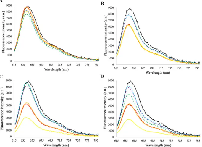
Tryptophan (Trp) and Tyrosine (Tyr) are two amino acids that have intrinsic fluorescence properties and in the native folded state of a protein they are generally located within the core of the protein, whereas in a partially folded or unfolded state, they become exposed to solvent. Therefore, this indicates that they are sensitive to their en-vironment changes and allows to know if proteins are folded or un-folded, associated with a decrease in the quantum yield and successive decrease influorescence caused by protein denaturation. Different ex-citation wavelengths were used: 280 nm, related to Trp and Tyr fluorescence; 295 nm that selectively excites Trp residues and 609 nm which could appear if some quenching process occurs (Rodrigues et al., 2019;Vernès et al., 2015). The emission wavelength was set between 615 and 795 nm, since PC emits fluorescence around 640 nm (Léa Vernès et al., 2015). Thus, the fluorescence spectra of PC were de-termined after the application of thermal treatments using the thermal conventional and OH methods. The solution containing the untreated protein was used as a control.
For 280 and 295 nm wavelength, thefluorescence spectra (data not shown) presented a large amount of noise and it was not possible to visualize the peaks relative to the Trp and Tyr amino acids. On the other hand, a peak between 620 and 650 nm was observed in all samples, possibly due to the quenching process.
Fig. 1shows the evolution offluorescence spectra as a consequence of thermal treatments with conventional or OH. The initialfluorescence of the untreated protein is approximately 9000 a.u. For conventional heating, it was verified that an increase in the severity of the treatment, in terms of both exposure time and temperature, leads to a decrease in the intrinsic fluorescence of PC. This decrease is clearly more accen-tuated for temperatures above 37 °C, particularly for higher times (60 min), which corroborates data from other authors who describe that the temperature of denaturation of PC is approx. 44 °C (Falkeborg et al., 2018;Jespersen et al., 2005).Martelli, Folli, Visai, Daglia, and Ferrari (2014)have assumed a two-state protein folding and obtained a cal-culated denatured temperature of 57.5 °C at pH 7, though changes in the normalized OD were visible after 45 °C, in the conditions used. Conversely, for OH this significant reduction in the fluorescence in-tensity with temperature increase was not observed, leading to a pro-tective effect of moderate electric fields against heating. For this reason, OH can be used as a promising tool for PC recovery, allowing the use of higher temperatures without affecting the protein physicochemical properties.
Other researchers have previously studied the effect of OH on milk protein stability.Rodrigues et al. (2019)studied the electricfield effects on β-lactoglobulin thermal unfolding and demonstrated that the OH treated samples presented significantly higher fluorescent intensities when compared to the conventional treatment, which reinforce our hypothesis that OH increases the thermal proteins stability. However, eventually OH applied at higher temperatures for shorter times may lead to similar or better results.
These tests with the commercial PC allowed to validate the perimental temperatures and times to be tested in the subsequent ex-traction assays.
3.1.2. Circular dichroism (CD) spectroscopy
It is expected that for temperatures above its denaturation tem-perature (45 °C), the PC secondary structure suffers irreversible changes (Jespersen et al., 2005). However, the application of PC as a colorant in
food industry often requires protein to withstand the high temperatures and long times involved in the production processes. Thus, the appli-cation of electricfields using OH can be potentially used as an emerging technology to preserve protein secondary structure at elevated tem-peratures, that cannot be maintained when conventional treatments are used (Rodrigues et al., 2019).
CD spectroscopy was used to assess PC secondary structure and see how it changed with conventional and ohmic heating conditions. In this sense, CD spectrum of native PC in the UV region (190–260 nm) was used to monitor conformational changes in the polypeptide backbone major elements (i.e.,α-helix and β-sheet) which have characteristic CD spectra.
The CD spectra profile for the protein in its native form showed a negative peak at 210 nm related to theα-helix structure and an intense peak at 190 nm related to theβ-sheet configuration (de Figueiredo Furtado et al., 2018;Saraiva et al., 2017). The temperature increase caused changes in its secondary structure identified by the decrease in peak sharpness at 210 nm. On the other hand, it was observed an in-crease in the peak at 190 nm both in conventional and ohmic treatment (Fig. 2.). These results were reinforced by quantifying the protein sec-ondary structures using the online software, which gave the conversion betweenα-helix and β-sheet structures over time. From Table 1it is observed that with conventional heating, both temperature increase and exposure time cause an increase in the percentage of irregular structures, which may occur due to a possible protein denaturation caused by a decrease in its stability. However, by applying a moderate electricfield to achieve heating, there is a decrease in irregular struc-tures, an increase ofβ-sheet content and a decrease/absence of α-helix. Thus, it can be hypothesized that the use of OH gives greater protein stability (supported by the presence of a more defined secondary structure) in higher temperature environments, which under conven-tional heating would cause its denaturation.
3.2. Extraction of C-phycocyanin from Spirulina
3.2.1. Nutritional characterization of Spirulina
Spirulina is commonly used in various industrial sectors due to the compounds that it can supply, mainly because its high protein content and the ease that it is digested and absorbed by the organism (Léa Vernès et al., 2015).
Table 2summarizes the nutritional and elemental composition of Spirulina used in this research study. In addition to proteins, which represent about 57% of its composition, Spirulina has other compounds of interest: lipids (11%); carbohydrates (15%);fiber (3%) as well as minerals (K, P, Ca, Fe, Mg), so it is commonly used as a dietary sup-plement (Soni et al., 2017; Léa;Vernès et al., 2015). These results are in accordance with other chemical studies conducted on Spirulina grown elsewhere. In a study byAndrade et al., 2019it can be observed that Spirulina sp. LEB 18 is a rich source of protein (aprox. 50%), carbohy-drates and lipids, obtaining values similar to those used in our study. In another work, about the nutritional composition of Spirulina, it was shown that it is not only a rich source of protein, but also in minerals, fatty acids, carotenoids and vitamins (Gutiérrez-Salmeán, Fabila-Castillo, & Chamorro-Cevallos, 2015).
3.2.2. C-phycocyanin extraction
As previously mentioned, the main objective of this study is to promote the efficient extraction of PC (blue colorant) without changing the characteristics of this compound/product, enhancing its application in the food industry.
The extraction of PC from microalgae has already been performed by various methods (e.g., physical, mechanical, chemical and biolo-gical). However, their total extraction is usually hampered by the ri-gidity of the cell wall (Vernès et al., 2015). Therefore, the application of strategies that allows full access to the intracellular medium is essential to facilitate the extraction of specific components. Although traditional
methods (e.g., sonication) induce cell wall disruption, they have some drawbacks because of the use of a high energy consumption and long extraction time (Kissoudi et al., 2018;Vernès et al., 2015). On the other hand, cell disintegration should be done avoiding the use of processing conditions that could negatively affect the quality of the extracts and have a negative impact in the product value (Postma et al., 2016). Currently, one of the frequently used methods is the freeze-thawing process, in which the sample is subjected to successive freeze/thaw cycles that promote cell wall disruption, being a simple technique that does not imply the use of organic solvents. In the case of PC extraction, this method becomes even more advantageous as this is a water-soluble protein. Freeze-thawing extraction was then used as a conventional reference methodology, providing the expected maximum extractable amount of PC from Spirulina (41.90 ± 0.23 mg/gdw Spirulina) (Table 3).
When conventional heating was applied as extraction method (Fig. 3.A), yield of PC was always lower than with freeze-thawing. At 30 °C and 37 °C protein extraction increases as the exposure time in-crease (p < 0.05). At 44 °C, the maximum PC yield (29.36 ± 10.12 mg/gdw Spirulina) is achieved in the first 30 min and does not sig-nificantly increase over time, which indicates that higher temperatures allow protein extraction in shorter time intervals (due to higher di ffu-sion coefficients). However, at 51 °C (above the denaturation tem-perature) a decrease in the protein yield is observed, probably due to protein denaturation.
Using OH (Fig. 3.B) at 30 °C, the PC extraction increased with an increase in the exposure time and the maximum concentration (45.54 ± 1.93 mg/gdwSpirulina) was achieved after 120 min and was above the value obtained with reference method. However, at 37 and
44 °C the maximum PC yield was achieved in thefirst 60 and 30 min, respectively. After 30 min at 51 °C, which is above PC denaturation temperature, the maximum extraction yield was 31.67 ± 1.59 mg/gdw Spirulina and remained constant over the experiment time. This yield is lower compared to results at 37 and 44 °C, due to protein denaturation. However it is only around 25% lower when compared to freeze-thawing extraction (where no denaturation problems are expected as no high temperatures where applied) and significantly higher than the obtained with conventional heating. PC is a phycobiliprotein, with two sub-units (α and β chains) and a chromophore (phycocyanobilin) is covalently attached to the apoprotein via cysteine residues (Martelli et al., 2014). PC is usually arranged in large granules and the position and intensity of the absorption maximum (that will also determine the overall color) is strongly dependent on the state of aggregation, that in turn is also dependent on pH and protein concentration (Jespersen et al., 2005). Therefore, it is expected that the protein denaturation will strongly influence the state of aggregation, on the one hand, and the protein solubility on the other (as more hydrophobic zones of the protein will be exposed). Therefore, this increase in the extraction yield observed for OH can be caused both by higher protein solubility in comparison with the CE, and by a higher amount of extracted protein with the blue coloring functionality active (as thus a higher amount of protein de-tected), as the quantification is colorometric. Furthermore, an electro-poration effect may also be present, thus facilitating the migration of the extracted compound towards the solvent.
Looking at the obtained results, it is important to appreciate that in all the tested conditions, OH showed higher efficiency in protein ex-traction, compared to conventional heating, mitigating the negative effects caused by the temperature applied during the process. Fig. 1. Fluorescence spectra of C-phycocyanin before (untreated) and after thermal treatments with or without the application of an electricfield (conventional and OH, respectively) at different exposure times (15, 30, 60 min) and temperatures (30 °C: A; 37 °C: B; 44 °C: C; 51 °C: D). Excitation wavelength was 609 nm. Untreated; Conv 15 min; Conv 30 min; Conv 60 min; OH 15 min; OH 30 min; OH 60 min.
The OH treatment increase the protein concentration with less time consumption and promotes the protein stability compared to the other studied extraction technologies (Fig. 3). Moreover, it is known that OH technology reduces energy consumption by approximately half
compared to the traditional heating process, demonstrating that OH is a more energy efficient process. These data have been previously re-ported by our group, both on a laboratory and industrial scale ( Ferreira-Santos et al., 2019; R.N.;Pereira & Vicente, 2010). Freeze-thawing Fig. 2. Circular dichroism spectra of C-phycocyanin before (untreated) and after thermal treatments with or without the application of an electricfield (conventional treatment:A, B, C, D and ohmic heating: E, F, G, H) at different exposure times and temperatures (30 °C: A, E; 37 °C: B, F; 44 °C: C, G; 51 °C: D, H). Untreated; Conv 15 min; Conv 30 min; Conv 60 min; OH 15 min; OH 30 min; OH 60 min.
method is traditionally used, but it is a process that requires a lot of manpower and time/energy-consuming. On the other hand, the enzy-matic process (with lysozymes) is efficient in the rupture of cyano-bacteria cell wall and is suitable for large scale industrial protein ex-traction, but it is an expensive process (Moraes et al., 2011;Zhu et al., 2007).
Our results have been compared with those obtained in the litera-ture with other methods such as sonication, glass bead homogenization and enzymatic action using lysozymes (Table 4). However, differences between methods and publications should be regarded with extra care. Extraction conditions are usually quite different and differences, for instance, in biomass to solvent ratios, type of solvent and type of bio-mass used (fresh vs. dried) can lead to huge differences in the overall extraction results. Furthermore, the initial extractable amount of PC in the cells depend on the growth conditions and harvest stage, and ab-solute values may not be comparable.İlter et al. (2018)studied the C-phycocyanin extraction from Spirulina platensis using ultrasonication and microwaves extraction methods and obtained higher yields com-pared with our work (92.48 ± 0.35 and 96.65 ± 015 mg/gdw, re-spectively). However, biomass to solvent ratios used were much lower (optima achieved with values ranging between 1 and 1.7 g/100 mL). Other researchers used electric technology (PEF) for C-phycocyanin recovery obtaining a maximum yield of 151.94 ± 14.22 mg/g, with
fresh biomass and 1 g:1000 mL of biomass to solvent ratio (Martínez et al., 2017) and 25–85 mg/g (Jaeschke et al., 2019), depending on the electricfield applied, also with fresh biomass. These contents are in the range of those obtain in our work, being differences ascribed to the voltage intensity applied in the extraction process (25 kV/cm to 40 kV/ cm used in PEF and a maximum of 7 V/cm in OH) and to different biomass processing and biomass to solvent ratios. However, these comparisons should be carefully made as it would be necessary to take into account the chemical composition of used Spirulina and express the results also as percentage recovery of total protein content.
Table 1
C-phycocyanin secondary structure (α-helix, β-sheets and irregular structures) before (untreated) and after the application of thermal treatments (conventional treatment and ohmic heating) with different exposure times (15, 30 and 60 min) and temperatures (30, 37, 44, 51 °C).
Temperature (°C) 30 37 44 51 Time (min) 15 30 60 15 30 60 15 30 60 15 30 60 Conventional Untreated ⍺-helix 0.06 0.04 0.02 0.01 0.02 0 0 0.03 0.02 0.03 0.03 0.07 0.01 β-sheets 0.47 0.49 0.5 0.48 0.5 0.46 0.47 0.47 0.5 0.47 0.49 0.49 0.51 Irregular 0.51 0.51 0.53 0.5 0.54 0.53 0.54 0.53 0.49 0.54 0.52 0.52 0.52 Ohmic heating ⍺-helix 0.02 0 0 0 0 0 0 0 0 0.02 0 0 β-sheets 0.52 0.57 0.61 0.55 0.59 0.64 0.52 0.58 0.62 0.53 0.57 0.62 Irregular 0.5 0.5 0.49 0.48 0.47 0.44 0.5 0.47 0.47 0.51 0.51 0.44 Table 2
Proximal (chemical and nutritional) and elemental (minerals) composition of Spirulina platensis used in this study.
Proximate composition (g/100g Spirulina) Elemental composition (mg/g Spirulina)
Protein 56.39 ± 0.22 0.99 ± 0.00 Calcium Total carbohydrates 14.82 ± 0.20 3.40 ± 0.01 Magnesium Dietary Fibre 3.08 ± 0.38 10.39 ± 0.22 Phosphorous Lipids 11.32 ± 0.65 30.82 ± 0.31 Potassium Ash 7.74 ± 0.09 17.45 ± 0.07 Sodium Moisture 9.73 ± 0.03 0.02 ± 0.00 Zinc < DL Copper 0.36 ± 0.00 Iron 0.02 ± 0.00 Manganese ND Selenium
< DL, below detection limit; ND, not determined. Table 3
Biocompounds recovery from Spirulina with freeze-thawing extrac-tion method.
Compounds Quantity
PC (mg/gdwSpirulina) 41.90 ± 0.23
TPC (mg GAE/gdwSpirulina) 9.08 ± 0.33
TC (mg GLcE/gdwSpirulina) 26.01 ± 1.63
PC, C-phycocyanin; TPC, total phenolic compounds; TC, total car-bohydrates.
Fig. 3. C-phycocyanin recovery (mg/gdwof Spirulina) for conventional heating (A) and ohmic heating (B) according to different temperatures (30, 37, 44, 51 °C) and exposure times (30, 60, 90, 120 min). Error bars represent mean ± SD and the line in the graphs indicates the concentration value ob-tained using the freeze-thawing method. 30 °C; 37 °C; 44 °C; 51 °C.
3.2.3. Total phenolics and carbohydrates extraction
Though the extraction of PC to be used in food or pharmaceuticals formulations as natural dye was the main focus of this study, it is in-teresting to study also the effect of the extraction technology in other compounds with potential high-added value present in this cyano-bacteria. The extraction and quantification of these compounds, such as phenolics and carbohydrates, provides information on the potential use of OH to extract also other compounds using Spirulina as raw material, considering the concept of circular economy and zero waste generation. Phenolic compounds and carbohydrates (in addition to proteins) are also found in the intracellular environment (’t Lam et al., 2017). Thus, the disruption and extraction process would allow to obtain not only proteins, but also other intracellular compounds, that can also be in-teresting. The extraction results of TPC and TC for the freeze-thawing, conventional thermal and OH-assisted methods, are presented in Table 3,Fig. 4andFig. 5.
It is evident that the amount of TPC extracted by freeze-thawing method is similar to that obtained with conventional thermal extrac-tion. The maximum phenolic compounds recovery (11.11 ± 1.17 mg GAE/gdwSpirulina) obtained using OH at 37 °C for 90 min, was 24% and 18% higher than using conventional thermal treatment at same con-ditions (8.43 ± 0.18 mg GAE/gdw Spirulina) and freeze-thawing method (9.08 ± 0.33 GAE/gdw Spirulina) (p < 0.05), respectively (Fig. 4).
Furthermore, the thermal treatment using electric fields (OH as-sisted extraction) also extracted higher amounts of TC at 51 °C for 60 min (46.01 ± 2.31 mg GLcE/gdw) comparatively with the max-imum extracted for conventional thermal and freeze-thawing methods
(23.71 ± 4.38 and 26.01 ± 1.63 mg GLcE/gdw Spirulina, respec-tively), representing an increase of approx. 73% (Fig. 5). In view of this, OH assisted extraction demonstrates a significant increase (p < 0.05) in both carbohydrates and phenolic compounds recovery when com-pared with the conventional methods.
The application of electricfields for biocompounds recovery from microalgae similar to cyanobacterium Spirulina was already studied by Postma et al. (2016), using PEF. Their results showed that a small fraction (< 5%) of the TC present in the microalgae Chlorella vulgaris was released between 25 and 55 °C using conventional treatments, while using PEF treatment the TC yields were between 22 and 25%. Also, Chaiklahan et al. (2013) studied the optimization of poly-saccharides extraction from Spirulina sp. and revealed that the main sugar present in Spirulina extracts is glucose.
In this paper the total phenolic content was also quantified, re-sulting in 12.88 ± 0.14 mg GAE/g sample for extracts produced at 50 °C, values in accordance with our results. On the other hand,İlter et al. (2018)studied the influence of different methods in the recovery of phenolic compounds from Spirulina platensis and obtained a con-centrations of 226.40 ± 7.35 mg GAE/L using an Turrax homogenizer, 368.24 ± 0.49 mg GAE/L using ultrasounds and 183.12 ± 1.75 mg GAE/L using microwaves. These results are lower than the maximum concentration obtained in this study: 453.92 ± 16.67, 484.89 ± 53.17 and 699.54 ± 166.06 mg GAE/L using freeze-thawing, conventional thermal treatment (at 44 °C, 60 min) and OH (at 37 °C, 120 min), respectively.
It is worth mentioning that when phenolic compounds and carbo-hydrates are analyzed by UV–visible spectrophotometry, they absorbe Table 4
Comparation of literature C-phycocyanin extraction methods in terms of extract concentration and overall yield.
Extraction method PC concentration (mg/mL) PC yield (mg/gspirulina) Temperature (°C) Time (h) Solid:liquid ratio
(w:v)
References
Biological Lysozyme 4.08 ± 0.08 ——————————— RT 24–30 1:40 Zhu et al. (2007)
——————————— < 2 RT ND ND Moraes et al. (2011)
0.230 ——————————— 30 25 1:25 Izadi and Fazilati (2018)
Chemical Sodium phosphate 0.60 ± 0.04 ——————————— ND 24 1:25 Nagar, Sharma, and Kumar (2018)
——————————— 137 ± 0.27 ND ND ND Saran, Puri, Jasuja, and Sharma (2016)
4.20 ± 0.72 ——————————— RT 24 1:25 Silveira, Burkert, Costa, Burkert, and Kalil (2007)
Sodium chloride 3.32 ± 0.26 ——————————— 30 24 1:25 Silveira et al. (2007)
Distilled water 3.73 ± 0.12 ——————————— 30 24 1:25 Silveira et al. (2007)
——————————— 88.00 ± 0.11 ND ND ND Saran et al. (2016)
0.55 ± 0.03 ——————————— 42 ND 1:25 Nagar et al. (2018)
Calcium chloride 3.48 ± 0.74 ——————————— 30 24 1:25 Silveira et al. (2007)
Sodium acetate 1.84 ± 0.23 ——————————— 30 24 1:25 Silveira et al. (2007)
Hydrochloric acid ——————————— < 2 RT ND ND Moraes et al. (2011)
Physical Maceration (with sodium phosphate)
0.61 ± 0.03 ——————————— 4 24 1:25 Kamble et al., 2013
Maceration (with water) 0.57 ± 0.05 ——————————— 4 24 1:25 Kamble et al. (2013)
Microwaves ——————————— 96.65 ± 015 40 ± 2 0.5 1:100 İlter et al. (2018)
Sonication 2.13 ± 0.09 ——————————— RT 0.66 1:40 Zhu et al. (2007)
——————————— 92.48 ± 0.35 25 ± 2 0.25 1.100 İlter et al. (2018)
0.25 ± 0.04 ——————————— ND 0.67 1:25 Nagar et al. (2018)
0.26 ± 0.05 ——————————— ND 0.67 1:25 Kamble et al. (2013)
0.238 ——————————— 4 > 25 1:25 Izadi and Fazilati (2018)
——————————— 0.57 ND ND ND Moraes et al. (2011)
Glass bead grinding 2.25 ± 0.10 ——————————— RT 4 1:40 Zhu et al. (2007)
Pulsed electricfield ——————————— 151.94 ± 14.22 40 2.5 1:1000 Martínez et al. (2017)
——————————— 25–80 25 30 ND Jaeschke et al. (2019)
Homogenization ——————————— 94.13 ± 0.61 25 ± 2 0.33 1:100 İlter et al. (2018)
——————————— 19.66 ND ND ND Saran et al. (2016)
Freeze-thawing ——————————— 146 −20/4 9 ND Saran et al. (2016)
——————————— 29.66 ± 0.52 −20/RT 72 1:50 Chittapun et al. (2020)
Water batch treatment 1.83 ± 0.54 29.36 ± 10.12 44 0.5 1:20 Current study Freeze-thawing 2.10 ± 0.01 41.90 ± 0.23 −20/RT 22 1:20
Ohmic heating 2.28 ± 0.10 45.54 ± 1.93 44 0.5 1:20
at wavelengths below 600 nm (Cheung, Meenu, Yu, & Xu, 2019; Olennikov & Tankhaeva, 2006). As mentioned previously, PC obtains its maximum absorption between 610 and 620 nm, which is propor-tional to its concentration and 650 nm is the wavelength at which the class of allophycocyanin absorbs (LéaVernès et al., 2015). In this sense, it is assumed that the presence of these bio-compounds does not affect the quantification of PC.
On the other hand, with these results (PC, TPC and TC), we can propose the use of multi-functional extracts, or after a separation pro-cess, take advantage of the various fractions for industrial applications. For example, phenolic compounds are widely used in the food industry as preservatives because they are antioxidant and antimicrobial agents. On the other hand, carbohydrates have prebiotic capacity and can be used as nutraceuticals.
Thus, it can be commented that OH is a versatile and suitable technique for obtaining various types of biocompounds from these cy-anobacteria, allowing to reduce the extraction time and increase the concentrations, compared to the traditional methods used.
3.2.4. Microscopy analysis
Spirulina in its natural form (wet state) presents multicellular fila-ments like regular spirals wrapped. However Spirulina is usually pro-cessed in the dry form. Dry Spirulina presents shorterfilaments due to fragmentation after the harvest treatment, without affecting the cell structure. Before the application of thermal treatments, which always involve the rehydration of Spirulina, slight filaments fragmentation occurs, leading to a loss in the regular and complete spirals structure
and giving origin tofilaments with different sizes. Furthermore, prior to thermal extraction, some membrane proteins may solubilize, facil-itating the exit of compounds (Vernès et al., 2019).
In this work, this was our starting point for processing, as all treatments were tested in rehydrated samples (Fig. 6.A and E). From the optical microscopy images (Fig. 6. A, B, C and D) we can observe a fragmentation offilaments and also a clear disintegration of the Spir-ulina cell wall, especially when the freeze-thawing method is applied (process that takes a long time; in this case it took approx. 22h) and OH (30 min at 44 °C), represented inFig. 6. B and D, respectively. The appearance of much smaller and less colored (less greenish) fragments is visible and this should facilitate the process of intracellular bio-compounds extraction. This breakdown in the cell structure is also observed, although less intense, after conventional heating at 44 °C for 30 min in comparison with the original Spirulina structure (Fig. 6. A).
When aqueous Spirulina solutions are observed by fluorescence using a bright orange-fluorescent dye (TRITC) (Fig. 6. E, F, G and H), we can observe that the characteristic cyanobacteria autofluorescence (in red) caused by the existence of phycobilins in the cell cytoplasm, is altered after different thermal processes which cause the extraction of C-phycocyanin and other intracellular compounds. Clearly, untreated Spirulina has a much higherfluorescence intensity than samples treated with thermal methods (seeFig. 6. F, G and H). These results confirm the above reported data of C-phycocyanin, which corroborate that the OH and the freeze-thawing method demonstrate greater efficiency in the extraction yield when compared to the conventional thermal method.
In comparison with the conventional thermal heating, OH clearly Fig. 4. Total phenolic compounds (TPC, mg GAE/gdwSpirulina) for conventional thermal treatment (A) and ohmic heating (B) at different temperatures (30, 37, 44, 51 °C) and exposure times (30, 60, 90, 120 min). Error bars represent mean ± SD and the line in the graphs indicates the concentration value obtained using the freeze-thawing method. 30 °C; 37 °C; 44 °C; 51 °C.
Fig. 5. Total carbohydrates quantification (TC, mg GLcE/gdwSpirulina) for conventional thermal treatment (A) and ohmic heating (B) subjected to different tem-peratures (30, 37, 44, 51 °C) and exposure times (30, 60, 90, 120 min). Error bars represent mean ± SD and the line in the graphs indicates the concentration value obtained using the freeze-thawing method. 30 °C; 37 °C; 44 °C; 51 °C.
provoked a higher degree of disruption and disintegration, indicating that temperature alone is not the only cause for this and an electric effect must be present. Furthermore, this disruption resulted in much smaller and more homogeneous filamentous fragments, which may indicate that the electric current may have destabilized the cyano-bacterium macrostructure also at specific more susceptible sites, and not only by non-specific thermal effects. This effect may also be the cause for the differences between the freeze thawing and OH. In fact, though freeze thawing seems to induce higher disintegration degree, ohmic heating disintegration seems to be slightly more homogeneous, with a narrower range of fragments size.
Other authors have also studied processes to promote Spirulina cell wall rupture. Recently,Martínez et al. (2017)used PEF and observed by light microscopy that the Spirulina cell wall is fragmented when the applied energy is high (25 kV/cm), obtaining extracts rich in C-phy-cocyanin. They have also concluded that, while other technologies in-duce unspecific and, sometimes complete, cell destruction, PEF treat-ment caused only the destruction of the macrostructural cylindrical filaments of S. platensis (trichomes), but not so much of the cells itself, which may also be the case here. If this is the case, then there is also probably an electroporation effect on the cells as OH drove to higher extraction yields, under certain conditions, of PC in comparison with freeze thawing, though the structural disintegration was much higher in this last case.
Thus, OH method can be used to promote mild disruption, frag-mentation and disintegration of cyanobacteria cell's structure while inducing electro-permeation effects, and clearly shows very promising results for using this technique to obtain high value-added products (such as natural colorants).
4. Conclusions
This study investigated the possibility of using ohmic heating as-sisted extraction for the recovery of C-phycocyanin Spirulina. Results showed that moderate electricfields associated to OH and extraction temperature below 51 °C, do not induce deleterious effects on C-phy-cocyanin, providing indeed an additional protection against protein denaturation. Therefore, they could be successfully applied in the
extraction of this compound from Spirulina, with increases of ca. 36% in the protein yield comparatively to conventional heating.
Using OH the PC stability increases when subjected to adverse temperature conditions without affecting its physicochemical proper-ties (i.e.,fluorescence and secondary structure). The PC was recovered with high yield (45.54 ± 1.93 mg/gdwSpirulina) over a short period of time (30 min at 44 °C or 60 min at 37 °C) when compared to the conventional heating (maximum of 29.36 ± 10.12 mg/gdw) and freeze-thawing (41.90 ± 0.23mg/gdw) methods. The application of moderate electricfields allowed also the extraction of other valuable intracellular compounds, such as phenolic compounds and carbohy-drates. Microscopic observation confirmed the effect of temperature associated with moderate electricfields (OH) promotes cell structure disintegration and electroporation effects facilitating the extraction of intracellular compounds.
Thus, it can be concluded that the OH treatment may improve in-tracellular compounds extraction, and reduce the process time, water and energy consumption compared to freeze-thawing and conventional heating based methods. This technique shows potential use at industrial scale to obtain high value-added products from cyanobacterium/mi-croalgae species.
CRediT authorship contribution statement
Pedro Ferreira-Santos: Conceptualization, Methodology, Investigation, Formal analysis, Data curation, Writing - original draft, Writing - review & editing. Rafaela Nunes: Methodology, Investigation, Data curation, Formal analysis, Writing - original draft, Writing - review & editing.Filomena De Biasio: Conceptualization, Writing - review & editing, Funding acquisition, Resources, Supervision. Giorgia Spigno: Conceptualization, Writing - review & editing, Funding acquisition, Resources, Supervision. Domenico Gorgoglione: Conceptualization, Funding acquisition, Resources. José A. Teixeira: Conceptualization, Writing - review & editing, Funding acquisition, Formal analysis, Supervision, Resources. Cristina M.R. Rocha: Conceptualization, Methodology, Investigation, Resources, Formal analysis, Writing - review & editing, Supervision, Funding ac-quisition.
Fig. 6. Images of optical (A, B, C, D) andfluorescence (E, F, G, H) microscopy (100X) of untreated (A, E), freeze-thawing treated (B, F), conventional thermal treated (C, G) and OH treated (D, H) cells of Spirulina platensis. Red images represent the cyanobacteria autofluorescence using TRITC filter. Scale bar of 100 μm applies to all images. (For interpretation of the references to color in thisfigure legend, the reader is referred to the Web version of this article.)
Declaration of competing interest
The authors have no competing interests to declare.
Acknowledgments
This study was supported by the Portuguese Foundation for Science and Technology (FCT) under the scope of the strategic funding of UIDB/ 04469/2020 unit and BioTecNorte operation (NORTE-01-0145-FEDER-000004) funded by the European Regional Development Fund under the scope of Norte2020 - Programa Operacional Regional do Norte. The study was also supported by the European Union's Horizon 2020 re-search and innovation programme under the Marie Skłodowska-Curie grant (MSCA-RISE; FODIAC; 778388) and project OH2O – POCI-01-0145-FEDER-029145 (FCT and COMPETE2020). Pedro Santos is re-cipient of a fellowship supported by a doctoral advanced training (call NORTE-69-2015-15), funded by the European Social Fund under the scope of Norte2020 - Programa Operacional Regional do Norte (NORTE-08-5369-FSE-000036).
References
Andrade, B. B., Cardoso, L. G., Assis, D. de J., Costa, J. A. V., Druzian, J. I., & da Cunha Lima, S. T. (2019). Production and characterization of Spirulina sp. LEB 18 cultured in reused Zarrouk's medium in a raceway-type bioreactor. Bioresource Technology, 284(January), 340–348.https://doi.org/10.1016/j.biortech.2019.03.144. Batista, A. P., Gouveia, L., Bandarra, N. M., Franco, J. M., & Raymundo, A. (2013).
Comparison of microalgal biomass profiles as novel functional ingredient for food products. Algal Research, 2(2), 164–173.https://doi.org/10.1016/j.algal.2013.01. 004.
Bekhit, A. E. D. A., Cheng, V. J., Zhang, H., Mros, S., Mohamed Ahmed, I. A., Al-Juhaimi, F. Y., et al. (2019). Effect of extraction system and grape variety on anti-influenza compounds from wine production residue. Food Control, 99, 180–189.https://doi. org/10.1016/j.foodcont.2018.12.036.
Chaiklahan, R., Chirasuwan, N., Triratana, P., Loha, V., Tia, S., & Bunnag, B. (2013). Polysaccharide extraction from Spirulina sp. and its antioxidant capacity. International Journal of Biological Macromolecules, 58, 73–78.https://doi.org/10. 1016/j.ijbiomac.2013.03.046.
Chemat, F., Rombaut, N., Meullemiestre, A., Turk, M., Périno, S., Fabiano-Tixier, A.-S., et al. (2017). Review of green food processing techniques. Preservation, transfor-mation, and extraction. Innovative Food Science & Emerging Technologies, 41, 357–377.
https://doi.org/10.1016/j.ifset.2017.04.016ï.
Chen, W., Xu, J., Yu, Q., Yuan, Z., Kong, X., Sun, Y., et al. (2020). Structural insights reveal the effective Spirulina platensis cell wall dissociation methods for multi-output recovery. Bioresource Technology, 300(October 2019), 122628.https://doi.org/10. 1016/j.biortech.2019.122628.
Cheung, Y., Meenu, M., Yu, X., & Xu, B. (2019). Phenolic acids andflavonoids profiles of commercial honey from different floral sources and geographic sources. International Journal of Food Properties, 22(1), 290–308.https://doi.org/10.1080/10942912.2019. 1579835.
Chittapun, S., Jonjaroen, V., Khumrangsee, K., & Charoenrat, T. (2020). C-phycocyanin extraction from two freshwater cyanobacteria by freeze thaw and pulsed electricfield techniques to improve extraction efficiency and purity. Algal Research, 46(January), 101789.https://doi.org/10.1016/j.algal.2020.101789.
Dai, J., & Mumper, R. J. (2010). Plant phenolics: Extraction, analysis and their anti-oxidant and anticancer properties. Molecules, 15(10), 7313–7352.https://doi.org/10. 3390/molecules15107313.
Falkeborg, M. F., Roda-Serrat, M. C., Burnæs, K. L., & Nielsen, A. L. D. (2018). Stabilising phycocyanin by anionic micelles. Food Chemistry, 239, 771–780.https://doi.org/10. 1016/j.foodchem.2017.07.007.
Ferreira-Santos, P., Genisheva, Z., Pereira, R. N., Teixeira, J. A., & Rocha, C. M. R. (2019). Moderate electricfields as a potential tool for sustainable recovery of phenolic compounds from Pinus pinaster bark. ACS Sustainable Chemistry & Engineering, 7(9), 8816–8826.https://doi.org/10.1021/acssuschemeng.9b00780.
de Figueiredo Furtado, G., Pereira, R. N. C., Vicente, A. A., & Cunha, R. L. (2018). Cold gel-like emulsions of lactoferrin subjected to ohmic heating. Food Research International, 103(October 2017), 371–379.https://doi.org/10.1016/j.foodres.2017. 10.061.
Goettel, M., Eing, C., Gusbeth, C., Straessner, R., & Frey, W. (2013). Pulsed electricfield assisted extraction of intracellular valuables from microalgae. Algal Research, 2(4), 401–408.https://doi.org/10.1016/j.algal.2013.07.004.
Graziani, G., Schiavo, S., Nicolai, M. A., Buono, S., Fogliano, V., Pinto, G., et al. (2013). Microalgae as human food: Chemical and nutritional characteristics of the thermo-acidophilic microalga Galdieria sulphuraria. Food and Function, 4(1), 144–152.
https://doi.org/10.1039/c2fo30198a.
Gutiérrez-Salmeán, G., Fabila-Castillo, L., & Chamorro-Cevallos, G. (2015). Aspectos nutricionales y toxicológicos de spirulina (arthrospira). Nutricion Hospitalaria, 32(1), 34–40.https://doi.org/10.3305/nh.2015.32.1.9001.
Izadi, M., & Fazilati, M. (2018). Extraction and purification of phycocyanin from spirulina
platensis and evaluating its antioxidant and anti- inflammatory activity. Asian Journal of Green Chemistry, 2(4), 364–379.https://doi.org/10.22034/ajgc.2018.63597. İlter, I., Akyıl, S., Demirel, Z., Koç, M., Conk-Dalay, M., & Kaymak-Ertekin, F. (2018).
Optimization of phycocyanin extraction from Spirulina platensis using different techniques. Journal of Food Composition and Analysis, 70(August 2017), 78–88.
https://doi.org/10.1016/j.jfca.2018.04.007.
Jaeschke, D. P., Mercali, G. D., Marczak, L. D. F., Müller, G., Frey, W., & Gusbeth, C. (2019). Extraction of valuable compounds from Arthrospira platensis using pulsed electricfield treatment. Bioresource Technology, 283(March), 207–212.https://doi. org/10.1016/j.biortech.2019.03.035.
Janek, T., Czyżnikowska, Łuczyński, J., Gudiña, E. J., Rodrigues, L. R., & Gałęzowska, J. (2017). Physicochemical study of biomolecular interactions between lysosomotropic surfactants and bovine serum albumin. Colloids and Surfaces B: Biointerfaces, 159, 750–758.https://doi.org/10.1016/j.colsurfb.2017.08.046.
Jespersen, L., Strømdahl, L. D., Olsen, K., & Skibsted, L. H. (2005). Heat and light stability of three natural blue colorants for use in confectionery and beverages. European Food Research and Technology, 220(3–4), 261–266. https://doi.org/10.1007/s00217-004-1062-7.
Kamble, S. P., Gaikar, R. B., Padalia, R. B., & Shinde, K. D. (2013). Extraction and pur-ification of C-phycocyanin from dry Spirulina powder and evaluating its antioxidant, anticoagulation and prevention of DNA damage activity. Journal of Applied Pharmaceutical Science, 3(8), 149–153.https://doi.org/10.7324/JAPS.2013.3826. Kissoudi, M., Sarakatsianos, I., & Samanidou, V. (2018). Isolation and purification of food-grade C-phycocyanin from Arthrospira platensis and its determination in con-fectionery by HPLC with diode array detection. Journal of Separation Science, 41(4), 975–981.https://doi.org/10.1002/jssc.201701151.
Kumar, D., Dhar, D. W., Pabbi, S., Kumar, N., & Walia, S. (2014). Extraction and pur-ification of C-phycocyanin from Spirulina platensis (CCC540). Indian Journal of Plant Physiology, 19(2), 184–188.https://doi.org/10.1007/s40502-014-0094-7. Lu, D., Zhang, M., Wang, S., Cai, J., Zhou, X., & Zhu, C. (2010). Nutritional
character-ization and changes in quality of Salicornia bigelovii Torr. during storage. Lebensmittel-Wissenschaft und -Technologie- Food Science and Technology, 43(3), 519–524.https://doi.org/10.1016/j.lwt.2009.09.021.
Martelli, G., Folli, C., Visai, L., Daglia, M., & Ferrari, D. (2014). Thermal stability im-provement of blue colorant C-Phycocyanin from Spirulina platensis for food industry applications. Process Biochemistry, 49(1), 154–159.https://doi.org/10.1016/j. procbio.2013.10.008.
Martínez, J. M., Luengo, E., Saldaña, G., Álvarez, I., & Raso, J. (2017). C-phycocyanin extraction assisted by pulsed electricfield from Artrosphira platensis. Food Research International, 99, 1042–1047.https://doi.org/10.1016/j.foodres.2016.09.029. Masuko, T., Minami, A., Iwasaki, N., Majima, T., Nishimura, S. I., & Lee, Y. C. (2005).
Carbohydrate analysis by a phenol-sulfuric acid method in microplate format. Analytical Biochemistry.https://doi.org/10.1016/j.ab.2004.12.001.
Mena-García, A., Ruiz-Matute, A. I., Soria, A. C., & Sanz, M. L. (2019, October 1). Green techniques for extraction of bioactive carbohydrates. TrAC - trends in Analytical Chemistry. Elsevier B.Vhttps://doi.org/10.1016/j.trac.2019.07.023.
Moraes, C. C., Sala, L., Cerveira, G. P., & Kalil, S. J. (2011). C-Phycocyanin extraction from Spirulina platensis wet biomass. Brazilian Journal of Chemical Engineering, 28(1), 45–49.https://doi.org/10.1590/S0104-66322011000100006.
Nagar, S., Sharma, N., & Kumar, S. (2018). C-phycocyanin extraction and purification from spirulina platensis. Research Inventy: International Journal of Engineering Science, 8(2), 60–63. Retrieved fromhttp://www.researchinventy.com/papers/v8i2/ I08026063.pdf.
Olennikov, D. N., & Tankhaeva, L. M. (2006). Absorption spectra of carbohydrates and related compounds in H 2SO4. Chemistry of Natural Compounds, 42(3), 262–264.
https://doi.org/10.1007/s10600-006-0095-5.
Papadaki, S., Kyriakopoulou, K., Tzovenis, I., & Krokida, M. (2017). Environmental im-pact of phycocyanin recovery from Spirulina platensis cyanobacterium. Innovative Food Science & Emerging Technologies, 44, 217–223.https://doi.org/10.1016/j.ifset. 2017.02.014.
Pataro, G., Barca, G. M. J., Pereira, R. N., Vicente, A. A., Teixeira, J. A., & Ferrari, G. (2014). Quantification of metal release from stainless steel electrodes during con-ventional and pulsed ohmic heating. Innovative Food Science & Emerging Technologies, 21, 66–73.https://doi.org/10.1016/j.ifset.2013.11.009.
Pereira, R. N., Rodrigues, R. M., Genisheva, Z., Oliveira, H., de Freitas, V., Teixeira, J. A., et al. (2016). Effects of ohmic heating on extraction of food-grade phytochemicals from colored potato. Lebensmittel-Wissenschaft und -Technologie- Food Science and Technology, 74, 493–503.https://doi.org/10.1016/J.LWT.2016.07.074.
Pereira, R. N., & Vicente, A. A. (2010). Environmental impact of novel thermal and non-thermal technologies in food processing. Food Research International, 43(7), 1936–1943.https://doi.org/10.1016/J.FOODRES.2009.09.013.
Postma, P. R., Pataro, G., Capitoli, M., Barbosa, M. J., Wijffels, R. H., Eppink, M. H. M., et al. (2016). Selective extraction of intracellular components from the microalga Chlorella vulgaris by combined pulsed electricfield-temperature treatment. Bioresource Technology, 203, 80–88.https://doi.org/10.1016/j.biortech.2015.12.012. Rocha, C. M. R., Genisheva, Z., Ferreira-Santos, P., Rodrigues, R., Vicente, A. A., Teixeira, J. A., et al. (2018). Electricfield-based technologies for valorization of bioresources. Bioresource Technology, 254, 325–339.https://doi.org/10.1016/J.BIORTECH.2018. 01.068.
Rodrigues, R. M., Vicente, A. A., Petersen, S. B., & Pereira, R. N. (2019). Electricfield effects on β-lactoglobulin thermal unfolding as a function of pH – impact on protein functionality. Innovative Food Science & Emerging Technologies, 52(October 2018), 1–7.
https://doi.org/10.1016/j.ifset.2018.11.010.
Saraiva, C. S., dos Reis Coimbra, J. S., de Carvalho Teixeira, A. V. N., de Oliveira, E. B., Teófílo, R. F., da Costa, A. R., et al. (2017). Formation and characterization of su-pramolecular structures ofβ-lactoglobulin and lactoferrin proteins. Food Research
International, 100(April), 674–681.https://doi.org/10.1016/j.foodres.2017.07.065.
Saran, S., Puri, N., Jasuja, N. D., & Sharma, G. (2016). Optimization , purification and characterization of phycocyanin from Spirulina optimization , purification and char-acterization of phycocyanin from Spirulina platensis. (March).
Selig, M. J., Malchione, N. M., Gamaleldin, S., Padilla-Zakour, O. I., & Abbaspourrad, A. (2018). Protection of blue color in a spirulina derived phycocyanin extract from proteolytic and thermal degradation via complexation with beet-pectin. Food Hydrocolloids, 74, 46–52.https://doi.org/10.1016/j.foodhyd.2017.07.027. Sharma, A., Goyal, R., & Sharma, L. (2016). Potential biological efficacy of Pinus plant
species against oxidative, inflammatory and microbial disorders. BMC Complementary and Alternative Medicine, 16(1), 1–11.https://doi.org/10.1186/s12906-016-1011-6. Silveira, S. T., Burkert, J. F. M., Costa, J. A. V., Burkert, C. A. V., & Kalil, S. J. (2007).
Optimization of phycocyanin extraction from Spirulina platensis using factorial de-sign. Bioresource Technology, 98(8), 1629–1634.https://doi.org/10.1016/j.biortech. 2006.05.050.
Sivasankari, S., Ravindran, D., & Naganandhini (2014). Comparison of different extrac-tion methods for phycocyanin extracextrac-tion and yield from spirulina platensis. International Journal of Current Microbiology and Applied Sciences, 3(8), 904–909. Soni, R. A., Sudhakar, K., & Rana, R. S. (2017). Spirulina– from growth to nutritional
product: A review. Trends in Food Science & Technology, 69, 157–171.https://doi.org/ 10.1016/j.tifs.2017.09.010.
Tibbetts, S. M., Milley, J. E., & Lall, S. P. (2015). Chemical composition and nutritional
properties of freshwater and marine microalgal biomass cultured in photobioreactors. Journal of Applied Phycology, 27(3), 1109–1119. https://doi.org/10.1007/s10811-014-0428-x.
Vernès, L., Abert-Vian, M., El Maâtaoui, M., Tao, Y., Bornard, I., & Chemat, F. (2019). Application of ultrasound for green extraction of proteins from spirulina. Mechanism, optimization, modeling, and industrial prospects. Ultrasonics Sonochemistry, 54(January), 48–60.https://doi.org/10.1016/j.ultsonch.2019.02.016.
Vernès, L., Granvillain, P., Chemat, F., & Vian, M. (2015). Phycocyanin from arthrospira platensis. Production, extraction and analysis. Current Biotechnology, 4(4), 481–491.
https://doi.org/10.2174/2211550104666151006002418.
Wiedemann, C., Bellstedt, P., & Görlach, M. (2013). Capito - a web server-based analysis and plotting tool for circular dichroism data. Bioinformatics, 29(14), 1750–1757.
https://doi.org/10.1093/bioinformatics/btt278.
Zhu, Y., Chen, X. B., Wang, K. B., Li, Y. X., Bai, K. Z., Kuang, T. Y., et al. (2007). A simple method for extracting C-phycocyanin from Spirulina platensis using Klebsiella pneumoniae. Applied Microbiology and Biotechnology, 74(1), 244–248.https://doi. org/10.1007/s00253-006-0636-7.
’t Lam, G. P., Postma, P. R., Fernandes, D. A., Timmermans, R. A. H., Vermuë, M. H., Barbosa, M. J., et al. (2017). Pulsed Electric Field for protein release of the micro-algae Chlorella vulgaris and Neochloris oleoabundans. Algal Research, 24, 181–187.