A NEW SPECIES OF THE SUBFAMILY DEVINOPHOCINAE
(CARNIVORA, PHOCIDAE) FROM THE CENTRAL PARATETHYS
IRINA A. KORETSKY & SULMAN J. RAHMAT
Received: July 16, 2014; accepted: December 18, 2014
Key words: Phocidae, Devinophoca, Miocene, Badenian, Cen-tral Europe, Vienna Basin.
Abstract. Several excavations in Slovakia resulted in the finding and description of a new species of the extinct phocid subfamily De-vinophocinae from the early Badenian, early Middle Miocene (16.26-14.89 Ma). Material of Devinophoca, including the skull, mandibles and teeth, presents distinguishing characters of the subfamily as well as mixed characters with the three extant phocid subfamilies (Cystophor-inae, Monachinae and Phocinae). Detailed descriptions of dentition reveal that true seals ranging from 16 million years ago to the present have a generally uniform dental formula within each subfamily, based on total number of incisors: Phocinae (10 incisors; 3/2), Cystophorinae (6 incisors; 2/1); Monachinae (8 incisors; 2/2), and Devinophocinae (also 8 incisors as Monachinae, but in different combination: 3/1). The newly described Devinophoca emryi is represented by the second known skull of this subfamily, with D. claytoni being the first. Certain derived characters in pinnipeds were clearly noticeable on this skull, supporting the phylogenetic analysis that showed D. claytoni as its sister taxon. Stratigraphical examinations suggest that these Paratethyan seals (D. claytoni and D. emryi) from the Vienna Basin, specifically from the DevõÂnska Nova Ves-Bonanza locality, occupied shallow mar-ine water with coral-reef zones. Over time, they transitioned from a humid, tropical shallow shore zone in the early Badenian (16.26 ± 14.89 Ma) to a subtropical climate in the middle Badenian (14.89 -13.82 Ma) and a warm temperate climate during the late Badenian (13.82 -12.73 Ma).
Introduction
During excavations at the base of the Male Kar-paty Mountains, specifically at the Bonanza site nearthe junction of the Morava and Danube rivers (Fig. 1), a single skull and numerous teeth, mandibles and multiple postcranial bones were collected. The assemblage com-prises a mixture of marine and terrestrial vertebrates derived from the southern slope of DevõÂnska Kobyla
Hill (geographic coordinates of the site are 48ë 12' 34'' N and 16ë 58'22'' E), located on the opposite side of the hill and situated not farfrom the village of DevõÂnska Nova Ves (= Neudorf an der March, vicinity of Brati-slava, western Slovakia). In addition to terrestrial and marine mammals, fossils from different species of fish and frogs were also found at this site. A brief descrip-tion and a list of all species of vertebrates from this site have been previously published (Holec et al. 1987; Holec & Sabol 1996; Holec et al. 1997; Schultz 2004; Fejfar& Sabol 2009). Additionally, detailed descrip-tions of the geology and history of this site and nearby localities can be found in Koretsky and Holec (2002) and Domning and Pervesler (2012).
The found material was discovered in 1984 by the amateurpaleontologist SÏ. MeÂszaÂrosÏ, who called this lo-cation ``Bonanza'' (dated to the Middle Miocene, early Badenian, lowerpart of MN 6). He and anotherama-teur paleontologist, Miroslav Hornacek, donated all theircollected material to the Slovak National Museum, including the skull of Devinophoca claytoni previously described by Koretsky and Holec (2002).
A scientific excavation at this site was performed in 1997-98 through the help of the Paleobiology De-partment of the Smithsonian Institution (Washington, D.C., USA) in collaboration with the Department of Geology and Paleontology, Faculty of Natural Sciences, Comenius University, and with the Department of Pa-leontology of the Slovakian National Museum (Bratisla-va, Slovak Republic), leading to the discovery of cranial and multiple mandibularand postcranial elements.
Here, we describe a new species of the Subfamily Devinophocinae from the early Badenian, early Middle
Laboratory of Evolutionary Biology, Department of Anatomy, College of Medicine, Howard University 520 W St. NW, Washington, DC 20059 (USA). E-mail: [email protected]; [email protected]
Miocene (16.26-14.89 Ma). As mentioned in numerous previous publications (Koretsky 2001; Koretsky & Gri-gorescu 2002; Koretsky & Holec 2002; Koretsky & Rahmat 2013), we accept a ``traditional'' classification of seals (Simpson 1945; Scheffer1958; Chapskii 1955, 1974; King 1964; Heptneret al. 1976), in which the Family Phocidae is divided into three extant subfami-lies: Phocinae, Monachinae, and Cystophorinae. Although this classification opposes the current view of some investigators (Wyss 1994; Muizon 1982, 1992; King 1983; Berta & Sumich 1999; Amson & Muizon 2014), detailed discussion of interpreting systematics and taxonomic history of seals has been provided in Koretsky and Rahmat (2013: 325-327).
In this paper, we focus only on the skull, mand-ibles, and teeth (collected by I.A. Koretsky and R.J. Emry). Descriptions of postcranial elements will be de-tailed in a separate paper.
Abbreviations
SNMZ, Department of Paleontology, Slovakian National Mu-seum, Bratislava, Slovak Republic; USNM, National Museum of Nat-ural History, Smithsonian Institution, Washington, D.C., USA.
Systematic Paleontology
Order Carnivora Bowdich, 1821 Superfamily Phocoidea Gray, 1825
Family Phocidae Gray, 1825
Subfamily Devinophocinae Koretsky & Holec, 2002 Type Genus: Type and only included genus by monotypy, Devinophoca
Koretsky & Holec, 2002.
Distribution: Early Badenian (MN 6), early Middle Miocene (16.26 -14.89 Ma); Central Paratethys, Vienna Basin of western Slova-kia.
Emended Diagnosis: Dental formula I3/1, C1/1, P4/4, M1/1 (presenting a unique combination of incisors that differ from Phocinae, Monachinae and Cystophorinae). Alveoli of upper incisors form wide u-shaped arcade; P2/2 ± M1/1 double-rooted (as in Phocinae, Mona-chinae, and Cystophorinae), with posterior root larger than anterior; pre-orbital part of maxilla with wide, pronounced concavity (similar to Monachinae); antorbital process well defined; frontal contacts of nasal bones much shorter than maxillary contact (shared with Phocinae); interorbital space slightly broader anteriorly than posteriorly; interor-bital width less than 25% of skull width at mastoid processes (as in Cystophorinae); sagittal crest very well developed (more than in Mona-chinae), and does not form triangle with lambdoidal crests; diameter of infraorbital foramen less than diameter of alveolus of upper canine (as in Monachus schauinslandi, unlike in Phocinae and Cystophorinae); anterior palatal foramina oval and deep (as in Cystophorinae), with well-pronounced palatal groove; anteroposterior length of tympanic Fig. 1 - A) Map of Slovakia. B) Loca-tion and C) Outcrop of the DevõÂnska Nova Ves Bonanza site (~ 16.5 Ma) nearBratisla-va, Slovakia (Map modifed afterFejfar& Sabol 2009).
bullae greater than distance between them (similar to Phocinae and Cystophorinae); width of mastoid process less than half length of tym-panic bulla; mastoid convexity does not turn ventrally behind mastoid process (similar to Phocinae).
Body of mandible shallow; mental process absent; symphyseal part not pronounced, symphysis reaches middle of alveolus p2; poster-ior(=distal) alveoli biggerthan anterior(=mesial); postcanine teeth multicusped; alveoli of p4 biggerthan those of m1; teeth with long metaconid and strongly developed basal cingulum; no diastemata.
Devinophoca emryisp. n. Figs 2, 3, 4, 6, 7; Tabs 1-4
Etymology: ``emryi'' in honor of Dr. Robert J. Emry (National Museum of Natural History, Smithsonian Institution, Washington, D.C.), in recognition of his contributions to the discovery and collect-ing of this fossil material.
Holotype: Incomplete skull without teeth; USNM 553684 of the Museum of Natural History. The closed sutures suggest a fully mature individual.
Paratypes: In addition to the holotype, the following specimens were found in the DevõÂnska Nova Ves ± Bonanza site: R. mandible (USNM 553687), with p2 and p3; L. mandible (SNMZ 25502), without teeth; R. mandible (USNM 553683), with p2 and p3; R. condyloid process of mandible (SNMZ 25503). Morphologically, the condyloid processes of these mandibles fit perfectly into the glenoid fossa of the
D. emryi skull and their length correspond to the size of this skull, and not to the D. claytoni skull.
Six isolated cheek teeth: right P1 (SNMZ 14529), left P3 (SNMZ 14529), and left P4 (SNMZ 14529); right p1 (SNMZ 14529); left p1 (SNMZ 14529), left p4 (SNMZ 14698).
Type Locality: The holotype and the paratypes (see below) are from the locality of the Stockerau lime plant, Bonanza DevõÂnska Ko-byla, outskirts of Bratislava, Slovak Republic.
Range: Badenian, early Middle Miocene (16.26 -14.89 Ma); Central Paratethys, Vienna Basin of western Slovakia.
Diagnosis: Infraorbital foramen oval and large (in contrast to D. claytoni where it is small and circular); length of palatine process shorter, but length of tooth-row longer than in D. claytoni; anteropos-terior length of tympanic bulla shorter than in D. claytoni, same as groove which extends anterolaterally from stylomastoid foramen be-tween meatal tube of bulla and mastoid process; distance bebe-tween base of paroccipital process and bulla greater than in D. claytoni, and base of paroccipital process larger. I3 much larger than I2, which in turn is larger than I1; upper canine roots relatively large and crown projected more anteriorly than ventrally.
Body of mandible thin and shallow, with long, wide, and oval teeth; metaconid and basal cingulum strongly developed; mental pro-cess absent; symphyseal part not pronounced, symphysis reaches mid-dle of alveolus p2; postcanine toothrow oriented parallel to axis of mandible; posterior alveoli larger than anterior; postcanine teeth multi-cusped; p4 shorter, but wider than m1; no diastemata.
Fig. 2 - Skull of the holotype of Devinophoca emryi sp. n. (Early-Middle Miocene; ~16 Ma; Badenian, Vienna Basin, Slovakia; holotype, USNM 553684) in A) dorsal, B) ventral; C) lateral, D) caudal, and E) cranial views.
Description and comparisons
The complete description of the Devinophoca skull has been detailed for D. claytoni in Koretsky and Holec (2002). All of the teeth of the holotype of D. emryi have fallen out. The cranial material (Fig. 2) and mandibles are well preserved and bear morpholo-gical and phylogenetic information, making possible a precise assessment of the affinities of Devinophoca em-ryi. In the following description and discussion, mor-phological implications are based on several previous publications on extant seals and other carnivores (Ho-well 1928; Milleret al. 1964; PieÂrard 1971; Koretsky & Holec 2002; Amson & Muizon 2014).
While the palatal parts of the premaxilla-maxil-lary sutures are fused and completely obliterated in D. claytoni, in the new species there is a groove, likely for the neurovascular bundle. The nasal bones, the palatal bone (Fig. 2A, B), parts of the palatine processes of the maxilla and a segment of the orbital part of the frontal bones are missing in D. emryi. While both species have equal condylobasal lengths, D. claytoni has a shorter palatine process and longer tooth-row than D. emryi. Part of the basicranium is broken away also, and the vomer, pterygoid, and presphenoid bones are missing. Overall, the D. emryi skull is more complete than that of D. claytoni. Both jugal (= zygomatic) bones are miss-ing, the paroccipital (= jugular) processes are broken away, but found separate from the skull, and the su-praoccipital part of the occipital shield is mostly pre-sent, in contrast to the D. claytoni skull.
As in D. claytoni, the in-fraorbital foramen in D. emryi (Fig. 2E) is located above the posterior P4 - anterior M1; but instead of being small and circu-lar(as in D. claytoni), it is oval and large in D. emryi.
In contrast to Phocinae (Chapskii 1974), but similarto Devinophocinae, the diameter of the alveolus of the upperca-nine is 13.4 mm (in D. claytoni 13.2 mm), which is greater than the diameter of the infraorbital foramen (= 10.7 mm).
The palatine process of the maxilla (Fig. 2B) is partially bro-ken, and flattened. Similarto D. claytoni, the anterior palatal for-amina (= fissurae palatinae) ar e located between the canines and are oval and deep, with a big groove directed towards the in-cisors, in contrast to the condi-tion stated by Wozencraft (1989) forotherphocids. Between the canines, the palate becomes wider(21.5 mm), opposite to D. claytoni where it is narrow (18.5 mm). The lingual alveolarmargins of the canines and incisors are at the same level as those of the cheek teeth. From the anterior palatal foramina to the level of P2, the palate is flat. Posterior to P2 is a shallow antero-poster-iorly-aligned groove (= sulcus palatinus) that is charac-terized as a primitive condition among phocids by Wyss and Flynn (1993). The posterior border of the horizon-tal plate of the maxilla is sharp-edged, turning ventrally about 2 mm behind M1. Posterior to M1, the ventro-lateral border of the maxilla (inside the orbits) is missing.
On the ventral surface of the skull, the basiocci-pital bone is rectangularly shaped between the tympanic bullae (Figs. 2B, 3A) and is continuous with the basi-sphenoid, in contrast to Hadrokirus martini (Amson & Muizon 2014), which has a slight transverse ridge separ-ating these bones. There exists a vertical ridge located approximately in the middle of the basioccipital and basisphenoid between the bullae.
The general structure of the tympanic bulla ap-pears the same in both species. The anteroposterior length of the tympanic bulla in D. emryi is 1.30 (in D. claytoni - 1.38) times greater than the distance between the bullae (27.4 mm), similarto that of Cystophorinae and Phocinae. The length of the D. emryi auditory bulla is 35.7 mm (in D. claytoni - 37.5 mm), which is 2.7 times greater than the anteroposterior width of the glenoid fossa (13.0 mm; in D. claytoni - 11 mm), similarto
Fig. 3 - Detailed views of basicrania of A) Devinophoca emryi sp. n. (Early-Middle Miocene; ~16 Ma; Badenian, Vienna Basin, Slovakia; holotype, USNM 553684) and B) Leptophoca lenis (Late-Early Miocene; ~18 Ma; Calvert Formation, Maryland, USA; CMM-V 2021) in ventral view.
Abbreviations: BO = basioccipital; BS = basisphenoid; cf = carotid foramen; cma = anterior musculotubular canal; ec = ectotympanic; en = entotympanic; fpl = posterior lacerate foramen; fpg = postglenoid foramen; fsm = stylomastoid foramen; g = glenoid fossa; jg = jugal process; mp = mastoid process; occ = occipital condyle; pop = para-occipital process; tb = tympanic bulla.
phocines where it is 2.5-3.0 times larger. The long axes of the bullae are parallel to the sagittal plane (in contrast to D. claytoni, where they are slightly oblique).
The tympanic bulla of D. emryi appears triangular on both sides in ventral view, with a smooth, convex ventral surface. The right side displays inflation of both the ectotympanic and entotympanic portions of the bul-la (Fig. 3A), also observed in Lobodon carcinophagus and Phoca vitulina (Amson & Muizon 2014). However, the left bulla has an inflated ectotympanic and caudally flattened entotympanic, a condition similarto what has been described in Leptophoca lenis (Fig. 3B; Koretsky 2001) and Puijila darwini (Rybczynski et al. 2009). The flatterentotympanic in D. emryi is in contrast to the more inflated condition of Mustelinae and other Phoci-dae described by Wozencraft (1989). Overall, this skull shows a mixture of characters from the three extant subfamilies (Cystophorinae, Monachinae and Phoci-nae), likely explaining the slight osteological differences between right and left sides of the basicranium.
Similarto D. claytoni (Koretsky & Holec 2002), the lateral portion of the tympanic bulla extends as a long tube, with a prominent ventral lip forming the ventral margin of the external auditory meatus on both sides. The opening of the external auditory meatus is oval on the left side and round on the right. The rim of the external auditory meatus is separated from the mastoid process by a deep notch on both sides (similar to other carnivores). In contrast to other phocids stu-died by Mitchell and Tedford (1973), this notch is sepa-rated from the well-defined groove, extending antero-laterally from the stylomastoid foramen along the side of the external auditory meatus, by a wide and promi-nent lip. Similarto D. claytoni, this groove has a differ-ent prolongation on either side of the skull, but starts from the vagina processus hyoidei (forthis terminology see Mitchell 1966 and Burns & Fay 1970). On the right side of the skull, this groove is shorter and disappears at about 1/3 the length of the meatal tube (in D. claytoni at the middle), while on the left side (Fig. 3), the groove continues almost to the edge of the meatal tube (in contrast to D. claytoni, where it disappears at the lateral 1/3 of the tube). This groove is present in all phocids and absent in all otarioids, including enaliarctines (Kor-etsky & Holec 2002). Another observed primitive char-acteris that the pit forthe tympanohyal ligament is medial to and separated from the stylomastoid foramen. The musculotubularcanal (= canalis musculotu-baris, cma) is located in the anteromedial corner of the bulla. Caudo-lateral to the pterygoid hamulus is a single opening, the foramen ovale. This contrasts with other fossil seals such as L. lenis (Fig. 3B; Koretsky 2001) and enaliarctines (Barnes 1979) which have paired openings (foramen ovale and petrotympanic fissure) and canines which also have paired openings, the foramen ovale and
posterior alar foramen (Miller 1964). Modern seals pos-sess dual openings as well, suggesting that a single open-ing may be the primitive condition.
The carotid foramen (Fig. 3) is separated from the posterior lacerate foramen by a thick wall, similar to L. lenis (Koretsky 2001), and in contrast to D. claytoni (Koretsky & Holec 2002), where only a tiny lip sepa-rates these openings. The posteromedial position of the carotid foramen in D. emryi is similarto the Recent Mirounga angustirostris and contrasts with the position in Puijila darwini (Rybczynski et al. 2009), where it is located at the caudal end of the bulla and in Enhydra lutris, where it is located in the middle of the medial side of the bulla. The carotid canal is concealed in the poster-omedial wall of the bulla, anterior to the posterior la-cerate foramen, similar to both L. lenis (Koretsky 2001) and D. claytoni (Koretsky & Holec 2002). In contrast to phocines (Berta & Wyss 1994), in D. claytoni and D. emryi, the posterior opening and the posteromedial pro-cess of the carotid canal are visible in ventral view. In both Devinophoca species, the posterior aperture of the carotid canal is in the same horizontal plane as (=lateral to) the basioccipital, opens in a posteromedial direction, and has a fully formed margin on both bullae at its medial side. This appears to be the primitive condition in seals, in contrast to that of later phocids in which the posterior opening of the carotid canal is directed ventrally.
The structure of the mastoid process is similar in both Devinophoca species (see description in Koretsky & Holec 2002: 171). Posteriorly, the bulla is separated from the base of the paroccipital process by a distance of 14.6 mm (in D. claytoni by 10 mm) and contacts both the mastoid and exoccipital bones. In contrast to D. claytoni, the base of the paroccipital (= jugular) process is large, but the tip of the process is broken away. The D. emryi mastoid is narrow and forms a pronounced prominence anterolateral to the bulla (also seen in L. lenis; Koretsky 2001). The mastoids of E. lutris and P. darwini appearto be shaped like a hook ventrally. Si-milarto L. lenis, the mastoid of D. emryi is not so inflated that it obscures the bulla in lateral view, which contrasts with the condition seen in most phocines (Chapskii 1974; Ray 1976; King 1983). The hypoglossal foramen is located caudal to the posterior lacerate fora-men in the exoccipital and is clearly visible on one side, but partially destroyed on the other.
Similarto D. claytoni, the posterior lacerate fora-men in D. emryi does not reach the base of the paroc-cipital process as in other phocids (Mitchell & Tedford 1973). At the posteromedial corner of the bulla, the left posterior lacerate foramen is transversely bilobed and formed of two fenestrae through which a septum is visible. The right posterior lacerate foramen is not bi-lobed, but is a single large opening, the likely primitive
(= ancestral) condition seen also in E. lutris, Desmato-phoca brachycephala (Family Otariidae, Subfamily Des-matophocinae), and Phoca groenlandica. In M. angusti-rostris, this septum is incomplete, as the posterior lacer-ate foramen is not fully bilobed. This contrasts with D. claytoni, where both posterior lacerate foramina are bi-lobed. The carotid foramen does not open into a com-mon fossa with the posterior lacerate foramen (fpl, Fig. 3A), as seen in ursids, otariids, and primitive musteloids (see Mitchell & Tedford 1973; Tedford 1977; Wolsan 1993), but is separated via a thick semicircular ridge (also seen in L. lenis). There also are differences in the shape of the posterior lacerate foramen in both devino-phocine seals, as the anterior part of the left posterior lacerate foramen is expanded anteromedially in D. em-ryi and anteroposteriorly in D. claytoni, while its pos-terior part extends pospos-teriorly in D. emryi and medio-laterally (= transversely) in D. claytoni. On the left side, the posterior extremity of the petrosal is visible inside the posterior lacerate foramen, behind the bulla (King 1964; Burns & Fay 1970; Ray 1976; Berta & Wyss 1994). On the lateral margin of the basioccipital, a groove for the inferior petrosal sinus is present that is deeper and wideron the left side.
In contrast to the opinion of Wyss and Flynn (1993), Koretsky (2001) stated the presence of a post-glenoid foramen is likely a primitive condition in Pho-cinae. Examination of this 16 million-year-old skull re-veals the presence of a postglenoid foramen, which has also been described in L. lenis (Koretsky 2001), D.
clay-toni (Koretsky & Holec 2002) and P. darwini (Rybczynski et al. 2009).
The glenoid fossa (g) mea-sures 13 mm anteroposteriorly and 18 mm transversely (11 mm x 23.5 mm in D. claytoni). The postglenoid process itself is lo-cated about 11.4 mm anteriorly of the meatal tube (in D. claytoni - 4 mm). The jugal process (jg) of the squamosal ascends anteriorly, as a long tapered process; the length of this process in front of the glenoid fossa is greater in D. emryi (42.3 mm) than in D. clay-toni (36 mm). The jugal is extre-mely low and thin, with its dor-sal edge forming a sharp crest on the ventral border of the orbit. Its anterodorsal margin reaches the posterior edges of the infra-orbital foramen, but well ventral to the small preorbital process of the maxilla.
The sagittal crest is poorly developed, indicating a weak origin for the temporalis muscle. The occipital shield is partially broken away (Fig. 2D), but recon-structions of the supraoccipital and lambdoidal crests appearsimilarin both species. The occipital condyles are 26 mm apart in the superior part of the foramen magnum and are 16.1 mm apart inferior to the foramen (in D. claytoni 27 mm and 18 mm respectively).
According to Mitchell and Tedford (1973), a un-ique phocid feature, especially well-developed in Pho-cinae and clearly visible in D. claytoni and D. emryi, is the inflation of the lateral side of the squamosal between the paroccipital and mastoid processes, joining the two in a crest. Overall, D. emryi has some of the most pri-mitive cranial characters seen in seals and differs signif-icantly from modern seals.
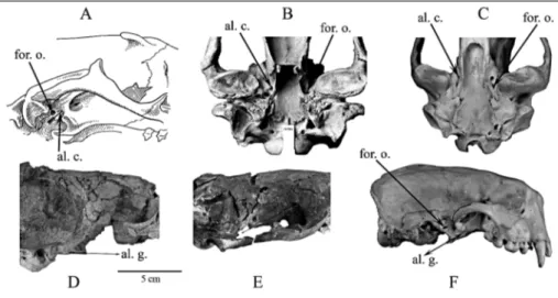
The alisphenoid canal is a tube in the pterygoid strut between the palate and basicranium that transmits the internal maxillary artery (= maxillary artery), a branch from the external carotid artery, deep to the neck of the mandible (Fig. 4). Fossil phocids (Fig. 4D, E) and recent mustelids (Fig. 4F) share a derived character by lacking an alisphenoid canal. However, in primitive Leptophoca and Devinophoca there exists a very shallow groove instead of a proper alisphenoid canal. A com-pletely formed alisphenoid canal is present in Otarioi-dea (Fig. 4A, B), Ursidae (Fig. 4C), Canidae, and most Procyonidae (Chapskii 1955; McLaren 1960; Heptner et al. 1976; Barnes & Hirota 1994). In one specimen of one species of Allodesmus (Barnes 1972), the alisphenoid
Fig. 4 - Carnivoran skulls showing location of the alisphenoid canal (indicated by arrows) and alisphenoid groove (indicated by double-headed arrows) in: A) generalized otariid re-presentative; B) Southern sea lion (Otaria byronia); C) Brown bear (Ursus arctos); D) Devinophoca emryi sp. n. (Early-Middle Miocene; ~16 Ma; Badenian, Vienna Basin, Slovakia; holotype, USNM 553684); E) Leptophoca lenis (Late-Early Miocene; ~18 Ma; Calvert Formation, MD, USA; CMM-V 2021) and F) Sea otter (Enhydra lutris). Abbreviations: al. c. = alisphenoid canal; al. g. = alisphenoid groove; for. o. = foramen ovale.
canal is not fully formed, but the track of the internal maxillary artery is clearly displayed on the bone. The canal is fully formed in other Allodesminae, such as Brachyallodesmus packardi and otherspecies of Allodes-mus (Barnes 1972).
Enaliarctos, recovered from the western coast of North America, is considered to be the most primitive otariid pinniped (from Late Oligocene - Early Miocene, ca. 24±22 Ma.) by some (Mitchell & Tedford 1973; Berta 1991) and has a fully developed alisphenoid canal. An-other semi-aquatic carnivore (from the Arctic Circle) that may represent a morphological link in early pinni-pedimorph evolution is Puijila darwini (from the Early Miocene, 21 to 24 Ma, Aquitanian, European mammal zones MN1-3), which also has an alisphenoid canal and small postglenoid foramen. Puijila is hypothesized to belong to some group of ``otterlike protopinnipeds'' or aquatic carnivores (Rybczynski et al. 2009).
Recent works (DemeÂre et al. 2003; Amson & Muizon 2014) acknowledge the importance of the ali-sphenoid canal as a morphological character. The
ab-sence of a proper alisphenoid canal in D. emryi and D. claytoni supports Tedford's (1976) suggestion that lack of this character relates phocids to the early mem-bers of mustelids rather than to ursids (Fig. 4), or at least supports a paraphyletic origin of Pinnipedia.
Detailed descriptions of the morphology of the middle and innerearregions of D. emryi and compara-tive analyses of these regions in other pinnipeds will be presented in a separate study using modern imaging techniques.
Ontogenetic changes. As previously mentioned by Koretsky (1987, 2001), many cranial and postcranial features are significantly related to individual, ontoge-netic, and sexual variability. Despite similar condylo-basal and total lengths in both devinophocine skulls, the general proportions of the skulls are different. To eliminate any uncertainties about description of a new species, ontogenetic changes of seal skulls were exam-ined.
According to a described set of characters (Kor-etsky 2001: 25), subadult individuals have swollen
tym-claytoni emryi sp.n
1. Total length 119.9 119.5
2. Condylobasal length 119.3 118.0
3. Length of palatine process 71.0 81.0 4. Length of rostral part. measured from
antero-upper corner of orbit 63.5 60.0
5. Length of braincase. measured from posterior
corner of orbit 69.0 70.0
6. Length of tympanic bulla 38.5 33.6
7. Length of tooth-row, P1 to M1 49.0 (L.); 53.2 (R.) 46.9 8. Length of tooth-row, P2 to P4 32.5 (L.); 34.5 (R.) 28.9 9. Maximum diameter of infraorbital foramen 7.5 (L.); 11.2 (R.) 12.1 (L.); 11.2 (R.)
10. Length of temporal fossa 61.5 61.5
11. Width of rostrum across canines 40.0 47.7
12. Maximal infraorbital width 25.5 22.0
13. Minimal infraorbital width 14.0 19.4
14. Width of skull across of zygomatic process of
squamosal 124.0 127.7
15. Width of braincase 88.0 78.3
16. Mastoid width 113.0 102.3
17. Width of palatine process between P1's 19.1 20.8 18. Maximum width of palatine process 55.5 40.9 19. Maximum width of infraorbital foramen 9.0 (L.);10.0 (R.) 7.2 (L.. R.)
20. Width of tympanic bulla 49.3 35.7
21. Width of rostrum 37.0 45.8
22. Height of skull in region of tympanic bulla 80.0 81.7 23. Distance from center of stylomastoid foramen
to center of postglenoid foramen 15.1 19.2
Ratios of measurements meas. 14/ meas. 1 1.03 1.07 meas. 4/ meas. 5 0.92 0.86 meas. 18/ meas. 9 6.17 3.56 meas. 10/ meas. 21 1.66 1.34 meas. 8/ meas. 18 0.59 0.71 Devinophoca Characters Tab. 1 - Cranial measurements (in
mm) of the skull of Devino-phoca.
panic bullae and the rostral portion of the skull is rela-tively short compared with those of older animals. In the D. emryi skull, the rostral part of the skull is not shortened and the tympanic bullae are less swollen (see Tab. 1), suggesting this fossil was an adult. An addi-tional characteristic of subadult individuals is the ar-rangement of incisors in the form of a half-circle, lo-cated at the alveolar margin of the premaxillae. In adult animals, this arrangement generally changes into a straight row with a bony prominence in front of the incisors. Both Devinophoca skulls have this adult ar-rangement.
The overall length of the skull increases as the rostral part grows, significantly influencing ratios of lengths of facial and cerebral parts of the skull in young and adult individuals. The total length and condylobasal length are the same in both species, but the length of the palatine process and braincase is longer in D. emryi, while the rostral part of skull is shorter in D. emryi (Tab. 1).
In accordance with the development of the facial part of the skull and with strengthening of the
muscu-lature during ontogeny, relative increases occur in: zy-gomatic (= jugal) width; width of the rostrum at the level of the uppercanines; width of the palate at P1; greatest width of the palatine bone; and numerous other measurements. All of these measurements are larger in D. emryi than in D. claytoni.
Lastly, based on obliterated cranial sutures and evaluation of the mentioned ontogenetic characters, the skull of D. emryi belongs to an adult individual. Consequently, the proportions of this skull do not cor-respond to the set of ontogenetic characters.
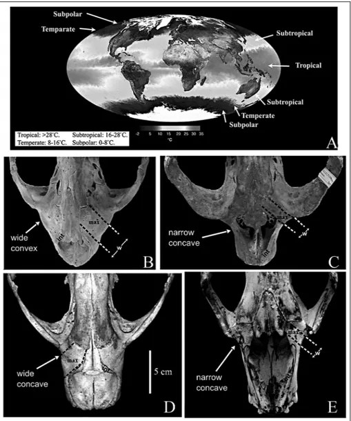
Enlargement of maxilla. In the devinophocine skulls, the preorbital parts of the maxillae (Fig. 2A) between the nasal aperture and the orbits are wide and concave, similarto Monachinae (Chapskii 1974; Koretsky & Rahmat 2013; Fig. 5). While Wyss (1987) and Berta and Sumich (1999) demonstrated that the contribution of an enlarged maxilla to the orbital region is a feature common to all ``pinnipeds'' and used this character to support monophyly, it must be noted that the enlargement of the maxilla is a multistate character
Fig. 5 - A) World map showing oceanic climate zones, for discussion of typical feed-ing distribution of modern true seals (adapted and modified from NASA/ GSFC, MODIS Land Rapid Response Team data). Dorsal view of rostral part of the skull with enlarge-ment of the maxilla in: B) Phocinae, Harbour seal (Phoca vitulina), deep benthic feeders in Tempe-rate zone; C) Cystophori-nae, Hooded seal (Cysto-phora cristata), bottom fee-ders in more shallow waters in Temperate zone; D) Monachinae, Caribbean monk seal (Monachus tropi-calis), feeders on planktonic crustaceans in shallow waterin Subtropical belt; and E) Lobodontinii, Crab-eater seal (Lobodon carcinophagus), feeders on large fish and warm blooded animals in Subpolarzone. Abbreviations: int = nasal process of intermaxilla; max = maxilla; w = width of exposed portion of max-illary bone.
(compare the matrices by Berta & Wyss 1994: 37 and Koretsky 2001).
The generally pronounced convex (Fig. 5B) nat-ure of the maxillary enlargement has been noted to be typical only forphocines (Burns & Fay 1970). In cysto-phorines, the maxilla has a narrow concave shape (Fig. 5C); while in monachines (Fig. 5D) it has a widely con-cave shape (Chapskii 1974; Koretsky & Rahmat 2013). In Devinophocinae, the maxilla is similarin shape to that of Monachinae (Koretsky & Holec 2002). Ursids and lutrines do not show maxillary enlargements, and are thus similar to other terrestrial mammals. Moreover, some authors state that the phocid condition is not a phylogenetically useful character, but can be attributed to a lateral expansion of the maxilloturbinals, consid-ered to be an adaptation to efficiency in warming of inspired air (Muizon & Hendey 1980; Bininda-Emonds & Russell 1996).
From the same locality, we found isolated mand-ibles and numerous individual teeth, with some corre-sponding morphologically and anatomically with teeth in situ in the mandible and skull of D. emryi, while others associate perfectly to teeth in situ in the mandible and skull of D. claytoni. We plan to describe the mand-ible, teeth and some postcranial elements of D. claytoni in the nearfuture.
Upper teeth. The skull, several hemi-mandibles, and three isolated upper teeth (Fig. 6A-C, J-O) men-tioned in this paperwere found nearthe same locality, and because the upper postcanines have root morphol-ogy that corresponds to the shape and size of the alveoli on the maxilla, all of them are identified as belonging to D. emryi. The upperdental formula, shape, position of the cingulum and arrangements of incisor alveoli and
Fig. 6 - Devinophoca emryi, sp. n., referred cheek teeth. Right upperP1 (SNMZ 14529) in A) labial, B) lingual and C) occlusal views. Right lowerp1 (SNMZ 14529) in D) lingual E) labial and F) occlusal views.
Left lowerp1 (SNMZ 14529) in G) lingual, H) labial and I) occlusal views.
Left upperP3 (SNMZ 14529) in J) lingual, K) labial and L) occlusal views.
Left upperP4 (SNMZ 14529) in M) lingual N) labial and O) occlusal views. Lowerleft p4 (SNMZ 14698) in P) lingual Q) labial and R) occlusal views.
length width
length
width
I1
3.0
3.6
I2
3.0
3.7
I3
5.7
C
13.2
10.5
11.8
11.5
P1
6.5
5.6
6.2
5.2
P2
10.0
6.5
5.2
7.2
P3
10.0
8.0
9.6
7.0
P4
10.6
8.2
11.0 - 11.1 5.7 - 7.1
M1
9.3
7.5
8.0
4.7
D. claytoni
D. emryi sp. n.
Teeth
Tab. 2 - Measurements (in mm) of the upper dentition of Devinophoca.
canine alveoli in D. emryi is the same as in D. claytoni (see Koretsky & Holec 2002: 172).
The crown of an isolated right P1 (SNMZ 14529) has a single root (Fig. 6A-C; Tab. 2), oval in cross sec-tion and with a central cusp which is slightly worn. The crown of this tooth is 6.8 mm long, has a conical central cusp and a cuspidate lingual cingulum bearing diminu-tive anterior and slightly larger posterior cusps. The gum line (on P1, P3, and P4), parallel to the lower mar-gin of the enamel, was oblique to the long axis of the root. However, when the tooth is correctly inserted into the alveolus of the holotype maxilla, the gum line is parallel to the margin of the dentary. The complete crown is irregularly shaped in occlusal view, with a flattened labial side, and is inflated distolingually on its lingual side. The cingulum, bearing minute cuspules, encircles the crown and is much smaller on the labial side, whereas, on the lingual side of the crown the cin-gulum is more prominent, and wrinkled.
All uppercheek teeth show slight wearand both of the otherisolated uppercheek teeth are double-rooted (except P1, single-double-rooted), with posterior roots much larger than anterior (Fig. 6J-O). While the anterior roots are round in cross section, the posterior roots are expanded transversely, but not bilobed as in D. claytoni. The crowns of the P3 (SNMZ 14529; Fig. 6J-L) and P4 (SNMZ 14529; Fig. 6M-O) are semi-oval in occlusal view, slightly worn as in the P1 (Fig. 6A-C), and inflated distolingually on its labial side as in P1, but also flat-tened.
The crown of a left P3 (SNMZ 14529; Fig. 6J-L) has a very prominent lingual cingulum, with only a minute anterior cingular cusp (the parastyle) and slightly larger two posterior cusps. The cingulum on a labial side extends at its mid-length, but without any carina as observed in Monachinae (Amson and Muizon 2014).
Characters
n
X
Range
Total length
1
131.5
Length of toothrow i1 - m1
1
53.4
Length of toothrow p1 - p4
3
34.7
33.3 - 36.7
Length of toothrow p1 - m1
3
45.3
44.4 - 46.8
Depth under m1
3
19.3
18.8 - 20.0
Depth under p2
2
20.2
20.0 - 20.4
Depth behind m1
3
16.4
15.9 - 16.7
Depth between p3 - p4
3
17.5
16.7 - 17.9
Thickness of mandible under m1
3
8.5
8.3 - 8.7
Height of ramus
2
48.4
44.9 - 51.8
Length of condyle
3
21.7
20.5 - 23.8
Length of retromandibular space
3
12.6
9.7 - 14.2
Length of symphysis
1
23.0
Condylar height
3
29.0
21.7 - 36.0
i1 length
1
4.6
width
1
3.8
c length
width
1
8.9
p1 length
4
6.2
4.8 - 6.9
width
4
5.0
4.8 - 5.1
p2 length
3
9.4
9.0 - 9.6
width
3
5.4
4.8 - 5.9
p3 length
3
11.0
10.0 - 13.0
width
3
5.7
5.2 - 6.3
p4 length
4
10.8
9.6 - 12.7
width
4
5.2
4.2 - 6.1
m1 length
3
11.3
10.5 - 12.3
width
3
4.0
3.5 - 4.4
Tab. 3 - Means (X) and range for measurements (mm) of numberin sample (n) of the mandibles and lower dentition of Devinophoca emryi sp. n.
The crown of the left P4 (SNMZ 14529; Fig. 6M-O) is even more inflated than in P3 and has one anterior accessory cusp and one slightly larger posterior addi-tional cusp. A likely cuspidate lingual cingulum was present, but showed some wear.
D. emryi, in contrast to D. claytoni, does not have two fused posterior roots or a carnassial notch on the metastyle blade of P4. Also, M1 has a regular double root according to the alveoli, not triangular with three cusps and three roots as seen in D. claytoni. As pre-viously mentioned (Koretsky & Holec 2002), no other phocid has teeth with three cusps and three roots in a triangular arrangement.
Mandible. The body of several mandibles of D. emryi (Fig. 7; Tab. 3) are flattened, thin, and low (vary-ing from 42.8-52.2 mm high), the retromolar space is elongated and the condyloid process is especially short and narrow (21.1 x 8.2 mm; 24.0 x 8.6 mm). The con-dyloid process is elevated, slightly above the level of the tooth row.
The coronoid process (cor. p.) is very thin and long, with its anterior edge strongly sharpened toward the apex of the process and its posterior edge slightly concave. The masseteric fossa is shallow and narrow (almost indistinct), but very well outlined, which indi-cates a sizeable insertion for the masseter muscle (PieÂr-ard 1971; Amson & Muizon 2014), especially on its
ventral edge. The size, width and depth of the fossa resemble those of Leptophoca lenis (Koretsky 2001; Koretsky et al. 2012) and differ from the deep and wide masseteric fossa described in Monachus and Cystophora (Koretsky & Rahmat 2013).
On the medial surface of the ramus, the mandib-ular foramen is located anterior to the apex of the cor-onoid process. The foramen and canal are both very well developed, similarto L. lenis (Phocinae), but in contrast to Hadrokirus martini (Monachinae) from the Early Pliocene (Amson & Muizon 2014).
The symphyseal part of the mandible is partially broken off, but reaches the middle of the alveoli of p2; the chin prominence and diastemata are absent.
Lower teeth. The alveoli of the lowercanines are significantly smallerthan the uppercanines (9 mm: 15 mm respectively), and oval in cross section, but partially destroyed. The cheek tooth row is oriented parallel to the axis of the symphyseal part of the mandible (except p2, where the anterior alveoli is oriented more labial than the posterior). The alveoli are round with the pos-terior being larger than the anpos-terior.
The crowns of the cheek teeth are wide and swol-len. The p2-p4 are multicusped, double-rooted, and have one ortwo additional cusps on a basal cingulum. The posterior alveoli of the postcanines are slightly lar-ger than the anterior. The m1 is double-rooted as well.
Fig. 7 - Mandible of Devinophoca emryi sp. n. (Early-Middle Miocene; ~16 Ma; Badenian, Vienna Basin, Slovakia; USNM 553687), in A) la-bial, B) lingual, and C) oc-clusal views.
Abbreviations: ap = angular process; con. p. = condyloid process; cor. p. = coronoid process; men. f = mental foramen; m = molar; mf = mandibularforamen; ms = mandibularsymphysis; p = premolar.
The basal cingulum of m1 is very well developed, espe-cially on the lingual side. The length of the m1 alveoli is shorter than p4.
Two isolated lowercheek teeth, a right
(SNMZ14529; Fig. 6D-F) and a left (SNMZ14529; Fig. 6G-I), were identified as p1 of D. emryi due to the presence of a single root that is slightly curved, which corresponds to the shape and size of the alveoli on both mandibles. The crown of a right p1 tooth is 16.7 mm long with a conical main cusp (protoconid) and a cus-pidate lingual cingulum bearing a small posterior cusp (hypoconid). The gum line, parallel to the lower margin of the enamel, was oblique to the long (= mesiodistal) axis of the root, but when the tooth is correctly inserted into the p1 alveolus of the mandible, the gum line is parallel to the dorsal margin of the dentary. The outline of the crown of p1 is triangular in occlusal view (Figs 6F, I), with a slightly flattened labial side; the tooth has a single root, which is oval in cross section as well. The cingulum encircles the complete tooth, widening on the lingual side. Near the base of the crown, there are very small additional posterior and anterior cusps.
The crown of p2 (in situ) is oval in occlusal view (Fig. 7C), the tooth is double-rooted, and the labial side of the crown is convex. A cingulum is located on the lingual side. The main cusp (protoconid) is slightly turned caudally. The two small cusps are located on the posteriorside of the majorcusp, and one small cusp is located anterior to the main cusp. In addition to the prominent metaconid, are a less prominent posterior cingularcusp (hypoconid) and tiny anteriorcingular cusp (paraconid). All cusps are entirely preserved. The crown is 10 mm long, 7.5 mm high, and has the same types of derived characters as the p1 described above: prominent and cuspidate lingual cingulum and less prominent basal cingulum (Barnes 1988). The p2 tooth is situated oblique to the axis of the tooth-row, with the posterior root larger than the anterior.
The overall morphology of p3 (in situ) is compar-able to p2 (Fig. 7). Despite the similarsize of p2 and p3 (p3 has a crown 10 mm long, 8.3 mm high), both sec-ondary cusps are larger and more prominent, especially the metaconid, and the main cusp is not turned caudally. The tooth is positioned parallel to the axis of the tooth-row. The posterior root is larger than the anterior.
One isolated left tooth (SNMZ14698; Fig. 6P-R) has a root morphology that corresponds to the alveolus of p4 in the paratype mandible. The crown of p4 is oval in occlusal view, and the buccal side is convex. A very short cingulum is located on the lingual side, restricted to the level of the main cusp. The basal cingulum is not developed at all. The main cusp (protoconid) is directed dorsally, inflated, and conical. Two additional small cusps (metaconids), located on both sides of the major cusp, are intact. The presence of four cusps in a row on
this tooth of D. emryi strongly suggests that it is p4. The two roots of the tooth are wide and oval in cross-sec-tion, with the posterior root wider than the anterior.
The double-rooted m1 alveoli were much smaller than those of p4. In contrast to other postcanines, the posterior alveoli are smaller than the anterior, and oval. Discussion and conclusions
As previously noted, the skulls of Devinophoca present distinguishing, taxon-specific characters as well as mixed characters with the other three extant subfa-milies (see Koretsky & Holec 2002), such as: a thin and low mandibularbody (as in Phocinae) and a total of 8 incisors (as in Monachinae, but with different incisors formula). Thus, the two different species of the Subfam-ily Devinophocinae have three upper incisors (similar to Phocinae, primitive condition) and one lower incisor (similar to Cystophorinae, derived condition).
Studies of Miocene Phocidae overthe last century (i.e. from Alekseev 1924 up to Koretsky & Rahmat 2013), as well as the new material described here from early Middle Miocene sediments, further support pre-vious views on dental morphology (Ognev 1935; Chapskii 1955, 1974; Scheffer1958; Heptneret al. 1976). Detailed descriptions of dentition conclude that true seals ranging from 16 million years ago up to the present have generally uniform incisor formulae within each subfamily, based on total numberof the incisors: Phocinae (10 incisors; 3/2), Cystophorinae (6 incisors; 2/1); Monachinae (8 incisors 2/2); and Devinophocinae (also 8 incisors as Monachinae, but in different combi-nation: 3/1).
Although the skull of the newly described Devi-nophoca emryi is incomplete, certain derived characters in pinnipeds were clearly noticeable on it, such as: re-duced upperfirst molar, loss of the fovea between the upper fourth premolar and first molar, closely spaced mastoid and paroccipital processes, and a reduced naso-labial fossa.
Only phocines (with a convex maxilla) have a circumpolar distribution in Arctic regions (latitude 300-800N) and in the temperate zones of the Northern Hemisphere (Fig. 5A). Cystophorines (excluding Mir-ounga), which have a narrow, concave maxilla, are dis-tributed similarly to phocines (latitude 450-800N). Mon-achines (wide, concave maxilla) are primarily thermo-philic seals, distributed in the subtropical belt (200-400), and Lobodontini (narrow, concave maxilla; Fig. 5E) and Mirounga (long, concave maxilla) are circumpolar in the Southern Hemisphere above latitude 300S (King 1964; Heptneret al. 1976; Berta & Sumich 1999). Based on this distribution pattern, seals that adapted to cold and moderately cold waters, in which the surface
tempera-ture does not exceed 20ëC, have both types of maxillae (concave and convex).
Therefore, the enlargement of the maxilla, and consequently the choanae, as an adaptation forbreath-ing cold air is not well supported by the pattern of geographical distribution. We suggest another theory, which may betterexplain the differences in size and shape of the maxilla.
Different types of diet are reflected in the general organization of seals (Riedman 1990). Some species of Phocinae are mainly benthic feeders, while others prey on fish and cephalopods and can dive in deep waterup to 300 meters (Heptner et al. 1976; Bigg 1981; Riedman 1990; Koretsky 2001; Berta & Adam, 2001). Cysto-phorines primarily rely on bottom feeding in deep water, but not as deep as phocines. Mirounga, the dee-pest diveramong seals (up to 1,000 m; Le Boeuf & Laws 1994), has a disproportionately elongated maxilla. Mona-chine seals feed chiefly in shallowerwater, eating planktonic crustaceans (Lobodon), or large fish, warm-blooded animals including birds (penguins), and krill like the leopard seal (Hydrurga).
The biomechanical and ecological significance of the traits defining each group give us at least some cor-relation to morphology. However, it can be suggested that the shape of the maxilla is an adaptive character for diving (providing increased flow rate, not air storage) and should not be involved in discussions of phyloge-netic relationships at higher taxonomic levels (Bechly 2000).
Based on the stratigraphy of the site (Shultz 2004; Hoheneggeret al. 2008; Domning & Pervesler2012), the Paratethyan seals from the Vienna Basin, specifically from the Bonanza locality (Devinophoca claytoni and D. emryi), occupied transitional shallow marine waters with coral-reef zones. Moreover, previous studies by Hoheneggeret al. (2008) and Domning & Pervesler
(2012), as well as examinations of diagnostic marine and terrestrial faunas of the region (Holec & Sabol 1996; Holec et al. 1997; Shultz 2004; Fejfar& Sabol 2009), support a transition from a humid, tropical shal-low shore zone in the early Badenian (16.26 - 14.89 Ma) to a subtropical climate in the middle Badenian (14.89 -13.82 Ma) and a warm temperate climate during late Badenian (13.82 -12.73Ma).
Phylogenetic analysis
The data matrix for the 40 included characters is shown in Tab. 4. Cranial, mandibular and dental char-acters and character-states for Phocidae: 0, designates the most primitive state among the taxa studied; 1-2, derived states; ?, unknown or missing data. Some char-acters have the opposite polarity to that of Berta and Wyss (1994) and Burns and Fay (1970), while some characters have the same polarity as that of Chapskii (1974). Some characters were updated and modified from Koretsky (2001), Koretsky and Grigorescu (2002), Koretsky and Rahmat (2013).
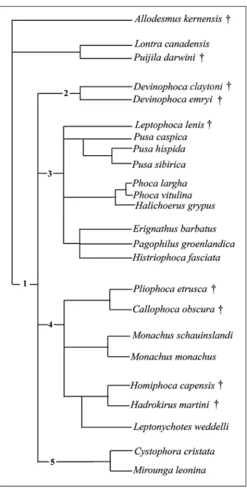
The analysis was done with NONA from Golo-boff (1999) and Winclada from Nixon (1999) using a heuristic search of the phocid taxa of 40 unweighted characters. This is the resulting single Wagner most par-simonious tree generated by Winclada with 179 steps long, having Consistency Index of 0.57, and Retention Index 0.64.
The matrix of character-state data for 20 species of fossil and modern phocids is given in Tab. 4; in addi-tion, the following taxa were used as outgroups: fossil representatives, such as Allodesmus and Puijila, and Re-cent mustelid Lontra.
Skull
1. Tympanic bulla: (0) small; (1) large.
Allodesmus kelloggi 0 1 0 1 0 0 1 1 1 1 0 2 2 1 0 1 0 0 1 0 0 0 0 2 1 0 0 0 1 0 1 0 0 0 0 1 0 0 1 1 Lontra canadensis 1 0 1 0 1 1 1 0 2 0 1 0 0 1 0 1 2 1 0 0 0 0 1 0 0 1 1 0 0 0 0 0 0 0 1 1 0 0 1 1 Puijila darwini 0 1 0 1 1 0 1 0 0 0 1 2 1 1 1 1 0 1 1 1 0 0 1 1 0 0 0 1 0 0 0 0 1 1 1 1 0 2 1 1 Devinophoca claytoni 0 0 1 0 0 0 0 0 0 0 1 2 1 0 0 ? 1 1 0 0 0 ? 1 0 0 1 0 0 0 0 0 0 ? ? ? ? ? ? ? ? Devinophoca emryi 0 0 1 0 1 0 0 0 0 ? ? 2 0 0 0 1 0 0 1 0 0 3 1 0 0 1 0 0 0 0 0 0 1 0 1 1 0 0 2 0 Cystophora cristata 1 0 1 1 0 1 1 1 2 0 0 1 1 1 0 0 1 1 0 0 0 2 0 1 1 0 1 1 1 0 1 1 0 1 1 0 2 1 1 0 Mirounga leonina 1 0 1 1 1 1 0 0 2 1 0 1 0 1 0 0 2 1 0 0 0 2 0 1 1 0 0 1 1 0 1 1 2 0 1 2 2 2 2 1 Monachus monachus 0 0 1 0 1 1 0 1 1 1 1 2 2 0 1 0 0 0 0 0 0 1 1 1 0 1 0 0 0 1 0 0 1 2 1 1 2 2 2 1 Monachus schauinslandi 0 0 1 0 1 1 0 1 0 1 1 2 2 0 1 0 0 0 0 1 1 1 1 1 0 0 0 0 0 1 1 0 1 1 1 1 1 2 2 1 Callophoca obscura 1 0 0 0 0 0 1 1 1 1 1 2 2 0 0 0 1 0 1 0 0 1 1 1 0 0 1 0 0 1 0 2 0 1 1 1 0 2 1 0 Phoca vitulina 1 0 1 0 0 0 0 0 1 0 0 0 1 0 0 0 0 1 0 2 1 0 1 1 0 0 1 1 0 0 0 0 0 1 0 0 0 0 0 1 Erignathus barbatus 1 1 1 1 0 1 0 0 0 0 0 0 1 0 1 0 0 1 0 2 1 0 1 1 1 1 1 1 0 1 0 0 0 1 1 1 1 1 2 0 Leptophoca lenis 0 0 ? 0 0 0 1 1 1 0 0 0 0 0 0 1 0 1 1 1 1 0 1 1 0 1 0 0 0 0 0 0 1 1 1 1 0 0 0 0 Pliophoca etrusca 1 0 ? 1 0 0 1 0 1 1 1 2 2 0 0 0 2 1 1 1 1 1 1 1 0 0 1 0 0 1 0 1 1 2 0 0 0 1 0 0 Homiphoca capensis 0 0 1 0 0 0 0 0 0 1 1 2 2 0 0 1 2 0 1 1 1 1 1 0 1 0 1 0 ? 1 1 0 0 1 1 0 1 1 0 1 Phoca largha 0 1 0 0 0 1 0 0 0 1 1 0 1 1 0 1 0 0 0 0 0 0 1 1 0 1 0 1 0 0 1 2 1 0 0 0 0 0 0 1 Pusa hispida 1 1 1 0 0 1 0 0 0 1 0 0 2 1 0 0 0 1 0 1 0 0 1 0 1 1 0 1 0 0 1 0 0 0 0 0 2 0 2 0 Halichoerus grypus 0 0 1 0 0 0 0 0 0 0 1 0 1 1 0 1 0 1 1 1 0 0 1 1 0 1 1 1 0 0 1 0 0 1 1 0 1 1 2 0 Pagophilus groenlandica 1 1 0 0 0 0 0 0 0 1 0 0 1 0 0 0 2 0 0 2 0 0 1 0 0 1 1 1 0 0 0 0 1 1 0 1 1 1 2 0 Hadrokirus martini 0 0 0 1 1 0 1 0 0 1 0 2 1 1 1 0 0 1 1 1 0 ? 1 1 0 1 0 0 0 1 1 2 2 0 1 2 2 1 2 1 Character number 5 6 10 1112 13 14 15 16 3132 33 34 35 36 3738 39 40 1 2 3 4 7 8 Species 9 17 18 1920 2122 23 24 25 2627 28 29 30
2. External auditory meatus: (0) inframeatal lip is well devel-oped; (1) poorly developed.
3. Mastoid process: (0) not united with paroccipital process; (1) united with paroccipital process.
4. Mastoid process: (0) axis of mastoid convexity is not directed ventrally; (1) directed ventrally.
5. Mastoid process: (0) not strongly pronounced prominence lateral to auditory bullae; (1) pronounced.
6. Mastoid process: (0) narrow (width of the process is less than the length of the process itself); (1) wide (Chapskii 1974: 301; in con-trast to Berta and Wyss 1994: 48).
7. Mastoid process: (0) bulbous; (1) cylindrical
8. Mastoid process: (0) width less than or equal to one-half of length of tympanic bulla; (1) width greater than one-half of length of tympanic bulla.
9. Mastoid convexity: (0) not turned down; (1) moderately turned down behind the mastoid process; (2) directed sharply down-ward behind the mastoid process.
10. Nasal bones: (0) anterior ends form one common termina-tion; (1) anterior ends separated.
11. Nasal bone: (0) maxillary contact longer than frontal con-tact; (1) frontal and maxillary contacts almost equal in length.
12. Maxilla: (0) has very pronounced convexity anterior to the orbits; (1) has short concavity; (2) has long concavity (Chapskii 1974: 299; in contrast to Berta and Wyss 1994: 46).
13. Anterior palatine foramina: (0) round and deep; (1) oval and shallow; (2) indistinctively marked (Burns and Fay, 1970).
14. Palatal groove: (0) present; (1) absent.
15. Palatal process of maxillary bone: (0) flat; (1) convex. 16. Oval foramen: (0) hidden under hamular process of ptery-goid bones; (1) exposed.
17. Interorbital width: (0) less than 25.0% of mastoid width of skull; (1) less than 30.0% but equal to orgreaterthan 25.0% of mastoid width; (2) equal to or greater than 30.0% of mastoid width (Burns and Fay 1970: 370; Chapskii 1974: 299).
18. Jugular process: (0) well developed, large hook shape; (1) poorly developed (as a small conical projection) or absent.
19. Rostrum: (0) short, relative to skull; (1) elongated (Chapskii 1974: 299).
20. Diameter of infraorbital foramen: (0) less than diameter of alveolus of uppercanine; (1) equal to diameterof uppercanine alveolus; (2) greater than diameter of alveolus of upper canine.
21. Anteroposterior length of auditory bullae: (0) equal to or greater than distance between them; (1) less than distance between them (Burns and Fay 1970: 382; Chapskii 1974: 300).
Teeth
22. Numberof incisors: (0) 3/2; (1) 2/2; (2) 2/1 (Chapskii 1974: 289; in contrast to Burns and Fay 1970: 380); (3) 3/1.
23. Roots of postcanine teeth (P,p 2 ± P,p 3): (0) one (fused); (1) two (Berta and Wyss 1994: 51).
24. Roots of P,p 4: (0) three; (1) two; (2) one.
25. Crowns of postcanine teeth: (0) multicusped; (1) single-cusped.
26. Dimensions of postcanine teeth relative to longitudinal dia-meterof alveolus of uppercanine: (0) more than 60.0%; (1) less than 60.0 % orsub-equal.
27. Longitudinal diameterof alveolus of uppercanine com-pared with maximal width of infraorbital foramen: (0) sub-equal in size; (1) more than one-half of maximal width.
28. Basal cingulum of postcanine teeth: (0) well developed; (1) not developed.
29. Number of additional cusps of premolars: (0) more than two; (1) no additional cusps.
30. Premolar: (0) aligned parallel to axis of tooth-row; (1) seated obliquely.
31. Upper incisors: (0) arranged in a curved arcade; (1) arranged in a straight line.
32. Second and third upper incisors: (0) third larger than sec-ond; (1) second larger than third, (2) all upper incisors equal in size.
Mandible
33. Mandibularsymphysis: (0) continues at least to the middle of the alveolus of p3; (1) reaches only to the alveolus of p2; (2) reaches only to the alveolus of p1.
34. Lateral outline of symphyseal region: (0) square, symphysis thin; (1) rounded, symphysis thick; (2) straight, symphysis thick.
35. Chin prominence: (0) pronounced; (1) absent or weakly developed.
36. Chin prominence: (0) extends from the anterior or poster-ior alveolus of p2 to the posterposter-ior or anterposter-ior alveolus of p4; (1) extends from the anterior alveolus of p2 to the anterior alveolus of p3; (2) extends from the anterior alveolus of p2 to the posterior alveolus of m1.
Fig. 8 - The single, most parsimonious Wagner tree generated by Winclada with 179 steps long, CI = 0.57, and RI = 0.64.
37. Maximum height of mandibularbody: (0) between p2 and p3; (1) in the middle of or at the posterior portion of p2; (2) situated between alveoli p4 and m1 orposteriorto alveolus of m1.
38. Diastemata and tooth alveoli: (0) alveoli small with equal diastemata; (1) alveoli round and large, with equal diastemata between them; (2) alveoli shallow, and diastemata unequal.
39. Alveoli of p4 and m1: (0) alveoli similarin size; (1) alveoli of p4 smallerthan alveoli of m1; (2) alveoli of p4 largerthan alveoli of m1 (unordered character).
40. Retromandibular space: (0) long; (1) short.
Points where the nodes of the present tree (Fig. 8) correspond to traditionally recognized phocid taxa are indicated. Only one new name is introduced here: in-clusion of Devinophoca emryi within the subfamily De-vinophocinae requires recognition of new species.
The nodes of the cladogram shown in Fig. 8 are supported by the following character transformations:
Node 1 (Family Phocidae): 25(0). This paraphy-letic group with an ancestral or primitive character (multicuspate crowns of postcanine teeth) is treated as plesiomorphic for the family. Also, unordered character 39(2): the alveoli of p4 larger than alveoli of m1, is shared with other members of the subfamilies Monachi-nae and PhociMonachi-nae.
Node 2 (subfamily Devinophocinae, possibly paraphyletic): 13(0); 22(3); 24(0). The anterior palatal foramina are round and deep; number of incisors. Para-phyly of D. claytoni: three fused roots of the postcanine teeth.
Node 3 (subfamily Phocinae): 20(1, 2); 21(1). The diameter of the infraorbital foramen is equal to or great-erthan the diameterof the alveolus of maxillary canine; the length of auditory bulla is less than the distance
between them. Also, character 20(1) is homoplasious in Monachus schauinslandi and Leptophoca lenis.
Node 4 (subfamily Monachinae): 11(1); 12(2); 13(2); 22(1); 31(1). The relative dimensions of the fron-tal and maxillary parts of the nasal bones; shape of the anterior palatine foramina; maxilla forms a long concav-ity; reduced number of incisors.
Node 5 (subfamily Cystophorinae): 9(2); 12(1); 22(2); 29(1); 32(1). The mastoid convexity directed shar-ply downward behind the mastoid process. Maxilla forms a short concavity; advance reduced number of incisors; no additional cusps on premolars. Second in-cisor is larger than third.
Acknowledgments. We thank Drs. Clayton E. Ray, Robert J. Emry, and Daryl P. Domning for their critical reviews and discussion of several drafts of this work; Steven J. Jabo and Peter Kroehler, for participation in excavation, preparation, and providing casts for study-ing; Dr. James G. Mead and Charles W. Potter, for permission to study the modern pinnipeds in the National Museum of Natural History, Smithsonian Institution; and Dr. Yuri A. Semenov of the Museum of Natural History, Kiev, Ukraine for taking an active role in this discov-ery. The authors also express their gratitude to reviewers of the article, whose comments helped to increase the scientific value of this paper, and Dr. Lawrence G. Barnes of the Natural History Museum of Los Angeles County for constructive critical reviews of the manuscript. The authors express their sincere thanks to Anna DurisÏova and Brani-slav MatousÏek, both of the Slovak National Museum of Natural His-tory, Bratislava and to Dr. Peter Holec, Department of Geology and Paleontology, Faculty of Sciences, Comenius University, Bratislava, for their cooperation and permission to study material under their care. We also thank Slovakian amateurpaleontologist SÏ. MeÂszaÂrosÏ forcollecting additional postcranial and some cranial material and donating it to this project. Most of the financial support for the excavation, international travel, preparation, and research for this project was provided by the National Geographic Society Grant #5927-97, and partially by the Re-mington Kellogg Fund of the Smithsonian Institution.
R E F E R E N C E S
Alekseev A.K. (1924) - Seals in the Sarmatian deposits of Southern Russia. J. Nauch.-Issle. kaf. v Odesse, 1: 26-34 [in Russian].
Amson E. & Muizon C. de. (2014) - A new durophagous phocid (Mammalia: Carnivora) from the late Neogene of Peru and considerations on monachine seals phy-logeny. J. Syst. Palaeontol., 12: 523-548.
Barnes L.G. (1972) - Miocene Desmatophocinae (Mamma-lia: Carnivora) from California. Univ. Calif. Publ. Geol. Sci., 89: 1-68.
Barnes L.G. (1979) - Fossil enaliarctine pinnipeds (Mamma-lia: Otariidae) from Pyramid Hill, Kern County, Ca-lifornia. Contrib. Sci. Nat. Hist. Mus. L.A. County 318: 1-41.
Barnes L.G. (1988) - A new fossil pinniped (Mammalia: Otariidae) from the Middle Miocene Sharktooth Hill Bonebed, California. Contrib. Sci, Nat. Hist. Mus. L.A. County, 396: 1-11.
Barnes L.G. & Hirota K. (1994) - Miocene pinnipeds of the otariid subfamily Allodesminae in the North Pacific Ocean: systematics and relationships. In: Barnes L.G., Hasegawa Y. & Inuzuka N. (Eds) -The Island Arc, Special Issue, Evolution and Biogeography of Fossil Marine Vertebrates in the Pacific Realm, Collected Papers from a Symposium Dedicated to the Memory of Arthur Remington Kellogg in the Year of the 100thAnniversary of his Birth. Proc. 29th Intern. Geol. Congress, Kyoto, Japan, 3: 329-360, 1992.
Bechly G. (2000) - Mainstream Cladistics versus Hennigian Phylogenetic Systematics. Stutt. Beitr. Naturk., Serie A(Biologie), 613, 11 pp.
Berta A. (1991) - New Enaliarctos (Pinnipedimorpha) from the Miocene of Oregon and the role of ``Enaliarctids'' in Pinniped Phylogeny. Smithsonian Contrib. Paleo-biol., 69: 1-33.
Berta A. (1994) - New specimens of the pinnipediform Ptero-narctos from the Miocene of Oregon. Smithsonian Contrib. Paleobiol., 78: 1-30.
Berta A. & Wyss A.R. (1994) - Pinniped Phylogeny. In: Berta A. & DemeÂre T.A. (Eds) - Contributions in Marine Mammal Paleontology Honoring Frank C. Whitmore, Jr. Proc. San Diego Soc. Nat. Hist., 29: 33-56.
Berta A. &. Sumich J.L. (1999) - Marine Mammals: Evolu-tionary Biology. Academic Press, San Diego, 494 pp. Berta A. & Adam P.J. (2001) - Evolutionary Biology of Pinnipeds. In: Mazin J.M. & de BuffreÂnil V. (Eds) -Secondary Adaptation of Tetrapods to Life in Water: 235-260. Verlag Dr. Friedrich Pfeil, MuÈnchen, Ger-many.
Bigg M. A. (1981) - Harbour seal - Phoca vitulina and P. largha. In: Ridgway S.H. & Harrison R.J. (Eds) -Handbook of marine mammals. 2 - Seals: 1-27. Aca-demic Press, Inc., London.
Bininda-Emonds O.R.P. & Russell A.P. (1996) - A morpho-logical perspective on the phylogenetic relationships of the extant phocid seals (Mammalia: Carnivora: Phocidae). Bonner Zool. Monogr., 41: 1-256.
Burns J.J. & Fay F.H. (1970) - Comparative morphology of the skull of the Ribbon seal, Histriophoca fasciata, with remarks on systematics of Phocidae. J. Zool. (London), 161: 363-394.
Chapskii K.K. (1955) - An attempt at revision of the sys-tematics and diagnostics of seals of the subfamily Phocinae. Trudy Zool. Inst. Akad. Nauk SSSR, 17: 160-199 [in Russian]. (English translation by Jeletzky, T.F. - Fish. Res. Board Can., Transl. Ser, 114, 57 pp., 1957).
Chapskii K.K. (1974) - In defense of classical taxonomy of the seals of the family Phocidae. Trudy Zool. Inst. Acad. Sci. USSR. 53: 282-334 [in Russian].
DemeÂre T.A., Berta A. & Adam P.J. (2003) - Pinnipedi-morph evolutionary biogeography. In: Flynn L.J. (Ed.) - Vertebrate Fossils and their Context, Contri-butions in Honor of Richard H. Tedford, Bull. Amer. Mus. Nat. Hist., 279: 32-76.
Domning D.P. & Pervesler P. (2012) - The sirenian Meta-xytherium (Mammalia: Dugongidae) in the Badenian (Middle Miocene) of Central Europe. Austrian J. Earth Sci., 105: 125-160.
FejfarO. & Sabol M. (2009) - Middle Miocene Plesiodimylus from the DevõÂnska Nova Ves-Fissures site (western Slovakia). Bull. Geosci. 84(4): 611-624.
Flynn J., Finarelli J., Zehr S., Hsu J. & Nedbal M. (2005) -Molecular phylogeny of the Carnivora (Mammalia): assessing the impact of increased sampling on resol-ving enigmatic relationships. System. Biol. 54(2): 317-337.
HeptnerV.G, Chapskii K.K. & Arseniev B.A. (1976) -Mammalia of the Soviet Union. Pinnipeds and Ceta-cea, Moscow, 2(3): 1-717 [in Russian].
Hohenegger J., Coric S., Khatun M., Pervessler P., Rogl F., Rupp Ch., Selge A., Uchman A. & Wagreich M. (2008) - Cyclostratigraphic dating in the Lower Ba-denian (Middle Miocene) of the Vienna Basin
(Aus-tria): the Beden-Sooss core. Intern. J. Earth Sci. 98: 915-930.
Holec P., Klembara J. & MeszaÂrosÏ S. (1987) - Discovery of new fauna of marine and terrestrial vertebrates in De-võÂnska Nova Ves. Geol. Carpath., 38: 349-356. Holec P. & Sabol M. (1996) - The Tertiary vertebrates from
DevõÂnska Kobyla. Miner. Slov., 28: 519-522 [in Slo-vak].
Holec P., Klembara J. & MeszaÂrosÏ S. (1997) - Fossils of the DeÂvinska Kobyla hill. In: FeraÂkova V. (Ed) - Flora of the DeÂvinska Kobyla hill, APOP-Edition, Bratislava, 639 pp. [in Slovak].
Howell A.B. (1928) - Contribution to the Comparative Anatomy of the Eared and Earless Seals (Genera Za-lophus and Phoca). Proc. U.S. Nat. Mus., 73: 1-142. King J.E. (1964) - Seals of the World. Brit. Mus. (Nat. Hist.),
London, 154 pp.
King J.E. (1983) - Seals of the World. Second Edition, Brit. Mus. (Nat. Hist.), Comstock Publishing Associates, Ithaca, New York, 240 pp.
Koretsky I.A. (1987) - Sexual dimorphism in the structure of the humerus and femur of Monachopsis pontica (Pin-nipedia: Phocinae). Vest. Zool., 4: 77-82 [in Russian]. Koretsky I.A. & Ray C.E. (1994) - Cryptophoca, new genus for Phoca maeotica (Mammalia: Pinnipedia: Phocinae) from Upper Miocene deposits in the Northern Black Sea region. Proc. Biol. Soc. Wash., 107: 17-26. Koretsky I.A. (2001) - Morphology and Systematics of
Mio-cene Phocinae (Mammalia: Carnivora) from Para-tethys and the North Atlantic Region. Geol. Hung., 54, 109 pp.
Koretsky I.A. & Grigorescu D. (2002) - The Fossil Monk Seal Pontophoca sarmatica (Alekseev) (Mammalia: Phocidae: Monachinae) from the Miocene of Eastern Europe. Smith. Contr. Paleobiol., 93: 149-162. Koretsky I.A. & Holec P. (2002) - A primitive seal
(Mam-malia: Phocidae) from the Early Middle Miocene of Central Paratethys. Smithsonian Contr. Paleobiol., 93: 163-178.
Koretsky I.A., Ray C.E. & Peters N. (2012) - A new species of Leptophoca (Carnivora, Phocidae, Phocinae) from both sides of the North Atlantic Ocean (Miocene seals of the Netherlands, part I). Deinsea Ann. Nat. Hist. Mus. Rotterdam, 15: 1-12.
Koretsky I.A. & Rahmat S. (2013) - First Record of fossil Cystophorinae (Carnivora, Phocidae): Middle Mio-cene seals from the Northern Paratethys. Riv. It. Pa-leontol. Strat., 119(3): 325-350.
Le Boeuf B.J. & Laws R.H. (1994) - Elephant seals: an in-troduction to the genus. In: Le Boeuf B.J. & Laws R.M. - Elephant seals. Population ecology, behavior and physiology: 1-26, Univ. California Press, Berke-ley (CA).
McLaren I.A. (1960) - Are the Pinnipedia biphyletic? Sys-tem. Zool., 9: 18-28.
MillerM.E., Christensen G.C. & Evans H.E. (1964) - Anat-omy of the dog. W.B. Saunders Company, Philadel-phia, London, 941 pp.
Mitchell E.D. (1966) - The Miocene pinniped Allodesmus. Univ. Calif. Publ. Geol. Sci. 61: 1-105.
Mitchell E.D. & Tedford R.H. (1973) - The Enaliarctinae a New Group of Extinct Aquatic Carnivora and a con-sideration of the Origin of the Otariidae. Bull. Amer. Mus. Nat. Hist., 151(3): 201-284.
Muizon C. de & Hendey Q.B. (1980) - Late Tertiary seals of the South Atlantic Ocean. Ann. South African Mus., 2: 91-128.
Muizon C. de (1981) - Premier signalement de Monachinae (Phocidae: Mammalia) dans le SaheÁlien (MioceÁne SupeÂrieue) d'Oran (AlgeÂrie). Palaeovertebrata, 11: 181-194.
Muizon C. de. (l982) - Les relations phylogenetiques des Lutrinae (Mustelidae, Mammalia). Geobios, mem. sp., 6: 259-272.
Muizon C. de. (1992) - PalaÈontologie. In: Duguy R. & Ro-bineau D. (Eds) - Handbuch derSaÈugetiere Europas, Band 6: MeeressaÈuger. Teil II: Robben - Pinnipedia: 34-41. In: NiethammerJ. & Krapp F. (Eds) - AULA-Verlag, Wiesbaden.
Ognev S.I. (1935) - Mammals of the USSR and adjacent countries. Carnivora. Glavpushnina, Moscow-Lenin-grad, 3, 752 pp. [in Russian] (English translations by Birron A. & Coles Z.S. for Israel Program for Scien-tific Translations, 1962).
PieÂrard J. (1971) - Osteology and myology of the Weddell seal Leptophoca weddelli (Lesson, 1826). In: Burt W.H (Ed.) - Antarctic Pinnipedia. Antarct. Res. Ser. Nat. Acad. Sci.-Nat. Res. Cen. 18, 53-108.
Ray C.E. (1976) - Phoca wymani and other Tertiary seals (Mammalia: Phocidae) described from the eastern sea-board of North America. Smithsonian Cont. Paleo-biol., 28: 1-36.
Riedman M. (1990) - The Pinnipeds: Seals, Sea Lions, and Walruses. University of California Press, Berkeley, Los Angeles, Oxford, 439 pp.
Rybczynski N., Dawson M.R., & Tedford R.H. (2009) - A semi-aquatic Arctic mammalian carnivore from the Miocene epoch and origin of Pinnipedia. Nature, 458 (7241): 1021-24.
SchefferV.B. (1958) - Seals, Sea Lions, and Walruses. A Re-view of the Pinnipedia. Stanford University Press, Stanford, California, 179 pp.
Schultz O. (2004) - A Triggerfish (Osteichthyes: Balistidae: Balistes) from the Badenian (Middle Miocene) of the Vienna and the Styrian Basin (Central Paratethys). Ann. Naturhist. Mus. Wien, 106A: 345-369.
Simpson G.G. (1945). The Principles of Classification and a Classification of Mammals. Bull. Amer. Mus. Nat. Hist. 85: 1-350.
Tedford R.H. (1976) - Relationship of pinnipeds to other carnivores (Mammalia). System. Zool., 25(4): 363-374. Wolsan M. (1993) - Phylogeny and classification of early European Mustelidae (Mammalia: Carnivora). Acta Theriologica, 38(4): 345-384.
Wozencraft C. (1989) - The phylogeny of the Recent Carni-vora. In: Gittleman J.L. (Ed.) - Carnivore behavior, ecology, and evolution: 495-535.
Wyss A.R. (1987) - The walrus auditory region and mono-phyly of pinnipeds. Am. Mus. Novitates 2871, 31 pp. Wyss A.R. & Flynn J.J. (1993) - A phylogenetic analysis and definition of the Carnivora. In: Szalay F. S., Novacek M. J. & McKenna M. C. (Eds) - Mammal phylogeny: Placentals: 32-52, Springer-Verlag, New York. Wyss A.R. (1994) - The evolution of body size in phocids:
some ontogenetic and phylogenetic observations. In: Berta A. & DemeÂre T.A. (Eds) - Contributions in Mar-ine Mammal Paleontology Honoring Frank C. Whit-more, Jr. Proc. San Diego Soc. Nat. Hist., 29: 69-77.