TESI DI DOTTORATO IN COTUTELA
UNIVERSITÀ DEGLI STUDI DI SASSARI DIPARTIMENTI DI MEDICINA VETERINARIAScuola di Dottorato in:
Riproduzione, Patologia, Allevamento e Benessere Animale
(XXXI CICLO)
UNIVERSITAT AUTÒNOMA DE BARCELONA FACULTAT DE VETERINÀRIA
Departament de Ciència Animal i dels Aliments
Producciòn Animal
RESVERATROL SUPPLEMENTATION DURING IN
VITRO MATURATION: EFFECT ON THE QUALITY
OF OOCYTES IN SPECIES OF VETERINARY
INTEREST
Supervisori: Dottorando: Prof. Luisa Bogliolo Anna Rita Piras Prof. Maria Teresa Paramio Nieto
Abstract
The quality of oocytes plays a pivotal role in determining in vitro embryo production (IVEP) outcomes. Different intrinsic and extrinsic factors can impair the quality of mammalian oocytes, affecting their developmental competence.
The aim of the study was to evaluate the effect of supplementation of resveratrol, a natural antioxidant, during in vitro maturation (IVM) on the quality of oocytes in species of veterinary interest (domestic cat, prepubertal goats and sheep).
Three studies were performed to test the potential beneficial influence of resveratrol to improve the in vitro developmental competence of poor quality oocytes such as under sub-optimal condition or with low developmental competence.
Specifically, the effect of resveratrol addition to the IVM medium was tested on:
-STUDY I: IVEP from domestic cat oocytes retrieved from ovaries stored at 4°C for 24 and 48h.
Oocytes retrieved from stored ovaries for 24 and 48h were IVM with or without 5μM resveratrol. Meiotic competence, intracellular levels of reactive oxygen species (ROS) and glutathione (GSH), blastocyst yield and the blastocyst cell number were evaluated. The results showed that resveratrol treatment had not effect on the meiotic competence of the oocytes. Resveratrol groups had lower (P<0.05) intracellular ROS levels and higher (P<0.05) GSH content compared to untreated oocytes, both at 24 and 48h. Moreover, resveratrol supplementation significantly increased blastocyst yield in 48h group and
-STUDY II: IVEP from prepubertal goat oocytes selected by brilliant cresyl blue (BCB) staining.
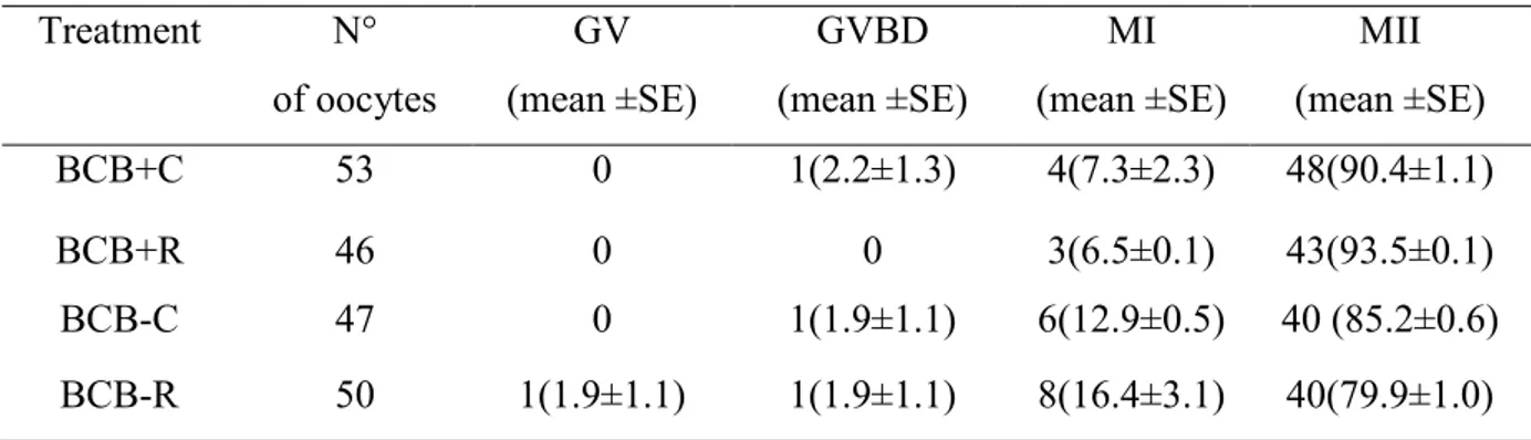
Oocytes were classified by BCB staining as BCB+ (fully grown oocytes ) or BCB- (growing oocytes) and IVM with or without 1μM resveratrol. ROS and GSH levels, mitochondrial activity and distribution and ATP content were analyzed in metaphase II oocytes (MII). After in vitro fertilization (IVF), the blastocyst rate and the blastocyst quality were assessed. No differences were found in ROS levels, ATP content and mitochondrial activity among groups. GSH levels were significantly higher in both BCB groups treated with resveratrol than their respective controls. Oocytes treated with resveratrol showed a higher proportion of clustered active mitochondria than control groups. The development to blastocyst stage was significantly higher in BCB+ oocytes matured with resveratrol compared with the other groups. No differences were observed in blastocyst quality among groups.
STUDY III: In vitro fertilization outcome of prepubertal sheep oocytes under cadmium exposure.
Oocytes were expose to 2μM cadmium (Cd) and IVM in the presence of different concentration of resveratrol: 0μM (Cd), 1 μM (Cd-Resv 1μM) and 2 μM (Cd-Resv 2μM). Oocytes matured in absence of Cd were used as control. Fertilization outcomes, cortical granules (CGs) and mitochondria distribution, mitochondria activity and ROS level were evaluated. Oocytes of control, Cd-Resv 1μM and Cd-Resv 2μM groups had higher normal fertilization compared to Cd group (P<0.05). The percentage of MII oocytes with CGs distributed in the cortex of the oocytes was higher (P<0.05) in control and Cd-Resv 1μM groups than Cd group. The percentage of MII oocytes that exhibited a homogeneous mitochondria distribution throughout the cytoplasm was higher in control, Cd-Resv 1μM and Cd-Resv 2μM groups than Cd group (P<0.05). Lower activity (P<0.05) of mitochondria was recorded in control and Cd-Resv 1μM oocytes compared to Cd oocytes. The intracellular ROS levels were lower in control, Cd-Resv 1μM and Cd-Resv 2μM groups than Cd group.
Taking all these results into account, we conclude that resveratrol supplementation during IVM constitutes a useful strategy to improve oocyte quality and IVEP outcome in species of veterinary interest. The mechanism underlying resveratrol effects included the regulation of bioenergetic/redox status of the oocytes by the modulation of ROS and GSH levels and mitochondria function and the distribution of cytoplasmic organelles.
Index
Chapter1:General background ... 7
Chapter2:Bibliographical revision ... 10
2.1 Acquisition of oocyte development competence ... 11
2.1.1 The final phase of oocyte maturation ... 12
2.2 Methods of oocyte quality assessment ... 15
2.2.1 Morphological assessment of oocytes ... 15
2.2.2 Brilliant cresyl blue selection ... 17
2.2.3 Cumulus cells transcriptome analysis ... 18
2.2.4 Follicular fluid analysis ... 18
2.3 Factors affecting oocyte quality ... 20
2.3.1 Donor Age ... 20
2.3.2 Environmental contaminant ... 22
2.3.3 ARTs procedures ... 23
2.4 Oxidative stress and oocyte quality ... 26
2.5 Utility of antioxidants during ARTs ... 30
2.5.1 Resveratrol ... 32
2.5.1.1 Resveratrol effect on oocyte quality and in vitro embryo production ... 38
2.5.1.2 Protective effect of Resveratrol on oocyte quality under sub-optimal conditions .... 39
Chapter 3:Objectives ... 41
Chapter 4:Resveratrol supplementation during in vitro maturation: effect on developmental competence of oocytes retrived from cat ovaries stored at 4°C for 24 and48h………...44
References:………67
Chapter 5:Resveratrol supplementation during in vitro maturation: effect on developmental competence of prepubertal goat oocytes selected by brilliant cresyl blue staining ... 73
References:………97
Chapter 6:Resveratrol supplementation during in vitro maturation of oocytes under cadmium exposure: effect on fertilization outcomes in the ovine model... 107
References: ……….128
Chapter 7:General discussion ... 133
Resveratrol supplementation during in vitro maturation: Effect on
the quality of oocytes in species of veterinary interest.
Chapter1:
1- General background
The application of Assisted Reproductive Technologies (ARTs) have experienced growing interest in human to treat infertility and in animal to increase the rate of selective breeding and in safeguard of endangered species. In the livestock industry, the use of methods such as artificial insemination (AI), in vitro production of embryos (IVP), embryo transfer (ET) and cryopreservation allow to increase the productivity of the animals and to obtain offspring from sub-fertile subjects of high genetic value (Wu and Zan, 2012). In vitro embryo production using oocytes derived from prepubertal donors in conjunction with in vitro embryo transfer, termed as juvenile in vitro embryo transfer (JIVET), is also applied with the aim of increasing the rate of genetic gain through a reduction of the generation gap (Morton, 2008; Paramio and Izquierdo, 2014). In the general interest of biodiversity conservation, ARTs represent an important tool for management and preservation of endangered species (Pukazhenthi et al., 2006; Andrabi and Maxwell, 2007; Cocchia et al., 2015). The possibility of producing embryos in vitro from germplasm of endangered species recovered even after death allows the propagation of genotypes that would otherwise be lost. However, despite the great potential application, the efficiency of ARTs and in particular the in vitro production of embryos is still low. The quality of oocytes plays a pivotal role in determining ARTs outcomes. The oocyte quality is unanimously defined as the oocyte ability to mature, to be fertilized and to develop to the blastocyst stage and give rise to healthy offspring (Duranthonn and Renard, 2001). The availability of good quality oocytes is a prerequisite to ensure satisfactory blastocyst yields in in vitro embryo production programs. Selection of oocytes with the best developmental potential has been the objective of intense research in the last years (Goovaerts et al., 2010; Labrecque and Sirard, 2014; Paramio and Izquierdo, 2016; Melo et al., 2017). The most widely used selection criterion is the morphological evaluation of the oocytes (size, homogeneity and regularity of the cytoplasm, number of cumulus cells). However, the still low efficiency of the IVP and the
In this regard, the selection with the brilliant cresyl blue (BCB), a vital dye that exploits the action of the enzyme G6PDH to separate growing oocytes from fully grown oocytes, has allowed to improve the in vitro production of embryos in different species (Opiela and Kątska-Książkiewicz, 2013a). The potential developmental competence of oocytes can be greatly affected by a large number of intrinsic and external factors including follicle size (Lonergan et al., 1994; Crozet et al., 1995; Bagg et al., 2007; Töpfer et al., 2016), donor’s age (Armstrong, 2001; Grupen et al., 2003; Tatone et al., 2008; Iwata et al., 2011; Mohammadzadeh et al., 2018), nutrition (Moussa et al., 2015), season (Al-Katanani et al., 2002; Comizzoli et al., 2003; Di Francesco et al., 2011; Mara et al., 2014), exposition to environmental contaminants (Gandolfi et al., 2002).
Moreover, the in vitro conditions themselves for their inadequacy to recreate the physiological environment where the oocyte matures and develops can contribute to affect the oocyte quality, thus decreasing the success of IVP protocols (Agarwal et al., 2014). All these factors act at multiple levels and may modify the bioenergetic/redox status of oocytes leading to oxidative stress. This condition can determine structural and molecular damages of the physiological arrangement of nuclear and cytoplasmic compartments of the female gamete impairing its quality (Combelles et al., 2009).
Major efforts have been made to ameliorate the quality of oocytes and to protect against oxidative damage (Zavareh et al., 2015). Among these approaches, the addition of antioxidants to the maturation medium represents a therapeutic strategy of research interest.
Chapter2:
2- Bibliographical revision
2.1 ACQUISITION OF OOCYTE DEVELOPMENT COMPETENCE
The acquisition of developmental competence of the oocyte is orchestrated by a complex series of events, which include molecular and morphological changes to both the oocyte and the surrounding follicle. Mammalian oocyte development begins during foetal life through to a diploid primordial germ cell (PGC) which differentiates into a specialized haploid cell, called oocyte (Picton, 2001).
The reservoir of female gametes is limited and defined in the foetal life in some species (e.g. primates, ruminants) or in early neonatal period in others ones (e.g. rodents, rabbits) (Fortune, 1994). The oocytes enclosed in primordial follicles are arrested at the prophase of the first meiotic division until puberty, when cyclically, few follicles are recruited to resume growth (Fortune, 1994). A series of autocrine and paracrine stimuli promote the exit of primordial follicles from the pool of follicles not growing towards the transition to primary follicles (Kawashima and Kawamura, 2018). In this phase the oocyte undergoes a series of morphological and structural changes: it increases in size, the cytoplasmic organelles begin to take on more mature forms, the granulosa cells proliferate and the zona pellucida appears (Paulini et al., 2014). Non-gonadotropic signals promote oocyte and follicle growth to the secondary follicle stage (Kawashima and Kawamura, 2018). The cytoplasm of the oocyte is enriched with organelles such as cortical granules, polyribosomes and a larger amount of lipid droplets (Paulini et al., 2014). Mitochondria elongated active form becomes more frequent and a well-developed endoplasmic reticulum and Golgi cisternae become aggregated forming a complex (Paulini et al., 2014). At this stage the oocyte is metabolically active and the mRNA synthesis begins (Fair et al., 1997). Also the somatic compartment undergoes changes, the granulosa cells become sensitive to gonadotropins thanks to the expression of the receptors for follicle-stimulating hormone (FSH) (Yamoto et al., 1992), the layer of the theca cell is formed accompanied by blood vessel network establishment (Orisaka et al., 2009). Follicular cells begin to secrete follicular fluid that fills the spaces between granulosa cells, the
When antrum is formed, the oocytes become able to resume meiosis. Although, the competence to complete meiosis and support embryo development after fertilization is acquired in dominant follicle. In the antral follicle, the oocyte is immersed in the follicular fluid which provides important regulatory substances produced by follicular cells or deriving from the blood, such as hormones, growth factors, lipoproteins and proteoglycans necessary for oocyte growth (Van Den Hurk and Zhao, 2005).The pre-antral and early-antral phases of folliculogenesis are mainly regulated by autocrine/paracrine factors while progression through antral and pre-ovulatory phase is gonadotropins dependent (Hutt and Albertini, 2007). Only follicles expressing luteinizing hormone (LH) receptors and that are sensitive to low FSH levels will become dominant follicles. These follicles produce large amount of estradiol and inhibin and are potentially able to ovulate, while the remaining follicles undergo atresia. In the dominant follicle (pre-ovulatory) the oocyte is arrested at the prophase I stage by the action of the cyclic adenosine monophosphate (cAMP). The pre-ovulatory LH peak induces the reduction of cAMP in the oocyte triggering a series of reactions that lead to the recovery of meiosis by the oocyte.
2.1.1 The final phase of oocyte maturation
The final phase of oocyte maturation is a complex process involving both the progression of the meiotic cycle and the reprogramming of cytoplasmic events of the fully-grown oocytes. Although nuclear and cytoplasmic maturation are distinct processes, they are interlinked and both events are required for appropriate acquisition of oocyte developmental competence. Nuclear maturation
However, only oocytes derived from large antral follicles are competent to advance beyond metaphase I and reach metaphase II (Wickramasinghe et al., 1991; De Smedt et al., 1994; Handel and Eppig, 1998; Marchal et al., 2002).
Instead, cow oocytes seem to acquire both competence at the same time, but the two events need different activators (Sirard et al., 1997). Once the oocyte becomes meiotically competent its arrest in prophase I is imposed by the action of granulosa cells and additional time is required to acquire complete developmental competence (Conti and Franciosi, 2018). Pincus and Enzmann (1935) first proved the involvement of granulosa cells in suppression of meiosis in oocytes derived from large antral follicles. In fact, when cumulus-oocyte complex (COC) is isolated from an antral follicle and cultured, it spontaneously resumes meiosis without needing hormonal stimulation (Pincus and Enzmann, 1935). This observation led to the hypothesis, later confirmed, that some factors produced by granular wall cells are necessary to block the meiotic progression of fully-grown mammalian oocytes.
Cyclic guanosine monophosphate (cGMP) produced by granulosa cells has been identified as key factor in meiosis arrest. Indeed, cGMP diffuses into the oocyte thought gap junctions and inhibits phosphodiesterase 3 (PDE3A) action. This enzyme is responsible for cyclic adenosine 3’, 5’ monophosphate (cAMP) degradation. Cyclic AMP is the main molecule responsible for oocyte arrest in prophase I. High concentration of cAMP due to inhibition of its degradation maintains a high protein kinase A (PKA) activity. In turn, PKA phosphorylates activating key cell cycle factors prevent maturation promoting factor (MPF) activation causing meiotic arrest (Conti and Franciosi, 2018).
In vivo, this arrest is maintained until the oocyte into pre-ovulatory follicle acquires complete developmental competence. LH surge induces rapid modification in mural granulosa cells leading to the shut off cGMP production. The drop-in cGMP levels actives PDE3A that degrades cAMP. The loose of the cAMP- induced arrest permits the oocyte to proceed through meiotic cycle until reaching metaphase II stage (Conti and Franciosi, 2018).
Cytoplasmic maturation
Cytoplasmic maturation gives the oocyte the ability to be activated, fertilized and support embryonic development (Eppig, 1996).
The mains events associated with the acquisition of oocyte cytoplasm competence are the intracellular reorganization of organelles, storage of mRNAs, proteins and transcription factors (Gosden et al., 1997; Zuccotti et al., 2011; Reader et al., 2017). During the growth, the oocyte transcripts, and accumulates mRNAs necessary not only for its maturation but also for the early embryo development (Zuccotti et al., 2011). It is well established that oocyte undergo ultrastructural changes during its growth and maturation. Cytoplasmic organelles redistribution take a place under the action of cytoskeletal microfilaments and microtubules in order to locate the organelles in the areas where their action is required. In particular, mitochondria distribution and activity is necessary for cytoplasmic maturation and subsequent embryo development (Stojkovic et al. 2001).
Mitochondria are the mainly producer of ATP in the cell and they are also involved in calcium control and redox homeostasis (Dumollard et al., 2007). Mitochondria number increases during oocyte growth from 10 units in the primordial germ cells to more 100000 units in the mature oocyte (Poulton and Marchington, 2002).Mitochondria replication ends during embryo cleavage. Therefore, an adequate number of mitochondria in the mature oocyte is crucial for embryo development. These organelles are distributed into each blastomere of the embryo in order to supply the energy necessary for the early embryo development (Dumollard et al., 2007). Moreover, cytoplasm maturation is associated with mitochondria re-distribution. Mitochondria move from peripheral localization in the immature oocyte to diffuse distribution throughout the cytoplasm in the mature oocyte (Reader et al., 2017). This reorganization is necessary not only to support oocyte function during maturation but also to ensure that each blastomere contains an adequate number of mitochondria (Dumollard et al., 2007). The
In the oocyte at the GV stage, ER is located in cortical region. As the oocyte progress through meiosis up to MII stage, ER forms small clusters and diffuse throughout the cytoplasm (Mehlmann et al., 1995).
Fertilization triggers a marked release of Ca2+ from ER leading to cortical granules exocytosis,
hardening of the zona pellucida and the beginning of embryonic development. Cortical granules migration and localization close to the plasma membrane is crucial for blocking polyspermy and for normal embryo development (Wessel et al., 2001).
2.2 METHODS OF OOCYTE QUALITY ASSESSMENT
Assessment of oocyte quality is an important matter of investigation in assisted reproduction technology (Goovaerts et al., 2010; Labrecque and Sirard, 2014; Melo et al., 2017).Current methods for assessing oocyte quality as morphological classification or molecular
techniques have significant limitations. Morphological classification of oocytes is highly
subjective and not always reflected the oocyte ability to develop (Wang and Sun, 2007). Molecular biology methods enable to identify markers of oocyte developmental potential, but they lead to destruction of the cell (Coticchio et al., 2004).
2.2.1 Morphological assessment of oocytes
Several studies have shown a relationship between the morphology of the cumulus-oocyte complex and oocyte developmental competence. Morphological characteristics as structure and number of cumulus cell layers, oocyte diameter and cytoplasm homogeneity allow to select oocytes with different grade of quality (Lasienė et al., 2009). Oocytes with several layers of compact cumulus cells and homogeneous cytoplasm are classified as good quality, grade A, oocytes and developed to blastocyst stage at higher rate than other classes of oocytes (Blondin and Sirard, 1995; Wood and Wildt, 1997; Warriach and Chohan, 2004; Nagano et al., 2006).
The morphology of the first polar body (PBI) has been also proposed as a marker for the evaluation of the quality of the human oocyte in the ICSI protocols (Coticchio et al., 2004; Borini et al., 2005). Ebner et al. (2000) showed that oocytes with the intact polar body had higher fertilization rates after ICSI, and gave rise to higher quality embryos (Ebner et al., 2000).
The correlation between PBI fragmentation and post-ovulatory aging of human oocytes has been suggested by Eichenlaub-Ritter et al. (1995). These Authors reported an increase of the implantation and the pregnancy rates, following the transfer of embryos derived from oocytes selected on the basis of the PBI morphology (Eichenlaub-ritter et al., 1995). However, results by other authors demonstrated no relationship between the morphological characteristics of the PBI and the rate of fertilization, the development of blastocysts and the quality of the embryo (Verlinsky et al., 2003; Ciotti et al., 2004).
Another marker of the oocyte quality is the morphology of the meiotic spindle of the oocyte arrested in the second metaphase stage. The meiotic spindle plays a pivotal role in the correct segregation of chromosomes in metaphase I and II, as well as in the fertilization process. Polarized light microscopy (Polscope) allows to analyze the macromolecular structures of the cell, such as spindle microtubules, on the basis of their birefringence (Liu et al., 2000). Several authors observed a positive correlation between the visualization of the birefringence of the meiotic spindle and the quality of the human oocyte. It has been reported that oocytes with a birefringent spindle have greater development competence after in vitro fertilization or ICSI compared to those without spindle birefringence (Wang et al., 2001 a, b; Shen et al., 2006; Fang et al., 2007). The morphological selection of oocytes is an easy and inexpensive practice applies routinely in the IVP laboratories. However, the still low efficiency of IVP programs suggests the need to find alternative methods.
2.2.2 Brilliant cresyl blue selection
The brilliant creasyl blue (BCB), a vital dye, has been proposed as good predictor of oocyte quality. BCB is a blue compound which is reduced by the action of glucosio-6-phosphate-dehidrogenase (G6PDH) in a colorless substance. Studies in mouse (De Schepper et al., 1985), rat (Tsutsumi et al., 1992) and cattle (Ferrandi et al., 1993) have been demonstrated that G6PDH activity decrease during oocyte growth. Indeed, growing oocytes having high G6PDH activity reduce BCB and present a colorless cytoplasm (BCB-), while grown oocytes with low G6PDH activity are unable to metabolize the stain and exhibit a blue cytoplasm (BCB+) (Ericsson et al., 1993). It has been demonstrated that BCB+ oocytes have larger diameter than BCB- oocyte (Rodrı́guez-González et al., 2002; Pujol et al., 2004; Catalá et al., 2011).
Furthermore, after IVM more BCB+ oocyte reach metaphase II than BCB- oocyte in goat (Rodrı́guez-González et al., 2002), sheep (Mohammadi-Sangcheshmeh et al., 2012), horse (Mohammadi-Sangcheshmeh et al., 2011), cattle (Alm et al., 2005), mouse (Wu et al., 2007) and human (Duarte Alcoba et al., 2018).
Several authors evaluated the percentage of oocytes selected by morphological assessment that were positive to BCB test. This percentage varies depending on the species and the laboratory from 50% to 79% (Roca et al., 1998; Rodrı́guez-González et al., 2002; Pujol et al., 2004; Alm et al., 2005; Ishizaki et al., 2009). These data suggest that morphological selection is not satisfactory to identify the most competent oocytes.
An increasing number of studies indicated that BCB test is a useful tool to select high quality oocytes for in vitro embryo production. Indeed, it has been widely demonstrated that fertilization and blastocyst production were significantly improved in BCB+ oocyte compared to BCB-.oocytes in cattle (Pujol et al., 2004; Silva et al., 2013) buffalo (Manjunatha et al., 2007) sheep (Catal et al., 2012; Mohammadi-Sangcheshmeh et al., 2012) mouse (Wu et al., 2007) and goat (Rodrı́guez-González et al., 2002).
However, the efficacy of BCB test as non-invasive method to assess oocytes quality is still a subject of debate. Wongsrikeao et al. (2006) showed that double exposure to BCB impaired fertilization and embryonic development in pig oocytes (Wongsrikeao et al., 2006). Moreover, an increased in chromosome abnormalities has been observe in the porcine oocytes exposed to BCB test (Pawlak et al., 2011). Furthermore, blastostocysts derived from bovine oocytes selected with BCB had higher caspase-3 activity than blastocysts of the control group, suggesting a possible negative effect of BCB staining (Opiela et al., 2010). Therefore, although the usefulness of BCB test as a method for selecting high-quality oocytes has been widely described, further studies are needed to evaluate a possible adverse effect.
2.2.3 Cumulus cells transcriptome analysis
It has well recognized the importance of thighs communication between oocyte and cumulus cells on oocyte quality (Albertini et al., 2003; Gilchrist et al., 2008). Transcriptome analysis of cumulus cells has been proposed as non-invasive approach to assess oocyte competence. Several studies, overall in human, have been aimed to find differences in gene expression of cumulus cells between high or low quality oocytes (Patrizio et al., 2007; Li et al., 2008). Although numerous genes have been candidates as biomarkers, no agreement has yet been reached on which are really predictors of oocyte competence (Dumesic et al., 2015). Currently, despite the great potential, the analysis of cumulus cells transcriptome is not yet routinely used in the human field (Goovaerts et al., 2010). Moreover, the need to separately analyze cumulus cells of each oocyte makes this technique laborious, time-consuming and expensive for the application in animal field (Goovaerts et al., 2010).
In recent years, the application of molecular, proteomic and metabolomic analyzes enabled to better characterize the complex composition of follicular fluid (Dumesic et al., 2015). Hormones, growth factors, proteins, peptides, amino acids, reactive oxygen species, antioxidants sugars and prostanoids are the main constituents of FF (Revelli et al., 2009). Several studies have been performed to identify any components differently expressed in the FF that can be associated with high or low oocyte development competence. Mendoza et al. (2002) reported high levels of LH and growth hormone (GH) in the FF of follicles from which the oocytes resulting in transferable embryos were derived (Mendoza et al., 2002). In cattle, FF analysis of individual follicles showed that lower content of palmitic acid and total fatty acids and higher levels of linoleic acid were present in FF of competent oocytes than that of incompetent oocytes (Matoba et al., 2014).
Furthermore, L-alanine, glycine and L-glutamate were positively correlated and urea was negatively correlated to blastocyst formation (Matoba et al., 2014). The concentration of D-Asp in human follicular fluid has been indicated as a marker for oocyte quality in patients undergoing IVF programs (D’Aniello et al., 2007). D'Aniello et al. (2007) reported a relationship between the age of patients and D-Asp levels in follicle fluid. Nicholas et al. (2005) described different levels of Insulin-like growth factor-blinding protein (IGFBP) in FF of bovine follicles in various development stages and demonstrated that correlation between the expression profiles of IGFBP in FF and oocyte developmental competence (Nicholas et al., 2005). The concentration of Insulin-like growth factor-1 (IGF-1) and IGFBP-1 in human FF has been associated with oocyte quality and pregnancy rate (Oosterhuis et al., 1998; Fried et al., 2003). The concentration of anti and pro-oxidized agents in FF has also been proposed as a possible predictive parameter of oocyte quality. Hight total antioxidant capacity (TAC) and low reactive oxygen species (ROS) levels in the FF has been positively related to the success rate of IVF and ICSI procedures and the quality of the embryos (Oyawoye et al., 2003; Nuñez-Calonge et al., 2016).
2.3 FACTORS AFFECTING OOCYTE QUALITY
Mounting evidence highlights the negative impact of multiple factors on the quality of the oocyte. In vivo influences as lifestyle factors, nutrition, age, exposition to environmental contaminants may compromise the developmental competence of oocytes. Similarly, the in vitro external environment that is associated with ART technique (e.g. in vitro culture condition, handling of gamete/embryo, exposition to sub optimal temperatures during cryopreservation) may determine a deterioration of oocyte quality.
2.3.1 Donor Age
Maternal age is one of the most important factors influencing the success of assisted reproductive technology both in human and in animal. It is well documented that advanced maternal age is linked to a decline of physiological fertility, IVF outcome and achievement of a normal pregnancy (Tatone et al., 2008; Cimadomo et al., 2018).
In animal field, the possibility of using juvenile subjects as a source of oocytes for the in vitro embryo production (JIVEP) is of great interest in breeding programmes. This technology offers the advantage of reducing the generational interval and increasing the rate of genetic gain. However, the low competence of oocytes derived from prepubertal animals makes the efficiency of this technique very low (O’Brien et al., 1997; S. Ledda, et al., 1997; Marchal et al., 2001; Palma et al., 2001; Leoni GG, et al., 2009). Several studies have been aimed to investigate the causes of sub-optimal developmental competence, after IVF, of oocytes derived from juvenile animals in comparison with adult ones.
Compared to the oocytes of adult donors, oocytes from prepubertal animals are smaller in diameter and, although have similar in vitro meiotic maturation ability, show a higher
It has been demonstrated that oocytes from prepubertal animals exhibit a precocious decline of protein synthesis and a reduced store of mRNA and proteins required to support normal fertilization and embryonic development (Amstrong, 2001; Morton, 2008). Jiao et al. (2013) demonstrated that oocytes from prepubertal animals have a decreased ability to synthesize glutathione that is reflect in their impaired ability to decondense sperm head, form male pronucleus and develop until blastocyst stage (Jiao et al., 2013). Moreover, difference in the quantity and distribution of cytoplasmic organelles between adult and prepubertal oocytes have been identified. O’Brien et al. (1996) reported that oocytes from juvenile ewes showed a reduction in the volume fraction and size of cortical granules and mitochondria in comparison with those of adult animals after in vitro maturation (O’ Brien et al. 1996). Cortical granules exocytosis is necessary for zona hardening reaction and polyspermy block. Higher frequency of polyspermy has been observed in oocytes from prepubertal than adult animals in goat ( Mogas et al., 1997), sheep (O’ Brien et al. 1996), pig (Marchal et al., 2001) and mouse (Jiao et al., 2013). Damiani et al. (1996) explained that high frequency of polyspermy in calf oocytes is due to an altered Ca2+ oscillations which in turn altered
exocytosis of cortical granules (Damiani et al., 1996). Mitochondria represent important organelles linked to oocyte quality and embryo development (Reader et al., 2017). Mitochondria provide the oocytes with energy in form of ATP, control of redox potential in the cytoplasm as well as intracellular Ca2+ level (Dumollard et al., 2007). In ovine (O’ Brien
et al., 1996) and bovine (Paz et al., 2001) models a lower volume density of mitochondria has been described in oocytes from prepubertal animals than adult ones. It has been observed that during maturation mitochondria are redistributed in the cytoplasm in bovine (Stojkovic et al.,2001), dog (Valentini et al., 2010), goat (Velilla et al., 2006), horse (Torner et al., 2007), human (Dell’Aquila et al., 2009) mice (Calarco, 1995) and pig (Torner et al.) oocytes. Leoni et al. (2015) for the first times observed differences of active mitochondria organization between sheep and lamb oocytes. At the GV stage both types of oocytes showed a fine or granular homogeneous distribution. After maturation most of the sheep oocytes presented a cluster mitochondrial organization while in the lamb oocytes persisted a fine configuration (Leoni et al., 2015).
2.3.2 Environmental contaminant
Exposure to environmental contaminants are also implicated in the reduction of reproductive performances. Currently, a large number of chemical pollutants as polychlorinated biphenyls (PCBs), organochlorine pesticides as 1,1,1-trichloro-2,2-bis (p-chlorophenyl)-ethane (DDT), polychlorinated dibenzodioxins (PCDDs), bisphenol A, phthalates, metals including cadmium, mercury and lead, have been identified as endocrine disruptors (EDs) (Schantz and Widholm, 2001; Brevini et al., 2005).
EDs are defined as exogenous agents that interfere with the synthesis, secretion, and activity of hormones involved in homeostasis, reproduction and developmental processes (Colborn et al., 1993). The exposure to EDs occurs as result of ingestion of contaminated water and food or breathing contaminated air (Brevini et al., 2005). The environmental concentration of these compounds is constantly increasing due to their long half-live. In addition, the stability and lipid solubility of EDs lead to their bioaccumulation in fat tissues, which could compromise human and animal health and reproduction processes. Environmental pollutants have been found in ovary and in the follicular fluid of human and animals (Kamarianos et al., 2003; Henson MC, 2004; Brevini et al., 2005 a; Petro et al., 2012).
Environmental contaminants may exert a negatively effect on primordial and primary follicles in the ovary compromising their healthy development to antral follicles (Hooser et al., 1994). During follicular growth, the finely regulated endocrine/paracrine interaction among oocytes, cumulus and follicular cells may be disrupted by the exposition to EDs hindering oocyte developmental ability (Brevini et al., 2005 a; Petro et al., 2012). In humans, high EDs levels in the follicular fluid have been associated with a decreased oocytes retrieval , in vitro fertilization and embryo development rate (Petro et al., 2012; AL-Hussaini et al., 2018)
Alterations of the oocytes nuclear maturation associated to spindle abnormalities have been documented after in vitro exposition to these chemicals (Alm et al., 1998; Leoni et al., 2002; Picard et al., 2003; Rossi et al., 2006; Nandi et al., 2010; Liu et al., 2014; Machtinger and Orvieto, 2014; Aker et al., 2018). Further recent findings also described the deleterious impact of these substances on oocyte cytoplasmic maturation. EDs exposure during in vitro maturation impaired transcript abundance as well as, mitochondria and cortical granules re-organization in bovine and swine oocyte (Pocar et al., 2006; Kalo and Roth, 2015, 2017). Furthermore, alteration of oxidative status and mitochondria activity of oocytes has been reported as consequence of pollutants exposition in bovine and ovine (Kalo and Roth, 2015; Martino et al., 2017).
2.3.3 ARTs procedures
The procedures and conditions to which the oocyte is exposed during ARTs may also contribute to reduce its developmental competence. In vivo, the oocyte is enclosed within the follicle, where an intense bi-directional communication between the oocyte and the surrounding environment modulates its growth and maturation. The follicular fluid derived from the bloodstream and from the secretion of cumulus and granulosa cells provides the oocytes with nutrients, growth factors, hormones, antioxidants and other undefined factors necessary for its development (Krisher, 2013). The in vitro culture condition cannot exhaustively simulate the follicular environment both for the formulation of maturation media and for the multitude of physicochemical factors that are present in the ex-vivo environment. During ART setting, oocytes are exposed to variation of pH and temperature, high oxygen tension, visible light and handling which can compromise the oocyte quality and the outcome of fertilization (Combelles et al., 2009; Agarwal et al., 2014). Exposition to different environmental conditions reflected in the lower fertilization and blastocyst rate of in vitro matured oocytes compared to in vivo matured oocytes, observed in different mammalian species (Wang et al., 1998; van de Leemput et al., 1999; Zeng et al., 2009; Sanfins et al., 2014; Arias-Álvarez et al., 2017).
Despite the oocyte resumes, the meiotic progression once removed from the follicle and cultured in vitro, the synchronous achievement of cytoplasmic maturation is not achieved. Comparative studies aimed to identify possible changes induced by in vitro maturation, revealed molecular and structural differences between in vitro and in vivo matured oocyte. Mouse oocytes matured in vitro showed a delayed and lower progression of development up to the blastocyst stage. This reduction in development competence has been associated with an alteration of cytoskeletal patterns of the oocyte and embryo, probably due to an asynchrony between nuclear and cytoplasmic maturation (Sanfins et al., 2014). The analysis of the global transcriptome of in vivo and in vitro matured bovine oocytes, identified alterations in the expression of the genes involved in cell cycle regulation, glucose metabolism, in the production of ATP, osmoregulation and stress-induced apoptosis (Katz-Jaffe et al., 2009). In addition, differences in the expression of redox-related gene in pig (Yuan et al., 2012) and rabbit (Arias-Álvarez et al., 2017) oocytes have been identified following in vitro maturation. Consistent with the results of the genetic analysis, other studies reported that in vitro matured oocytes had a lower activity of the two major proteins involved in the regulation of meiotic process as maturation promoting factor (MPF) and mitogen-activated protein kinase (MAPK) (Bogliolo et al., 2004), as well as a reduced content of ATP (Combelles and Albertini, 2003; Nishi et al., 2003) and GSH (Brad et al., 2003) compared to oocytes matured in vivo. The organization, number and activity of mitochondria are others important markers of cytoplasmic quality. Zeng et al. (2009) reported that rat oocyte matured in vitro exhibited a reduction in the mitochondrial DNA copy number and the intracytoplasmic ATP content (Zeng et al., 2009). Furthermore, the alteration in the mitochondria distribution has been described in rat (Zeng et al., 2009), rabbit (Arias-Álvarez et al., 2017) and swine (Sun et al., 2001) oocyte after in vitro maturation. All together, these results suggest a modification of oocyte quality in response to the surrounding in vitro environment, which could affect the in vitro embryo production efficiency.
The oocyte exposure to low temperature, the osmotic stress and the high concentration of cryoprotectancts may cause structural and molecular damage impairing oocyte developmental competence. A reduction in the ability to form blastocysts after in vitro fertilization of vitrified/thawed oocytes with respect to non-vitrified oocytes has been observed in various species such as porcine (Shi et al., 2007), bovine (Zhao et al., 2011), ovine (Shirazi et al., 2016), feline (Merlo et al., 2008). The cytoskeleton and in particular the meiotic spindle are sensitive to the low temperatures that can cause de-polymerization of microtubules and microfilament, with consequent scattering of the chromosomes and aneuploidies (Saunders and Parks, 1999; Bogliolo et al., 2007; Shi et al., 2007; Gomes et al., 2008; Luciano et al., 2009; Mikołajewska et al., 2012). Another factor that may impair the competence of vitrified/thawed oocytes is the oxidative stress generated during the cryopreservation process (Tatone et al., 2010). In fact, an increase in reactive oxygen species (Somfai et al., 2007; Tatone et al., 2011; Zhao et al., 2011; Dai et al., 2015; Succu et al., 2018) and a reduction of antioxidant system (Somfai et al., 2007; Dai et al., 2015; Cao et al., 2017; Succu et al., 2018) has been observed in vitrified oocytes, suggesting an alteration of the mechanisms involved in the regulation of the redox status of the oocyte. Other evidences indicated damage to the bioenergetic system of the oocyte.
Mitochondria are the main producers of oocyte energy, through oxidative phosphorylation. The negative effect of vitrification of mitochondria function, organization and their membrane potential has been described in human (Jones et al., 2004), fox (Cao et al., 2017), swine (Dai et al., 2015), ovine (Succu et al., 2018) and bovine (Rho et al., 2002) oocytes. Mitochondria damage has been associated with drop of ATP content, alteration in Ca2+
release, ROS production and decreased of oocytes developmental competence (Jones et al., 2004; Zhao et al., 2011; Dai et al., 2015; Succu et al., 2018).
2.4 OXIDATIVE STRESS AND OOCYTE QUALITY
Intrinsic and extrinsic factors can affect the quality of the oocyte by influencing its oxidative status. In physiological condition, the equilibrium between reactive oxygen species (ROS) production and degradation is maintained by the antioxidant defence system of the in vivo environment.
The reactive oxygen species such as superoxide anion (O2 -), hydroxyl radical (.OH), and
hydrogen peroxide (H2O2) are normally produced as by-products of cellular metabolism
(Kohen and Nyska, 2002; Zavareh et al., 2015). The process of oxidative phosphorylation by which the cell synthetizes adenosine triphosphate (ATP) is one of the main sources of ROS (Murphy, 2009; Zavareh et al., 2015). Approximately, 1 to 3% of electrons crossing the mitochondrial electron transport chain leak from the system at the level of the complex I and III and creates superoxide anion (O2-) (Kowaltowski and Vercesi, 1988; Rhoads et al.
2006; Murphy, 2009; Birben et al., 2012). Several other organelles such as peroxisomes and endoplasmic reticulum, and enzymes, as xanthine oxidase, lipoxygenase, nicotinamide adenine dinucleotide phosphate (NADPH) oxidase and cytochrome P450 enzyme, contribute to ROS generation (Lewis, 2002; Schrader and Fahimi, 2004; Santos et al., 2009; Manea, 2010).
At low concentration ROS regulate cellular functions and act as signaling molecules (Hancock ’ et al., 2001), whereas their over-production is detrimental for the oocyte. The maintenance of redox homeostasis within the oocyte is given by the collaboration of numerous enzymatic and non-enzymatic antioxidants. The main enzymatic antioxidants are superoxide dismutase (SOD), catalase and glutathione peroxidase (GSH-Px). The SOD and its cytoplasmic (Cu-SOD and Zn-SOD) and mitochondrial (Mn-SOD) variants by the dis-mutation reaction of O - to H O , represent the main defence system against superoxide
The GSH-Px performs its detoxifying activity thanks to the action of glutathione (GSH) that acts as a cofactor of different enzymes. In fact, the GSH gives an electron oxidizing to GSSG to convert H2O2 to H2O and scavenge other free radical.
GSH together with other non-enzymatic antioxidants such as vitamin E and carotenoids protect the cell membrane from lipid peroxidation (Birben et al., 2012) . The presence of a micro-environment in which the pro and anti-oxidant agents are in equilibrium is important to support the correct growth of the oocyte and the subsequent embryonic development. Tonic levels of ROS are required for modulate key cellular functions as cell cycle or apoptosis (Hancock et al., 2001). In the oocyte, a certain threshold level of ROS is required for resume meiosis from the diplotene-arrested stage (Pandey and Chaube, 2014). Pandey and Chaube (2014) reported that the reduction of intra-oocyte cAMP is associated with a moderate increase in H2O2 which, in turn, through phosphorylation and dephosphorization
events, could induce the inactivation of the Maturation Promoting Factor (MPF) and restarts the meiosis (Pandey and Chaube, 2014). Conversely, the excessive increased of H2O2 levels
inhibits oocyte maturation (Tamura et al., 2008), first polar body extrusion and promotes apoptosis in rat oocytes (Chaube et al., 2005). Tarín et al. (1996) hypothesized that the increase in the frequency of anomalies such as aneuploidy, the inhibition of first polar body extrusion and fragmentation in the aged mammalian oocytes was attributable to the negative effect of oxidative stress on cytoskeleton (Tarín, 1996). A recent study (Mihalas et al., 2017) underlined the contribution of the electrophilic aldehyde 4-hudroxynonenal (4HNE) produced by lipid peroxidation in the reduction of tubulin polymerization and in the destabilization of the meiotic spindle in mouse oocytes. In addition, oxidative stress induced mitochondrial damage, reduction of ATP levels, increase in cytosolic Ca2+ and fall in the
GSH/GSSG ratio (Tarín, 1996; Zhang et al., 2006). All these effects acting separately or synergistically can interfere with the assembly/disassembly processes of the microtubules and/or microfilaments inducing cytoskeletal alterations (Tarín, 1996; Zhang et al., 2006). The integrity of the meiotic spindle is necessary for the correct alignment and segregation of chromosomes during meiosis.
Oxidative stress may also lead to alterations of fertilization and embryo development process by affecting adequate cytoplasmic maturation. Mitochondrial dysfunction, low ATP levels (Zhang et al., 2006) and the alteration of Ca2+ oscillations (Takahashi et al., 2003) induced
by oxidative stress can interfere with key processes of fertilization such as migration of cortical granules and their exocitosis.
Appropriate glutathione content is crucial during fertilization and initial embryonic development (Luberda, 2005; Adeoye et al., 2017). The depletion of GSH levels prevented the decondensation of the sperm head and the formation of the male pronucleus in bovine oocyte (Sutovsky and Schatten, 1997), as well as, gave rise to abnormalities in female pronucleus in hamster oocytes (Zuelke et al., 1997a). Embryo development could be compromised by oxidative stress proportionally to the stress intensity (Gardiner and Reed, 1994; Fujitani et al., 1997; Sakatani et al., 2008; Bain et al., 2011).
The exposure of mouse oocytes 6 hours after fertilization to a moderate increasing of ROS levels induced a reduction in mitochondrial membrane potential and an increase in DNA damage with a consequent negative effect on blastocyst formation (Qian et al., 2016). Oxidative stress could also cause arrest, fragmentation and apoptosis in human (Yang et al., 1998), bovine (Velez-Pardo et al., 2007) and mouse (Noda et al., 1991) embryos (Bain et al., 2011).
Favetta et al. (2007) showed that bovine embryos cultured under 20% oxygen exhibited a 2-4 cell arrest 2-fold higher than those cultured under 5% oxygen. The increase of embryo arrest is associated with a 20-fold rise of ROS levels and to an increase in mRNA and protein levels of p66 shc but not p53 (Favetta et al., 2007).
In order to prevent oocyte oxidative damage during ARTs procedures therapeutic approaches should be established. In this sense, the oral antioxidant therapy and the supplementation of medium for culture during ARTs is an ongoing area of research interest (Agarwal et al., 2014).
2.5 UTILITY OF ANTIOXIDANTS DURING ARTs
The oxidative stress due to intrinsic and external factor may affect oocyte quality and ARTs outcome. In vivo, the oocyte is protected by endogen and exogenous antioxidant system. Indeed, the enzymatic (such as SOD, glutathione peroxidase, and catalase) and non-enzymatic (including taurine, hypotaurine, vitamin C, and glutathione) antioxidants present in the oocyte, in the ovary, follicles, follicular, tubal and peritoneal fluid and endometrial epithelium (Rakhit et al., 2013), preserve the oocyte to ROS injuries during all steps of its developmental process. These defence systems are loose during in vitro culture condition. The supplementation of environmental medium with antioxidant compounds represent a useful therapeutic approach to protect oocytes against oxidative damage. In the last years, the effect of several antioxidants has been tested in order to improve the efficiency of IVP system. The beneficial effect of adding cysteamine to the maturation medium has been demonstrated in several species (Deleuze and Goudet, 2010). Cysteamine promotes GSH synthesis by increasing the oocyte cysteine content. An increase of GSH levels in cysteamine-treated oocytes has been detected in buffalo (Gasparrini et al., 2003), dog (Hossein et al., 2007), goat (Zhou et al., 2008), mouse (De Matos et al., 2003), sheep (De Matos et al., 2002) and pig (Kobayashi et al., 2007). Furthermore, cysteamine promoted male pronuclear formation and embryo development in goat (Urdaneta et al., 2003; Zhou et al., 2008), buffalo (Gasparrini et al., 2003; Anand et al., 2008), pig (Bing et al., 2001; Kobayashi et al., 2007) and sheep (De Matos et al., 2002). Hu et al. (2012) reported the beneficial effect of Vitamin C supplementation to the maturation medium on the competence of pig oocytes (Hu et al., 2012). The rate of blastocyst formation after parthenogenetic activation and the total cell number/blastocyst were higher in oocytes treated with vitamin C compared to control. Similar findings have been described by Sovernigo et al. (2017) in bovine after in vitro fertilization (Sovernigo et al., 2017). The improved developmental competence of the oocyte induced by vitamin C has been associated with its antioxidant
In mice, treatment with melatonin at various stages of the in vitro embryo production process helped to improve oocyte maturation (Bahadori et al., 2013) and embryonic development (Ishizuka et al., 2000; Bahadori et al., 2013; He et al., 2016). Similarly, the addition of melatonin to the maturation medium increased embryonic development and improved the quality of embryos produced also from oocytes of prepubertal goats (Soto-Heras et al., 2018), bovine (Tian et al., 2014) and pig (Li et al., 2016). Melatonin protected the oocyte from oxidative stress by reducing the intracytoplasmic levels of ROS and increasing GSH levels (He et al., 2016; Li et al., 2016; Soto-Heras et al., 2018). He et al. (2016) reported that the use of melatonin during in vitro maturation regulated mitochondria activity, increased ATP production, promoted the assembly of the meiotic spindle and protected DNA from oxidative damage.
Interesting results have also been reported for L-carnitine. Recent studies in cattle (Knitlova et al., 2017) and sheep (Reader et al., 2015) described the positive effect of L-carnitine on the embryonic development of low-quality oocytes. In cattle, the use of L-carnitine during the in vitro maturation of oocytes deriving from small follicles (2-5 mm) improved fertilization and embryo development compared to control (Knitlova et al., 2017). Treatment with L-carnitine positively affected embryonic development compared to control and induced an increase in cytoplasmic volume of the lamb oocyte (Reader et al., 2015). Furthermore, Wu et al. (2011) observed that L-carnitine improved pig oocytes developmental competence following parthenogenetic activation, promoting redox homeostasis of the oocyte and embryo (Wu et al., 2011). Several other antioxidants such as quercetin (Kang et al., 2013; Banihosseini et al., 2017; Sovernigo et al., 2017), superoxide dismutase (Ochota et al., 2016), and Coenzyme Q10 (Abdulhasan et al., 2017; Liang et al., 2017) protected the oocyte against oxidative stress and improved embryo development.
2.5.1 Resveratrol
In 1940, resveratrol (3,5,4’-trihydroxystilbene) was isolated for the first time from the root of white hellbore (Takaoka, 1940). Two decades later, the growing interest in the biomedical properties of chinese herbs led to the identification of resveratrol in the medicinal plant Polygonum Cuspidatum (Nonomura et al., 1963; Timmers et al., 2012). However, the real interest in resveratrol began in the 90s when its presence was discovered in large quantities in red wine (Siemann and Creasy, 1992; Timmers et al., 2012). In fact, the involvement of resveratrol in the cardio-protective action of red wine known as French paradox was soon suggested (Meishiang Jang et al., 1997; Timmers et al., 2012). Further studies led to the discovery of the multiple biological effects of resveratrol such as inflammatory, anti-cancer, blood glucose-lowering and antioxidant (Kuršvietienė et al., 2016; Timmers et al., 2012). The identification in 2003 of resveratrol as a powerful activator of SIRT1 (Howitz et al., 2003; Timmers et al., 2012), a NAD+ - dependent deacetylases belonging Sirtuin family, led to an increased interest on this stilbene. In fact, Sirt1 acts, inside the cell as the master controller of metabolism, gene silencing, energy homeostasis, genomic stability, and cell survival, thanks to the multiplicity of targets on which it acts (Canto and Auwerx, 2012). To date, numerous research groups are involved in the study of the effects and mechanisms of action of resveratrol.
Chemistry
Resveratrol is a phytoalexin produced by plants in response to biotic stresses such as fungal and bacterial infections or UV radiation (Murakami et al., 2013) . Resveratrol is mainly produced by the shikimic pathway in which the p-coumaroyl CoA, its precursor, is produced starting from phenylalanine (Donnez et al., 2009). The p-coumaroyl CoA and 3 molecules of malonyl CoA are condensed by the enzyme stilbene synthase to form resveratrol (Fig.2) (Donnez et al., 2009). Resveratrol is present both in cis and trans- configuration; however, the trans isoform is more stable and bioactive as well as the predominant one (Fig.3)
Fig.2. Representative image of biosynthetic pathway from phenylalanine to resveratrol.
Adapted from (Soleas et al., 1997).
Antioxidant property of resveratrol
One of the multiplicity biological activities attributed to resveratrol is represented by its antioxidant activity. In vitro studies have shown that resveratrol exerts its antioxidant action mainly through three mechanisms:
1) free radical scavenger
2) regulation of enzymatic and non-enzymatic antioxidants
3) reduction of mitochondrial ROS production (Xia et al., 2017; Truong et al., 2018). Thanks to its polyphenolic nature, resveratrol is able to exert a free radical scavenger function against a variety of oxidant such as hydroxyl radical (●OH), superoxide anion (O2●¯), singlet oxygen (1O2) hydrogen peroxide (H2O2), nitrogen oxide (NO●), and
peroxynitrite (Truong et al., 2018).
Antioxidant properties is conferred to the presence of phenolic rings with three hydroxyl groups in positions 3, 4, and 5, and with conjugated double bonds, as well as potential for electron delocalization in the structural molecule (Truong et al., 2018; Papuc et al., 2017). Indeed, it has been demonstrated that hydrogen donation (or atom hydrogen abstraction) is one of the mechanisms that explain the antioxidant activity of trans-resveratrol.
Two ways of hydrogen abstraction are possible: 1) from para-OH group located in position 4’ and 2) from a meta-OH located in position 3 or 5 (Fig 4) (Papuc et al., 2017). In the first case, the delocalization of the unpaired electron occurs on the whole molecule generating a more stable semiquinone than the one produced when the unpaired electron is delocalized in a single ring. Thus, it is believed that the para- OH path is the most favorable mechanism to resveratrol scavenger action. Another important mechanism by which resveratrol exerts its antioxidant action is the regulation of endogenous antioxidant defense system (Papuc et al., 2017). Resveratrol works by activating molecular targets involved in the regulation of
Recent findings have shown that SIRT1 up-regulated SOD2 through the activation of the transcription factor FOXO1 (Xia et al., 2017). Instead, the activation of FOXO3a causes the up-regulation of CAT. Resveratrol treatment was also associated with the up-regulation of NAD(P)H: quinone oxidoreductase 1 (NQO1), and ɣ-glutamylcysteine synthetase (Xia et al., 2017). Although the direct target of resveratrol in this process has not yet been identified, the involvement of Nrf2 has been demonstrated. Furthermore, resveratrol increased intracellular content of glutathione, the major non-enzymatic antioxidant of the cell (Xia et al., 2017). As well known, within the cell one of the main ROS-producing system is constituted to the electron transport chain of mitochondria (mtETC) (Murphy, 2009; Zavareh et al., 2015). There is a growing evidence that most of the O2●¯ generated by mitochondria
occurs at the levels of NADH dehydrogenase and coenzyme Q, respectively, in the complexes I and III of electron transport chain (Kowaltowski and Vercesi, 1988; Rhoads et al. 2006; Murphy, 2009; Birben et al., 2012, Xia et al., 2017). To counteract the continuous production of O2●¯ that occurs physiologically at the mitochondrial level, this organelle is
equipped with an efficient antioxidant system (Xia et al., 2017). However, when the rate of electrons leaving the mtETC through terminal oxidases is slowed down or the rate of electron passing through the chain exceeds its capacity, an overproduction of ROS occurs. The beneficial effect of resveratrol on mitochondria function has been described in different cell types. Resveratrol through SIRT1 activates PGC-1α by stimulating mitochondrial biosynthesis (Ungvari et al., 2011). The increase in mitochondria content reduces the flow of electrons per unit of mitochondria by decreasing the production of ROS.
Fig.4. Representative image of two possible mechanism for scavenging action of trans-resveratrol from (Papuc et al.,
2017). A) hydrogen abstraction from the para-OH group and unpaired electron delocalization over the entire molecule. B) hydrogen abstraction from the meta-OH group (position 3 or 5) and unpaired electron delocalization in a single ring.
Fig.5. Schematic representation of the mechanisms through which resveratrol up-regulates endogenous antioxidant systems
2.5.1.1 Resveratrol effect on oocyte quality and in vitro embryo production
The antioxidant properties of resveratrol and its ability to regulate numerous cellular processes have led numerous researchers to investigate its effect on in vitro maturation of oocyte and embryonic development. In vitro culture conditions may affect oocyte quality leading to poor embryo development compared with in vivo produced embryos.
An increasing number of evidence indicate that resveratrol is a useful agent for oocyte maturation and subsequent embryonic development. Supplementation of culture medium with resveratrol had beneficial effect on in vitro embryo production in different domestic species (Galeati and Spinaci, 2015,) as demonstrated by improved blastocysts rate, hatching blastocyst rate, and number of blastocyst cells after in vitro maturation and fertilization (Galeati and Spinaci, 2015).
Resveratrol promoted nuclear maturation of bovine and goat oocytes (Mukherjee et al., 2014; Wang et al., 2014) by inducing progesterone secretion from cumulus cells that, in turn, enhanced the expression of the Mos/ MEK/p42 MAP kinase cascade genes (Wang et al., 2014).
Beneficial effect of resveratrol on cytoplasm maturation of oocytes and subsequent embryo development has been also described. Downregulation of apoptosis-related genes such as Bax/Bcl-2, Bak, and Caspase-3 expressions in matured oocytes resulted in the significant improvement of the blastocyst formation rate and blastocyst quality after parthenogenetic activation or in vitro fertilization (Lee et al., 2010; Kwak et al., 2012; Sugiyama et al., 2015).
Furthermore, the increase of the mitochondria number, mitochondria activity and ATP content of oocytes under resveratrol treatment underlined its positive effect on oocyte quality (Takeo et al., 2014).
The molecular mechanisms of resveratrol's effects on oocyte quality might be mediated by SIRT1 activation. Sato et al. (2014) demonstrated that activation of SIRT1 by resveratrol enhanced the biosynthesis and degradation of mitochondria in porcine oocytes (Sato et al., 2014). Resveratrol also increased ATP content and mitochondrial membrane potential, thereby improving mitochondrial function and the developmental ability of oocytes (Sato et al., 2014).
In addition to its positive influence on bioenergetic status of the oocyte, resveratrol is also an antioxidant. Resveratrol addition to the IVM medium has been associated with a reduction of intracellular ROS levels and an increase of GSH content in cattle (Wang et al., 2014), goat (Mukherjee et al., 2014) and porcine (Kwak et al., 2012) oocytes. Moreover, the up-regulation of the expression of other antioxidant enzymes as CAT, GPx4 and SOD1 has been demonstrated in porcine oocytes (Kwak et al., 2012).
Taken together, all these data clearly indicated that the use of resveratrol during in vitro maturation may be an effective treatment to improve the oocyte developmental potential and the in vitro embryo production outcome in different mammalian species.
2.5.1.2 Protective effect of Resveratrol on oocyte quality under sub-optimal conditions
Recent studies provide new evidences that resveratrol may be considered a useful candidate to protect the oocytes from the deterioration of their quality induced by sub-optimal conditions.
Using a murine model Jia et al. (2018) proved that the negative impact of diet-induced obesity on the quality of oocytes can be alleviated by the addition of resveratrol.
Specifically, resveratrol attenuated the obesity-induced oxidative stress, mitochondrial misdistribution and abnormal spindle and chromosome arrangement of mouse oocytes (Jia et al., 2018).
Administration of resveratrol can also be a useful strategy to protect oocytes against postovulatory aging in vivo and in vitro as reported in the mouse (Liang et al., 2018) and the porcine model (Ma et al., 2015). Such effect was mediated by the prevention of ROS production (Liang et al., 2018) and the activation of SIRT1 expression which counteract the decline of the oocyte quality.
Moreover, both in vivo and in vitro experiments, showed that resveratrol can protect rat and mouse oocytes against toxic substances such as methylglyoxal, 2-bromopropane, and ethanol through preventing oxidative damage and apoptosis (Huang et al., 2007, 2011; Chan, 2011; Liu et al., 2013). Promising results have also been reported with regard to resveratrol ability to alleviate the negative impact of cryopreservation on the female gamete. Giaretta et al. (2013) showed that resveratrol supplementation to the IVM medium improved the resistance of MII porcine oocytes to damage induced by vitrification by modulating the apoptotic process (Giaretta et al., 2013).
Beneficial effect of resveratrol has been also reported in vitrified/warmed prepubertal bovine oocytes, (Sprícigo et al., 2017) and immature vitrified/warmed porcine oocytes (Santos et al., 2018).
Resveratrol can also protect the oocytes against the harmful effects induced by hyperthermia. The oxidative stress generated by heat stress compromised the quality of the oocyte and determined a reduction of embryo development (Nabenishi et al., 2012). Li et al. (2016), demonstrated that resveratrol treatment during in vitro maturation improved nuclear
Chapter 3:
3- Objectives
The objective of the study was to evaluate the effect of resveratrol supplementation during in vitro maturation on the quality of oocytes in species of veterinary interest (domestic cat, prepubertal goats and sheep).
The research includes three different experimental studies which were performed to test the potential beneficial influence of resveratrol to improve the in vitro developmental competence of poor quality oocytes such as oocytes with low developmental competence (oocytes from prepubertal animals) or oocytes under sub-optimal condition (oocytes retrieved from cold stored ovaries and oocytes exposed to environmental contaminants).
SPECIFIC OBJECTIVES
To evaluate the effect of resveratrol addition to the in vitro maturation medium on:
STUDY I: in vitro embryo production from domestic cat oocytes retrieved from ovaries stored at 4°C for 24 and 48h
STUDY II: in vitro embryo production from prepubertal goat oocytes selected by brilliant cresyl blue (BCB) staining
STUDY III: In vitro fertilization outcome of prepubertal sheep oocytes under cadmium exposure
Chapter 4:
STUDY I
Resveratrol supplementation during in vitro maturation: Effect on
developmental competence of oocytes retrieved from domestic cat
ABSTRACT
Background: Feline oocytes have a unique tolerance to cold storage at 4°C. Domestic cat
oocytes collected from ovaries stored at 4°C up to 72h maintain the ability to mature in vitro. However, the extension of the storage period beyond 24h decreases the oocyte ability to produce blastocysts after in vitro fertilization. Oxidative stress of in vitro matured oocytes is one of the factors affecting their potential developmental competence following ovary storage for more than 24h.
Aim: To evaluate whether supplementation of maturation medium with resveratrol would
improve the in vitro embryo production after cold storage of cat ovaries up to 48h.
Methods: Ovaries were recovered from domestic queens during ovariectomy and stored at
4°C for 0 (control), 24 and 48h. After recovery cumulus-oocyte complexes (COCs) were cultured in maturation medium supplemented with 0 (24h-; 48h- groups) and 5 µM resveratrol (24h+; 48h+ groups) for 24h. COCs collected from fresh ovaries were matured in vitro (IVM) in standard condition as control. The meiotic competence and intracellular levels of reactive oxygen species (ROS) and glutathione (GSH) of oocytes were evaluated. After IVM, oocytes were fertilized in vitro and presumptive zygote culture for 7 days. The percentages of embryos cleaved and developing to the blastocyst stage and the blastocyst cell number were determined.
Results
There were no significant differences in the maturation rates of oocytes among the groups, irrespective of resveratrol supplementation.
The levels of GSH were similar between 24h+ and 24h- groups and higher (P<0.05) in 48h+ group respect to its counterpart 48h-. Significantly increased (P<0.05) GSH levels were recorded both in 24h+ and 48h+ oocytes compared to those of fresh control ones. Resveratrol supplementation decreased significantly (P<0.05) intracellular ROS levels in both 24h+ and 48h+ stored groups compared with their respective counterparts (24h-; 48h-).