“Sapienza” Università di Roma Dipartimento di Neurologia e Psichiatria
Tesi di Dottorato
Parkinson's disease and degenerative parkinsonism: relationship
between clinical and neurobiological aspects
Candidata: Dott.ssa Scatozza Rossella Relatore: Prof.re Giuseppe Meco Correlatore: Dott.ssa Marcella Valente
INDICE
Introduction 3
1. Dementia in Parkinson's disease 5
1.1Clinical Features 5
1.2 Neuropathology of cognitive disorders in Parkinson's disease 8
1.3 Alzheimer-like pathological changes 9
1.4 Vascular white matter lesion load 10
2. Monitoring cerebral blood flow: cerebral autoregulation and
neurovascular coupling 12
2.1 Cerebral Autoregulation 12
2.2 Neurovascular Coupling 13
3. Cerebral Vasoreactivity: methods of evaluation 17
3.1 Vasoreactivity to CO2 18
4. Cerebral hemodynamics and cognitive functions 21
5. Objectives of the study 27
6. Materials and Methods 27
6.1 MCI in Parkinson's disease 28
6.2 Eco-colordoppler of the neck vessels, transcranial Doppler ultrasound at baseline and Breath-Holding unit test
Breath-Holding unit test using a GE LOGIQ P5 pro.
31 7. Statistical Analysis 32 8. Results 32 9. Discussion 36 10. Conclusions 40 41
Introduction
Parkinson's disease (PD) has been classically considered as discrete disease entity, characterized clinically by rigidity, tremor and bradykinesia, and from the neuropathological point of view, the neuronal degeneration of the substantia nigra of the midbrain and the presence in the neuronal population surviving in this same region of inclusions containing α-synuclein (α-syn) in the cell bodies and synaptic processes (respectively known as Lewy bodies and Lewy neurites).
However remain valid this core descriptive, the progressive acquisitions on different aspects of Parkinson's disease tend to recognize now the disease as a more complex clinical-pathologic entity: a "protean" multiorgan disease caused by the synergistic convergence of different pathophysiological moments.
Consistent with this vision "extended" the degenerative process, the cognitive impairment in Parkinson's disease is considered to be the result of complex interactions between neurobiological and pathophysiological variables, rather than the expression of a linear degenerative process.
Clinically, some nuanced cognitive dysfunction are typically found already in the early stages of Parkinson's disease; such dysfunctions tend to evolve in a typical cognitive impairment only in a subset of patients to progress, in some cases, within a framework of dementia.
The cognitive alterations most frequently found at the onset of the disease are due to impairment of the fronto-striatal systems, manifested by alterations of executive
functions, planning, goal-directed behaviors and working-memory(1).
An initial deterioration of visuo-spatial expression of an involvement by the pathological process of the posterior brain structures, is another aspect of cognitive potentially involved already in the early stages of the disease, although they are not necessarily the expression of a full-blown dementia.
Extremely heterogeneous even the progression of cognitive impairment in patients with Parkinson's disease.
Based on the functions involved, we observe at least two patterns of progression: a relatively slow decline in cognitive function of "nature" fronto-striatal and a more rapid decline when to be involved are the functions classically attributed to the posterior structures(2).
However it is plausible that such a division would be an oversimplification; in fact the cognitive decline in patients with Parkinson's disease has a more considerable variability, probably reflecting the complex interaction of different substrates pathophysiological, neuropathological and genetic studies.
1. Dementia in Parkinson's disease
Cognitive impairment in Parkinson's disease is a primary determinant associated with a significant reduction in quality of life, with a greater risk of developing psychiatric disorders, with a greater risk of institutionalization and reduced life expectancy.
Studies in patients with Parkinson's disease to accept the frequency showed considerable variability.
Two studies have investigated the incidence of dementia in patients with Parkinson's disease in the period pre-levodopa: Lewy (1923) observed that 54 of 70 patients investigated (77%) developed dementia and Monroe reported that the painting in approximately one third of patients Parkinson's disease.
A recent review estimated the prevalence of Parkinson-dementia (PDD) in 40% of patients in a sample of 4336 individuals included in 27 studies. However, with the use of different methodological criteria, other authors have reported a given
prevalence of 31.3% .(3)
How common is dementia in Parkinson's disease is still a matter of discussion.
Two longitudinal studies are now available: a study of 149 patients on PD de-novo Sydney where it was reported a prevalence of dementia of 28% after 5 years, 48%
after 15 years and 83% after 20 years(4) and the study Stavanger where the
prevalence of PDD increased up to 78% after 8 years of observation(5).
1.1 Clinical Features
A certain degree of cognitive impairment in PD is potentially present in the early stages, can also be observed in patients not on dopaminergic therapy, although it is generally not very noticeable to the patient and the neurologist.
extensive neuropsychological battery. The authors found cognitive deficits in 23.5% of patients with Parkinson's disease; performance on tests of psychomotor speed, language functions, features attentional/executive, memory functions and visuo-spatial abilities are aspects results in deficit. While in the domain of language disorders were relatively rare (22%), alterations in attention functions/execution were found in nearly all of the subgroup of patients whose cognitive deficits emerged.
The same authors emphasized as the age of onset of the disease is an independent risk factor in the development of cognitive impairment in patients with Parkinson's disease. The figure is in according with previous studies indicate that it is predictive of the development of cognitive impairment in patients with Parkinson's disease, age of onset of the disease rather than the duration.
In a study by P. Marini et al.(7) on a small group of patients with Parkinson's disease
de-novo has been observed that some executive dysfunction tend to decline after the administration of levodopa.
A recent study(8) investigated the association between cognitive and motor
symptoms in a cohort of patients, identifying a strong correlation in patients suffering from non-demented Parkinson's disease between cognitive deficits and non-motor symptoms levodopa-responsive (impaired gait, postural abnormalities and dysarthria); the data therefore stresses the important role of extra nigro-striatal systems in the development of cognitive impairment.
The initial cognitive impairment in patients with Parkinson's disease, non-demented, include a wide range of ailments, mostly in relation to deficit of frontal-executive functions; they may be detected through the use of specific psychometric instruments:
- Verbal fluency and phonemic alternating
- Sustained attention: Stroop test and Wisconsin card sorting test - Working memory: digit span backward
- Set-shifting, acquisition and conceptualization: Trail-Making Test, Wisconsin Card Sorting Test, Tower of London, WAIS similarities.
- Speed of cognitive processing: Digit Symbol test. - Verbal and visual memory: words and figure of Rey - Test of Watch
- Visuo-spatial ability: mental rotation of shapes, orientation lines.
With the progression of the disease, in addition to a further deterioration of the domains mentioned above, may arise deficit regarding the following areas:
- Verbal fluency semantics: semantic fluency by category - Name
- Recognition
- Constructional apraxia and a copy of the figure: Pentagons .
However the cognitive impairment in Parkinson's disease remains extremely heterogeneous in terms of both temporal progression of that dysfunctional profile. Besides the already mentioned clinical phenotype of Parkinson's disease, also the presence of psychiatric disorders is a risk factor for the development of cognitive impairment; in particular visual hallucinations have been associated with alterations in cognitive profile in the domains of verbal learning and attention(9). Comparing
groups of patients suffering from non-demented Parkinson's disease who presented with visual hallucinations (VH) with a control group of patients suffering from Parkinson's disease, but without VH, some authors have observed the correlation of the psychiatric disorder with different non-motor symptoms, such as depression, anxiety and sleep disorders.
Cognitive deficits, as well as neuropsychiatric disorders, are both believed to be extra nigro-striatal symptoms. This aspect supports the hypothesis that patients
with PD and visual hallucinations, are a subgroup of patients at high risk for developing dementia, even when cognitively intact. The symptom is in fact due to a complex interaction of various neurotransmitter systems.
Similarly, the potential role of an imbalance between different neurotransmitter pathways is also suggested in the genesis of cognitive impairment, as supported by
recent scientific evidence. Monastero et al.(10) assessed the neuropsychiatric profile
of 410 patients in a large cohort of patients with non-demented Parkinson's disease: the authors observed that neuropsychiatric disorders were more frequent in patients with PD and mild cognitive impairment, in particular in patients in whom emerged dysfunction borne of multiple domains.
1.2 Neuropathology of cognitive disorders in Parkinson's disease :
a-Synucleinopatia
The presence of intracytoplasmic neuronal inclusions (inclusions known as Lewy Bodies) and confined to neuronal processes (known as Lewy neurites, LN) is the pathognomonic neuropathological feature of Parkinson's disease. The involvement of the substantia nigra with the destruction of dopaminergic neurons in the pars compacta is unanimously considered to be the hallmark of Parkinson's disease. A study carried out to examine the neuropathological substrates of cognitive
dysfunction in Parkinson's disease(11) indicates that the extension of cortical
neuropathological inclusions typical of so-called "synucleinopathies" is the element that most correlates with cognitive impairment; the data would therefore be in line with the hypothesis of Braak on the progression of alpha-synuclein.
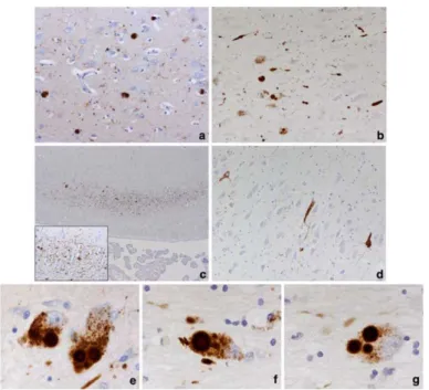
Figure from Kalaitzakis ME (Acta Neuropathol 2009). Neuropathology in cortical and subcortical regions. A) pathological changes (cortical LB) in the cingulate gyrus in a case PDD. B) LB and LN pathology in the basal nucleus of Meynert (NBM) of a case PDD. C) selective susceptibility of field CA2 (Cornu Ammon) hippocampal alterations LN-like. The inset shows the network of LNs. D) neurofibrillary pathology in the hippocampal area CA2 of a case PDD E) 'classical' multiple LBs in pigmented neurons of the substantia nigra medial. A single LB in a pigmented neuron is also evident. A single (f) and multiple (g) LB in a neuron in the enlargement of the NBM.
1.3 Alzheimer-like pathological changes
Although several studies indicate that cortical neuropathological alterations similar to those seen in Alzheimer's disease are considered an important factor in the development of dementia in Parkinson's disease, the real meaning of plaques of beta-amyloid (Aß) and neurofibrillary tangles in dementia Parkinson's disease is still
the subject of research(12), since there is no consistent correlation between cortical
Aß load and assessments of cognitive deficits in patients with clinically determined PDD.
However in the study by Irwin (2012) previously cited, the authors observed through a sub-analysis of the PDD group, the presence of beta-amyloid plaques and neurofibrillary tangles may influence cognitive impairment and disease progression in a subset of patients; specifically the presence of Alzheimer-like changes seem associated with cognitive decline in the group of patients at onset of illness in old
age and with a reduced free interval between onset of motor symptoms and onset of cognitive impairment.
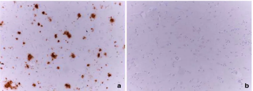
Figure from Kalaitzakis ME (Acta Neuropathol 2009). Immunohistological analysis for the deposition of b-amyloid in the caudate nucleus. A) Numerous deposition of Ab in the caudate nucleus in a case PD with dementia. B) caudate nucleus, without Ab deposits in the event a non-demented PD. Magnification 920 Parkinson's disease PD, Ab amyloid b peptide.
1.4 Vascular white matter lesion load
The white matter lesion load has been classically associated with motor disability and cognitive dysfunction in elderly subjects not suffering from dementia or Parkinson's disease; therefore vascular disease may have an additive effect on clinic sintomotology in Parkinson's disease, including cognitive deficits. A review of
Bohnen NI(13) highlights this concept.
Figure by Bohnen NI, Nat Rev Neurol, 2011
A recent study by Gonzalez-Redondo R.(14) analyzed the vascular lesion load by
means of magnetic resonance brain imaging (MRI) in a large cohort of patients with PD; to identify a possible relationship between silent infarcts and dementia or MCI in this population has been structured assessment transverse and longitudinal. The authors found no association between white matter hyperintensities and global cognitive profile in patients with Parkinson's disease; also the presence of vascular lesion load was not a negative prognostic index after a mean follow-up of 30 months.
2. Monitoring cerebral blood flow: cerebral autoregulation and
neurovascular coupling
Cerebral blood flow (CBF) is regulated by two major homeostatic physiological systems, cerebral autoregulation that maintains a relatively constant CBF to changes in systemic blood pressure and cerebral perfusion pressure and neurovascular coupling, consisting of the neurovascular unit, which adapts the cerebral blood flow to neuronal activity in the brain. These two systems work in part with the same physiological mechanisms.
2.1 Cerebral Autoregulation
The cerebral circulation has a fundamental homeostatic mechanism called self-regulation that is the ability to maintain cerebral blood flow (CBF) relatively constant in terms of variability acute or chronic systemic blood pressure and cerebral perfusion pressure, ensuring, within certain limits, a adequate metabolic supply to the brain tissue; therefore unlike other tissues exempt from autoregulation, in the brain there is a nonlinear relationship between systemic arterial pressure and flow. The self-regulating mechanism is able to maintain the cerebral flow relatively constant within the limits of mean systemic arterial pressure between 60 and 150 mmHg; beyond these limits the flow becomes passively dependent on perfusion pressure, causing ischemia if it is reduced or if edema increases. The principal mechanism employed to keep the cerebral blood flow constant when the perfusion pressure is represented mainly from changes in the local vascular resistance according to the formula:
Regional blood flow (rCBF) = cerebral perfusion pressure (CPP) / resistance cerebrovascular regional (rCVR).
2.2 Neurovascular Coupling
The Neurovascular Coupling is the process in which the neurovascular unit, represented by complex-vessel wall endothelium of small blood vessels, neurons, astrocytes and glial cells interact with each other by modulating the complex relationship between neuronal activity, hemodynamic factors and intracellular signals. These interactions allow you to synchronize the neuronal brain perfusion with the request, allowing an adequate metabolic supply to the brain areas in relation to the functional demand; in addition to the trophic support the neurovascular unit is involved in immune surveillance in trade at the level of the blood-brain barrier and hemostatic balance.
Currently the physiological mechanisms implicated in the regulation of regional cerebral blood flow are represented by:
. myogenic mechanism . neurogenic mechanism . metabolic mechanism
These mechanisms may act alone or in synergy with each other.
In mechanism myogenic vascular tone is modified in response to the mechanical tension on the vessel wall induced by the pressure and the amount of flow. The small vessels and arterioles (vessels more accountable vascular resistance) constrict or dilate in response respectively to an increase or reduction in the transmural pressure gradient with every heartbeat. Increased pressure within the vessel mechanically generate a greater circumferential wall tension and relax the smooth muscle fibers of the muscle layer, thus resulting in depolarization of the cell membrane and contraction of the muscle cell. An increase in flow within the vessel generates a voltage increase grazing (shear stress) on endothelial cells, resulting in
the relax of smooth muscle cell with a mechanism Nitric oxide (NO) -mediate. The flow-induced muscle relaxation allows to answer the need for increased local flow. The metabolic regulation provides that changes in molecules and metabolic substrates such as O2, CO2, ATP, glucose, glutamate, at the level of the tissue microenvironment generate vessel regulators regional adaptations, physiological condition exists as a direct relationship between cerebral blood flow and cerebral metabolism. The increase of neuronal activity in physiological conditions produces an increase in production and release of glutamate, adenosine triphosphate (ATP) and an increase in the activity of cyclooxygenase-2. The activation of NMDA receptors for glutamate increases the production of NO (vasodilator) to increase the activity of Nitric Oxide Synthase. Also through the glutamate binding to its receptors and metabotropic at the membrane causes an increase in astrocytic Phospholipasi C, with an increase in consensual inositol triphosphate, release of calcium from the sarcoplasmic reticulum and activation of calcium-dependent potassium channels. The resulting in hyperpolarization of the cell membrane, caused by the leakage of potassium ions will be responsible for the reduced contractility of the muscle cell itself. The COX-2 resulting in an increase of prostaglandins in the extracellular space produces vasodilation. The increase of ATP determines increase in the concentration of adenosine that in turn activates the receptors A2b placed both on the membrane astrocytic that the membrane of the smooth muscle cell. The activation of these receptors leads to the increase of the concentration of cAMP with consequent vasodilatation. The CO2 is believed to work with PH-mediated mechanism.
The adjustment uses a neurogenic component extrinsic and intrinsic.
Extrinsic component: Several studies have demonstrated the innervation of vessels extraparenchymal located at the base and the surface of the brain, the ganglia of origin of these endings perivascular and vasoactive substances released from the jar. It is believed that the extrinsic innervation finds its origin in the superior cervical
ganglion of the sympathetic fibers which originate containing norepinephrine and neuropeptide Y from the sphenopalatine and otic ganglion from which they came parasympathetic fibers containing vasoactive intestinal peptide (VIP) and nitric oxide synthase (NOS) and the trigeminal ganglion, from which originate sensory fibers containing calcitonin gene-related peptide (CGRP), substance P, the neurokinina A and pituitary adenylate cyclase-activating polypeptide. Once inside these vessels into the brain parenchyma lose the autonomic component and finished the spaces of Virchow-Robin vessels respond to a central neuronal control (intrinsic innervation). The role of the sympathetic system is mainly linked to the ability to move the upper limit of the curve of self-regulation in response to increases in systemic arterial pressure in the protection of brain tissue. The parasympathetic system, which mediates vasodilator responses, does not appear to play a significant role on the physiological mechanism of autoregulation of cerebral blood flow while it seems implicated in pathological conditions such as ischemic stroke and migraine. The trigeminal-vascular system, whose fibers are the only sensory innervation of brain vessels, appears to mediate CGRP-mediated vasodilator responses to vasoconstrictor stimuli. The trigeminal vascular afferents play an important role in the pathogenesis of migraine.
Intrinsic component: the intrinsic regulation of small intraparenchymal cerebral vessels is mediated by the neurovascular unit, which receives nerve fibers from basic subcortical systems and local cortical interneurons, although it should be noted that it is not yet well-defined neuronal network that acts functionally complete in synergy with the neurovascular unit. Neural networks, to date the best studied, which project to the small cerebral vessels and also to the surrounding astrocytes are cholinergic pathways from the basal nucleus of Meynert, noradrenergic pathways from the locus coeruleus and serotonergic pathways from the raphe nucleus. Electrical or chemical stimulation of these nuclei elicits profound
changes in cortical cerebral blood flow. M5 muscarinic receptors present on the vascular wall (endothelium and smooth muscle cells) that astrocytes mediate vasodilator responses.
Serotonergic 5-HT 1b median vasoconstrictive responses. It been shown that stimulation of noradrenergic neurons of the locus coerulus causes reduction of cortical CBF and that the target of perivascular cortical noradrenergic fibers is represented mainly by astrocytes. It is also shown that by stimulating specific subsets of cortical GABAergic interneurons are generated vasoconstrictive or vasodilatory responses of local microvessels; in particular interneurons producing VIP or NOS causing vasodilation, while those producing somatostatin caused vasoconstriction.
diabetes mellitus, smoking, obesity, stroke and degenerative diseases such as the Alzheimer's disease.
BROKERS INVOLVED IN THE REGULATION OF SMALL VESSELS CORTICAL (Spronck B. 2012)
3. Cerebral Vasoreactivity: methods of evaluation
Cerebral Vasoreactivity (CVR) reflects the ability of small cerebral vessels to adapt to changes in the microenvironment, to metabolic demand and to pressure changes, through the regulation of local blood flow. In neuroscience, there are several non-invasive techniques and vasomotor stimuli for the estimation of CVR; between the techniques include the TCD (transcranial Doppler), PET (positron emission tomography), SPECT (Positron Emission single-photon), the NIRS (infrared spectroscopy), the CASL MRI 3D (3D-continuous arterial spin labeling), the MRI BOLD (blood oxygenation level dependent) and Angio-RM 2D time of flight.
These techniques, however, are based on different parameters (blood flow or velocity of flow at the level of the great vessels, volume of flow, tissue perfusion), use different variables and have a different temporal and spatial resolution. Consequently, the estimation of CVR may differ widely between the different approaches. The main vasoactive stimuli are represented by chemical stimuli (CO2, O2), pharmacological (acetazolamide) or causing a transient reduction in blood pressure. It is believed that CO2 represents the stimulus more appropriate in terms of ease of use and in terms of standardization.
3.1 Vasoreactivity to CO2
Carbon dioxide (CO2) is a potent vasodilatory stimulus that modulates vascular tone during normal breathing during sleep and during hyperventilation. The CO2-VCR is measured in terms of changes in cerebral blood flow (CBF) or flow velocity (BFV) in response to vasoconstrictive stimuli (hypocapnia and hyperventilation) or vasodilators (hypercapnia, acetazolamide). The VCR is calculated as the percentage change in flow due to changes in CO2. Blood flow increases linearly over the
The method most commonly used in the clinic to measure the CO2-VCR is transcranial Doppler for the ease of execution. The advanced imaging techniques sovramenzionate than the TCD used to assess vasoreactivity in specific brain areas and vascular but lack in temporal resolution. The tests used for the assessment of CO2-VCR using transcranial doppler are represented by the Breath-holding, the test with a mixture of CO2, by re-breathing and test with acetazolamide.
Breath-holding test: this test for the measurement of CVR was demonstrated for the first time since Ratnatunga and Adiseshiah in 1990 and subsequently used clinically
to assess the risk of stroke in pz suffering from carotid artery stenosis.(15,16)
Methodology: after a period of normoventilation, asks the patient to hold their breath for at least 30 seconds. It is important that the apnea beginning with a normal breath to avoid the phenomenon of Valsalva and therefore the increase of the intracranial pressure. Through a bilateral monitoring by TCD are measured values of mean flow velocity (VMF) at the level of the stretch M1 middle cerebral artery bilaterally in rest conditions and after 4 seconds of breath at the end apnea. During the experiment are monitored by a capnometer the pCO2, the blood pressure (BP) and heart rate (HR). The Breath Holding index (BHI) is obtained by dividing the percentage increase of the speed of flow that occurs during the BH for the period of time (seconds) in which the subject is able to hold his breath after a normal inhalation according to the formula:
BHI: [(MFVBH- MFVbase) / MFVbase) x 100 / sec of BH].
This method has been validated in numerous studies, it is also a non-invasive, well tolerated and easy to perform. The main limitations of the method are represented by:
. need for adequate cooperation of the subject.
. insufficient change in the pCO2 (prolonged apnea causes in fact an increase of only 3-4 mmHg pCO2)
. not perfect reproducibility: interindividual variability with which it establishes hypercapnia and apnea post does not make clear correlation between blood levels of PCO2 and time of apnea; apnea is not the only factor that influences the PCO2 "current" are called upon other factors such as the metabolism of the subject, the size of the lungs (influenced by age, sex, weight) and the presence of bronchopulmonary disease. However, the use of capnometer can sensitize the method.
Testing with inhalation of CO2: in the reactivity test is used to CO2 inhalation of mixtures of CO2 at a concentration that varies from 2, 5 or 7%.
By registering with bilateral TCD, are measured values of average velocity in the middle cerebral arteries of the two sides at rest and at the end of the test. During the experiment is also measured by a capnometer pCO2, and monitored the PA and FC. At each steady state value of MFV is compared with the value of the end-tidal CO2. The cerebral vasomotor response (VMR) to CO2 is calculated as the percentage increase of MFV measured after 90 seconds of inhalation of the mixture of CO2 compared to the rest condition in accordance with the formula:
[(MFV MFVCO2- base) / MFVCO2] x100.
The test also provides a simple method to CO2, non-invasive and validated.(17)
The re-breathing consists in allowing the patient to breathe the same air that is exhaled within a closed system. With this method you do not have reached a stady of blood concentration of CO2 and it is difficult to correlate changes in flow with changes in PaCO2.
The acetazolamide test consists of a test drug that involves the intravenous infusion of 500 mg or 2 g of acetazolamide, a carbonic anhydrase inhibitor, the enzyme that catalyzes both dissociation and the formation of carbonic acid (H2CO3); the inhibition of this enzyme is responsible for a marked cerebral acidosis. The reduction of brain pH thus obtained causes vasodilation. The effects are expressed within the
first minute of infusion and end after 30 minutes, reaching a plateau at 15 minutes. The drug in its intravenous formulation is not yet available in Italy. Compared to the previous two techniques, it is an invasive method, a longer duration and with important side effects.(18)
In addition, another limitation inherent in the TCD is to give an overall measure of cerebrovascular reactivity going to sample the proximal vessels; methods of functional magnetic resonance imaging (fMRI) are able to analyze functional changes in regional cerebral perfusion.
4. Cerebral hemodynamics and cognitive functions
In recent years the relationship between cerebral hemodynamics and cognitive performance has become a major challenge in clinical practice in relation to the worldwide growth of the elderly population. The presence of cardiovascular risk factors in middle-aged individuals is considered to be a potential marker for the early identification of individuals at risk of dementia. Hypertension and diabetes mellitus are risk factors for vascular prevalent; hypertension affects more than one-third of the world population and 366 million people are suffering from diabetes. Among the subjects that have more than 65 years of age approximately 70% are suffering from hypertension and 26% diabetes. Although the pathophysiological link between cerebral hemodynamics, reduced cerebrovascular reserve and dementia is complex, several epidemiological studies, neurophysiological and clinical-instrumental showed that microvascular damage associated with hypertension, diabetes, and other vascular risk factors, advanced age is responsible alteration of the neurovascular coupling resulting in regional hypoperfusion and neurodegeneration. Genetic factors, inflammation, autonomic disorders, neurodegeneration (m. Alzheimer's, Parkinson-dementia, dementia with Lewy
bodies), ischemic and hemorrhagic stroke are other overriding factors in the development of dementia.
Studies measuring CBF in response to hypercapnia with transcranial doppler(19) and
method of inhalation of 133 Xe (20) showed reduced vasoreactivity in subjects with
type 2 diabetes compared with controls healthy. Other studies have shown an increased pulsatility index in diabetic subjects highlighted an increase in vascular resistance and microvascular damage. Studies carried out with high-resolution MRI (8 TESLA) showed the damage of the small vessels, microinfarcts, microhemorrhages, iron deposits and diffuse leukoaraiosis (white matter hyperintensities WMHS) in subjects with hypertension.
From a histopathological point of view of the deep periventricular white matter lesions showed partial demyelination, astrogliosis, dilatation of the perivascular
spaces, activated macrophages and fibrohyalinosis(21). Isolated lesions and spread of
the deep white matter were associated with heart attacks arteriolar sclerosis
incomplete and complete(22). The small vessel disease brain and the WMHS were
associated with cognitive impairment in several studies (23,24); it is also considered
that the same factors affecting the white matter may cause hypoperfusion at the level of the gray matter and functional disconnection cortico-subcortical and that this process may precede cognitive decline even many years. More studies have shown the strong association between WMHS with diabetes and hypertension. Although vascular inflammation related to glucose toxicity, lipotoxicity and neurotoxicity plays an important role in damaging the neurovascular unit and the blood-brain barrier by promoting neuronal loss. The combined action of vascular risk factors cause oxidative stress in the mitochondria and endoplasmic reticulum causing the release of pro-inflammatory cytokines and activation of pro-apoptotic pathways; these events cause endothelial dysfunction with decrease of NO synthesis and impaired cerebrovascular reactivity. Recently it has been shown that the
presence of intercellular adhesion molecule (ICAM) and vascular cell adhesion molecule (VCAM) were associated with impaired cerebral vasoreactivity and atrophy of the gray matter in several regions of the brain in type 2 diabetic standing and in
hypertensive subjects (25) and that adhesion molecules were also associated with
worsening of cognitive sphere.
In recent years, much emphasis is given to the relationship between cerebrovascular damage and neurodegeneration, in particular on the role (causal summation or synergistic) of vascular risk factors and the neurovascular unit damage on neuronal loss.
There is extensive evidence epidemiological, clinical, neuropathological and pharmacological evidence that an "overlap" between vascular dementia and Alzheirmer's disease (AD).
Vascular risk factors such as hypertension, diabetes, hypercholesterolemia, and obesity are associated with an increased risk of AD and accelerate the clinical expression. White matter lesions (white matter lesions) and lacunar infarcts are associated with amyloid plaques and neurofibrillary tangles in the more than 40% of elderly patients with dementia. The control of vascular risk factors in patients with
AD slows the progression of WMLs (26). The use of drugs angiotensin receptor
blockers reduce the risk of AD(27). Several studies have shown alterations of cerebral
vasoreactivity in Alzheimer's dementia (28,29,30,31); AD patients with impaired
cerebrovascular reactivity to CO2 undergo a more rapid cognitive decline.
Experimental studies have shown that β-amyloid (Abeta) has important effects on the vessels and that hypoxia-ischemia is a potent modulator dell'amiloidogenesi
brain (32). The blood-brain barrier plays a critical role in the clearance of β-amyloid
(33,34). Genetic factors (such as the ApoE4 genotype) can alter the regulation of
cerebral blood flow, contributing to white matter lesions (WMLs), and deposition of amyloid β-. Ischemia can promote the accumulation of β-amyloid through reduced
vascular clearance of this peptide; also ischemia-hypoxia promotes the cleavage of Aβ from APP precursor (Amyloid Precursor Protein) through the up-regulation of β-secretase and epressione; the increased production and decreased clearance of Aβ can increase the deposition in the brain and encourage the formation of amyloid plaques and amyloid angiopathy.
In turn, elevated brain levels of Aβ and amyloid plaques are found in patients with
cerebrovascular insufficiency and cognitive impairment (35) and it is assumed that
the damage of the vessel wall may damage the Aβ-induced vasomotor regulation and cerebrovascular reserve ; in favor of this hypothesis, some studies have shown that focal ischemia produced large infarcts in mice over-expressing APP (36,37) in
relation to Aβ-mediated vascular dysregulation. Other pathophysiological hypothesis argue that the deficit of the cholinergic neurons of the basal nucleus of Meynert produces a deficit of intrinsic neurogenic control of CBF.
From the most recent acquisitions can therefore be assumed that there is a synergistic pathogenetic or at least a synergistic effect between vascular damage and neurodegenerative disease.
Conceptual model of pathogenesis: the vascular risk factors through processes of oxidative stress and inflammation cause alteration neurovascular unit (resulting in hypoperfusion) and neurotoxicity with evolution in atrophy and functional decline (Novak V.2012)
Limited data in the literature between cerebrovascular reactivity (VCR) and Parkinson's disease. The studies were structured framing the deficit cerebral autoregulation as a dysfunction of the autonomic system, with results only partially
concordant(39,40). A recent study that investigated the VCR using functional magnetic
resonance imaging (3T blood oxygenation level-dependent-BOLD) and hypercapnic stimulus showed no significant differences before and after loading of levodopa,
among patients with Parkinson's disease and healthy controls (41); However, the
5. Objectives of the study
Given the recent findings that identify a possible association between alterations in cerebral hemodynamics and cognitive performance decay of both primary and secondary origin, the objective of this study was to evaluate the possible relationship between cognitive impairment and alterations in cerebrovascular reactivity in patients suffering from Parkinson's disease.
6. Materials and Methods
It was assessed a population of 34 patients, 22 male and 12 female subjects suffering from Parkinson's disease according to UK Brain Bank Criteria pertaining to the Parkinson's Disease Center of the Department of Neurology and Psychiatry of the' University of Rome "Sapienza ". Of each patient were recorded medical history-personal data such as gender, age, age of onset of motor symptoms and dosage of dopaminergic therapy. Were not considered were to be enrolled patients with signs and symptoms of atypical parkinsonism, comorbidity, neurological pathologies can change the cerebrovascular reactivity as previous cerebrovascular events, changes in carotid atheromatous with significant hemodynamic intracranial stenosis, moderate to severe hypertension, diabetes mellitus; were also excluded patients with no or questionable response to dopamine replacement therapy, cognitive impairment (MMSE <24) occurred within one year of onset of motor symptoms, in patients treated with anticholinergic drugs, major depression. The study population was classified into two groups according to the presence of mild cognitive
impairment (MCI) according to Diagnostic Criteria for PD-MC (42). All pieces have
given their informed consent. Each patient was subjected to: 1. clinical, neuropsychological and motor assessment:
Cognitive assessment:
- Mini Mental State Examination (Folstein, 1975) for global cognitive function. - Ten point clock test.
- Test for the assessment of memory functions: immediate and delayed memory for prose.
- Test for the assessment of executive / attentional: TMT-A and TMT-B, Digit Span - Assessment of phonological verbal fluency.
-Test for the assessment of visuo-spatial functions: Copy Immediate and Deferred Figure of Rey, test figures tangled.
Praxic-test functions.
In addition, the test was administred structured SCOPA-AUT for evaluation of autonomic functions.
6.1 MCI in Parkinson's disease
A diagnosis of MCI is distinct from that of dementia. A person with a diagnosis of MCI has altered cognitive function with objective and subjective indicators of deterioration, without there being necessarily an impact on the complex functions of everyday life.
The existence of a continuum that goes from normal cognitive aging to frank dementia (Alzheimer's disease) and passing through a phase of failure has been postulated in 1995 by Petersen. With a priori hypothesis and applying the same
methodological criteria used for Alzheimer's disease, Caviness (43) has incorporated
the concept of MCI in Parkinson's disease. A review of 'critique' of this concept was published in 2011 by the Movement Disorders Society (MDS). The condition, which is considered a risk factor for the development of dementia, is found to be rather frequent (27% prevalence) and heterogeneous in terms of number and cognitive domains involved.
However, we must stress that major factors contributing to the overestimation of cognitive impairment in Parkinson's disease: depressive symptoms, pre-morbid personality, motor performance, social isolation, side effects attributable to treatment, and the presence of methodological biases, both within and between
groups psychometric analyzes, can limit the validity of the construct PD-MCI (44), in
particular a pathology where it would seem evident that the cognitive deterioration cannot recognize a single disease process. Therefore, we recognize that the term
MCI in PD (PD-MCI) should not be accepted without these appropriate conditions(45).
Criteria for the Diagnosis of PD-MCI, from Litvan I (Mov Dis, 2012) I. Inclusion criteria
Diagnosis of Parkinson’s disease as based on the UK PD Brain Bank
Criteria
Gradual decline, in the context of established PD, in cognitive ability
reported by either the patient or informant, or observed by the clinician
Cognitive deficits on either formal neuropsychological testing or a scale
of global cognitive abilities (detailed in section III)
Cognitive deficits are not sufficient to interfere significantly with
functional independence, although subtle difficulties on complex functional tasks may be present
II. Exclusion criteria
Diagnosis of PD dementia based on MDS Task Force proposed criteria
Other primary explanations for cognitive impairment (e.g., delirium,
stroke, major depression, metabolic abnormalities, adverse effects of medication, or head trauma)
Other PD-associated comorbid conditions (e.g., motor impairment or
severe anxiety, depression, excessive daytime sleepiness, or psychosis) that, in the opinion of the clinician, significantly influence cognitive testing
III. Specific guidelines for PD-MCI level I and level II categories A. Level I (abbreviated assessment)
Impairment on a scale of global cognitive abilities validated for
use in PD or
Impairment on at least two tests, when a limited battery of
neuropsychological tests is performed (i.e., the battery includes less than two tests within each of the five cognitive domains, or less than five cognitive domains are assessed)
B. Level II (comprehensive assessment)
Neuropsychological testing that includes two tests within each of
the five cognitive domains (i.e., attention and working memory, executive, language, memory, and visuo-spatial)
Impairment on at least two neuropsychological tests, represented
by either two impaired tests in one cognitive domain or one impaired test in two different cognitive domains
Impairment on neuropsychological tests may be demonstrated
by:
Performance approximately 1 to 2 SDs below appropriate
norms or
Significant decline demonstrated on serial cognitive testing
or
Subtype classification for PD-MCI (optional, requires two tests for each of the five cognitive domains assessed and is strongly suggested for research purposes)
PD-MCI single-domain—abnormalities on two tests within a single
cognitive domain (specify the domain), with other domains unimpaired or
PD-MCI multiple-domain—abnormalities on at least one test in two or
more cognitive domains (specify the domains)
6.2 Ecocolodoppler of the neck vessels, transcranial Doppler ultrasound
at baseline and Breath-Holding unit test using a GE LOGIQ P5 pro.
TCD AND WITH CEREBRAL Vasoreactivity BREATH- HOLDING TEST
For the assessment of cerebral vasoreactivity was used a probe of 2 MHz with recording of the flow through the acoustic window of time, the proximal segment (M1) of the middle cerebral artery and the proximal segment (P1) posterior cerebral artery by a experienced operator and not informed about the clinical characteristics of the subject to be examined. The measurements were made in a quiet room, with the subject lying in the supine position, in the absence of visual stimulation or sound. Blood pressure and heart rate were measured at the beginning and at the end of each experiment. The values of mean flow velocity (VMF) at the M1 portion of middle cerebral artery and posterior cerebral artery P1 segment were recorded at baseline and after 4 seconds of breath at the end of an apnea lasting 30 seconds (stimulus to get the hypercapnia). A capnometer (Capnodig; Drager, Lubeck, Germany) recorded the pCO2 during the entire duration of the maneuver to confirm the increase of the gas in the blood.
ll Breath Holding Index was calculated according to the formula: BHI: [(MFVBH- MFVbase) / MFVbase) x 100 / sec of BH]
Each subject was submitted to the test 3 times at 5 minute intervals to allow the VMF to return to baseline; for each individual value of BHI obtained the first evidence is consistent with that of the other two tests. The BHI used in the analysis is the average of the three tests. With the Doppler ultrasound examination of the neck vessels and transcranial Doppler ultrasound were excluded significant stenosis and has been calculated pulsatility index of Gosling (PI) to assess indirectly the resistances in the microcirculation.
Magnetic Resonance Imaging of the brain
The neuroimaging protocol included standard sequences (FLAIR, DP-T2-T1-weighted), using 1.5 T MRI unit (Siemens, Erlangen, Germany). The lesion load was
assessed using T2 and Flair and applying the semiquantitative visual scale CHS(46).
7. Statistical Analysis
For the analysis of the data was the software SPSS 15. It was used the Student t for continuous variables and the chi-square test for non-continuous variables.
8. Results
The groups concerned could be homogeneous for age and years of illness; there were no statistically significant differences in the degree of impaired autonomic and motor disabilities, although patients with PD-MCI present average values greater than SCOPA-AUT and UPDRS Part III; according to matching procedures the MMSE was higher in subjects with cognitive impairment. The neuro-cognitive analysis of domains examined (conceptualization, executive functions, memory, attention, visual-spatial functions) showed a prevalent alteration of attention and visual-spatial functions in the population studied. The analysis of neuroimaging has documented a comparison of the degree of suffering microvascular assessed by semiquantitative
visual scale CHS [Table 1] and indirectly with the pulsatility index between the two groups of patients.
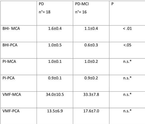
The hemodynamic parameters measured by TCD showed no significant differences at baseline of VMF MCA and PCA. After hypercapnic stimulus there was a statistically significant difference in cerebrovascular reactivity, resulting in reduced PD-MCI group (BHI- MCA PD vs PD-MCI: 1.6±0.4 vs 1.1 ±0.4 p <0.01; BHI-PCA PD vs PD-MCI: 1.0 ± 0.5 vs 0.6 ± 0.3 p <0.05) . [Table 2] PD n°=18 PD-MCI n°=16 P Genere (M/F) (11/7) (11/5) Età media 70.1±6.8 70.5±6,9 n.s.* Durata di malattia 7.3± 3,6 8.8±6.5 n.s.* UPDRS parte III 17.3±5,7 20,8±5.1 n.s.* LED (levodopa equivalent dose) 673.8 ±296.1 856.6±617.2 n.s.* SCOPA-AUT 13,5±6.9 17,6±7.0 n.s.* SCOPA-AUT Item 14-16 1,4±1,5 1.9±1.6 n.s.* Carico lesionale CHS 2,3±0.9 2,4±0.9 n.s.* Ipertensione arteriosa (SI/NO) (9/9) (6/10) n.s.* MMSE 28,1±1,2 23,9±2,5 <0.01
TABLE 1 Demographic data and clinical
PD n°= 18 PD-MCI n°= 16 P BHI- MCA 1.6±0.4 1.1±0.4 < .01 BHI-PCA 1.0±0.5 0.6±0.3 <.05 PI-MCA 1.0±0.1 1.0±0.2 n.s.* PI-PCA 0.9±0.1 0.9±0.2 n.s.* VMF-MCA 34.0±10.5 33.3±7.8 n.s.* VMF-PCA 13.5±6.9 17.6±7.0 n.s.*
TABLE 2. Rating transcranial
*n.s.= not significant
t-student;
p < 0.01
PD – Ctrl PD - MCI
t-student;
p < 0.05
PD - Ctrl PD – MCI
9. Discussion
The proposed study represents the first evaluation of cerebrovascular reactivity in patients with no-Alzheimer degenerative cognitive impairment; the results are consistent with a reduction of vasomotor reserve, identified by BHI, in patients with Parkinson's disease (PD) and cognitive impairment, when compared to a control group of patients with PD.
Recent acquisitions, with appropriate methodological approaches and samples, suggest that some parameters of cerebral vasoreactivity, after hypercapnic stimulus, is compromised in patients with primary dementia and, possibly, even in patients with MCI.
Therefore, the data for which the vascular dysregulation is a feature frequently observed in neurodegenerative diseases such as Alzheimer's dementia, although difficult to define, emphasizes the potential role of involution brain at the base of the reduced reactivity cerebrovascular.
In fact, the structural and functional integrity of brain tissue depends on the delicate balance between neuronal energy demands tax the activity and substrate availability through the bloodstream, indicating how the neurovascular coupling is a crucial mechanism for maintaining the functional integrity, mutually depends on the elements that constitute it.
Therefore we cannot exclude that the involution of the structures primarily involved in the disease process, in particular by extending the cortex, could lead to inadequate modulation of cerebral vasoreactivity, probably resulting in an impairment of the physiological signaling between the elements of the neurovascular unit.
In line with this hypothesis, some authors have found, through the use of Voxel-Based Morphometry (RM automated method), circumscribed areas of cortical
atrophy already evident in patients with MCI and PD(47).
Regions with areas of gray matter atrophy in patients with (A) Parkinson's disease (PD) without cognitive impairment (PD-N), (B) PD with MCI and (C) PD with dementia (PD-D). (Melzer TR. J Neurol Neurosurg Psychiatry 2012).
Moreover, as the real impact of Alzheimer-like neuropathological alterations in dementia of the Parkinson's disease is yet to be determined, it could be argued that the data obtained can be attributed to pathophysiological mechanisms similar to those proposed in Alzheimer's disease, recognizing a loss of Aβ-induced vascular wall. However, studies carried out by means of PET-imaging showed a high degree
of β-amyloid load in only a small percentage of patients with PDD (48) ; Therefore,
Another pathophysiologic mechanism potentially implicated in the reduction of cerebrovascular reactivity is represented by a deficiency of the cholinergic system; in fact it would seem the neurotransmitter implicated in the neurogenic control of cerebral blood flow. Several lines of evidence using PET studies emphasize the impairment of the cholinergic system in patients with Parkinson's disease and
cognitive impairment (49,50) and as such degeneration appears spatially consistent
with a reduction in metabolic activity(51).
Image adapted from Klein et al. (2010) 0.44 (A) brain areas with significant reduction of [11C] MP4A absorption in PDD compared to PD. (B) the brain areas with a significant reduction in [18F] FDG PDD compared with controls. CMRglc, cerebral metabolic rate of glucose. (Ray and Stafella, Mov Dis, 2012).
The impact of vascular disease on cognitive dysfunction in Parkinson's disease has not been widely accepted; the possible contribution of vascular degeneration has been suggested in some studies of ultrastructural evaluation of the cerebral microcirculation in patients with PD, assuming alterations similar to those observed in Alzheimer's disease (52,53). Although the selection of the sample in the present
study has been carried out in order to minimize the possible confounding role of cardiovascular risk factors and the groups in question were found to be homogeneous with respect to the parameters established for estimating the
vascular damage (white matter lesion load, index pulsatility), we cannot exclude that the observed data can recognize the contribution of ultrastructural alterations in the microcirculation.
The pathophysiological mechanisms that modulate the relationship between cerebral hemodynamics, cerebrovascular reserve and cognitive deficits remain poorly characterized.
However, the emerging model of an altered neurovascular coupling resulting in dysfunction of the microcirculation, regional hypoperfusion and neurodegeneration
could integrate the relationship between vascular changes and cognitive decline(54).
The previously conducted studies on cerebrovascular reactivity in patients with Parkinson's disease have interpreted this aspect as a potential expression of autonomic dysfunction, but does not establish a specific profile or hemodynamic parameter.
In our study, the autonomic aspects were evaluated by SCOPA-Aut, observing, although not reaching statistical significance, mean scores higher in the group of patients with cognitive impairment. In line with other authors, one could hypothesize that alterations in cerebrovascular reactivity may be secondary to a dysregulation in neurogenic vascular control in the context of a systemic autonomic dysfunction. However, it is possible that the relationship between autonomic disorders and cerebrovascular reactivity may represent the expression of a more obvious spread of the disease process, rather than an interdependent phenomenon.
Furthermore, previous studies carried out with different methods, have found no change in the cerebral reactivity in relation to the administration of L-Dopa; however in our population MCI-PD patients tend to assume major doses of LED and, considering the tendency to have a higher value all'UPDRS although did not reach a statistical significance, we cannot exclude that the severity of the disease per se has contributed to the results observed.
Some limitations of the study require cautious interpretation in the generalization of the observed data, in particular:
1) the small sample size
2) as expressed previously, the BHI for the evaluation of cerebrovascular reactivity, while being generally well tolerated and widely validated, presents inherent limitations to the method
3) The integration with more sophisticated neuroimaging methods could provide more sensitive
4) for the definition of MCI in Parkinson's disease is a construct limited.
10. Conclusions
The neurovascular coupling is a module whose function is required for the integrity of the structures that constitute it. The evidence of abnormal cerebrovascular reactivity in patients with Parkinson's disease and cognitive impairment, compared to the control group of PD patients, may indicate how the involution of the primitive neural systems may lead to a negative modulatory effect neurovascular homeostasis. Further evidence is needed to adequately characterize the role of hemodynamic factors in neurodegenerative processes.
REFERENCES
1) Pagonabarraga J, Gómez-Ansón B, Rotger R, Llebaria G, García-Sánchez C, Pascual-Sedano B, Gironell A, Delfino M, Ruscalleda J, Kulisevsky J.Spectroscopic changes associated with mild cognitive impairment and dementia in Parkinson's disease. Dement Geriatr Cogn Disord. 2012;34(5-6):312-8.
2) Williams-Gray CH, Evans JR, Goris A, Foltynie T, Ban M, Robbins TW, Brayne C, Kolachana BS, Weinberger DR, Sawcer SJ, Barker RA. The distinct cognitive syndromes of Parkinson's disease: 5 year follow-up of the CamPaIGN cohort. Brain. 2009 Nov;132(Pt 11):2958-69.
3) Aarsland D, Zaccai J, Brayne C. A systematic review of prevalence studies of dementia in Parkinson's disease. Mov Disord. 2005 Oct;20(10):1255-63.
4) Hely MA, Reid WG, Adena MA, Halliday GM, Morris JG. The Sydney multicenter study of Parkinson's disease: the inevitability of dementia at 20 years. Mov Disord. 2008 Apr 30;23(6):837-44.
5) Aarsland D, Andersen K, Larsen JP, Lolk A, Kragh-Sørensen P. Prevalence and characteristics of dementia in Parkinson disease: an 8-year prospective study. Arch Neurol. 2003 Mar;60(3):387-92.
6) Muslimovic D, Post B, Speelman JD, Schmand B. Cognitive profile of patients with newly diagnosed Parkinson disease. Neurology. 2005 Oct 25;65(8):1239-45.
7) Marini P, Ramat S, Ginestroni A, Paganini M. Deficit of short-term memory in newly diagnosed untreated parkinsonian patients: reversal after L-dopa therapy. Neurol Sci. 2003 Oct;24(3):184-5.
8) Pfeiffer HC, Løkkegaard A, Zoetmulder M, Friberg L, Werdelin L. Cognitive impairment in early-stage non-demented Parkinson's disease patients. Acta Neurol Scand. 2013 Oct 11.
9) Hepp DH, da Hora CC, Koene T, Uitdehaag BM, van den Heuvel OA, Klein M, van de Berg WD, Berendse HW, Foncke EM. Cognitive correlates of visual hallucinations in non-demented Parkinson's disease patients. Parkinsonism Relat Disord. 2013 Sep;19(9):795-9.
10) Monastero R, Di Fiore P, Ventimiglia GD, Camarda R, Camarda C. The neuropsychiatric profile of Parkinson's disease subjects with and without mild cognitive impairment. J Neural Transm. 2013 Apr;120(4):607-11.
11) Irwin DJ, White MT, Toledo JB, Xie SX, Robinson JL, Van Deerlin V, Lee VM, Leverenz JB, Montine TJ, Duda JE, Hurtig HI, Trojanowski JQ. Neuropathologic substrates of Parkinson disease dementia. Ann Neurol. 2012 Oct;72(4):587-98.
12) Kalaitzakis ME, Pearce RK. The morbid anatomy of dementia in Parkinson's disease. Acta Neuropathol. 2009 Nov;118(5):587-98.
13) Bohnen NI, Albin RL. White matter lesions in Parkinson disease.Nat Rev Neurol. 2011 Apr;7(4):229-36.
14) González-Redondo R, Toledo J, Clavero P, Lamet I, García-García D, García-Eulate R, Martínez-Lage P, Rodríguez-Oroz MC. The impact of silent vascular brain burden in cognitive impairment in Parkinson's disease. Eur J Neurol. 2012 Aug;19(8):1100-7. 15) Markus HS & Harrison MJ Estimation of cerebrovascular reactivity using transcranial Doppler,including the use of breath-holding as the vasodilatory stimulus. Stroke (1992)23, 668–67.
16) Silvestrini M, Pasqualetti P, Baruffaldi R, Bartolini M, Handouk Y, Matteis M, Moffa F, Provinciali L, Vernieri F.Cerebrovascular reactivity and cognitive decline in patients with Alzheimer disease. Stroke. 2006 Apr;37(4):1010-5.
17) Ringelstein EB, Sievers C, Ecker S, Schneider PA & Otis SM. Noninvasive assessment of CO2-induced cerebral vasomotor response in normal individuals and patients with internal carotid artery occlusions. Stroke (1988)19, 963–969.
18) Ringelstein EB, Van Eyck S & Mertens I. Evaluation of cerebral vasomotor reactivity by various vasodilating stimuli: comparison of CO2 to acetazolamide. J Cereb Blood Flow Metab (1992)12, 162–168.
19) Fülesdi B, Limburg M, Bereczki D, Káplár M, Molnár C, Kappelmayer J, Neuwirth G, Csiba L. Cerebrovascular reactivity and reserve capacity in type II diabetes mellitus.J Diabetes Complications. 1999 Jul-Aug; 13(4):191-9.
20) Griffith DN, Saimbi S, Lewis C, Tolfree S, Betteridge DJ. Abnormal cerebrovascular carbon dioxide reactivity in people with diabetes. Diabet Med. 1987 May-Jun; 4(3):217-20.
21) Gouw AA, Seewann A, van der Flier WM, Barkhof F, Rozemuller AM, Scheltens P, Geurts JJ. Heterogeneity of small vessel disease: a systematic review of MRI and histopathology correlations. J Neurol Neurosurg Psychiatry. 2011;82:126–135.
22) Gouw AA, van der Flier WM, Pantoni L, Inzitari D, Erkinjuntti T, Wahlund LO, Waldemar G, Schmidt R, Fazekas F, Scheltens P, Barkhof F, LADIS study group On the etiology of incident brain lacunes: longitudinal observations from the LADIS study. Stroke. 2008 Nov; 39(11):3083-5.
23) deGroot JC, de Leeuw FE, Ouderk M, Hofman A, Jolles J, Breteler MM. Cerebral white matter lesions and subjective cognitive dysfunction: the Rotterdam Scan Study. Neurology. 2001;56:1539–1541.
24) Vermeer SE, Prins ND, den Heijer T, Koudstaal PJ, Breteler MM. Silent brain infarcts and the risk of dementia and cognitive decline. N Engl J Med. 2003;27:1215– 1222.
25) Novak V, Zhao P, Manor B, Sejdic E, Alsop D, Abduljalil A, Roberson PK, Munshi M, Novak P. Adhesion molecules, altered vasoreactivity, and brain atrophy in type 2 diabetes. Diabetes Care. 2011 Nov; 34(11):2438-41.
26) Richard E, Gouw AA, Scheltens P, van Gool WA. Vascular care in patients with Alzheimer disease with cerebrovascular lesions slows progression of white matter lesions on MRI: the evaluation of vascular care in Alzheimer's disease (EVA) study. Stroke. 2010 Mar;41(3):554-6.
27) Li NC, Lee A, Whitmer RA, Kivipelto M, Lawler E, Kazis LE, Wolozin B. Use of angiotensin receptor blockers and risk of dementia in a predominantly male population: prospectivecohort analysis.BMJ. 2010 Jan 12;340:b5465. doi: 10.1136/bmj.b5465.
28) den Abeelen AS, Lagro J, van Beek AH, Claassen JA. Impaired cerebral autoregulation and vasomotor reactivity in sporadic Alzheimer's disease. Curr Alzheimer Res. 2014 Jan;11(1):11-7.
29) Claassen JA, Diaz-Arrastia R, Martin-Cook K, Levine BD, Zhang R. Altered cerebral hemodynamics in early Alzheimer disease: a pilot study using transcranial Doppler. J Alzheimers Dis. 2009;17(3):621-9.
30) Glodzik L, Randall C, Rusinek H, de Leon MJ. Cerebrovascular reactivity to carbon dioxide in Alzheimer's disease. J Alzheimers Dis. 2013;35(3):427-40. doi: 10.3233/JAD-122011. Review.
31) Vicenzini E, Ricciardi M.C, Altieri M, Puccinelli F, Bonaffini N, Di Piero V, Lenzi G.L. Cerebrovascular Reactivity in Degenerative and Vascular Dementia: A Transcranial Doppler Study. Eur Neurol 2007;58:84–89.
32) Ladecola C. Neurovascular regulation in the normal brain and in Alzheimer's disease. Nat Rev Neurosci. 2004 May;5(5):347-60.
33) Zlokovic B.V. Review The blood-brain barrier in health and chronic neurodegenerative disorders. Neuron. 2008 Jan 24; 57(2):178-201.
34) Kalaria RN. Review The blood-brain barrier and cerebral microcirculation in Alzheimer disease. Cerebrovasc Brain Metab Rev. 1992 Fall; 4(3):226-60.
35) Lewis H, Beher D, Cookson N, Oakley A, Piggott M, Morris CM, Jaros E, Perry R, Ince P, Kenny RA, Ballard CG, Shearman MS, Kalaria RN. Quantification of Alzheimer pathology in ageing and dementia: age-related accumulation of amyloid-beta(42) peptide in vascular dementia.Neuropathol Appl Neurobiol. 2006 Apr; 32(2):103-18. 36) Koistinaho M, Kettunen MI, Goldsteins G, Keinänen R, Salminen A, Ort M, Bures J, Liu D, Kauppinen RA, Higgins LS, Koistinaho J. Beta-amyloid precursor protein transgenic mice that harbor diffuse A beta deposits but do not form plaques show increased ischemic vulnerability: role of inflammation.Proc Natl Acad Sci U S A. 2002 Feb 5; 99(3):1610-5.
37) Zhang F, Eckman C, Younkin S, Hsiao KK, Iadecola C. Increased susceptibility to ischemic brain damage in transgenic mice overexpressing the amyloid precursor protein. J Neurosci. 1997 Oct 15; 17(20):7655-61.
38) Iadecola C. The overlap between neurodegenerative and vascular factors in the pathogenesis of dementia. Acta Neuropathol. 2010 Sep;120(3):287-96.
39) Vokatch N, Grötzsch H, Mermillod B, Burkhard PR, Sztajzel R. Is cerebral autoregulation impaired in Parkinson's disease? A transcranial Doppler study. J Neurol Sci. 2007 Mar 15;254(1-2):49-53.
40) Gurevich T, Gur AY, Bornstein NM, Giladi N, Korczyn AD. Cerebral vasomotor reactivity in Parkinson's disease, multiple system atrophy and pure autonomic failure. J Neurol Sci. 2006 Apr 15;243(1-2):57-60. Epub 2006 Jan 24.
41) Krainik A, Maillet A, Fleury V, Sahin M, Troprès I, Lamalle L, Thobois S, Fraix V, Villien M, Warnking J, Pollak P, Pinto S, Krack P. Levodopa does not change cerebral vasoreactivity in Parkinson's disease. Mov Disord. 2013 Apr;28(4):469-75.
42) Litvan I, Goldman JG, Tröster AI, Schmand BA, Weintraub D, Petersen RC, Mollenhauer B, Adler CH, Marder K, Williams-Gray CH, Aarsland D, Kulisevsky J, Rodriguez-Oroz MC, Burn DJ, Barker RA, Emre M. Diagnostic criteria for mild cognitive impairment in Parkinson's disease: Movement Disorder Society Task Force guidelines. Mov Disord. 2012 Mar;27(3):349-56.
43) Caviness JN, Driver-Dunckley E, Connor DJ, Sabbagh MN, Hentz JG, Noble B, Evidente VG, Shill HA, Adler CH. Defining mild cognitive impairment in Parkinson's disease. Mov Disord. 2007 Jul 15;22(9):1272-7.
44) Korczyn AD. Mild cognitive impairment in Parkinson's disease. J Neural Transm. 2013 Apr;120(4):517-21.
45) Korczyn AD. Mild cognitive impairment in Parkinson's disease. J Neural
Transm. 2013 Apr;120(4):517-21. doi: 10.1007/s00702-013-1006-0. Epub 2013 Mar 19. Review.
46) Bryan RN, Manolio TA, Schertz LD, Jungreis C, Poirier VC, Elster AD, Kronmal RA. A method for using MR to evaluate the effects of cardiovascular disease on the brain: the cardiovascular health study. AJNR Am J Neuroradiol. 1994 Oct;15(9):1625-33.
47) Melzer TR, Watts R, MacAskill MR, Pitcher TL, Livingston L, Keenan RJ, Dalrymple-Alford JC, Anderson TJ. Grey matter atrophy in cognitively impaired Parkinson's disease. J Neurol Neurosurg Psychiatry. 2012 Feb;83(2):188-94.
48) Edison P, Rowe CC, Rinne JO, Ng S, Ahmed I, Kemppainen N, Villemagne VL, O'Keefe G, Någren K, Chaudhury KR, Masters CL, Brooks DJ.Amyloid load in Parkinson's disease dementia and Lewy body dementia measured with [11C]PIB positron emission tomography. J Neurol Neurosurg Psychiatry. 2008 Dec;79(12):1331-8.
49) Bohnen NI, Kaufer DI, Hendrickson R et al. Cognitive correlates of cortical cholinergic denervation in Parkinson’s disease and parkinsonian dementia. J Neurol. 2006 Feb; 253(2):242-7.
50) Hilker R, Thomas AV, Klein JC et al. Dementia in Parkinson disease: functional imaging of cholinergic and dopaminergic pathways. Neurology. 2005 Dec 13;65(11): 1716-2.
51) Klein JC, Eggers C, Kalbe E et al. Neurotrasmitter changes in dementia with Lewy bodies and Parkinson disease dementia in vivo. Neurology 2010 Mar 16; 74(11):885-92.
52) Guan J, Pavlovic D, Dalkie N et al. Vascular degeneration in Parkinson’s disease. Brain Pathol.2013 Mar; 23(2): 154-64.
53) Farkas E, De Jong G, Aprò E et al. Similar ultrastructural breakdown of cerebrocortical capillaries in Alzheimer’s disease, Parkinson’s disease and Experimental Hypertension. Ann NY Acad Sci. 2000 Apr; 903:72-82.
54) Novak V. Cognition and Hemodynamics. Curr Cardiovasc Risk Rep. 2012 October; 6(5): 380–396.