ABSTRACT:
Cardiomyopathy is a manifestation of transthyretin
amyloidosis (ATTR), which is an underrecognized systemic disease
whereby the transthyretin protein misfolds to form fibrils that
deposit in various tissues and organs. ATTR amyloidosis is debilitating
and associated with poor life expectancy, especially in those with
cardiac dysfunction, but a variety of treatment options have recently
become available. Considered a rare disease, ATTR amyloidosis
may be more prevalent than thought, particularly in older persons.
Diagnosis is often delayed because of a lack of disease awareness
and the heterogeneity of symptoms at presentation. Given the recent
availability of effective treatments, early recognition and diagnosis
are especially critical because treatment is likely more effective earlier
in the disease course. The Amyloidosis Research Consortium recently
convened a group of experts in ATTR amyloidosis who, through an
iterative process, agreed on best practices for suspicion, diagnosis,
and characterization of disease. This review describes these consensus
recommendations for ATTR associated with cardiomyopathy as
a resource to aid cardiologists and others in the recognition and
diagnosis of ATTR associated with cardiomyopathy. Included in
this review is an overview of red flag signs and symptoms and a
recommended diagnostic approach, including testing for monoclonal
protein, scintigraphy, or biopsy and, if ATTR associated with
cardiomyopathy is identified, TTR genotyping.
ADVANCES IN HEART FAILURE
Expert Consensus Recommendations
for the Suspicion and Diagnosis of
Transthyretin Cardiac Amyloidosis
© 2019 American Heart Association, Inc.
Mathew S. Maurer, MD
Sabahat Bokhari, MD
Thibaud Damy, MD, PhD
Sharmila Dorbala, MD
Brian M. Drachman, MD
Marianna Fontana, PhD
Martha Grogan, MD
Arnt V. Kristen, MD
Isabelle Lousada, MA
Jose Nativi-Nicolau, MD
Candida Cristina Quarta,
MD, PhD
Claudio Rapezzi, MD
Frederick L. Ruberg, MD
Ronald Witteles, MD
Giampaolo Merlini, MD
https://www.ahajournals.org/journal/ circheartfailureKey Words: amyloid
◼ cardiomyopathies ◼ diagnosis ◼ heart failure ◼ rare diseases
T
ransthyretin amyloidosis (ATTR) is a disease caused
by abnormal fibrils derived from TTR
(transthyre-tin), a protein produced mainly by the liver, which
aggregate and deposit in tissues and organs.
1Cardiomy-opathy is a common manifestation of ATTR amyloidosis
(ATTR associated with cardiomyopathy [ATTR-CM]) and
is associated with a particularly poor life expectancy of
2 to 6 years after diagnosis.
2Patients with ATTR-CM
experience debilitating physical symptoms common to
heart failure (HF), such as exercise intolerance and
fa-tigue, which result in decreased functional capacity,
di-minished quality of life, and eventual death.
3ATTR-CM
can be acquired through aggregation of wild-type TTR
(ATTRwt) or inherited from a variety of genetic variants
of TTR (mutant transthyretin amyloidosis [ATTRm]; also
known as hereditary ATTR).
ATTRm is considered rare and is transmitted in an
autosomal-dominant manner and with variable
pene-trance. Certain variants typically result in
cardiomyopa-thy, whereas others typically result in polyneuropacardiomyopa-thy,
although cardiomyopathy and polyneuropathy
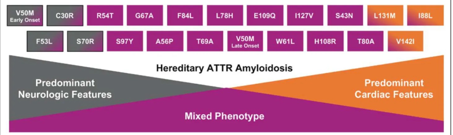
mani-festations may overlap (Figure 1). The prevalence of
car-diomyopathy among persons with ATTRm is estimated
at ≈40 000 of the 50 000 persons with ATTRm globally,
4but this may be an underestimate.
The most common worldwide TTR variant, Val122Ile
(or pV142I), occurs in ≈3% to 4% of black Americans,
with undefined phenotypic penetrance.
5,6This
Val122I-le TTR variant manifests predominantly as
cardiomy-opathy,
7and 1 estimate shows 10% of black
Ameri-cans with HF who are older than 60 are carriers of the
Val122Ile TTR variant.
8Thr60Ala, another common TTR
variant, often manifests as a mixed phenotype,
includ-ing cardiomyopathy, polyneuropathy, and
gastrointes-tinal dysfunction and is present in ≈1% of persons in
northwest Ireland.
9The Val30Met variant is the most
common cause of ATTRm with polyneuropathy;
how-ever, late-onset ATTRm in patients of the Val30Met
variant typically manifests as cardiomyopathy.
Pheno-typic penetrance of ATTRm is clearly age dependent;
thus, ascertainment of population prevalence varies
depending on age.
The true prevalence of ATTRwt is unknown; it may
be relatively high compared with the prevalence of
ATTRm. In autopsy studies, ≈25% of the hearts of
per-sons 80 years or older contained wild-type TTR fibrils,
regardless of the presence of symptoms.
4,10Studies
using nonbiopsy approaches to diagnosis demonstrate
a TTR prevalence of 16% among patients undergoing
percutaneous aortic valve replacement for severe aortic
stenosis,
1113% among patients with heart failure with
a preserved ejection fraction,
125% among patients
with presumed hypertrophic cardiomyopathy,
13and 7%
to 8% among patients with carpal tunnel syndrome on
biopsy of the tenosynovial tissue.
14Furthermore, ≈1%
to 3% of persons older than 75 showed myocardial
retention of the diphosphono-1,2-propanodicarboxylic
acid, which is indicative of TTR cardiac amyloidosis.
15,16Delays in diagnosis of ATTR-CM amyloidosis
com-monly occur because of physician- and disease-related
reasons, including fragmented knowledge among
dif-ferent specialists and subspecialists, shortage of centers
and specialists dedicated to disease management,
erro-neous belief that it is an incurable disease, perceived
rarity of the condition, intrinsic phenotypic and
geno-typic heterogeneity, and, in some cases, the necessity of
target organ tissue histological diagnosis.
12,17,18The Amyloidosis Research Consortium recently led the
development of a comprehensive set of consensus
rec-ommendations for the suspicion and diagnosis of ATTR
amyloidosis. These recommendations were developed
in collaboration with companies conducting research in
ATTR amyloidosis (GSK, Ionis, Pfizer, and Alnylam) and
through an iterative process with key specialists in
amy-loidosis. They also reflect collaboration and consensus
among key amyloidosis experts of best practices for
diagnosis and characterization of the disease.
This review describes the consensus
recommenda-tions for ATTR-CM amyloidosis with a goal of providing
clinicians with an overview of key aspects of ATTR-CM
Figure 1. Genotype–phenotype correlations in mutant transthyretin amyloidosis. ATTR, transthyretin amyloidosis. Reprinted with permission from Akcea Therapeutics.
diagnosis to help facilitate rapid and accurate
identifi-cation of the disease. Focus is placed on disease
presen-tation, characterization, and challenges for early and
accurate diagnosis.
MISDIAGNOSIS AND RAISING
SUSPICION
Misdiagnosis
Because they are considered rare and typically manifest
with heterogeneous symptoms similar to those of other
more common diseases, ATTRm and ATTRwt
amyloi-dosis can be difficult to diagnose. Unexplained
senso-rimotor neuropathy or autonomic symptoms, such as
orthostasis, erectile dysfunction, sweating
abnormali-ties, and diarrhea, may lead to many lengthy and
unfo-cused medical evaluations before amyloid is discovered.
Depending on the mutation, patients with ATTR-CM
show common signs and symptoms of HF, such as
dyspnea, orthopnea, paroxysmal nocturnal dyspnea,
edema, fatigue, exercise intolerance, dizziness/syncope,
palpitations, electrical conduction abnormalities, and
arrhythmias. Therefore, ATTR-CM is sometimes
mistak-enly diagnosed as hypertrophic cardiomyopathy
13or as
generic, undifferentiated heart failure with preserved
ejection fraction rather than as amyloidosis.
12It is significant that in addition to symptoms of
cardiomyopathy, other systemic phenotypes such as
polyneuropathy and gastrointestinal disorders may be
present. Because of the age-dependent development
of ATTR-CM, many patients have true comorbid
con-ditions including hypertension, diabetes mellitus,
isch-emic heart disease, or aortic stenosis (particularly low
flow-low gradient) before amyloidosis develops. In this
context, a high degree of clinical suspicion is necessary
to identify incident ATTR-CM.
Signs and Symptoms
The spectrum of clinical presentations in patients with
ATTR amyloidosis obliges all clinicians to be aware of
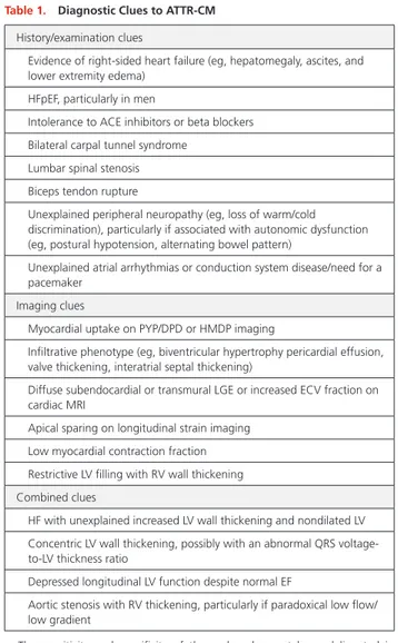
common disease patterns (Table 1; Figure 2),
addition-al clues, and commonly affected populations.
Suspi-cion of ATTR-CM should be triggered in older persons
who have been hospitalized for HF, elevated troponin
levels, or levels of NT-proBNP (N-terminal pro-brain
natriuretic peptide) that are out of proportion to
the clinical context. Other hints of ATTR amyloidosis
include hypertension that resolves over time and an
intolerance of ACE (angiotensin-converting enzyme)
inhibitors, angiotensin receptor blockers, or β
block-ers. In addition, though not infrequent in the general
population, carpal tunnel syndrome occurs particularly
frequently among males with ATTR.
19Lumbar spinal
stenosis,
20,21previous orthopedic procedures,
22and
spontaneous biceps tendon rupture
23may also be
ear-ly indicators of ATTR-CM.
Biomarkers
No plasma or urinary biomarker is available for the
diag-nosis of ATTR. Nevertheless, in the clinical arena, the
combination of very high plasma levels of NT-proBNP
(disproportionate compared with the degree of HF)
and elevated troponin levels in a patient with
echocar-diographic hypertrophic phenotype is strongly
sugges-tive of amyloidotic cardiomyopathy and can prompt
the diagnostic workup. NT-proBNP is a biomarker that
is elevated early in ATTRm amyloidosis before cardiac
symptoms appear, especially among asymptomatic
car-riers of a TTR gene mutation or patients with
neuro-logical symptoms only.
24In addition, the usefulness of
Table 1. Diagnostic Clues to ATTR-CM
History/examination clues
Evidence of right-sided heart failure (eg, hepatomegaly, ascites, and lower extremity edema)
HFpEF, particularly in men
Intolerance to ACE inhibitors or beta blockers Bilateral carpal tunnel syndrome
Lumbar spinal stenosis Biceps tendon rupture
Unexplained peripheral neuropathy (eg, loss of warm/cold discrimination), particularly if associated with autonomic dysfunction (eg, postural hypotension, alternating bowel pattern)
Unexplained atrial arrhythmias or conduction system disease/need for a pacemaker
Imaging clues
Myocardial uptake on PYP/DPD or HMDP imaging
Infiltrative phenotype (eg, biventricular hypertrophy pericardial effusion, valve thickening, interatrial septal thickening)
Diffuse subendocardial or transmural LGE or increased ECV fraction on cardiac MRI
Apical sparing on longitudinal strain imaging Low myocardial contraction fraction Restrictive LV filling with RV wall thickening Combined clues
HF with unexplained increased LV wall thickening and nondilated LV Concentric LV wall thickening, possibly with an abnormal QRS
voltage-to-LV thickness ratio
Depressed longitudinal LV function despite normal EF
Aortic stenosis with RV thickening, particularly if paradoxical low flow/ low gradient
The sensitivity and specificity of these clues has not been delineated in population-based samples with heart failure. ACE indicates angiotensin-converting enzyme; ATTR-CM, transthyretin amyloidosis with predominant cardiomyopathy; DPD, diphosphono-1,2-propanodicarboxylic acid; ECV, extracellular volume; EF, ejection fraction; HF, heart failure; HFpEF, heart failure with preserved ejection fraction; HMDP, hydroxymethylene diphosphonate; LGE, late gadolinium enhancement; LV, left ventricular; MRI, magnetic resonance imaging; PYP, pyrophosphate; and RV, right ventricular.
circulating retinol binding protein 4 in conjunction with
electrocardiographic and echocardiographic measures
to identify patients with HF who have ATTR cardiac
amyloidosis from the Val122Ile mutation has recently
been reported.
25Electrocardiography and Cardiac Imaging
Electrocardiography is a broadly available screening
test, and findings may reveal abnormalities associated
with ATTR-CM (eg, low voltage) that are classically
described in patients with cardiac amyloidosis.
26–28However, low voltage is less common in cardiac
amy-loidosis than a pseudoinfarct pattern of Q waves
unrelated to prior myocardial infarctions.
27Given that
low QRS voltage has been seen in ≈50% of patients
with AL amyloidosis and in ≈25% of patients with
ATTR amyloidosis, its usefulness as a screening test is
limited by low sensitivity.
27More commonly, cardiac
amyloidosis is hallmarked by QRS voltages that are
disproportionate to the thickness of the left
ventricu-lar (LV) wall, which can be assessed using a ratio of
QRS voltage to LV wall thickness.
29,30The presence of
left ventricular hypertrophy on electrocardiography
does not exclude ATTR-CM.
Echocardiography is cost-effective, commonly
available, relatively quick to perform, and better
than most other imaging techniques at identifying
diastolic dysfunction. Although all are not invariably
present, classic echocardiography findings of
infiltra-tive disease include LV wall thickening, small LV cavity
size, biatrial enlargement, thickened valves, elevated
right ventricular systolic pressure and atrial septum
thickness, granular sparkling appearance of the
myo-cardial wall, perimyo-cardial effusion, restrictive
transmi-tral Doppler filling pattern, and reduced ventricular
strain, apical-to-basal strain ratio >2.1, LV ejection
fraction-to-strain ratio >4 (Figures 2 and 3).
26,31,32Combining electrocardiography (low voltage) and
echocardiography (LV septal thickness above the
upper limit of normal, especially >12 mm) is
particu-larly useful for increased clinical suspicion of
ATTR-CM
33(Table 1; Figure 2).
Similarly, cardiac magnetic resonance (CMR) can
show detailed information about systolic function and
cardiac structure (Figure 3). The advantage of CMR is
its unique ability to enable tissue characterization,
34,35allowing it to differentiate amyloidosis from
nonamy-loid wall-thickening disorders. On tissue
characteriza-tion, the typical CMR findings of cardiac
amyloido-sis include diffuse subendocardial or transmural late
gadolinium enhancement on late gadolinium imaging
with nulling of the blood pool and elevated native T1
and extracellular volume on T1 mapping sequences.
T1 mapping, a relatively new and quantitative CMR
technique, with native T1 and extracellular volume,
has the potential to longitudinally monitor disease
progression.
35,36Myocardial scintigraphy with bone avid tracers
99m
technetium pyrophosphate (
99mTc-pyrophosphate),
99mtechnetium
3,3-diphosphono-1,2-propanodicarbox-ylic acid (
99mTc-diphosphono-1,2-propanodicarboxylic
acid), and
99mtechnetium hydroxymethylene
diphos-phonate (
99mTc-hydroxymethylene diphosphonate) has
high sensitivity and specificity for ATTR-CM and may
help with the early diagnosis of ATTR-CM (Figures 4
and 5).
37–43Ease of access, simplicity of imaging,
rela-tively low cost, and specificity for cardiac ATTR
amy-loid deposits are some of the advantages of myocardial
scintigraphy compared with echocardiography, CMR,
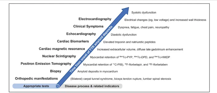
Figure 2. Proposed timeline of appropriate diagnostic tests based on typical disease process.
11C-PIB indicates Pittsburgh compound B; 99mTc-DPD, 99mtechnetium-3,3-diphosphono-1,2-propanodicarboxylic acid; 99mTc-HMDP, hydroxymethylene diphosphonate; 99mTc-PYP, technetium pyrophosphate; ATTR-CM, transthyretin amyloidosis with predominant cardiomyopathy (either wild-type or hereditary); CA, cardiac
amyloi-dosis; ECG, electrocardiography; LVST, left ventricular septal thickness; and TTR, transthyretin.
and endomyocardial biopsy. In addition, the utility of
these agents in identifying ATTR-CM before increases
in wall thickness are observed or electrocardiographic
voltage is reduced
15,41,44,45suggests that they may be
useful for early identification of affected individuals.
Molecular imaging with targeted amyloid-binding
posi-tron emission tomography radiotracers
11C-Pittsburgh
compound B (
11C-PIB),
18F-florbetapir, and
18F-florbe-taben is an emerging quantitative diagnostic approach
that may distinguish cardiac amyloidosis from other
forms of heart disease.
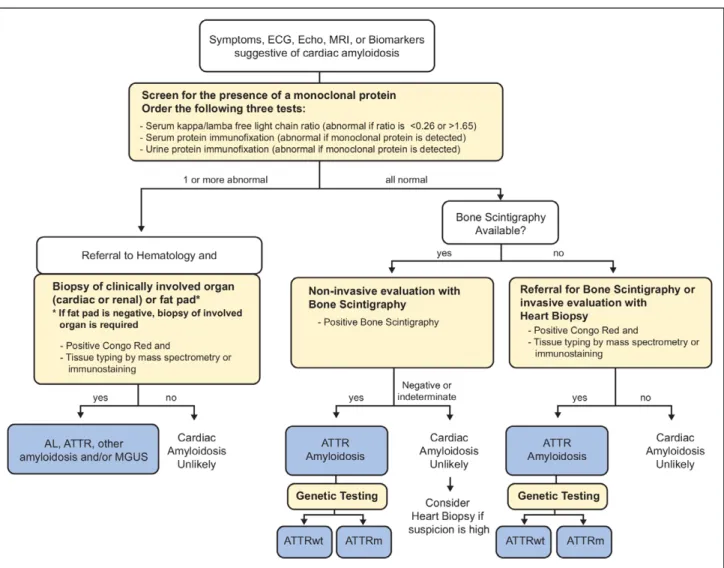
46–49DIAGNOSIS AND ASSESSMENT
A diagnostic approach for patients with suspected
car-diac amyloidosis should include testing for monoclonal
protein followed by scintigraphy or biopsy (Figure 6).
50Clinicians should note that up to 40% of patients
with ATTR-CA can have a monoclonal gammopathy of
unknown significance
51and in this setting scintigraphy
alone cannot ensure a diagnosis with 100%
specific-ity. Nuclear imaging can also be performed
concur-rent to AL assessment, even in the case of a detected
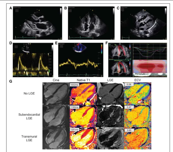
Figure 3. Typical echocardiography and cardiac magnetic resonance (CMR) findings in a patient with cardiac amyloidosis.
Parasternal longitudinal axis (A) and apical 4-chamber (B) echocardiography views show considerably increased left ventricular wall thickness in the absence of ventricular dilation; the myocardial walls seem hyperechogenic. Other characteristic findings include biatrial enlargement and thickening of valve leaflets (A and
B) of the interatrial septum (B) and right ventricle-free wall (C). A generalized small pericardial effusion is also noticeable (A–C). The profile of left ventricle (LV)
filling (D) is restrictive, with markedly elevated E wave, reduced A wave, and decreased deceleration time. A decreased E’ wave measurement can be observed on lateral wall tissue Doppler imaging (E). The longitudinal systolic function is impaired with decreased S’ measurement on lateral wall tissue Doppler imaging (E) and markedly reduced longitudinal strain evident on the apical 4-chamber view (F). LV longitudinal strain (F) is preserved at the LV apex but is significantly impaired at the midbasal segments. Each colored curve shows longitudinal strain at 1 of the 6 LV measured segments. Dotted line is the mean. Color map represents the 6 LV segments, with time corresponding to the x axis. The bullseye appearance (with apex at the center of the color-coding map) is typical of cardiac amyloidosis. Cardiac magnetic resonance images (G) include 4-chamber cine, corresponding native T1 maps, late gadolinium enhancement (LGE) image with phase-sensitive reconstruction, and extracellular volume (ECV) maps in a patient with no cardiac amyloidosis (upper row) and 2 patients with cardiac amyloidosis (middle row and bottom row). In the upper row, the patient with no cardiac amyloidosis has no LGE and normal native T1 and ECV maps; in the middle row, the patient with car-diac amyloidosis has subendocardial LGE, elevated T1 values, and elevated ECV values; in the bottom row, the patient with carcar-diac amyloidosis has a high carcar-diac amyloid load, with transmural LGE, high native T1 values, and high ECV values.
monoclonal gammopathy, for additive information.
However, in the context of monoclonal
gammopa-thy of unknown significance, endomyocardial biopsy
is necessary to definitively diagnose ATTR-CM. If no
monoclonal protein is detected and a diagnosis of AL
cardiac amyloidosis is excluded, radionuclide
scintigra-phy alone, without myocardial biopsy, can be used to
diagnose ATTR-CM.
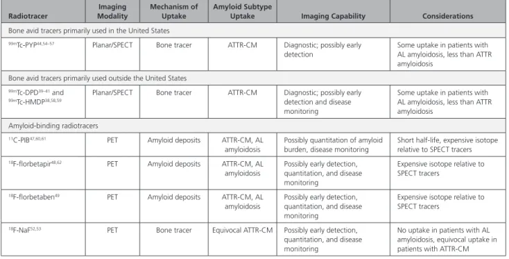
37Among all radiotracers that have
been tested, diphosphono-1,2-propanodicarboxylic
acid, pyrophosphate, and hydroxymethylene
diphos-phonate are recommended for the diagnosis of
amy-loidosis (Table 2).
52,53The radiotracer
123I-metaiodoben-zylguanidine can detect sympathetic innervation of the
heart and may indicate cardiac amyloid,
63–66although
123I-metaiodobenzylguanidine imaging is also abnormal
in other cardiac conditions and is not specific enough
to diagnose ATTR-CM. If ATTR-CM is identified, TTR
genotyping should be performed.
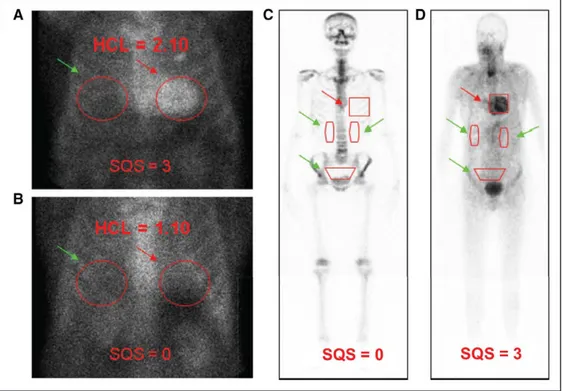
Figure 4.99mTechnetium imaging procedures for cardiac amyloidosis.
SPECT imaging to identify myocardial retention of technetium-based isotopes is particularly useful in discriminating blood pool on planar scans that result in in a false-positive test from myocardial uptake of the isotope indicative of transthyretin amyloidosis with cardiomyopathy. 99mTechnetium-pyrophosphate imaging for
transthyretin cardiac amyloidosis. Adapted with permission of the American Society of Nuclear Cardiology. ©2018, American Society of Nuclear Cardiology.
Figure 5. Semiquantitative approach to 99mTc-PYP/DPD/HMDP imaging in cardiac amyloidosis.
Semiquantitative methods to generate heart-to-contralateral (HCL) ratios with a target region of interest (ROI) over the heart (A, B, red arrows) mirrored over the contralateral chest for a background ROI (A, B, green arrows). An HCL ratio of >1.5 on 1-h imaging is diagnostic of transthyretin amyloidosis with predominant cardiomyopathy (ATTR-CM). Comparatively, 99mTc DPD includes a whole-body scan, 25 to 30 mCi of radiotracer, 200 min of study time, with heart-to-whole-body
ratios generated by a target ROI over the heart (C, D, red arrows) as well as background ROIs over the kidneys and bladder (C, D, green arrows). C and D, Adapted from Perugini et al39 with permission. Copyright ©2005, Journal of the American College of Cardiology.99mTc hydroxymethylene diphosphonate was validated for
diagnosing ATTR CA, and representative scans show diffuse myocardial uptake in a patient with cardiac transthyretin amyloidosis at baseline. 99mTc-DPD indicates 99mtechnetium-3,3-diphosphono-1,2-propanodicarboxylic acid; 99mTc-HMDP, hydroxymethylene diphosphonate; 99mTc-PYP, technetium pyrophosphate.
If monoclonal protein is detected, assessments should
include amyloid typing of a tissue biopsy from a clinically
affected organ (eg, endomyocardial biopsy if the heart
is clinically affected), abdominal fat, or bone marrow,
depending on availability and expertise at the clinic. TTR
genotyping should be performed if a diagnosis of ATTR
amyloidosis is made on biopsy. Endomyocardial biopsy
is invasive, carries a small risk for serious complications,
and requires technical expertise, whereas fat pad biopsy
is less invasive and poses little risk but has varying
sen-sitivity in ATTR-CM (with roughly 45% sensen-sitivity for
ATTRm and roughly 15% sensitivity for ATTRwt).
67Given
the high false-negative rate from biopsies of nonclinically
involved sites (eg, fat pad, bone marrow), further
evalu-ation is warranted even in the presence of a negative
biopsy from such sites if clinical suspicion remains
elevat-ed. In such cases, biopsy of a clinically affected organ (eg,
endomyocardial biopsy) is imperative. Endomyocardial
biopsy assessment with Congo red staining has ≈100%
specificity and sensitivity for detecting amyloid deposits
and is still considered the gold standard in situations with
equivocal noninvasive findings.
68Regardless of the site
of biopsy, amyloid deposits must then undergo either
immunofluorescence or mass spectrometry to confirm
the amyloidosis subtype (eg, AL or ATTR).
In patients with confirmed ATTR amyloidosis, TTR gene
sequencing is necessary even if they do not have a
fam-ily history of amyloidosis or evidence of polyneuropathy
because the penetrance of ATTRm varies among the
vari-ants and families. If a TTR variant is detected, genetic
counseling for relatives of the affected patient is indicated.
Prognostic Stratification Using
Biomarkers
Natriuretic peptides and cardiac troponins, though not
specific markers of ATTR-CM, are well established to
assess risk (eg, Mayo staging) and to evaluate response
to treatment in patients with AL amyloidosis.
69–71How-ever, evidence in AL amyloidosis may not apply to ATTR
Figure 6. Diagnostic algorithm for patients with suspected cardiac amyloidosis.
Note that urine protein electrophoresis with immunofixation can be performed on spot or 24-h urine collection. AL indicates light chain amyloidosis; ATTR, transthyretin amyloidosis; ATTRm, mutant transthyretin amyloidosis; ATTRwt, wild-type transthyretin amyloidosis; ECG, electrocardiography; Echo, echocardiogram; MGUS, monoclonal gammopathy of undetermined significance; MRI, magnetic resonance imaging. Adapted from Nativi-Nicolau and Maurer50 with permission.
Copyright ©2018, Wolters Kluwer Health, Inc.
amyloidosis because the 2 diseases have substantially
different biologies.
29,72,73Different staging systems for
ATTRwt amyloidosis and ATTR-CM have been proposed.
A proposed system in patients with ATTRwt includes
NT-proBNP (>3000 pg/mL) and troponin T (>0.05 ng/mL).
74The most recently proposed system for staging ATTR-CM
(including ATTRwt and ATTRm) uses NT-proBNP (>3000
pg/mL) and estimated glomerular filtration rate (<45 mL/
minute).
75Staging in both systems is defined such that
stage 1 does not meet either threshold, stage 2 meets 1
of the 2 thresholds, and stage 3 meets both thresholds.
FUTURE DIRECTIONS
To reduce the delays in diagnosis of this important health
problem, specific programs for screening or early
iden-tification should be studied that leverage techniques
having appropriate sensitivity and specificity as well as
favorable cost/benefit. Implementation of appropriate
screening programs for ATTR-CM will need to include
factors such as whether there are methods and
facili-ties available for diagnosis, whether certain diagnostic
tests are acceptable to those at risk, and the particular
test characteristics, as well as cost and benefits of
treat-ments and when appropriate timing would be for the
intervention. Given that prognosis is highly dependent
on the underlying cardiac dysfunction coupled with
the recently available treatments for ATTR amyloidosis,
screening programs may become important.
CONCLUSIONS
ATTR amyloidosis is a progressive disease associated with
increased morbidity and mortality and occurs in inherited
(ATTRm or hereditary ATTR) or acquired (ATTRwt) forms.
Disease-related cardiac dysfunction in patients with ATTR
amyloidosis is associated with particularly poor outcomes
and is a manifestation of many of the genetic variants
and the wild-type form of ATTR amyloidosis.
Diagnosis of ATTR-CM is often missed or mistaken as
hypertrophic cardiomyopathy or heart failure with
pre-served ejection fraction of unknown cause. Although
considered a rare disease, the true prevalence of
ATTR-CM is unclear and is likely higher than appreciated.
Physicians should consider systemic signs and
symp-toms along with evidence from biomarkers and
imag-ing to build suspicion for ATTR-CM. To facilitate early
diagnosis of ATTR-CM, evaluation of myocardial uptake
on bone scintigraphy should be considered in patients
with HF, unexplained neuropathy, family history of
amy-loidosis, or unexplained increased LV wall thickness.
Appropriate evidence on echocardiography or cardiac
MRI—combined with no light chain clone, grade ≥2
myocardial uptake of
99mTc-pyrophosphate,
diphos-phono-1,2-propanodicarboxylic acid, and
hydroxy-methylene diphosphonate—is diagnostic of ATTR-CM,
in which case endomyocardial biopsy is unnecessary.
Genetic testing should be performed to differentiate
ATTRm from ATTRwt causes of ATTR-CM.
Table 2. Radiotracers for Imaging of Cardiac Amyloidosis
Radiotracer Imaging Modality Mechanism of Uptake Amyloid Subtype
Uptake Imaging Capability Considerations
Bone avid tracers primarily used in the United States
99mTc-PYP44,54–57 Planar/SPECT Bone tracer ATTR-CM Diagnostic; possibly early
detection
Some uptake in patients with AL amyloidosis, less than ATTR amyloidosis
Bone avid tracers primarily used outside the United States
99mTc-DPD39–41 and 99mTc-HMDP38,58,59
Planar/SPECT Bone tracer ATTR-CM Diagnostic; possibly early detection and disease monitoring
Some uptake in patients with AL amyloidosis, less than ATTR amyloidosis
Amyloid-binding radiotracers
11C-PIB47,60,61 PET Amyloid deposits ATTR-CM, AL
amyloidosis
Possibly quantitation of amyloid burden, disease monitoring
Short half-life, expensive isotope relative to SPECT tracers
18F-florbetapir48,62 PET Amyloid deposits ATTR-CM, AL
amyloidosis
Possibly early detection, quantitation, and disease monitoring
Expensive isotope relative to SPECT tracers
18F-florbetaben49 PET Amyloid deposits ATTR-CM, AL
amyloidosis
Possibly early detection, quantitation, and disease monitoring
Expensive isotope relative to SPECT tracers
18F-NaF52,53 PET Bone tracer Equivocal ATTR-CM Possibly early detection,
quantitation, and disease monitoring
No uptake in patients with AL amyloidosis, equivocal uptake in patients with ATTR-CM
The tracers 99mTc-MDP and 99mTc-aprotinin are not recommended. 11C-PIB indicates Pittsburgh compound B; 18F-NaF, sodium fluoride; 99mTc-DPD, 99m
technetium-3,3-diphosphono-1,2-propanodicarboxylic acid; 99mTc-HMDP, hydroxymethylene diphosphonate; 99mTc-PYP, technetium pyrophosphate; 123I, iodine-123; AL, amyloid
light chain; ATTR-CM, transthyretin amyloidosis with predominant cardiomyopathy (either wild-type or hereditary); PET, positron emission tomography; and SPECT, single-photon emission computed tomography.
The consensus recommendations described in this
review were developed with the goal of providing
cli-nicians with an overview of key aspects of ATTR-CM
diagnosis. We hope these recommendations facilitate
early, rapid, and accurate identification of ATTR-CM to
allow implementation of targeted, disease-modifying
treatment and improved outcomes for patients.
ARTICLE INFORMATION
Correspondence
Mathew S. Maurer, MD, Columbia University Medical Center, 622 W 168th St, New York, NY 10032. Email [email protected]
Affiliations
Division of Cardiology, Department of Medicine, Columbia University Medical Center, New York (M.S.M., S.B.). Department of Cardiology, Center for Cardiac Amyloidosis, GRC Amyloid Research Institute, DHU A-TVB, APHP CHU Henri Mon-dor and Université Paris Est Créteil, France (T.D.). Division of Nuclear Medicine and Molecular Imaging, Department of Radiology, Brigham and Women’s Hos-pital, Boston, MA (S.D.). Department of Cardiovascular Medicine, University of Pennsylvania Perelman School of Medicine, Philadelphia (B.M.D.). National Amy-loidosis Centre, Division of Medicine, University College London, United Kingdom (M.F., C.C.Q.). Cardiovascular Medicine, Mayo Clinic, Rochester, MN (M.G.). De-partment of Cardiology, Amyloidosis Center, University of Heidelberg, Germany (A.V.K.). Amyloidosis Research Consortium, Newton, MA (I.L.). Division of Car-diovascular Medicine, University of Utah Health, Salt Lake City (J.N.-N.). Depart-ment of ExperiDepart-mental, Diagnostic and Specialty Medicine, Alma Mater Studiorum University of Bologna, Italy (C.C.Q., C.R.). Cardiovascular Center, Boston University School of Medicine, Boston Medical Center, MA (F.L.R.). Stanford Amyloid Center, Division of Cardiovascular Medicine, Stanford University School of Medicine, CA (R.W.). Amyloidosis Center Foundation IRCCS Policlinico San Matteo, Italy (G.M.). Department of Molecular Medicine, University of Pavia, Italy (G.M.).
Acknowledgments
Funding support for these recommendations was received from GSK, Ionis, Pfizer, and Alnylam; these companies were not involved in writing this manu-script. Medical writing and editorial assistance was provided by ApotheCom (San Francisco, CA).
Sources of Funding
This review was sponsored by the Amyloidosis Research Consortium.
Disclosures
Dr Maurer served on the steering committee of the Tafamidis in Transthyretin Cardiomyopathy Clinical Trial. His institution received research support for clini-cal studies from Pfizer and Alnylam. He has served on advisory boards or Data and Safety Monitoring Boards for Akcea, Ionis, Prothena, Pfizer, Alnylam, Eidos, GSK. Dr Bokhari receives honoraria from Pfizer. Dr Damy’s institution received research support for clinical studies from Pfizer and Alnylam. He has served on advisory boards or DSMBs for Akcea, Ionis, Prothena, Pfizer, Alnylam, and GSK. Dr Dorbala received consulting fees from Pfizer, GEHC, and Advanced Accelera-tor Applications. Dr Drachman received consulting fees from Pfizer and Alnylam (not significant). Dr Fontana received research grants from GSK, salary from BHF FS/18/21/33447, and honoraria from Pfizer, Alnylam, and Prothena (modest). Dr Grogan received research funding from Alnylam, Eidos, Pfizer, and Prothena. Dr Kristen received consulting fees from Akcea EIDOS and honoraria/travel sup-port from Pfizer, Akcea, and Alnylam. I. Lousada received an honorarium from Akcea. Dr Nativi-Nicolau received research funding for clinical studies from Pfiz-er, Akcea, and Eidos and served on advisory boards for Alnylam, Ionis, Akcea, and Pfizer. Dr Quarta received honoraria from Alnylam. Dr Rapezzi received research grants from Pfizer and honoraria from Pfizer and Alnylam. Dr Ruberg received consulting fees from Pfizer and GSK and grant support from Eidos (sig-nificant). Dr Witteles received advisory board honoraria from Pfizer, Alnylam, and Eidos and clinical trial research support from Pfizer and Eidos. Dr Merlini received honoraria from Janssen, honoraria and travel support from Prothena,
travel support from Celgene, and consulting fees from Millennium, Pfizer, Jans-sen, Prothena, and Ionis.
REFERENCES
1. Castaño A, Drachman BM, Judge D, Maurer MS. Natural history and therapy of TTR-cardiac amyloidosis: emerging disease-modifying therapies from organ transplantation to stabilizer and silencer drugs. Heart Fail Rev. 2015;20:163–178. doi: 10.1007/s10741-014-9462-7
2. Patel KS, Hawkins PN. Cardiac amyloidosis: where are we today? J Intern
Med. 2015;278:126–144. doi: 10.1111/joim.12383
3. Brunjes DL, Castano A, Clemons A, Rubin J, Maurer MS. Transthyretin cardiac amyloidosis in older Americans. J Card Fail. 2016;22:996–1003. doi: 10.1016/j.cardfail.2016.10.008
4. Hawkins PN, Ando Y, Dispenzeri A, Gonzalez-Duarte A, Adams D, Suhr OB. Evolving landscape in the management of transthyretin amyloidosis. Ann
Med. 2015;47:625–638. doi: 10.3109/07853890.2015.1068949
5. Jacobson DR, Pastore R, Pool S, Malendowicz S, Kane I, Shivji A, Embury SH, Ballas SK, Buxbaum JN. Revised transthyretin Ile 122 allele frequency in African-Americans. Hum Genet. 1996;98:236–238. doi: 10.1007/s004390050199
6. Quarta CC, Buxbaum JN, Shah AM, Falk RH, Claggett B, Kitzman DW, Mosley TH, Butler KR, Boerwinkle E, Solomon SD. The amyloidogenic V122I transthyretin variant in elderly black Americans. N Engl J Med. 2015;372:21–29. doi: 10.1056/NEJMoa1404852
7. Jacobson DR, Pastore RD, Yaghoubian R, Kane I, Gallo G, Buck FS, Buxbaum JN. Variant-sequence transthyretin (isoleucine 122) in late-onset cardiac amyloidosis in black Americans. N Engl J Med. 1997;336:466– 473. doi: 10.1056/NEJM199702133360703
8. Buxbaum J, Jacobson DR, Tagoe C, Alexander A, Kitzman DW, Greenberg B, Thaneemit-Chen S, Lavori P. Transthyretin V122I in African Americans with congestive heart failure. J Am Coll Cardiol. 2006;47:1724–1725. doi: 10.1016/j.jacc.2006.01.042
9. Reilly MM, Staunton H, Harding AE. Familial amyloid polyneuropathy (TTR ala 60) in north west Ireland: a clinical, genetic, and epidemiological study.
J Neurol Neurosurg Psychiatry. 1995;59:45–49. doi: 10.1136/jnnp.59.1.45
10. Tanskanen M, Peuralinna T, Polvikoski T, Notkola IL, Sulkava R, Hardy J, Singleton A, Kiuru-Enari S, Paetau A, Tienari PJ, Myllykangas L. Senile sys-temic amyloidosis affects 25% of the very aged and associates with genet-ic variation in alpha2-macroglobulin and tau: a population-based autopsy study. Ann Med. 2008;40:232–239. doi: 10.1080/07853890701842988 11. Castaño A, Narotsky DL, Hamid N, Khalique OK, Morgenstern R, DeLuca A,
Rubin J, Chiuzan C, Nazif T, Vahl T, George I, Kodali S, Leon MB, Hahn R, Bokhari S, Maurer MS. Unveiling transthyretin cardiac amyloidosis and its predictors among elderly patients with severe aortic stenosis undergoing transcatheter aortic valve replacement. Eur Heart J. 2017;38:2879–2887. doi: 10.1093/eurheartj/ehx350
12. González-López E, Gallego-Delgado M, Guzzo-Merello G, de Haro- Del Moral FJ, Cobo-Marcos M, Robles C, Bornstein B, Salas C, Lara-Pezzi E, Alonso-Pulpon L, Garcia-Pavia P. Wild-type transthyretin amyloidosis as a cause of heart failure with preserved ejection fraction. Eur Heart J. 2015;36:2585–2594. doi: 10.1093/eurheartj/ehv338
13. Damy T, Costes B, Hagège AA, Donal E, Eicher JC, Slama M, Guellich A, Rappeneau S, Gueffet JP, Logeart D, Planté-Bordeneuve V, Bouvaist H, Huttin O, Mulak G, Dubois-Randé JL, Goossens M, Canoui-Poitrine F, Buxbaum JN. Prevalence and clinical phenotype of hereditary transthyre-tin amyloid cardiomyopathy in patients with increased left ventricular wall thickness. Eur Heart J. 2016;37:1826–1834. doi: 10.1093/eurheartj/ehv583 14. Sperry BW, Reyes BA, Ikram A, Donnelly JP, Phelan D, Jaber WA, Shapiro D, Evans PJ, Maschke S, Kilpatrick SE, Tan CD, Rodriguez ER, Monteiro C, Tang WHW, Kelly JW, Seitz WH Jr, Hanna M. Tenosynovial and cardiac amyloidosis in patients undergoing carpal tunnel release. J Am Coll
Car-diol. 2018;72:2040–2050. doi: 10.1016/j.jacc.2018.07.092
15. Longhi S, Guidalotti PL, Quarta CC, Gagliardi C, Milandri A, Lorenzini M, Potena L, Leone O, Bartolomei I, Pastorelli F, Salvi F, Rapezzi C. Identification of TTR-related subclinical amyloidosis with 99mTc-DPD scintigraphy. JACC
Cardiovasc Imaging. 2014;7:531–532. doi: 10.1016/j.jcmg.2014.03.004
16. Mohamed-Salem L, Santos-Mateo JJ, Sanchez-Serna J, Hernández- Vicente Á, Reyes-Marle R, Castellón Sánchez MI, Claver-Valderas MA, Gonzalez-Vioque E, Haro-Del Moral FJ, García-Pavía P, Pascual-Figal DA. Prevalence of wild type ATTR assessed as myocardial uptake in bone scan in the elderly population. Int J Cardiol. 2018;270:192–196. doi: 10.1016/j.ijcard.2018.06.006
17. Galant NJ, Westermark P, Higaki JN, Chakrabartty A. Transthyretin amy-loidosis: an under-recognized neuropathy and cardiomyopathy. Clin Sci
(Lond). 2017;131:395–409. doi: 10.1042/CS20160413
18. Lousada I, Comenzo RL, Landau H, Guthrie S, Merlini G. Patient expe-rience with hereditary and wild-type transthyretin amyloidosis: a survey from the Amyloidosis Research Consortium. Paper Presented at: First Eu-ropean Congress on Hereditary ATTR Amyloidosis; November 2–3, 2015; Paris, France.
19. Sekijima Y, Uchiyama S, Tojo K, Sano K, Shimizu Y, Imaeda T, Hoshii Y, Kato H, Ikeda S. High prevalence of wild-type transthyretin deposition in patients with idiopathic carpal tunnel syndrome: a common cause of carpal tunnel syndrome in the elderly. Hum Pathol. 2011;42:1785–1791. doi: 10.1016/j.humpath.2011.03.004
20. Yanagisawa A, Ueda M, Sueyoshi T, Okada T, Fujimoto T, Ogi Y, Kitagawa K, Tasaki M, Misumi Y, Oshima T, Jono H, Obayashi K, Hirakawa K, Uchida H, Westermark P, Ando Y, Mizuta H. Amyloid deposits derived from trans-thyretin in the ligamentum flavum as related to lumbar spinal canal steno-sis. Mod Pathol. 2015;28:201–207. doi: 10.1038/modpathol.2014.102 21. Westermark P, Westermark GT, Suhr OB, Berg S. Transthyretin-derived
amyloidosis: probably a common cause of lumbar spinal stenosis. Ups J
Med Sci. 2014;119:223–228. doi: 10.3109/03009734.2014.895786
22. Rubin J, Alvarez J, Teruya S, Castano A, Lehman RA, Weidenbaum M, Geller JA, Helmke S, Maurer MS. Hip and knee arthroplasty are common among patients with transthyretin cardiac amyloidosis, occurring years be-fore cardiac amyloid diagnosis: can we identify affected patients earlier?
Amyloid. 2017;24:226–230. doi: 10.1080/13506129.2017.1375908
23. Geller HI, Singh A, Alexander KM, Mirto TM, Falk RH. Association be-tween ruptured distal biceps tendon and wild-type transthyretin cardiac amyloidosis. JAMA. 2017;318:962–963. doi: 10.1001/jama.2017.9236 24. Damy T, Deux JF, Moutereau S, Guendouz S, Mohty D, Rappeneau S,
Guellich A, Hittinger L, Loric S, Lefaucheur JP, Plante-Bordeneuve V. Role of natriuretic peptide to predict cardiac abnormalities in patients with hereditary transthyretin amyloidosis. Amyloid. 2013;20:212–220. doi: 10.3109/13506129.2013.825240
25. Arvanitis M, Koch CM, Chan GG, Torres-Arancivia C, LaValley MP, Jacobson DR, Berk JL, Connors LH, Ruberg FL. Identification of trans-thyretin cardiac amyloidosis using serum retinol-binding protein 4 and a clinical prediction model. JAMA Cardiol. 2017;2:305–313. doi: 10.1001/jamacardio.2016.5864
26. Rapezzi C, Quarta CC, Riva L, Longhi S, Gallelli I, Lorenzini M, Ciliberti P, Biagini E, Salvi F, Branzi A. Transthyretin-related amyloidoses and the heart: a clinical overview. Nat Rev Cardiol. 2010;7:398–408. doi: 10.1038/nrcardio.2010.67
27. Ruberg FL, Berk JL. Transthyretin (TTR) cardiac amyloidosis. Circulation. 2012;126:1286–1300. doi: 10.1161/CIRCULATIONAHA.111.078915 28. Quarta CC, Kruger JL, Falk RH. Cardiac amyloidosis. Circulation.
2012;126:e178–e182. doi: 10.1161/CIRCULATIONAHA.111.069195 29. Quarta CC, Solomon SD, Uraizee I, Kruger J, Longhi S, Ferlito M, Gagliardi C,
Milandri A, Rapezzi C, Falk RH. Left ventricular structure and function in transthyretin-related versus light-chain cardiac amyloidosis. Circulation. 2014;129:1840–1849. doi: 10.1161/CIRCULATIONAHA.113.006242 30. Carroll JD, Gaasch WH, McAdam KP. Amyloid cardiomyopathy:
character-ization by a distinctive voltage/mass relation. Am J Cardiol. 1982;49:9–13. doi: 10.1016/0002-9149(82)90270-3
31. Falk RH. Diagnosis and management of the cardiac amyloidoses.
Circula-tion. 2005;112:2047–2060. doi: 10.1161/CIRCULATIONAHA.104.489187
32. Maurer MS, Elliott P, Comenzo R, Semigran M, Rapezzi C. Address-ing common questions encountered in the diagnosis and manage-ment of cardiac amyloidosis. Circulation. 2017;135:1357–1377. doi: 10.1161/CIRCULATIONAHA.116.024438
33. Di Bella G, Minutoli F, Piaggi P, Casale M, Mazzeo A, Zito C, Oreto G, Baldari S, Vita G, Pingitore A, Khandheria BK, Carerj S. Usefulness of com-bining electrocardiographic and echocardiographic findings and brain na-triuretic peptide in early detection of cardiac amyloidosis in subjects with transthyretin gene mutation. Am J Cardiol. 2015;116:1122–1127. doi: 10.1016/j.amjcard.2015.07.008
34. Maceira AM, Joshi J, Prasad SK, Moon JC, Perugini E, Harding I, Sheppard MN, Poole-Wilson PA, Hawkins PN, Pennell DJ. Cardiovascular magnetic resonance in cardiac amyloidosis. Circulation. 2005;111:186– 193. doi: 10.1161/01.CIR.0000152819.97857.9D
35. Martinez-Naharro A, Treibel TA, Abdel-Gadir A, Bulluck H, Zumbo G, Knight DS, Kotecha T, Francis R, Hutt DF, Rezk T, Rosmini S, Quarta CC, Whelan CJ, Kellman P, Gillmore JD, Moon JC, Hawkins PN, Fontana M. Magnetic resonance in transthyretin cardiac amyloidosis. J Am Coll
Car-diol. 2017;70:466–477. doi: 10.1016/j.jacc.2017.05.053
36. Martinez-Naharro A, Kotecha T, Norrington K, Boldrini M, Rezk T, Quarta C, Treibel TA, Whelan CJ, Knight DS, Kellman P, Ruberg FL, Gillmore JD, Moon JC, Hawkins PN, Fontana M. Native T1 and extracellular volume in transthyretin amyloidosis. JACC Cardiovasc Imaging. 2019;12:810–819. doi: 10.1016/j.jcmg.2018.02.006
37. Gillmore JD, Maurer MS, Falk RH, Merlini G, Damy T, Dispenzieri A, Wechalekar AD, Berk JL, Quarta CC, Grogan M, Lachmann HJ, Bokhari S, Castano A, Dorbala S, Johnson GB, Glaudemans AW, Rezk T, Fontana M, Palladini G, Milani P, Guidalotti PL, Flatman K, Lane T, Vonberg FW, Whelan CJ, Moon JC, Ruberg FL, Miller EJ, Hutt DF, Hazenberg BP, Rapezzi C, Hawkins PN. Nonbiopsy diagnosis of cardiac transthyretin amyloidosis.
Circulation. 2016;133:2404–2412. doi: 10.1161/CIRCULATIONAHA.
116.021612
38. Galat A, Rosso J, Guellich A, Van Der Gucht A, Rappeneau S, Bodez D, Guendouz S, Tissot CM, Hittinger L, Dubois-Randé JL, Plante-Bordeneuve V, Itti E, Meignan M, Damy T. Usefulness of (99m)Tc-HMDP scintigraphy for the etiologic diagnosis and prognosis of cardiac amyloidosis. Amyloid. 2015;22:210–220. doi: 10.3109/13506129.2015.1072089
39. Perugini E, Guidalotti PL, Salvi F, Cooke RM, Pettinato C, Riva L, Leone O, Farsad M, Ciliberti P, Bacchi-Reggiani L, Fallani F, Branzi A, Rapezzi C. Noninvasive etiologic diagnosis of cardiac amyloidosis using 99mTc-3,3-diphosphono-1,2-propanodicarboxylic acid scintigraphy. J Am Coll
Car-diol. 2005;46:1076–1084. doi: 10.1016/j.jacc.2005.05.073
40. Rapezzi C, Quarta CC, Guidalotti PL, Longhi S, Pettinato C, Leone O, Ferlini A, Salvi F, Gallo P, Gagliardi C, Branzi A. Usefulness and limi-tations of 99mTc-3,3-diphosphono-1,2-propanodicarboxylic acid scintigraphy in the aetiological diagnosis of amyloidotic cardio-myopathy. Eur J Nucl Med Mol Imaging. 2011;38:470–478. doi: 10.1007/s00259-010-1642-7
41. Rapezzi C, Quarta CC, Guidalotti PL, Pettinato C, Fanti S, Leone O, Ferlini A, Longhi S, Lorenzini M, Reggiani LB, Gagliardi C, Gallo P, Villani C, Salvi F. Role of (99m)Tc-DPD scintigraphy in diagnosis and prognosis of hereditary transthyretin-related cardiac amyloidosis. JACC Cardiovasc
Im-aging. 2011;4:659–670. doi: 10.1016/j.jcmg.2011.03.016
42. Pilebro B, Suhr OB, Näslund U, Westermark P, Lindqvist P, Sundström T. (99m)Tc-DPD uptake reflects amyloid fibril composition in heredi-tary transthyretin amyloidosis. Ups J Med Sci. 2016;121:17–24. doi: 10.3109/03009734.2015.1122687
43. Falk RH, Quarta CC, Dorbala S. How to image cardiac amyloidosis. Circ
Cardiovasc Imaging. 2014;7:552–562. doi: 10.1161/CIRCIMAGING.
113.001396
44. Haq M, Pawar S, Berk JL, Miller EJ, Ruberg FL. Can 99mTc-pyrophosphate aid in early detection of cardiac involvement in asymptomatic variant TTR amyloidosis? JACC Cardiovasc Imaging. 2017;10:713–714. doi: 10.1016/j.jcmg.2016.06.003
45. Glaudemans AW, van Rheenen RW, van den Berg MP, Noordzij W, Koole M, Blokzijl H, Dierckx RA, Slart RH, Hazenberg BP. Bone scintigra-phy with (99m)technetium-hydroxymethylene diphosphonate allows early diagnosis of cardiac involvement in patients with transthyretin-derived systemic amyloidosis. Amyloid. 2014;21:35–44. doi: 10.3109/13506129. 2013.871250
46. Park MA, Padera RF, Belanger A, Dubey S, Hwang DH, Veeranna V, Falk RH, Di Carli MF, Dorbala S. 18F-florbetapir binds specifically to myocardial light chain and transthyretin amyloid deposits: autoradiography study.
Circ Cardiovasc Imaging. 2015;8:e002954. doi: 10.1161/CIRCIMAGING.
114.002954
47. Antoni G, Lubberink M, Estrada S, Axelsson J, Carlson K, Lindsjö L, Kero T, Långström B, Granstam SO, Rosengren S, Vedin O, Wassberg C, Wikström G, Westermark P, Sörensen J. In vivo visualization of amyloid de-posits in the heart with 11C-PIB and PET. J Nucl Med. 2013;54:213–220. doi: 10.2967/jnumed.111.102053
48. Dorbala S, Vangala D, Semer J, Strader C, Bruyere JR Jr, Di Carli MF, Moore SC, Falk RH. Imaging cardiac amyloidosis: a pilot study using ¹⁸F-florbetapir positron emission tomography. Eur J Nucl Med Mol Imaging. 2014;41:1652–1662. doi: 10.1007/s00259-014-2787-6
49. Law WP, Wang WY, Moore PT, Mollee PN, Ng AC. Cardiac amyloid imag-ing with 18F-florbetaben PET: a pilot study. J Nucl Med. 2016;57:1733– 1739. doi: 10.2967/jnumed.115.169870
50. Nativi-Nicolau J, Maurer MS. Amyloidosis cardiomyopathy: update in the diagnosis and treatment of the most common types. Curr Opin Cardiol. 2018;33:571–579. doi: 10.1097/HCO.0000000000000547
51. Phull P, Sanchorawala V, Connors LH, Doros G, Ruberg FL, Berk JL, Sarosiek S. Monoclonal gammopathy of undetermined significance in systemic transthyretin amyloidosis (ATTR). Amyloid. 2018;25:62–67. doi: 10.1080/13506129.2018.1436048
52. Van Der Gucht A, Galat A, Rosso J, Guellich A, Garot J, Bodez D, Plante-Bordeneuve V, Hittinger L, Dubois-Randé JL, Evangelista E, Sasanelli M, Chalaye J, Meignan M, Itti E, Damy T. [18F]-NaF PET/CT imaging in cardiac amyloidosis. J Nucl Cardiol. 2016;23:846–849. doi: 10.1007/s12350-015-0287-0
53. Gagliardi C, Tabacchi E, Bonfiglioli R, Diodato S, Nanni C, Guidalotti P, Lorenzini M, Lodi F, Milandri A, Rapezzi C, Fanti S. Does the etiology of cardiac amyloidosis determine the myocardial uptake of [18F]-NaF PET/CT? J
Nucl Cardiol. 2017;24:746–749. doi: 10.1007/s12350-016-0457-8
54. Bokhari S, Morgenstern R, Weinberg R, Kinkhabwala M, Panagiotou D, Castano A, DeLuca A, Kontak A, Jin Z, Maurer MS. Standardization of 99mTechnetium pyrophosphate imaging methodology to diag-nose TTR cardiac amyloidosis. J Nucl Cardiol. 2018;25:181–190. doi: 10.1007/s12350-016-0610-4
55. Bokhari S, Castaño A, Pozniakoff T, Deslisle S, Latif F, Maurer MS. (99m)Tc-pyrophosphate scintigraphy for differentiating light-chain cardiac amyloidosis from the transthyretin-related familial and senile cardiac amyloidoses. Circ Cardiovasc Imaging. 2013;6:195–201. doi: 10.1161/CIRCIMAGING.112.000132
56. Castano A, Haq M, Narotsky DL, Goldsmith J, Weinberg RL, Morgenstern R, Pozniakoff T, Ruberg FL, Miller EJ, Berk JL, Dispenzieri A, Grogan M, Johnson G, Bokhari S, Maurer MS. Multicenter study of planar techne-tium 99m pyrophosphate cardiac imaging: predicting survival for patients with ATTR cardiac amyloidosis. JAMA Cardiol. 2016;1:880–889. doi: 10.1001/jamacardio.2016.2839
57. Castaño A, DeLuca A, Weinberg R, Pozniakoff T, Blaner WS, Pirmohamed A, Bettencourt B, Gollob J, Karsten V, Vest JA, Chiuzan C, Maurer MS, Bokhari S. Serial scanning with technetium pyrophosphate (99mTc-PYP) in advanced ATTR cardiac amyloidosis. J Nucl Cardiol. 2016;23:1355–1363. doi: 10.1007/s12350-015-0261-x
58. Wadhwa SS, Nour R. Tc-99m HDP uptake in cardiac amyloidosis. Clin Nucl
Med. 1999;24:156–158. doi: 10.1097/00003072-199903000-00002
59. Kulhanek J, Movahed A. Uptake of technetium 99m HDP in cardiac amy-loidosis. Int J Cardiovasc Imaging. 2003;19:225–227.
60. Lee SP, Lee ES, Choi H, Im HJ, Koh Y, Lee MH, Kwon JH, Paeng JC, Kim HK, Cheon GJ, Kim YJ, Kim I, Yoon SS, Seo JW, Sohn DW. 11C-Pittsburgh B PET imaging in cardiac amyloidosis. JACC Cardiovasc Imaging. 2015;8:50– 59. doi: 10.1016/j.jcmg.2014.09.018
61. Kero T, Lindsjö L, Sörensen J, Lubberink M. Accurate analysis and visualiza-tion of cardiac (11)C-PIB uptake in amyloidosis with semiautomatic soft-ware. J Nucl Cardiol. 2016;23:741–750. doi: 10.1007/s12350-015-0149-9 62. Osborne DR, Acuff SN, Stuckey A, Wall JS. A routine PET/CT protocol with
streamlined calculations for assessing cardiac amyloidosis using (18)F-flor-betapir. Front Cardiovasc Med. 2015;2:23. doi: 10.3389/fcvm.2015.00023 63. Nakata T, Shimamoto K, Yonekura S, Kobayashi N, Sugiyama T, Imai K,
Iimura O. Cardiac sympathetic denervation in transthyretin-related famil-ial amyloidotic polyneuropathy: detection with iodine-123-MIBG. J Nucl
Med. 1995;36:1040–1042.
64. Delahaye N, Dinanian S, Slama MS, Mzabi H, Samuel D, Adams D, Merlet P, Le Guludec D. Cardiac sympathetic denervation in familial amy-loid polyneuropathy assessed by iodine-123 metaiodobenzylguanidine scintigraphy and heart rate variability. Eur J Nucl Med. 1999;26:416–424. 65. Hongo M, Urushibata K, Kai R, Takahashi W, Koizumi T, Uchikawa S,
Imamura H, Kinoshita O, Owa M, Fujii T. Iodine-123
metaiodobenzylgua-nidine scintigraphic analysis of myocardial sympathetic innervation in pa-tients with AL (primary) amyloidosis. Am Heart J. 2002;144:122–129. doi: 10.1067/mhj.2002.123115
66. Noordzij W, Glaudemans AW, van Rheenen RW, Hazenberg BP, Tio RA, Dierckx RA, Slart RH. (123)I-Labelled metaiodobenzylguanidine for the evaluation of cardiac sympathetic denervation in early stage amyloidosis. Eur J Nucl Med Mol Imaging. 2012;39:1609–1617. doi: 10.1007/s00259-012-2187-8
67. Quarta CC, Gonzalez-Lopez E, Gilbertson JA, Botcher N, Rowczenio D, Petrie A, Rezk T, Youngstein T, Mahmood S, Sachchithanantham S, Lachmann HJ, Fontana M, Whelan CJ, Wechalekar AD, Hawkins PN, Gillmore JD. Diagnostic sensitivity of abdominal fat as-piration in cardiac amyloidosis. Eur Heart J. 2017;38:1905–1908. doi: 10.1093/eurheartj/ehx047
68. Adams D, Suhr OB, Hund E, Obici L, Tournev I, Campistol JM, Slama MS, Hazenberg BP, Coelho T; European Network for TTR-FAP (ATTReuNET). First European consensus for diagnosis, management, and treatment of transthyretin familial amyloid polyneuropathy. Curr Opin Neurol. 2016;29(suppl 1):S14–S26. doi: 10.1097/WCO.0000000000000289 69. Merlini G, Lousada I, Ando Y, Dispenzieri A, Gertz MA, Grogan M,
Maurer MS, Sanchorawala V, Wechalekar A, Palladini G, Comenzo RL. Rationale, application and clinical qualification for NT-proBNP as a sur-rogate end point in pivotal clinical trials in patients with AL amyloidosis.
Leukemia. 2016;30:1979–1986. doi: 10.1038/leu.2016.191
70. Lavatelli F, Albertini R, Di Fonzo A, Palladini G, Merlini G. Biochemical markers in early diagnosis and management of systemic amyloidoses. Clin
Chem Lab Med. 2014;52:1517–1531. doi: 10.1515/cclm-2014-0235
71. Kumar S, Dispenzieri A, Lacy MQ, Hayman SR, Buadi FK, Colby C, Laumann K, Zeldenrust SR, Leung N, Dingli D, Greipp PR, Lust JA, Russell SJ, Kyle RA, Rajkumar SV, Gertz MA. Revised prognostic staging system for light chain amyloidosis incorporating cardiac biomarkers and serum free light chain measurements. J Clin Oncol. 2012;30:989–995. doi: 10.1200/JCO.2011.38.5724
72. Cappelli F, Baldasseroni S, Bergesio F, Perlini S, Salinaro F, Padeletti L, Attanà P, Paoletti Perini A, Moggi Pignone A, Grifoni E, Fabbri A, Marchionni N, Gensini GF, Perfetto F. Echocardiographic and biohum-oral characteristics in patients with AL and TTR amyloidosis at diagnosis.
Clin Cardiol. 2015;38:69–75. doi: 10.1002/clc.22353
73. Pinney JH, Whelan CJ, Petrie A, Dungu J, Banypersad SM, Sattianayagam P, Wechalekar A, Gibbs SD, Venner CP, Wassef N, McCarthy CA, Gilbertson JA, Rowczenio D, Hawkins PN, Gillmore JD, Lachmann HJ. Senile systemic amyloidosis: clinical features at presentation and outcome. J Am Heart Assoc. 2013;2:e000098. doi: 10.1161/JAHA. 113.000098
74. Grogan M, Scott CG, Kyle RA, Zeldenrust SR, Gertz MA, Lin G, Klarich KW, Miller WL, Maleszewski JJ, Dispenzieri A. Natural history of wild-type transthyretin cardiac amyloidosis and risk stratification us-ing a novel stagus-ing system. J Am Coll Cardiol. 2016;68:1014–1020. doi: 10.1016/j.jacc.2016.06.033
75. Gillmore JD, Damy T, Fontana M, Hutchinson M, Lachmann HJ, Martinez-Naharro A, Quarta CC, Rezk T, Whelan CJ, Gonzalez-Lopez E, Lane T, Gilbertson JA, Rowczenio D, Petrie A, Hawkins PN. A new staging system for cardiac transthyretin amyloidosis. Eur Heart J. 2018;39:2799– 2806. doi: 10.1093/eurheartj/ehx589