International PhD program in Neuroscience
XXX cycle
Direct neuronal and microglia-mediated effects of group I metabotropic glutamate
receptors in neurodegenerative and neuroinflammatory processes
Doctorate thesis Martina Beneventano
Tutor: Prof. Agata Copani
Co-tutor: Prof Maria Angela Sortino Coordinator: Prof Salvatore Salomone
Table of contents
Abstract………. ... 1
Introduction……… ... 3
mGlu receptors structure and classification ... 3
Group I mGlu receptors ... 5
Group II mGlu receptors... 8
Group III mGlu receptors ... 9
Role of mGlu1 and mGlu5 receptors in neuronal damage ... 10
Retina and neurodegenerative processes ... 13
Neurodegeneration and neuroinflammation ... 15
Microglia ... 15
mGlu receptors in neuroinflammation ... 17
Cell communication through extracellular vesicles ... 18
Exosomes ... 19
Microvesicles ... 20
MVs as containers of bioactive molecules modulating cell functionality ... 22
MicroRNAs ... 24
Main objectives of the research project ... 27
Shedding of microvesicles from microglia contributes to the effects induced by metabotropic glutamate receptor 5 activation on neuronal death……….... 28
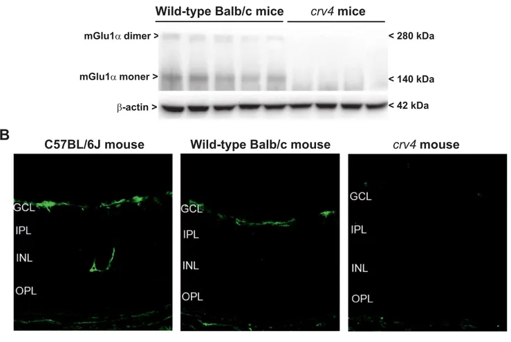
Pharmacological blockade of mGlu 1 metabotropic glutamate receptors is protective against excitotoxic retinal degeneration……… 60
Concluding remarks………...81
References………..84
Abstract
The group I of metabotropic glutamate (mGlu) receptor, mGlu1 and mGlu5 receptors are involved in neurodegenerative and in neuro-inflammatory processes. We have examined the role of mGlu5 receptor in a neuroinflammatory context. This receptor is expressed in glial cells and has been reported to play an anti-inflammatory role but also a pro-inflammatory effect. We were particularly involved in establishing whether mGlu5 receptor could modulate the inflammatory response in microglia, more specifically through the release of microvesicles (MVs). These have been shown to represent a more recent mode of intercellular communication and are extracellular vesicles, with a diameter of 100-1000 nm, that bud from the plasma membrane of cell of origin. MVs have been described in microglia where their formation occurs following activation of P2X7 purinergic receptors. We observed MVs development in pure microglial cultures, but, in the present study, they were more easily examined in the murine BV2 cell line. Activation of mGlu5 receptor attenuated the levels of pro-inflammatory cytokines, such as tumor necrosis factor (TNF) α, an effect that occurred only under conditions of activated microglia. Stimulation of mGlu5 increased also P2X7 receptor activation as shown by increased purinergic currents and by enhanced formation of MVs in response to the stable ATP analog, benzoyl-ATP. When MVs from mGlu5 receptor-stimulated microglia were transferred onto neurons, they increased neuronal toxicity induced by rotenone. Among possible factors released by MVs, in preliminary studies, we have identified miRNA146a as a potential mediator of this effect.
At the same time, the other member of group I mGlu receptor, mGlu1 receptor, was mainly analyzed considering its involvement in neurodegeneration. Specifically, we examined its role in an experimental setting of retinal ganglion cells (RGC) degeneration, a well-established model of several eye disorders including prematurity-induced retinal degeneration, glaucoma, and age-related macular degeneration. All these conditions are associated with excitotoxicity and it has been observed that activation of mGlu1 receptor significantly contributes to excitotoxic degeneration of RGCs. Thus, using postnatal day 9 wild type mice and knockout mice with a spontaneous recessive mutation
causing the lack of mGlu1 receptor protein (crv4 mice), we observed that mGlu1 receptor is primarily involved in RGC excitotoxic death induced by monosodium glutamate (MSG). Treatment with the mGlu1 receptor negative allosteric modulator, JNJ16258695, was in fact protective against retinal degeneration induced by MSG. In addition, MSG treatment did not produce any toxic effect in crv4 mice, lacking mGlu1 receptor.
Altogether these data confirm a role for mGlu1 and mGlu5 receptor in neurodegeneration and neuroinflammation, respectively and may serve as a starting point for the development of future possible pharmacological intervention targeting these receptors.
Introduction
Glutamate, the most abundant excitatory neurotransmitter in the central nervous system (CNS) acts through two distinct classes of receptors, ionotropic glutamate (iGlu) receptors and metabotropic glutamate (mGlu) receptors. iGlu receptors, namely N-methyl-D-aspartate (NMDA), α-amino-3-hydroxy-5-methyl-4-isoxazolepropionic acid (AMPA) and kainate receptors, are tetrameric ligand-gated ion channels and mediate fast responses to glutamate whereas mGlu receptors are G-protein coupled receptors (GPCRs) (Seeburg 1993; Pin and Duvoisin 1995; De Blasi 2001). Acting though these receptors glutamate is responsible for governingmany processes in the brain such as fast and slow excitatory neurotransmission, control of basal neuronal activity and synaptic plasticity.
(Sladeczek et al., 1985; Nicoletti et al.,1986; De Blasi et al., 2001).
mGlu receptors have been traditionally associated with the regulation of synaptic transmission and recognized to play a modulatory role. This has been also due to their localization at pre- as well as post-synaptic levels and to their not central position at the synapse. In addition, several mGlu receptors are also expressed in glial cells, suggesting their potential, indirect participation to neuronal function. In the last several years, however, mGlu receptors have long been implicated in mechanisms of neurodegeneration and neuroprotection so that their involvement in several diseases, disorders and processes has been well recognized. These include pain, learning and memory, anxiety, epilepsy, drug addiction, Parkinson’ s disease, schizophrenia, hypoxic brain damage, excitotoxic neuronal death and efforts have been made to develop drugs targeting these receptors as efficacious treatment for several CNS disorders (Nicoletti et al., 2011; Caraci et al., 2012; Bruno et al 2017;).
mGlu receptors structure and classification
As mentioned, mGlu receptors are GPCRs so they consist of an extracellular N-terminal domain, seven transmembrane-spanning domains, and an intracellular C-terminus. From a structural point of view, mGlu receptors are composed by a large extracellular N-terminal domain, also named Venus Flytrap domain (VFD) which contains two lobes able to create a clam shell-like structure. N-terminal
domain is characterized by the orthosteric binding site for glutamate. The two lobes responsible for the shell-like structure in this domain tend to close when the linkage between glutamate and its binding site occurs in VFD (Kunishima et al., 2000; Kniazeff et al., 2004; Muto et al., 2007). At this level, all mGlu receptors constitutively can form stable dimers through a disulfide bond (Muto et al., 2007). Further, by the presence of a cysteine-rich domain, it is possible to form an association between the VFD and the seven transmembrane-spanning domain modulating the signal transduction from the GPCRs (Kniazeff et al., 2004; Doumazane et al., 2013; Vafabakhsh et al., 2015). Interestingly, the orthosteric binding site is characterized by a high level of conservation and this is the reason why mGlu receptors can be bound by more than one ligand (reviewed in: (O'Brien and Conn, 2015). Several mGlu receptor selective ligands have been developed acting either at orthosteric or at allosteric sites, thus providing drugs with a modulatory function. Hence, positive allosteric modulators (PAM), able to potentiate the response of an orthosteric ligand, and negative allosteric modulators (NAM), or non-competitive antagonists, which induce the inhibition or a reduction of the response of an orthosteric ligand are now available (Gregory and Conn, 2015)
Through high affinity binding of NAMs and X-ray crystallography, 3D structures of the seven transmembrane-domain of the mGlu1 (Wu et al., 2014) and mGlu5 (Doré et al., 2014) receptors, it was possible to observe in more detail the allosteric binding region in the seven transmembrane domain. This finding provides an interesting platform for the development of new mGlu receptor ligands with more specific pharmacological profile and efficiency (Harpsoe et al., 2015).
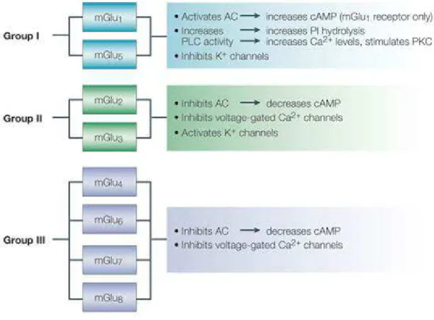
mGlu receptors form a family of eight subtypes (mGlu1 to -8), subdivided into three groups on the basis of amino acid sequence, pharmacological profile and transduction pathways (Table 1). They can be activated as individual, monomeric receptors but they also exist as dimers. In this case the formation of di-sulphide linkages between the N-terminal domains of the two individual receptors takes place (Romano et al.,1996).
Table 1. mGlu receptor family includes eight subtypes (mGlu1 to -8), subdivided into three groups
on the basis of amino acid sequence, pharmacological profile and transduction pathways (From Zhong et al., 2006; Nature Review).
Group I mGlu receptors
Group I includes mGlu1 and mGlu5 receptors, which are coupled to Gq/G11.These receptor subtypes are preferentially localized in the peripheral portion of dendritic spines (whereas all other mGlu receptor subtypes, with the exception of mGlu6 receptor, are preferentially localized in axon terminals where they negatively regulate glutamate release). In particular, high levels of mGlu5 receptor are found in forebrain regions such as the cerebral cortex, dorsal and ventral striatum (including the nucleus accumbens, olfactory bulb and tubercle, lateral septum, and hippocampus). (Romano et al., 1993). In contrast, expression levels of mGlu1 receptor in these areas are relatively low, with the exception of the hippocampus where it is widely expressed primarily in the CA3 region and dentate gyrus. The cerebellum, thalamus, hypothalamus, and pallidum represent regions in which it is
possible to observe high expression levels of mGlu1 and low levels of mGlu5 receptor expression. (Shigemoto et al., 1992).
Activation of the group I of mGlu receptors leads to the hydrolysis of phosphatidylinositol-4,5-bisphosphate (PtdIns-4,5-P2) with ensuing formation of inositol-1,4,5-trisphosphate (InsP3) and diacylglycerol (DAG). InsP3 determines the release of Ca2+ from intracellular stores whereas DAG is able to activate all isotypes of protein kinase C (PKC). The C-terminus domain of mGlu1 and mGlu5 receptors are associated, through homer proteins, to several membrane and intracellular proteins, such as signaling proteins (e.g., phospholipase-Cβ3, and the PtdIns-3-kinase enhancer, PIKE-L) and ion channels (e.g., NMDA receptors, InsP3 receptors, and TrpC6 channels) (Brakeman et al., 1997). PIKE-L links mGlu1 and mGlu5 receptors to the activation of the PtdIns-3-kinase pathway, which is able to promote neuronal survival by inhibiting glycogen synthase kinase-3β (GSK3β) (Brazil and Hemmings, 2001). Although mGlu1 and mGlu5 receptors are in some cases involved in the same signal transduction mechanisms, they can exert very different behaviors in the context of brain injury. In fact mGlu1 receptor agonists are able to induce an increase of necrotic cell death (Allen et al., 2000), whereas selective antagonists of the same receptors tend to exert a neuroprotective effect in vitro and in vivo (Faden et al., 2001; Fei et al., 2006; Szydlowska et al,. 2007). In contrast, stimulation of mGlu5 receptor has been shown to cause inhibition of caspase dependent neuronal apoptosis in cell culture models (Movsesyan et al., 2004). (S)-3,5-Dihydroxyphenylglycine (DHPG) is the first agonist which showed a specific selectivity for group I of mGlu receptors. It can be considered a potent agonist for both mGlu1 and mGlu5 receptors (Wiśniewski et al., 2002). In particular, DHPG exerts its agonist activity exclusively in the S-isomer. Some studies report that, under certain conditions, DHPG may also create an interaction with NMDA receptors. DHPG promotes PtdIns-4,5-P2 hydrolysis in a dose-dependent manner either in adult and neonate hippocampus, decreases levels of cAMP in the adult and enhances it in the neonate. DHPG can induce an increase of [Ca2+]i regulating multiple subtypes of Ca2+ channels and, being involved in a series of mechanisms, it can regulate the release of neurotransmitters. In
particular, based on the concentrations, DHPG can amplify or attenuate excitatory postsynaptic potentials (EPSPs) and it may induce long-term depression (LTD) and long-term potentiation (LTP) (Bear et al., 1996).
(RS)-2-chloro-5-hydroxyphenylglycine (CHPG), an analog of DHPG is considered a selective agonist of mGlu5 receptor. The use of PAM allows receptor activation in a more physiological way determining less receptor desensitization and/or tolerance than classical agonists. PAMs for mGlu 1 receptor were developed (Knoflach et al., 2001; Wichmann et al., 2002): S)-2-(4-fluoro-phenyl)-1-(toluene-4-sulfonyl)-pyrrolidine (Ro 67-7476), diphenylacetyl-carbamic acid ethyl ester (Ro 01-6128) and (9H-xanthene-9-carbonyl)-carbamic acid butyl ester (Ro 67-4853). Specifically these compounds do not cause an intrinsic agonist activity but significantly promote an agonist-induced response (Knoflach et al., 2001). The first positive allosteric modulators studied for mGlu5 receptor are 3,3′-difluorobenzaldazine (DFB) (O'Brien et al; 2003) and N-{4-chloro-2-[(1,3-dioxo-1,3-dihydro-2H-isoindol-2-yl)methyl]phenyl}-2-hydroxybenzamide (CPPHA) (O'Brien et al., 2004). DFB, as well as CPPHA, do not exhibit an intrinsic agonist activity but are able to amplify agonist
responses. 7-Hydroxyiminocyclopropan[b]chromen-1a-carboxylic acid ethyl ester (CPCCOEt) can
be defined as the prototypic non-competitive mGlu1 receptor antagonist (Ott et al., 2000). In particular, it acts selectively on mGlu1 receptor by inhibiting receptor activation non-competitively from a binding pocket into the transmembrane heptahelical domain. Since the discovery of CPCCOEt, (Hermans et al., 1998; Litschig et al., 1999), various selective NAMs for mGlu1 receptor were synthesized including (3aS,6aS)-6a-Naphtalen-2-ylmethyl-5-methyliden-hexahydro-cyclopental[c]furan-1-on (BAY36-7620), 1-(3,4-dihydro-2H-pyrano[2,3-b]quinolin-7-yl)-2-phenyl-1-ethanone (R214127) (Lavreysen et al., 2003), 1-ethyl-2-methyl-6-oxo-4-(1,2,4,5-tetrahydro-benzo[d]azepin-3-yl)-1,6-dihydro-pyrimidine-5-carbonitrile (EM-TBPC) (Malherbe et al., 2003a). Regarding negative modulators for mGlu5 receptor, 6-Methyl-2-(phenylazo)-3-pyridinol (SIB-1757) and (E)-2-methyl-6-(2-phenylethenyl)pyridine (SIB-1893) can be considered (Varney et al., 1999). These led to the identification of 2-methyl-6-(phenylethynyl)-pyridine (MPEP), which can be
considered a more potent ligand (Gasparini et al., 1999) and 3-[2-methyl-1,3-thiazol-4-yl)ethynyl]pyridine (MTEP) which shows a better selectivity and CNS bioavailability (Cosford et al., 2003b).
Group II mGlu receptors
Group II includes mGlu2 and mGlu3 receptors which are coupled to Gi/Go proteins. Their activation reduces intracellular cAMP formation and inhibits voltage-sensitive calcium channels. mGlu2 and mGlu3 receptors exhibit high homology and are predominantly expressed as heterodimers on presynaptic membranes of glutamatergic terminals where they negatively regulate the release of glutamate. However, localization of mGlu2 and -3 receptors in peripheral regions of the synapse has also been reported. Since these receptors are predominantly found as heterodimers, they are often indicated as mGlu2/3 receptors, although homodimers of each receptor subtype have been identified (Schoepp et al., 2001; Tamaru et al., 2001). mGlu2 and mGlu3 receptors are found in moderate to high abundance in the olfactory bulb, cerebral cortex, septal region, hippocampus, dorsal and ventral striatum, amygdala, and cerebellum (Ohishi et al., 1993). Several agonists for mGlu2/3 receptors have been developed including (2S,2′R,3′R)-2-(2′,3′-dicarboxycyclopropyl)glycine (DCGIV); l-carboxycyclopropyl-glycine (L-CCG-I); (2R,4R)-4-aminopyrrolidine-2,4-dicarboxylate (2R,4R-APDC); LY379268 and LY566332. Through some preliminary studies it has been discovered that Ro 67-6221 and Ro 71-8218 may exert an antagonist activity also for the group II of mGlu receptors. Moreover LY341495 and MGS 0039 are drugs that act as a selective antagonist blocking either mGlu2 and mGlu3 receptors. A series of mGlu2 receptor PAMs have been described (Johnson et al., 2003) including LY487379 which causes an amplification of glutamate potency (Schaffhauser et al., 2003; Koike et al., 2011) whereas RO4491533 acts as a negative allosteric modulator of either mGlu2 and mGlu3 receptors (Campo et al., 2011).
Group III includes mGlu4, mGlu6, mGlu7 and mGlu8 receptors, which are also coupled to Gi/Go proteins. The expression levels of Group III receptors in the brain are more limited than those of group I and II mGlu receptors. mGlu6 receptor expression is confined to the retina whereas mGlu4 receptors have a limited distribution in the CNS, being mostly expressed on presynaptic terminals at high levels in the cerebellum, basal ganglia, thalamus, and hippocampus (Nakajima et al., 1993); Corti et al., 2002). In contrast, mGlu7 receptors are expressed much more widely throughout the CNS including the cerebral cortex, striatum, hippocampus, hypothalamus, and cerebellum, where they are predominantly located to presynaptic terminals. Finally, mGlu8 receptors have a restricted distribution in the brain, primarily being confined to olfactory bulb, piriform cortex, hippocampus and thalamus (Kinzie et al., 1995; Saugstad et al., 1997). L-AP4 is an orthosteric, highly selective agonist of group III mGlu receptors. Orthosteric antagonists have been developed that show high selectivity but low potency (Niswender, 2016). More interesting has been the development of PAMs and NAMs for these receptors. (−)-N-Phenyl-7-(hydroximino)cyclopropa[b]chromen-1a-carboxamide ((−)-PHCCC), an analogue of the mGluR1 antagonist CPCCOEt, has been described as a PAM of mGluR4 (Maj et al., 2003; Marino et al., 2003). The antagonists of mGlu5 receptor, SIB-1893 and MPEP, behave as positive allosteric modulator for mGlu4 receptor (Mathiesen et al., 2003). In contrast, some mGlu5 receptor PAMs including DFB and CPPHA exert an antagonistic activity for mGlu4 and mGlu8 receptors (O'Brien et al., 2003, 2004) but it is not yet clear the mechanism through which a competitive or non-competitive interaction is possible. Finally, VU0155041 and VU0080421 have been identified as structurally distinct mGluR4 PAMs (reviewed in Niswender and Conn, 2010).
Role of mGlu1 and mGlu5 receptors in neuronal damage
As mentioned above, both mGlu1 and mGlu5 receptors are found in the extrasynaptic portion of dendritic spines. In this region they can link NMDA receptors via a chain of scaffolding proteins which include the long isoforms of Homer, Shank, and PSD95 (Tu et al., 1999). Specifically, mGlu5
receptors support the activation of NMDA receptors by relieving the Mg2+ blockade of the NMDA-gated ion channel or through other mechanisms (Doherty et al., 1997; Attucci et al., 2001; Mannaioni et al., 2001; Pisani et al., 2001). In this case it is legitimate to believe that pharmacological stimulation of mGlu1 and mGlu5 receptors determines neuronal cell damage by promoting the activation of NMDA receptors. However, the biological effects of these receptors are more complex. For this reason, the specific role of mGlu1 and mGlu5 receptors in mechanisms of neurodegeneration and neuroprotection has long been debated (Nicoletti et al., 1999; Bruno et al., 2001). Activation of mGlu1/5 receptors with DHPG or other orthosteric agonists may either increase or diminish excitotoxic neuronal death depending on several factors such as in vitro or in vivo models of neurodegeneration, nature of the toxic insult and functional state of the two receptors (reviewed by Nicoletti et al., 1999). For example, in mixed cortical cultures (one of the most widely used in vitro models for the study of excitotoxicity) DHPG induces an amplification of NMDA toxicity when applied only once (either before or during the NMDA pulse), but it is also able to exert a neuroprotective effect when applied for the second time after a brief pre-exposure; this situation may be due to an activity-dependent switch in receptor signaling from the stimulation of PtdIns-4,5-P2 hydrolysis to the inhibition of voltage-sensitive Ca2+ channels (Herrero et al., 1998; Bruno et al., 2001). This dual role of group-I mGlu receptors has been confirmed in several studies. Thus, it has been shown that mGlu1 receptors can both stimulate intracellular Ca2+ release and activate the PI3K pathway only if the C-terminus domain is intact. A strong increase in intracellular Ca2+ levels, for example in the presence of a toxic stimulation of NMDA receptors, can in fact lead to a calpain-mediated cleavage of the C-terminal portion of the mGlu1 receptor, hindering the receptor from activating the PI3K pathway but, at the same time, allowing the stimulation of PtdIns-4,5-P2 hydrolysis. All this can provoke an imbalance in favor of the neurotoxic signal which results into neuronal death (Xu et al., 2007).
A series of studies have shown that mGlu1 receptors may also interact with membrane estrogen receptors (ERs) in hypothalamic neurons, and this interaction is critical for the regulation of the
estrous cycle (Micevych and Mermelstein, 2008; Mermelstein, 2009). It has been observed that the interaction between mGlu1 receptors and membrane ERs occurs also in cortical neurons, where the protective effect of 17β-estradiol against β-amyloid toxicity is prevented by pharmacological blockade of mGlu1 receptors, and the protective activity of DHPG is prevented by pharmacological blockade of ERα (Spampinato et al., 2012). In contrast, interaction between the two receptors mediates an exacerbation of neurotoxicity, when stimulation of one or the other occurs after the toxic insult, such as that induced by a high concentration of NMDA (Spampinato 2012b). Taken together, these findings suggest that the precise role of mGlu1 and mGlu5 receptors in mechanisms of neurodegeneration and neuroprotection is very complex and depends heavily on the specific context. However, unlike the dual effect of receptor agonists, mGlu1 and mGlu5 receptor antagonists or NAMs are more consistently neuroprotective independently of the context and the nature of the toxic insult. For example, in in vitro and in vivo studies it has been shown that selective mGlu1 receptor antagonists exert a neuroprotective role by stimulating GABA release (Battaglia et al., 2001; Cozzi et al., 2002). It was also possible to observe a substantial protective action of mGlu1 receptor antagonists against hypoxic and ischemic neuronal damage (De Vry et al., 2001; Cozzi et al., 2002; Meli et al., 2002; Moroni et al., 2002; Pellegrini-Giampietro, 2003; Makarewicz et al., 2006; Kohara et al., 2008; Murotomi et al., 2008, 2010). More recent studies show more specifically the mechanism through which mGlu1 receptor antagonists exert a protective effect by means of the release of GABA. Activation of mGlu1 receptors stimulates PtsIns-4,5-P2 hydrolysis with ensuing formation of DAG. This one is in turn converted by DAG lipase into the endocannabinoid, 2-arachidonylglycerol (2-AG), which diffuses back to presynaptic terminals and activates type-1 cannabinoid (CB1) receptors, thereby inhibiting GABA release. So, mGlu1 receptor blockade could attenuate the amount of 2-AG produced during the development of ischemic damage (reviewed by Landucci et al., 2009). mGlu1 and mGlu5 receptors have also been shown to be involved in mechanisms of ischemic tolerance. Indeed, in organotypic hippocampal slices subjected to a protocol for ‘‘ischemic preconditioning’ ’ , neuroprotection induced by a non-lethal 10-min pre-exposure to oxygen/glucose deprivation (OGD)
was canceled by treatment with mGlu1, but not mGlu5, receptor antagonists. DHPG pre-treatment exerted also a protective effect against neuronal damage induced by a subsequent lethal exposure to OGD (pharmacological pre-conditioning) (Werner et al., 2007). In contrast, when hippocampal slices were subjected to a protocol of ‘‘ischemic post-conditioning’ ’ , in which a brief (3 min) period of OGD was delivered 5 min after a lethal episode of OGD, neuroprotection was abrogated by both mGlu1 and mGlu5 receptor antagonists. Stimulation of mGlu1 and mGlu5 receptors in the protocol of ischemic post-conditioning leads to neuroprotection through the activation of PI3K/Akt/GSK3β pathway (Scartabelli et al., 2008). Pharmacological blockade of mGlu5 receptors could induce a reduction of neuronal death in models of chronic neurodegenerative disorders, such as Parkinson’ s disease (PD) and amyotrophic lateral sclerosis (ALS). Indeed, higher levels of mGlu5 receptor immunoreactivity have been observed in the putamen of PD patients, and in the frontal cortex, hippocampus and putamen of patients with dementia with Lewy bodies, a disorder that has in common with PD the presence of intracellular aggregates of α-synuclein (Price et al., 2010). Increased levels of mGlu5 receptors have been also found in α-synuclein transgenic mice. Moreover, in mice and monkeys subjected to parkinsonian neurotoxin, 1-methyl-4-phenyl-1,2,3,6-tetrahy-dropyridine (MPTP), systemic exposure to mGlu5-receptor NAMs (MPEP and MTEP, respectively) exert a protective activity in nigro-striatal neuron against degeneration (Battaglia et al., 2004; Masilamoni et al., 2011). Treatment with MTEP also promotes an attenuation of MPTP-induced damage of noradrenergic neurons in the locus coeruleus of monkeys (Masilamoni et al., 2011). Pharmacological blockade or genetic deletion of mGlu5 receptors has also a protective action against 6-hydroxydopamine lesions of the nigro-striatal dopaminergic pathway (Armentero et al., 2006; Black et al., 2010). On these bases, it has been suggested that treatment with mGlu5 receptor NAMs in PD patients is important to both attenuate motor symptoms and to reduce nigro-striatal degeneration.
mGlu receptors are not only expressed in neurons, but also in glial cells and to get a clearer view on the role of mGlu5 receptors in mechanisms of neurodegeneration and neuroprotection attention has
been paid to the cross-talk between neurons and astrocytes and the role of these receptors. Specifically, mGlu5 receptors are present in astrocytes and their expression in astrocyte cultures tend to increase when cells acquire a reactive-like phenotype. Interestingly, pharmacological blockade of mGlu5 receptors reduces excitotoxic death of spinal motor neuron grown in culture medium from enriched reactive astrocytes (D’ Antoni et al., 2011). Moreover, pharmacological blockade of these receptors delays the onset of motor symptoms in mice which express a mutated form of human superoxide dismutase, an animal model of familial ALS (Rossi et al., 2008).
Retina and neurodegenerative processes
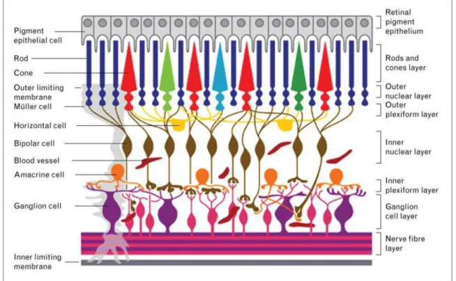
The retina can be defined as the sensory membrane of the eye which allows the vision through the reception of images by photoreceptors and that are then conveyed to the brain. The retina has a very complex organization, in fact it contains several neuronal cell types and interneurons (Hageman et al., 1991). It is characterized by three layers of nerve cell bodies (the outer nuclear layer composed by cell bodies of the rods and cones, the inner nuclear layer with cell bodies of the bipolar, horizontal and amacrine cells and the ganglion cell (RGC) layer that contains cell bodies of RGCs and displaced amacrine cells (Fig.1). Two layers of synapses are also present (Kolb et al., 1991). Neurodegenerative processes of retinal cells are at the basis of several eye diseases including glaucoma (Sathyamangalam et al., 2009), age-related macular degeneration (Shams Najafabadi et al., 2017), retinitis pigmentosa (Bennis et al., 2017), diabetic retinopathy (Jackson and Barber, 2010).
Fig. 1 Schematic representation of retinal organization: three layers of neural cells characterized by photoreceptor cells, bipolar cells, and ganglion cells, and a layer of pigmented epithelial cells (from Lorber et al., Curr Opin Ophthalmol 2016).
Within the retina mGlu1 and -5 receptors are present in the dendrites of ON-bipolar cells and in amacrine cells, but also in RGCs (Koulen et al., 1997; Brandstätter et al., 1998; Yang et al., 2004). However, stimulation of PtdIns-4,5-P2 hydrolysis in RGCs occurs only when mGlu1, but not mGlu5, receptor is activated (Romano et al., 2016). The increase of intracellular Ca2+ induced by mGlu1 receptor activation and its ability to modulate NMDA receptor function suggest a potential involvement of this receptor in excitotoxicity of RGCs.
Neurodegeneration and neuroinflammation
Growing evidence has clearly established that neurodegeneration is not merely a disease process that affects neurons, but that the surrounding environment plays a main role, taking an active part to neuronal fate with a complex event generally known as neuroinflammation. This may precede or accompany the neurodegenerative event and involves mainly glial cells but also peripheral cells of the immune system. Among glial cells, the most important role in neuroinflammation is played by microglia.
Microglia
Microglia are the immune cells of the CNS so their primary function is to maintain brain tissue homeostasis to provide the first line of defense during infection or brain injury and to promote tissue repair. Under normal conditions in the brain, microglia is generally named the ‘resting’ or ‘surveillant’ microglia (Nimmerjahn et al., 2005; Kettenmann et al., 2011). In this case microglial cells show a ramified morphology containing long and thin processes which allow the cells to communicate with surrounding neurons and other glial cells and continuously control the microenvironment to exert a guard function for incoming pathogens and brain alterations. When microglial cells detect a brain insult they act as inflammatory effector cell types and respond to pathogens and injury by becoming “activated”. Specifically, in response to many types of alarm signals (cytokines, material from apoptotic cells and exogenous viral factors) surveillant microglia rapidly undergo a morphological change assuming an ameboid shape, proliferate and migrate to the site of infection or injury in which they phagocyte and destroy pathogens as well as remove damaged cells and protein aggregates, and provide the secretion of cytokines, chemokines, prostaglandins, nitric oxide and reactive oxygen species, which help to increase and govern the immune response. However, microglia are also involved in the resolution of the inflammatory response through the production of anti-inflammatory cytokines such as interleukin (IL)-10. Thus, depending upon the nature of stimuli, the entity of activation and the duration of environmental signals,
In the latter case, the distinction between the M1 and M2 phenotype is not so clear. TBI determines in fact a wide range of responses both at pro-inflammatory and anti-inflammatory level so that after TBI a concomitant expression of pro- and anti-inflammatory genes can be observed. Interestingly, this microglial oscillatory profile between M1 and M2, has also been observed in LPS-stimulated conditions and ALS models of neuro-inflammation and spinal cord injury (Chiu et al., 2013). Therefore, microglia as well as macrophages do not exclusively assume an M1 or M2 phenotype but display a mixed phenotype likely due to the complex signaling events surrounding them (Morganti et al., 2016).
mGlu receptors in neuroinflammation
mGlu receptors are expressed in glial cells and a role for these receptors in neuroinflammation has also been considered. Accordingly, mGlu receptors have been identified as potential targets for the treatment of MS, the prototype of CNS disease with a prominent neuroinflammatory component. An overexpression of different subtypes of mGluRs in the brain of MS patients has been observed. mGlu1 receptors show in fact an enhanced axonal immunoreactivity and are expressed also in reactive microglia and astrocytes. Similarly, mGlu2/3 receptors are markedly overexpressed in astrocytes in acute and chronic active MS lesions and mGlu5 receptor expression is enhanced in reactive astrocytes. mGlu4 receptor also is overexpressed at the rim of chronic active lesions and a role for this receptor subtype has been characterized in detail using an animal model of multiple sclerosis, the experimental autoimmune encephalomyelitis (EAE) model, obtained in mice after immunization with the oligodendrocyte protein, Myelin Oligodendrocyte Glycoprotein (MOG)-35-55. The absence of the mGlu4 receptor enhances the severity of EAE, whereas treatment with a selective mGlu4 receptor enhancer is protective against EAE. This activity has been ascribed to a peripheral role of mGlu4 receptors on cells of the immune system, dendritic cells (DCs) and T lymphocytes. The activation of mGlu4 receptors is in fact able to modify the differentiation of naive
T cells towards the immune tolerant Treg cells, with a concomitant reduction of autoreactive TH17 cells. Interestingly, other mGlu receptors are expressed in cells of the immune system and may play a regulatory role as well. The protective effect mediated by mGlu4 receptor, however, are not exclusively due to immune cells as mGlu4 receptors are also expressed in glial cells and their activation protects oligodendrocytes against excitotoxicity (Spampinato et al., 2015).
Looking more specifically to group I mGlu receptors, it has been reported that mGlu5 receptor stimulation with the selective agonist, CHPG, causes a reduction of microglial activation and a decrease of associated pro-inflammatory factors release in vitro, which is mediated in part through inhibition of reduced nicotinamide adenine dinucleotide phosphate (NADPH) oxidase. These effects of delayed CHPG administration were associated with reduction of chronic neuroinflammation and ensuing neurodegeneration after experimental traumatic brain injury in mice. (Byrnes et al., 2009, 2009b).
In microglial cells, the selective mGlu5 selective agonist, CHPG, has been reported to reduce the production TNFα induced by LPS. On the other hand, a correlation between LPS and mGlu5 receptor has been suggested. LPS in microglial cells can in fact induce [Ca2+] oscillations in a Toll-like receptor 4 (TLR4)-independent manner, an effect sensitive to blockade by the mGlu5 receptor antagonist MTEP. These observations, together with immunocytochemistry showing that LPS co-localizes with mGlu5 receptor, suggest that mGlu5 receptor may mediate a proinflammatory action in microglia and that it may represent a novel target for modulating microglia-dependent neuroinflammation. ( Byrnes et al., 2012; Wang et al., 2012, 2013; Liu et al., 2014).
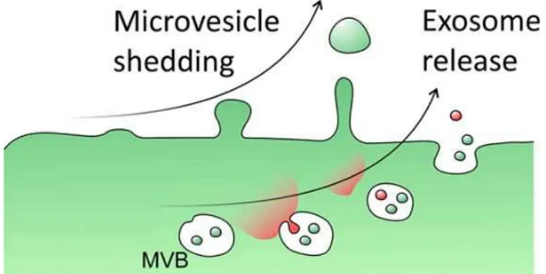
Cell communication through extracellular vesicles
Brain functions depend on coordinated interactions between neurons and glial cells that include microglia, astrocytes, and oligodendrocytes. These cells, either in normal or pathological condition, are able to release extracellular vesicles (EVs) (Chivet et al., 2014) which represent an important mode of intercellular communication. In fact they may represent a vehicle for transfer and delivery
of membrane and cytosolic proteins, lipids, mRNAs, and microRNAs (miRNAs) between cells (Lin et al., 2015). Formation of EVs is not restricted to CNS as they can be released by several cell types and are present in many biological fluids including blood, urine, saliva, amniotic fluid, breast milk and medium of cell cultures (Théry et al., 2002, 2006). EVs include exosomes and microvesicles (Fig 3).
Fig. 3. Differences in the formation of microvesicles and exosomes.
Exosomes
Exosomes are endocytic membrane-derived vesicles with a diameter between 30–100 nm. These small vesicles are contained in multivesicular bodies (MVBs) within the endosomal system and then they are secreted through MVB fusion with the plasma membrane (Prada et al., 2013). Although MVB fusion with the plasma membrane has not been described in detail yet, this process seems to involve several Rab GTPases, such as iRab5, Rab27, and Rab35 (Shifrin et al., 2013; Yuyama et al., 2012; Mittelbrunn et al., 2011). Exosomes are associated with a lot of cellular components among which messenger RNAs (mRNAs), microRNAs (miRNAs) and proteins. Exosomes derived from microglial cells express specific markers including major histocompatibility complexes (MHCs) class
II molecules, enzymes (cathepsin S) and integrins, which are involved in antigen presentation and pattern recognition receptors during innate immune response. Moreover exosomes are packed with tetraspanins, a family of transmembrane proteins including CD9, CD14, CD63 and CD81, which are particularly enriched in exosomes derived from microglia. Further, in microglia-derived exosomes, there is a high expression of lactate monocarboxylate transporter 1MCT-1 and CD13 (the surface-bound aminopeptidase which degrades enkephalins). Extrusion of Na, K-ATPase is also frequent in exosomes derived from several populations of cells. Stressful events in astrocytes, like heat or oxidative stress, were shown to promote the release of exosomes carrying the heat shock protein 70 (Hsp70) and synapsin 1 with ensuing pro-survival effect on neurons (Taylor et al., 2007). In addition exosomes may contain a distinct set of proteins, such as cytoskeletal proteins and the glycolytic protein glyceraldehyde 3-phosphate dehydrogenase (GAPDH).
Microvesicles
Microvesicles (MVs) are vesicles larger than exosomes (100–1000 nm) which bud directly from the plasma membrane of donor cells (Turola et al., 2012). Shedding of MVs usually involves a budding process in which surface blebs selectively accumulate cellular constituents that are packaged into MVs (Thery et al., 2009). These vesicles present a lot of cell surface receptors, intracellular signaling proteins and genetic materials derived from the cell of origin. Because MVs originate from different cell types, they are characterized by a different molecular profile which in turn represents the mirror of the differential proteins expression of various cells of origin. The heterogeneity of composition and biological activity of MVs is dependent on the state (resting or stimulated) of donor cells and on the agent employed for stimulation (Bernimoulin et al.,2009).
Blebbing of MVs can be induced by purinergic P2X7 receptor activation. Indeed a specialized type of MVs shedding has been found in cells which typically express the ATP receptor P2X7. P2X7 receptor is an ATP-gated ion channel highly expressed in immune cells. It is characterized by a long cytoplasmic C-terminus in which there are a lot of protein-protein interaction motifs. Dramatic
morphological changes occur in cells expressing P2X7 receptor during and subsequent to receptor stimulation. In particular these changes consist in rapid formation of membrane blebs associated to several intracellular signaling events such as the activation of protein kinases and other effector enzymes (Duan and Neary, 2006). In particular P2X7-induced blebbing is dependent upon p38 and ROCK activation. This process leads to local disassembly of cytoskeletal elements associated with the P2X7 C-terminus (Budagian et al.,2003; Morelli et al., 2003; Verhoef et al., 2003). Surface blebbing is preceded by loss of plasma membrane symmetry and exposure of phosphatidylserine (PS) at the outer leaflet of the plasma membrane, a process that occurs within the first few minutes of P2X7 receptor activation and is reversible (Hugel et al., 2005). Acidic sphingomyelinase (A-SMase) is also involved in MVs biogenesis. This enzyme hydrolizes sphingomyelin (SM) to the sphingolipid ceramide. After P2X7 stimulation, p38MAP kinase activation determines translocation of A-SMASE to the plasma membrane outer leaflet in which ceramide is formed. MVs budding can be caused by reorganization of ceramide in the bilayer and by local accumulation of the cone-shaped sphingolipid into the inner leaflet of the membrane. Thanks to its natural negative curvature, ceramide can promote the formation of subdomains with curvature different from the adjacent planar membrane. Further, hydrolysis of SM, which is known to present a considerable affinity for cholesterol could determine an increase of the efflux of cholesterol. This is considered the main factor implicated in membrane fluidity and structural integrity of the plasma membrane. This condition can contribute to membrane destabilization and promote the process of blebbing and MVs secretion (Bianco et al., 2009).
The expression of P2X7 receptor is evident in macrophages (Steinberg et al., 1987), mast cells (Cockcroft and Gomperts, 1979) and microglia (Visentin and Levi, 1997), where it controls the release of inflammatory cytokines, including IL-1β and IL-18 (Ferrari et al., 2006). This is accompanied by assembly of inflammasome which represents the protein complex able to activate IL-1β processing enzyme, caspase 1. In this way a rapid cytokine secretion occurs (Bianco et al.,2009). MVs blebbing occurs in reactive microglia (Fig. 4), for example following exposure to
LPS, and cytokines can be released into the environment in a ATP-dependent manner (Bianco et al., 2005).
Fig. 4. MVs shed from cultured primary microglia from rat cerebral cortex as shown by labelling with the fluorescent dye FM1-43 (Beneventano et al., 2017).
However, sustained P2X7 receptor activation can also be responsible for cell death. In contrast, several studies have shown that shedding of MVs from microglia, is not only induced by stimulation of P2X7 receptor through exogenous ATP but also by ATP endogenously released from healthy astrocytes in astrocyte-microglia co-cultures. Hence, although ATP is generally considered an alarm signal, these studies suggest that MVs can be released from microglia even in normal conditions, in the absence of cellular damage.
MVs as containers of bioactive molecules modulating cell functionality
One of the reasons for increasing interest on MVs is because the cells have several advantages when release cytokines through MVs rather than exporting these molecules directly. For example, secreted MVs containing IL-1β, can deliver the cytokine at considerable distance from the cell of origin, thus avoiding dispersal, degradation and dilution of the cytokine in the extracellular space. In addition, several different factors can be contained in MVs, so that biogenesis of MVs represent an important mechanism to mediate the assembly of multiple factors regulating biological responses.
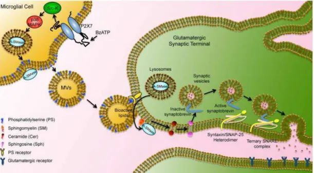
Microglia-released MVs influence brain cell functions, by propagating inflammation and causing neurodegeneration (Fig. 5).
Fig. 5. Schematic representation of microglial MVs interaction with neurons: blebbing of MVs induced by P2X7 stimulation by BzATP, preceded by loss of plasma membrane symmetry and exposure of PS at the outer leaflet of the plasma membrane; hydrolysis of SM to ceramide by ASMase which is translocated to plasma membrane outer leaflet after p38 MAPK activation; released MVs can then interact with target neuronal cell (from Turola et al., Front Physiol. 2012).
It was observed that MVs from reactive microglia induce an inflammatory reaction in target cells (Verderio et al., 2012a) and cause an immune-stimulatory activity in recipient microglial cells that determines an up-regulation of CD86 and expression of inflammatory genes (Verderio et al., 2012). Indeed, in several pathological conditions, a role for microglia-derived MVs has been recognized. For example, microglial MVs participate in the progression of AD by inducing the production of soluble amyloid β species and the propagation of such toxic forms (Joshi et al., 2014). In addition it has been observed that MVs derived from microglial cells are present in cerebrospinal fluids of rodents and interestingly, their concentration tend to increase in the course of EAE (Verderio et al., 2012). Accordingly, injection of MVs into the brain of mice with subclinical EAE promotes recruitment of inflammatory cells at the site of delivery whereas a reduction of MVs formation, as occurs in A-SMase knock out mice, protects against EAE (Verderio et al., 2012). In addition, microglia-released MVs enhance excitatory neurotransmission. In fact, MVs shed from microglia,
are able to interact with the plasma membrane of neurons evoking and increasing excitatory transmission. Interestingly, it was found that MVs modulate neurotransmission by inducing sphingolipid metabolism in neurons (Antonucci et al., 2012).
MicroRNA
MicroRNAs (miRNAs) are a class of small noncoding RNAs (containing about 22 nucleotides) which represent key post-transcriptional regulators of gene expression, hindering the translation of their target mRNA or promoting their degradation. Besides, they may also play a role as translation activators or impair transcription by binding to gene promoters (Faravelli and Corti, 2017). The majority of miRNAs are transcribed as pri-miRNAs by RNA polymerases II and III in the nucleus and cut into pre-miRNAs by the Drosha complex. The last stage of the process involves exportin 5 and Ran GTPase which transport pre-miRNAs to the cytoplasm where they undergo final processing by the RNAse III enzyme Dicer. In this way the formation of 22 nt double-stranded mature miRNAs with a 5’ phosphate end takes place. The mature miRNA is then incorporated into the RNA-induced silencing complex (RISC), which become able to repress translation. Although most of miRNAs are localized within the cell, it has been observed that some miRNAs, commonly termed extracellular miRNAs or circulating miRNAs, may occupy extracellular environment, comprising biological fluids and cell culture media.
miRNAs can modulate the development and function of the nervous system (Karthikeyan et al., 2016). Neural miRNAs are implicated in several steps of synaptic development, such as dendritogenesis (involving miR-132, miR-134 and miR-124), synapse generation and synapse maturation (miR-134 and miR-138 seem to be related to these events). Moreover it has been found that miRNA expression is modified in schizophrenia, as well as in bipolar disorder and major depression and anxiety disorders. Recently, some studies have led to the consideration of a link between miRNAs and inflammation. Recently it has been found that miR-92a is down-regulated after
activation of TLRs, and exerts a significant role in inflammatory contexts being essential for the production of cytokines in pro-inflammatory macrophages (M1 phenotype). miR-124 is implicated in the secretion of cytokines from reactive pro-inflammatory effector microglial cells (Ponomarev et al., 2011). In addition, genes associated to reactive microglial cells, among which genes encoding proteins related to TGF-β pathway (SMAD2), may represent targets of miR-155 (Louafi et al., 2010). In contrast, miR-Let7, under inflammatory conditions, can play an important role in microglial cell’ s functions being responsible of anti-inflammatory effects through repression of genes targeting downstream signaling pathways.
MiR-146a is widely expressed in different species and tissues, and is involved in immunity, inflammation and viral infections by regulating different target genes (Li et al., 2010).
When implicated in the regulation of inflammation and other processes that involve the innate immune system, miR-146, acts as a mediator of inflammation along with miR-155. Indeed the expression of miR-146 is upregulated by inflammatory factors such as IL-1) and TNF-α (Sheedy, and O'Neill, 2008). miR-146 dysregulates a number of targets which are mostly involved in toll-like receptor pathways that bring a cytokine response (Sonkoly et al., 2008). Accordingly, miR-146a appears to be preferentially expressed in cells of microglial lineage. A great up-regulation of miR-146a was observed in immortalized murine microglial cell line, BV2 cells, following TLR2 or TLR4 activation. It has also been suggested that miRNA146a can be involved in neurodegenerative disorders such as AD. In this pathological condition, phagocytosis of β–amyloid (Aβ) peptide by activated microglia is significantly increased in the presence of ligands that activate TLRs. In particular, TLR4 stimulation of BV2 cells resulted in activation and in the transcription of miR-146a. Then miR-146a may represent a potent modulator of microglial function by regulating the activation state of microglial cell during neurodegeneration (Saba et al., 2012). Accordingly, miRNA146a is upregulated in AD patients (Zhang et al., 2016) and in this condition, its targets include the low-density lipoprotein receptor-related protein-2 (Lrp2), a member of the LDLR family which exerts a
protective role in AD (Marzolo and Farfan, 2011). Overexpression of miRNA-146a in neuroblastoma SH-SY5Y cells induced in fact a significant reduction of Lrp2 expression, with ensuing decrease of Akt activation and induction of pro-apoptotic caspase-3, thereby causing cell apoptosis.
Another target of miR-146a is superoxide dismutase 2 (SOD2) which represents an endogenous antioxidant enzyme able to exert a protective role against reactive oxygen species (ROS) (Valko et al., 2006). Through interaction with SOD2 mRNA, miR-146a is involved in the post-transcriptional regulation of SOD2 expression (Pogue et al., 2011). Hence, miR-146a is up-regulated by H2O2-induced stress and down-regulates SOD2 protein expression whereas an antisense-miR-146a reverses the decrease of both the SOD2 level and cell viability in H2O2 treated PC12 cells (Ji et al., 2013).
Main objectives of the research project
My research has focused on the role of group I mGlu receptors, namely mGlu1 and mGlu5 receptors, in neurodegeneration and in neuroinflammation. The research has been carried out following two independent lines.
The major part of the work has been centered on the role of mGlu5 receptor in neuroinflammation, based mainly on the discrepancy existing in literature regarding a potential anti-inflammatory activity, but also a possible pro-inflammatory effect, mediated by this receptor subtype. Using microglia cells in culture, my aim was then to establish whether mGlu5 receptor activation in microglia could modify the neuroinflammatory process and mainly to establish whether this effect could involve MVs that are recognized as important novel mediators of cell-to-cell and also glia-to-neuron communication. The other part of the study has been centered on the role of mGlu1 receptor in neurodegeneration. In particular, we focused on neurodegenerative processes occurring during eye disorders and affecting RGC such as prematurity-induced retinal degeneration, glaucoma, and age-related macular degeneration. The main goal of the study was to establish the real role of mGlu1 receptor in excitotoxicity that is responsible for RGC death. For this purpose we used wild type and crv4 mice, that, because of spontaneous recessive mutation of the Grm1 gene, do not express mGlu1 receptors.
Front. Pharmacol., 09 November 2017 https://doi.org/10.3389/fphar.2017.00812
Shedding of microvesicles from microglia contributes to the effects induced by metabotropic glutamate receptor 5 activation on neuronal death
Martina Beneventano§1#, Simona Federica Spampinato§1, Sara Merlo1, Mariangela Chisari1, Paola Platania1, Marco Ragusa2, Michele Purrello2, Ferdinando Nicoletti3,4, Maria Angela Sortino1
Department of Biomedical and Biotechnological Sciences, section of 1Pharmacology and section of 2Biology and Genetics, University of Catania; 3Department of Physiology and Pharmacology, Sapienza University, Rome, 4I.R.C.C.S. Neuromed, Pozzilli, Italy
§these authors equally contributed to this work
# attending the PhD program in Neuroscience, University of Catania
Running title: mGlu5 receptor activation increases microglial microvesicles
Keywords: microglia, miRNA, extracellular vesicle; mGlu5; neuroinflammation; CHPG
Funding:
This work was supported by personal grants to MAS (University of Catania cod. 20130143075).
Address all correspondence to Maria Angela Sortino
Department of Biomedical and Biotechnological Sciences University of Catania
Via Santa Sofia 97, 95123 Catania, Italy [email protected]
Abstract
Metabotropic glutamate (mGlu) receptor 5 is involved in neuroinflammation and has been shown to mediate reduced inflammation and neurotoxicity and to modify microglia polarization. On the other hand, blockade of mGlu5 receptor results in inhibition of microglia activation. To dissect this controversy, we investigated whether microvesicles (MVs) released from microglia BV2 cells could contribute to the communication between microglia and neurons and whether this interaction was modulated by mGlu5 receptor. Activation of purinergic ionotropic P2X7 receptor with the stable ATP analog benzoyl-ATP (100 µM) caused rapid MVs shedding from BV2 cells. Ionic currents through P2X7 receptor increased in BV2 cells pretreated for 24 h with the mGlu5 receptor agonist CHPG (200 µM) as by patch-clamp recording. This increase was blunted when microglia cells were activated by exposure to LPS (0.1 µg/ml for 6 h). Accordingly, a greater amount of MVs formed after CHPG treatment, an effect prevented by the mGlu5 receptor antagonist MTEP (100 µM), as measured by expression of flotillin, a membrane protein enriched in MVs. Transferred MVs were internalized by SH-SY5Y neurons where they did not modify neuronal death induced by a low concentration of rotenone (0.1 µM for 24 h), but significantly increased rotenone neurotoxicity when shed from CHPG-treated BV2 cells. miR146a was increased in CHPG-treated MVs, an effect concealed in MVs from LPS-activated BV2 cells that showed per se an increase in miRNA146a levels. The present data support a role for microglia-shed MVs in mGlu5-mediated modulation of neuronal death and identify miRNAs as potential critical mediators of this interaction.
Introduction
Metabotropic glutamate receptors (mGlu) receptors are G protein coupled receptors involved in a variety of functions and processes at the central nervous system (CNS) including synaptic plasticity, neurodevelopment, neuronal excitability, pain, addiction, neurodegeneration, neuroinflammation (Conn & Pin, 1997; Nicoletti et al., 2011). Among the three classes of mGlu receptor subtypes, group 1 mGlu, which includes mGlu1 and mGlu 5 receptors, is highly expressed throughout the brain (Martin et al., 1992; Romano et al., 1995) and mGlu5 receptor shows high density also in the spinal cord (Berthele et al., 1999). They are both Gq-coupled receptors, thus their activation causes increase of intracellular Ca2+ and predominate at post-synaptic level where they participate to the modulation of synaptic excitability (Shigemoto et al., 1993; Lujan et al., 1996; Biber et al., 1999). Although initial studies made questionable the function of mGlu5 receptor with a dual role, either neuroprotective or neurotoxic, depending on neuronal type and experimental conditions, it is now clearer that mGlu5 receptor interacts with NMDA receptors and likely exerts a permissive role in NMDA excitotoxicity. Hence, several antagonistic modulators of mGlu5 receptor activity exhibit neuroprotective properties whereas mGlu5 receptor agonists are neurotoxic (Parmentier-Batteur et al., 2014). Among others, mGlu5 receptors are not only expressed in neurons, but also in glial cells (Biber et al., 1999). Interestingly, in the cortex, expression of this receptor subtype is even higher in astrocytes than in neurons (Zhang et al., 2014) and appears upregulated in reactive astrocytes (Aronica et al., 2000; Ferraguti et al., 2001; Dolan et al., 2003., Ribeiro et al., 2017). Cultured microglia also express mGlu5 receptor, and its activation has been reported to be associated with reduced inflammation and neurotoxicity (Byrnes et al., 2009) as well as changes in microglia polarization (Loane et al., 2014). However, again, anti-inflammatory effects secondary to activation of mGlu5 receptor are rather controversial as opposing results with reduction of microglia activation by blockade of group 1 mGlu receptors have also been reported (Hsieh et al., 2012). Surprisingly, mGlu5 receptor has also been suggested to act as an alternative binding site for lipopolysaccharide (LPS), thus mediating activation of microglia toward an inflammatory phenotype (Liu et al., 2014).
Microglia affects neuronal function by the release of several factors, but in more recent years an additional way of intercellular communication has been described in several cell types, including glial cells. Extracellular vesicles, either as microvesicles (MVs) or exosomes that differ in size, content and origin, form under basal conditions, or as in the case of microglia, upon stimulation of the purinergic P2X7 receptor. In particular, microglial-derived MVs, ranging from 100 to 1000 nm in diameter, are shed from the extracellular membrane and play as a cargo for cytokines, RNA, miRNA, etc. to neighboring cells. Although the precise kinetics of this interaction are not completely known, hedding of MVs from microglia is a well-established event (Turola et al., 2012; Prada et al., 2013) reported to mediate neurotoxicity by transferring and propagation of pro-inflammatory cytokines (Bianco et al., 2005), or through modulation of β-amyloid aggregation status (Joshi et al., 2014). Even more, MVs can be detected and are increased in the cerebrospinal fluid of humans and rodents under conditions of brain inflammation, suggesting their role in the development of the disease, by spreading the inflammatory process (Verderio et al., 2012).
On these bases we decided to investigate whether MVs could take part to the effects elicited by mGlu5 receptor activation in microglia and whether this alternative means of interaction between cells could account for the controversial response observed following activation of this receptor.
Materials and methods
Drugs and reagents
Cell culture plastics were provided by BD Falcon (Milan, Italy). Media, media supplements, serum, trypsin and antibiotics, were from Invitrogen Srl (Milan, Italy). (RS)-2-chloro-5-hydroxyphenylglycine (CHPG) Sodium Salt, 3,5-di(RS)-2-chloro-5-hydroxyphenylglycine (DHPG), and 3-((2-Methyl-1,3-thiazol-4-yl)ethynyl)pyridine (MTEP), were from Tocris (North Point, UK). 2'(3')-O-(4-Benzoylbenzoyl)adenosine-5'-triphosphate tri(triethylammonium) salt (Bz-ATP), lipopolysaccharide
(LPS), rotenone were from Sigma-Aldrich (St. Louis, MO, USA). The following primary antibodies were used: rabbit anti-metabotropic glutamate receptor 5 (1:500; Millipore, Billerica, MA, USA), rabbit anti-flotillin (1:500; Santa Cruz Biotechnology, CA, USA); goat anti-tumor necrosis factor (TNF) α, (1:100; Santa Cruz Biotechnology), FITC-conjugated mouse anti-TNFα, (1:10; Miltenyi Biotec, Macs, Berisc-Gladbach, Germany).
Cell cultures
Immortalized murine microglial BV2 cells were cultured at 37 °C, with 5% CO2, in Dulbecco’ s Modified Eagle’s medium (DMEM) supplemented with 5% fetal bovine serum (FBS), penicillin/streptomycin (100 U/ml-100 µg/ml). For the experiments cells were plated in 35 mm tissue culture dishes (200 k/ each). The human neuroblastoma SH-SY5Y cell line was cultured at 37 °C with 5% CO2, in DMEM F-12 supplemented with 10% FBS and penicillin/streptomycin (100 U/ml-100 µg/ml). For experiments, cells were plated in 35 mm culture dishes (300 k/each) or in 96 well plates (60 k/each). To achieve BV2 cells conditioned medium (CM), BV2 cells were exposed to either vehicle, CHPG (200 µM), LPS (0,1 µg/ml) or their combination, for 24 h, then washed and incubated with fresh medium for further 6 h. This particular paradigm was chosen in order to allow exposure to treatments for a reasonable time to induce effects before removal so that transferred CM did not contain any drug.
Shedding of microvesicles
BV2 cell cultures were treated with CHPG (200 µM, for 24 h), the selective mGlu5 receptor antagonist MTEP (100 μM added 30 min before CHPG), and with LPS (0,1 µg/ml, for 6 h). They were then stimulated with Bz-ATP 100 µM, 20 min at 37° C in Krebs-Ring-buffer (Sigma-Aldrich) and the supernatant was subjected to differential centrifugations at 4° C: 5 min at 300 x g to discard cells and debris (P1 pellet); 20 min at 1200 x g to obtain P2 vesicle fraction (P2); 60 min at 10 000 × g to obtain vesicle population (P3). The resulting pellets corresponding to microvesicles (P3; MVs)
was resuspended in 30 µl of protein solubilisation buffer MPER (Invitrogen srl) supplemented with protein inhibitors and loaded for gel separation by western blotting analysis. For this specific experiment, the total amount of protein from 8 x 35-mm dishes, resuspended in a small volume was then loaded into the gel in order to assess the whole flotillin, expression of MVs formation. Alternatively, MVs were processed for microRNA extraction or resuspended in DMEM/F12 medium and transferred on SH-SY5Y cells. In the latter case four 35-mm dishes per treatment group were used and, after centrifugation, resuspended in a final volume of 1200 µl that were then divided into 8 wells of a 96-well multiwell plate (150 µl/well). To achieve experimental consistent conditions, MVs were collected from BV2 cells plated and grown always at the same density and transferred on SH-SY5Y cells also maintained under constant conditions.
Western blot analysis
Protein extraction was performed using the M-PER Mammalian Protein Extraction Reagent (Thermo Fisher Scientific, Grand Island, New York) and protein concentration was determined with the Bradford reagent (Sigma-Aldrich). Forty to 50 μg of protein extract for each sample were diluted in loading buffer, denatured by boiling at 95 °C for 5 min. In the case of proteins extracted from MVs, an equal volume of the extracts was subjected to electrophoresis. Polyacrylamide-SDS gel at 10% or 4-15% were used. The proteins were then transferred onto nitrocellulose membranes (Hybond ECL, Amersham) through a semi-dry transfer apparatus for 60 min at 0.8 mA/cm2. After transfer, membranes were blocked with Odyssey blocking buffer (LI-COR Biotechnology GmbH, Bad Homburg, Germany) diluted 1:1 with phosphate-buffer, and then incubated with primary antibodies overnight, at 4 °C. Immunodetection was performed using specific fluorescent secondary antibodies IRDye©680 or IRDye©800-conjugated secondary anti-bodies (LI-COR). Detection of specific bands was carried out using the Licor Odyssey© infrared Imaging System (LI-COR). Band intensity was analyzed using the image processing software “ImageJ” developed by NIH and in public domain.
Densitometric analysis of band intensity was carried out with the Image J Software freely available on the web.
Cell viability assay
SH-SY5Y cell cultures were exposed to rotenone in the presence of either BV2-derived CM or MVs derived from BV2-treated cells. To test SH-SY5Y cell culture viability, 3- (4,5-dimethylthiazol-2-yl) -2,5-diphenyl tetrazolium bromide (MTT) assay was performed after 18 h treatments. Cells were exposed to MTT solution (5 mg/ml) for 90 min at 37 °C. DMSO was added to dissolve formazan crystals. The absorbance was measured at 540 nm using Varioskan TM LUX multimode microplate reader (ThermoFisher, Milan, Italy).
Flow cytometry
After treatments BV2 cells were harvested, fixed in PFA 4% and then washed and centrifuged at 300 x g for 10 min. Cells were incubated for 20 min at RT with 300 µl of Inside Stain Kit (Miltenyi Biotec) and then stained with FITC-conjugated anti-TNFα (1:10, Miltenyi Biotec) according to the manufacturer’ s instruction for 15 min, at RT. Cells were then washed and resuspended in 20 µl of PBS). Analyses were carried out using Amnis® imaging flow cytometer (Millipore).
RNA extraction
RNA extraction from BV2-derived MVs was performed using The RNeasy Micro Kit (Qiagen, Milan, Italy) according to the manufacturer’ s instruction. About 20 ng of RNAs were used for miRNA-specific reverse transcription (RT) to obtain miRNA-miRNA-specific cDNAs by using TaqMan™ MicroRNA Reverse Transcription Kit (Thermo Scientific), according to the manufacturer’ s instructions. Four-fifths of the cDNA total volumes were analyzed with quantitative real-time polymerase chain reaction (RT–PCR) using TaqMan MicroRNA Assays (Thermo Scientific). RT-PCRs were performed on a 7900 HT Fast Real Time PCR System (Applied Biosystem – Thermo Scientific), by using TaqMan™
Universal Master Mix II, no UNG (Thermo Scientific), according to the manufacturer’ s instructions. SnRNA U6 was used as reference gene. Expression fold changes were calculated by the 2 -ΔΔCt method.
Reactive oxygen species determination
Determination of reactive oxygen species (ROS) was performed using the 2',7'-dichlorodihydrofluorescein diacetate (H2-DCFDA) assay (Sigma-Aldrich). SH-SY5Y cell cultures, plated in 96 well plates and treated for 24 h as described in the result section, were loaded with carboxy-H2DCFDA at a final concentration of 1 μM and incubated for 30 min in the dark at 37 °C, with 5% CO2. After dye’ s removal, intracellular fluorescence (excitation 485 nm, emission 530 nm) was measured using Varioskan TM LUX multimode microplate reader. Results are given as fluorescence intensity normalized for the number of viable cells as measured by MTT assay.
Whole-cell patch-clamp recordings and analysis
Cells plated in 35 mm dishes were transferred from cell culture medium to an extracellular recording solution containing (in mM) the following components: 138 NaCl, 4 KCl, 2 CaCl2, 1 MgCl2, 10 glucose, 10 HEPES, at pH 7.25 with NaOH. Borosilicate patch pipette were pulled with P-1000 puller (Sutter Instruments, USA) and resistance was 3-5 MΩ when filled with an intracellular solution containing (in mM): 115 K+ gluconate, 20 KCl, 2 EGTA, 10 HEPES, 2 Mg-ATP, and 0.3 GTP-Na2, pH 7.25 adjusted with KOH. P2X7 receptor current was recorded in a low divalent cation extracellular solution to avoid blocking action of Ca2+ and Mg2+. This solution contained (in mM): 138 NaCl, 4 KCl, 0.1 CaCl2, 10 glucose, 10 HEPES at pH 7.25 with NaOH. Bz-ATP (100 µM) was added to this solution and application and removal of P2X7 receptor selective agonist and other drugs (as indicated in figure legends) were made locally by a gravity-driven multibarrel local perfusion system operated by solenoid valves (ValveBank II, Automated Scientific, USA) and controlled by computer. Exchange times were measured by the change in junction currents at the tip of an open
patch pipette and were estimated in 126.2 ± 9 ms. Experiments were performed at RT using an EPC7 Plus amplifier (HEKA Elektronik, Germany) in voltage-clamp configuration (holding potential, -70 mV). Data were acquired at 5 kHz and filtered at 1 kHz using a 7-pole Bessel filter and digitized with a low noise data acquisition system, Digidata 1440A (Molecular Devices, USA). Data were recorded and analyzed in pClamp 10 (Molecular Devices) and peak current was normalized to membrane capacitance (Cm) using Microsoft Excel.
Statistical analysis
All data are presented as mean ± SEM and are always derived from at least three independent experiments. Data were analyzed by Student's t-test or, where appropriate, one-way ANOVA followed by Neuman-Keuls test for significance. Plots, bar diagrams and statistical analysis were finalized with GraphPad Prism 5 (GraphPad Software, USA).
Results
Expression of mGlu5 receptor in microglial BV2 cells was examined by Western blot analysis. As shown in Fig 1A, microglial BV2 cells expressed mGlu5 receptor and the protein levels were not modified by activation of BV2 cells after exposure to LPS (0.1 µg/ml) for 24 h. The antinflammatory effect mediated by mGlu5 receptor was confirmed by assessment of TNF-α expression by Western blot and flow cytometry. Treatment for 24 h with the selective mGlu5 receptor agonist CHPG (200 μM) and exposure to LPS (0.1 µg/ml) during the last 6 h, significantly counteracted the LPS-induced increase of TNF-α expression as by Western blot (Fig. 1B) and by flow cytometry (Fig 1C,D) analyses. This specific time point (6 h LPS treatment) was chosen based on published data showing that a significant increase of proinflammatory cytokines (Minogue et al., 2012) and of FoxP3 (Chung et al., 2010) is observed within 6 h following LPS exposure in primary microglia and in BV2 cells, respectively. Specificity of this response was shown by prevention of the effect by the mGlu5 receptor antagonist, MTEP (100 µM), added 30 min before the agonist (Fig 1C,D). Changes induced by mGlu5
receptor stimulation in microglial cells modified the response of SH-SY5Y cells to a non specific toxic insult such as the mitochondrial electron transport inhibitor, rotenone. Transferring of CM from CHPG-pretreated BV2 cells, significantly increased SH-SY5Y neuronal death induced by rotenone (100 nM for 24 h; Fig. 1E). This effect was observed only with CM from quiescent microglia, but not after activation by exposure to LPS (0.1 µg/ml).
In order to dissect the large discrepancies existing in literature regarding the real role of mGlu5 receptor in neuroinflammation, we decided to investigate whether modulation of mGlu5 receptor could also affect shedding and content of MVs involved in the crosstalk between microglia and neurons. To this purpose, BV2 cells were exposed to the ATP stable analog Bz-ATP (100 μM), a P2X7 receptor agonist, for 20 min, a condition that causes blebbing of cell membrane and ensuing formation of MVs (Antonucci et al., 2012).
Before assessment of MVs formation, patch-clamp recording was performed in order to test purinergic current through ionotropic P2X7 receptors. BV2 cells exposed to vehicle or CHPG (200 μM) for 24 h were stimulated with Bz-ATP (100 μM) for 3 s in a solution with low divalent ions (0.1 mM Ca2+, 0 mM Mg2+) to avoid blocking action on channels (Norenberg et al., 2010). Currents elicited by P2X7 receptor activation were significantly increased in BV2 cells pre-treated with CHPG (Fig. 2A, upper panel and B). In contrast, when BV2 cells were activated by exposure to LPS for 6 h, pre-treatment with CHPG (200 µM, for 24 h) did not elicit any significant increase in current (Fig. 2A, lower panel and B). LPS per se was not able to modify P2X7 currents as well (Fig. 2A, lower panel and B). Membrane capacitance (Cm) showing no change in cell size following treatments is reported (Fig. 2C).
MVs shedding from BV2 cells was monitored by time-lapse microscopy following staining of cell membranes with the fluorescent dye FM1-43 (10 μM). MVs formation started soon after addition of Bz-ATP (1 min) and was monitored by sequential captures for up to 20 min (Fig 3A). Shed MVs were visible, close to the cell surface for the time points examined. Sequential images did not reveal any eye-detectable change in MVs shedding following mGlu5 receptor activation with the mixed