Selection of our books indexed in the Book Citation Index in Web of Science™ Core Collection (BKCI)
Interested in publishing with us?
Contact [email protected]
Numbers displayed above are based on latest data collected. For more information visit www.intechopen.com Open access books available
Countries delivered to Contributors from top 500 universities
International authors and editors
Our authors are among the
most cited scientists
Downloads
We are IntechOpen,
the world’s leading publisher of
Open Access books
Built by scientists, for scientists
12.2%
117,000
130M
TOP 1%
154
High Doses of Vitamin C and Leukemia: In Vitro Update
Domenico Mastrangelo, Lauretta Massai,
Giuseppe Fioritoni, Francesco Lo Coco,
Nèlida Noguera and Ugo Testa
Additional information is available at the end of the chapter http://dx.doi.org/10.5772/intechopen.71484
© 2016 The Author(s). Licensee InTech. This chapter is distributed under the terms of the Creative Commons Attribution License (http://creativecommons.org/licenses/by/3.0), which permits unrestricted use, distribution, and reproduction in any medium, provided the original work is properly cited.
Domenico Mastrangelo, Lauretta Massai,
Giuseppe Fioritoni, Francesco Lo Coco, Nèlida Noguera
and Ugo Testa
Additional information is available at the end of the chapter
Abstract
Vitamin C (ascorbic acid) is an essential nutrient with a number of beneficial effects on the human body. Although the majority of mammals can synthesize their own Vitamin C, humans and a few other species, do not produce it and depend on dietary sources for their Vitamin C supply. Among its many effects on cell function and metabolism, Vitamin C has shown, in vitro, a powerful anticancer effect against a number of human tumor cell lines, including myeloid leukemia. There are many different mechanistic explanations for the anticancer/anti-leukemic effects of Vitamin C and the aim of the present review is to illustrate these mechanisms, showing the results of some preliminary in vitro investiga-tions, and outlining their potential clinical relevance.
Keywords: Vitamin C, ascorbate, sodium ascorbate, high doses of ascorbate,
intravenous ascorbate, cancer, leukemia, antioxidants, pro-oxidants, free radicals, oxidative stress, redox balance
1. Introduction
Vitamin C is an essential nutrient with a number of beneficial functions, for the organism [1], such as
1. helping the metabolism of tyrosine, folic acid, and tryptophan; 2. increasing the elimination of cholesterol;
3. contributing to the synthesis of catecholamines;
4. helping the body to absorb and breakdown histamine;
© 2018 The Author(s). Licensee IntechOpen. This chapter is distributed under the terms of the Creative Commons Attribution License (http://creativecommons.org/licenses/by/3.0), which permits unrestricted use, distribution, and reproduction in any medium, provided the original work is properly cited.
5. enhancing the absorption of non-heme iron;
6. promoting the synthesis of collagen (its most widely known physiological function); 7. neutralizing free radicals (it is a reducing agent, “scavenger” of free radicals, and a
found-er among the natural antioxidants);
8. protecting DNA from damage due to free radicals and mutagens; 9. reducing the risk of premature death;
10. fighting off widespread environmental pollutants; and 11. preventing the development of nitrosamines.
Though ubiquitous, ascorbate is not produced by humans, guinea pigs, some primates, a particular type of fruit eating bat, the majority of fishes and birds [2], who depend on diet for the assumption and use of this fundamental nutrient.
2. Vitamin C and leukemia: historical background
The first mention of the therapeutic potentialities of Vitamin C in leukemia, can be found in the book “The healing Factor: Vitamin C against disease,” written by the biochemist Irwin Stone, in 1974 [3]. In his book, Stone refers to a study, performed in 1936 by Stephen and Hawley [4], demonstrating, for the first time, that when the blood is separated into plasma, red blood cells, and white blood cells, there is a 20- to 30-fold concentration of Vitamin C in the white blood cells, as compared to plasma.
Following this report, Barkhan and Howard, by studying a few cases of chronic myelogenous and lymphatic leukemia, added the evidence that leukemic patients have substantially lower than normal plasma levels of Vitamin C [5]. As noted by Stone, although this knowledge could suggest the use of Vitamin C as a therapeutic agent, in leukemia, the first clinical trials showed contrasting results, due to the inappropriately low doses administered.
Later on, Vogt, in a literature review [6], confirmed that there are high deficits of Vitamin C in leukemic patients, as also confirmed by the reports of Kyhos and Coll. [7] in 1945, and Waldo and Zipf, in 1955 [8].
According to Stone [3], leukemia reduces the body stores of Vitamin C to very low levels, and any residual Vitamin C circulating in the blood is scavenged and locked in the excessive num-bers of leukocytes characterizing this disorder. As a direct consequence, the plasma levels of Vitamin C are reduced to zero or close thereto, and tissues are no longer being supplied with this most important metabolite, since it is accumulated in leukocytes.
Stone [3] defined “biochemical scurvy” as the condition of insufficient Vitamin C supply to body tissues, and proposed that its correction required the administration of Vitamin C at a rate of 25 g or more per day.
In 2012, 76 years after the first observations on the “concentration” of Vitamin C in leuko-cytes, an investigation on 131 patients affected by different types of leukemia, definitively confirmed that leukemic patients have significant lower plasma Vitamin C than normal con-trols. The reduction of plasma Vitamin C levels in leukemia, as predicted by Stone, is due to an increased uptake and utilization by the actively proliferating leukocytes, leading to tissue biochemical scurvy and consequent increased tendency to bleeding and infections, which are the hallmark of this pathological condition [9, 10]. Interestingly, low plasma levels of Vitamin C, have been, very recently, found in around 30% of cases of Non-Hodgkin Lymphoma (NHL), particularly in patients with high bulk disease [11]. With the above data at hand, it is clear that leukemia can be viewed as a condition of functional Vitamin C deficiency, associated with biochemical scurvy, and therefore, all leukemic patients are suitable candidates for the treatment with this nutrient.
3. How much Vitamin C to treat leukemia? The concept of “mega-doses”
In 1949, Frederik Klenner first reported the successful treatment of bulbar poliomyelitis, with high doses of Vitamin C administered by intramuscular, intravenous, and oral route [12]. Klenner had also established clinical protocols using massive doses of Vitamin C to treat a number of different viral conditions, but only more than two decades later, Stone formally defined the concept and rationale for the use of “mega-doses” of Vitamin C. In particular, Stone observed that man and only a few other species do not produce their own Vitamin C, while the great majority of mammals do, according to their physiologic requirements [3]. This observation led the author to hypothesize that due to either insufficient intake or increased consumption of the nutrient, or both, man could easily undergo a condition that he defined “hypoascorbemia.” Hypoascorbemia is a reduced amount of circulating Vitamin C (also called “ascorbic acid”), due to the lack of the enzyme L-gulonolactone oxidase (GLO), as a conse-quence of an “inborn error of carbohydrate metabolism” [13–15]. This defect, now very well acknowledged and characterized [16], led Stone to hypothesize that to be in good health, man needs mega-doses of Vitamin C (several grams a day) [17, 18], rather than doses in the order of milligrams, as stated by the Recommended Daily Allowances (RDAs) [19].The rationale behind the use of mega-doses of Vitamin C was further refined by the chemist and twofold Nobel Prize, Linus Pauling. Pauling soon became an enthusiastic supporter of the use of this nutrient, in high doses, not only to prevent disease [20–23], but also to treat a number of pathologic conditions, ranging from common cold [24, 25] to cancer [26] and AIDS [27].
4. Intravenous Vitamin C and cancer
Studies on dose-concentration relationship in humans, performed by Levine and co-workers [28], revealed that at oral doses exceeding 250 mg/day, the plasma levels of Vitamin C reach a
plateau, and any further increase in the amount administered by mouth, does not determine significant increase in plasma concentration. This is due to multiple “control” mechanisms, including, among others, intestinal absorption, tissue accumulation, renal reabsorption and excretion, and utilization. On the contrary, the intravenous administration of high doses of Vitamin C, bypassing the above control mechanisms, allows plasma concentrations that are 100-fold or higher than maximally tolerated oral doses, and the peak could last for hours within the millimolar (mM) range [29].
More importantly, at plasma concentrations easily achievable by intravenous administration (5–10 mM for 1–2 h), Vitamin C induced death in 75% of 48 cancer cell lines tested in vitro [30], but had no toxic effect on human peripheral white blood cells, fibroblasts, or epithelial cells. This selective cytotoxic effect would be achieved since at high doses, parenteral ascorbate is a peroxide delivery system for the generation of sustainable ascorbate radical and H2O2. H2O2 would be produced in the extracellular space, with consequent oxidative damage to cancer cells [31, 32]. Therefore, Vitamin C in high doses would be cytotoxic to cancer cells because of its pro-oxidant, rather than anti-oxidant effect, even though some authors remark that the pro-oxidant activity of Vitamin C, may not be relevant, in vivo [33–35].
More recently, Yun and co-workers [36], by investigating the effects of high doses of Vitamin C on KRAS and BRAF mutants cells derived from colorectal cancer (CRC), have further refined the mechanistic explanation of the anticancer properties of Vitamin C. In particular, according to the authors, the death of KRAS and BRAF cell mutants of CRC is not caused by the Vitamin C itself, but rather, by its oxidized form, dehydroascorbic acid (DHAA). While Vitamin C enter cells though specific receptors, called sodium-Vitamin C co-transporters (SVCTs) [37], DHAA competes with glucose, for intracellular uptake by glucose transporters (GLUT), mainly 1 and 4 subtype receptors [38, 39].
Interestingly, both KRAS and BRAF activating mutations are responsible for the upregulation of GLUT1 expression in different types of cancer, including CRC [40, 41].
However, as reported by Yun and Coll. [36], the upregulation of GLUT-1 expression is not always associated with increased sensitivity of tumor cell lines to the cytotoxic effects of DHAA.
Further investigation into the metabolic makeup of KRAS and BRAF mutations CRC-derived cell lines, showed an accumulation of glycolytic intermediates upstream glyceraldehyde-3-phosphate-dehydrogenase (GAPDH) and a contemporary depletion of the metabolites downstream GAPDH. This finding indicates an inhibition or severe reduction of GAPDH activity, which appears to be the key of the cytotoxic effect of DHA.
In summary, the data reported by Yun and Coll. on the effect of DHAA on CRC cell lines, indicate that in glycolysis-addicted KRAS and BRAF mutated cell lines, high amounts of DHAA are transported into the cancer cells, through the overexpressed GLUT-1 receptors. The exceeding amounts of intracellular DHAA are then reduced again to Vitamin C with con-sequent consumption of glutathione (GSH), redox imbalance, and oxidative stress. Oxidative stress, in turn, causes GAPDH inactivation, with inhibition of glycolysis, and energetic crisis, ultimately leading to cell death [42].
More precisely, beyond being inactivated directly by ROS (including H2O2), GAPDH function is also hindered by the depletion of nicotinamide adenine dinucleotide (NAD+), caused by the activation of the DNA repairing enzyme, poly(ADP-ribose) polymerase (PARP), induced by damaged DNA. In fact, the increased production of ROS, in cancer cells, due to the high doses of Vitamin C, produces increased DNA damage and consequent activation of PARP. PARP, in turn, consumes NAD+ with consequent NAD+ depletion, ATP depletion, and cancer cell death [43].
5. High doses of Vitamin C and H
2O
2The view that Vitamin C in high concentrations, administered by intravenous infusion, acts as a pro-oxidant, leading to the formation of H2O2, thus inducing oxidative damage to cancer cells, is not new. In 1969, Benade and co-workers had already demonstrated that Vitamin C could selectively kill cancer cells, without harming normal cells. The authors suggested that the cytotoxic effect of ascorbate could be due (“in major part”) to the intracellular generation of toxic hydrogen peroxide produced upon oxidation of Vitamin C, by the cells. This view was corroborated by the fact that the toxicity of Vitamin C was greatly enhanced by the con-comitant administration of 3-amino-1, 2, 4-triazole (ATA) that inhibits the enzyme catalase, “thus decreasing or destroying the ability of the cancer cells to detoxify H2O2 effectively” [44]. Further
scientific reports confirmed that human cancer cells have low levels of antioxidant enzymes (including, among others, catalase and glutathione peroxidase), and therefore cannot detoxify hydrogen peroxide [45, 46].
According to the pro-oxidant theory, Vitamin C in high concentrations induces the produc-tion of H2O2 through a Fenton-like reaction. This reaction is the oxidation of organic sub-strates by iron and hydrogen peroxide, in which trivalent iron (Fe3+) plays a fundamental role.
However, since Fenton-like reactions are usually controlled, in vivo, because of iron seques-tration by metal binding proteins, the pro-oxidant effect of Vitamin C, in vivo, may be scarcely significant [33–35], and other mechanisms should be hypothesized.
Other authors, using two prostate cancer cell lines (LNCaP and PC-3) have shown that iron at physiological concentrations in cell culture medium and human plasma abrogates the anti-cancer/cytotoxic effects of Vitamin C. In particular, at physiological concentrations, iron pro-motes both production and decomposition of H2O2, the latter being mediated by a Fenton reaction, which prevents the accumulation of H2O2, thus abolishing the cytotoxic effect of Vitamin C. Therefore, for an optimal anticancer effect, Vitamin C should be administered with chelating agents, which remove iron from the medium [47].
On the other hand, Vitamin C readily undergoes pH-dependent autoxidation producing hydrogen peroxide, and catalytic metals only accelerate the oxidation process. Therefore, cata-lytic iron may not be strictly necessary for the production of H2O2. This auto oxidation process (oxidation in the absence of catalytic metals) occurs via the ascorbate di-anion (Asc2−). In par-ticular, at pH 7.0, 99.9% of ascorbate (Vitamin C) is in the form of mono-anion (AscH−). Asc2− increases by a factor ten, with one unit increase in the pH. Therefore, while the production of
H2O2 may be scarcely relevant in the absence of catalytic iron (as in the “Fenton chemistry”), it may become considerable when the concentration of ascorbate is in the order of the millimoles, as in the case of the use of Vitamin C as an anticancer compound [48].
Finally, accumulating evidence suggests that cancer cells produce high amounts of hydrogen peroxide [49], and hydrogen peroxide itself is a powerful carcinogen, associated with muta-genic potential [50]. Therefore, the role of Vitamin C as a pro-drug of hydrogen peroxide, to kill cancer cells, is still far from being fully elucidated.
6. Oral vs. intravenous Vitamin C
The pharmacokinetic studies of Levine and Padayatty [28, 29], on Vitamin C, indicate that after oral administration of 200 mg of the nutrient, the maximum plasma concentrations obtained, are not superior to 70–80 μM. This is due to a “tight control,” operated by several different mechanisms, including, among others: bioavailability, intestinal absorption, tissue accumula-tion, renal reabsorption and excreaccumula-tion, and utilization rate as a function of homeostasis. On the contrary, when Vitamin C is administered intravenously, “tight control” is bypassed, until renal excretion restores equilibrium, depending on the dose administered [51].
Therefore, according to these data, the intravenous administration of Vitamin C is the only way to achieve plasma concentrations in the order of millimoles, necessary to kill cancer cells. However, this view is in disagreement with the following evidences:
a. The results obtained by intravenous administration of Vitamin C, do not show the same
large effects reported by Robinson, feeding squamous cell carcinoma implanted mice, with large doses of the nutrient [52];
b. Abram Hoffer [53] used oral high doses of Vitamin C in cancer patients and obtained
es-sentially the same significant results as Cameron and Pauling, Cameron and Campbell [54–58], and Murata [59];
c. Although it is presently believed that only injected Vitamin C delivers the concentrations
needed to produce an anti-tumor effect, neither the legendary scientist Linus Pauling nor the consultant surgeon, Ewan Cameron, seemed to know the difference between oral and intravenous administration. In fact, in their clinical trial, the protocol started with a few days of 10 grams of intravenous Vitamin C, followed by 10 grams of oral Vitamin C for the whole life. Interestingly, Cameron and Campbell, who had already reported on the successful treatment of cancer with oral Vitamin C, had already observed that “… with
increasing experience, we tend now to believe that the intravenous regime is probably unnecessary as a routine measure, and need only to be employed in situations where vomiting, anorexia, or other complications of malignancy, preclude oral administration” [58];
d. Plasma concentrations above the 400 μM have been reported, after the administration of a
e. At times of stress or illnesses (including cancer), the body may absorb extra Vitamin C,
as demonstrated by the principle of “bowel tolerance” to the nutrient administered by mouth. According to this principle, when the body is saturated with Vitamin C, slight diarrhea may appear, due to intestinal elimination of the nutrient. However, during stress or disease, the amount of oral Vitamin C a patient can tolerate, before the appearance of diarrhea, increases in proportion with the severity of the condition [61];
f. This means that the “tight control” hypothesized by Levine and Padayatty, over the
plas-ma concentration of Vitamin C, is either inexistent or relative to disease conditions or stress. To achieve the maximum plasma levels, a typical person may need 20 g of oral Vitamin C spread throughout the day (3–4 g every 4 h); but cancer patients may require far more [62]. Such massive intake may result in plasma concentrations that the tumor may absorb, generating hydrogen peroxide that kills cancer cells;
g. More recently, the paradigm according to which antioxidants inhibit tumorigenesis
pre-dominantly by decreasing ROS-mediated DNA damage and mutations [63, 64] has been challenged by experimental data. Antioxidants such as N-acetylcysteine (NAC) and Vita-min C exerts their anti-tumorigenic activity by downregulating HIF-1α [65]. Interestingly, these data were obtained not by injecting, but by simply feeding mice with large amounts of NAC or Vitamin C. These findings validate the role of oral administration of Vitamin C (and other antioxidants) in fighting cancer.
7. Vitamin C and leukemia: an in vitro update
As we have previously demonstrated, high (“pharmacologic”) concentrations of Vitamin C (in the form of the sodium salt of ascorbic acid) are capable of eliciting a clear-cut pro-apop-totic/cytotoxic effect on human promyelocytic leukemia-derived cell lines (HL60), in vitro [66] (Figures 1 and 2). This effect is already evident at concentrations of Vitamin C of 1 mM in the culture medium, and it is proportional to the amount of Vitamin C.
Since clinical investigations using high doses of Vitamin C to treat cancer, have reported plasma levels of more than 30 [67], and up to 49 mM [68], it seems reasonable to conclude that using high amounts of Vitamin C, administered by intravenous injection, is not strictly neces-sary to kill cancer cells in APL.
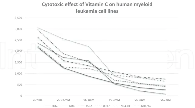
Further investigations in leukemia, performed by our research group, have shown that a plasma concentration of 3 mM of Vitamin C in the culture medium, is sufficient to kill more than a half of the cells in culture (LC50) in a number of different human myeloid leukemia cell lines [69] (Figures 3 and 4) (Table 1). It is of interest to consider that according to our protocol, the leukemic cells are exposed to Vitamin C for no more than 2 h, then accurately “washed,” re-suspended in fresh culture medium, without Vitamin C, and further incu-bated for additional 18–24 h, before the evaluation of cell survival and apoptosis. Given the results obtained, it is reasonable to conclude that the Vitamin C added to the culture medium (in the form of sodium ascorbate) is rapidly internalized by the leukemic cells, and
its “toxic” effects last for hours (days), even when the nutrient has been removed from the culture medium. This is in agreement with the notion that both normal and leukemic white blood cells tend to concentrate Vitamin C [70–73] to levels that are 10–100 fold higher than plasma [74, 75], and it is in contrast with the view that hydrogen peroxide forms outside the tumor cells [31, 32].
C 1mM 3mM 5mM
Figure 2. Viability profile of (Human) acute promyelocytic leukemia (APL) cell line (HL60) exposed for 2 h to increasing
concentrations of Viability profile of (Human) acute promyelocytic leukemia (APL) cell line (HL60) exposed for 2 h to increasing concentrations of Vitamin C. (Flow Cytometry analysis: see text). The percentage of dead cells in the plates is proportional to the concentration of Vitamin C in the medium. C = control (untreated) sample; 1 mM, 3 mM, 5 mM = Vitamin C at 1, 3, and 5 mM in the culture medium.
C 1mM 3mM
Hoechst/PI M.G.G.
5mM
Figure 1. The microphotographs refer to the cytomorphologic modifications of HL60 cell lines (human acute
promyelocytic leukemia—APL) exposed for 2 h to increasing concentrations of Vitamin C. It is evident that by increasing the concentration of Vitamin C in the medium (from 1 to 5 mM), APL cells show an increasing degree of morphologic alterations indicating progressive cell death (apoptosis, autophagy, autoschizis). With the Hoechst/PI fluorescent staining, vital cells are colored in blue, while dead/apoptotic cells are stained in red. M.G.G. = May Grunwald Giemsa cell staining; Hoechst33342/Propidium Iodide (PI) = Vital Staining; C = control (untreated) sample; 1 mM, 3 mM, 5 mM = Vitamin C at 1, 3, and 5 mM in the culture medium; original magnification: 400×.
Neutrophils, in particular, accumulate Vitamin C via the sodium-dependent Vitamin C co-transporter 2 (SVCT2) [76], and have intracellular levels of 1–2 mM [77]. Therefore, while there is agreement on the fact that in solid tumors, Vitamin C, initially oxidized to dehy-droascorbic acid (DHAA), is internalized by the cell, via GLUT 1 and 4, and finally reduced
0 500 1,000 1,500 2,000 2,500 3,000 3,500 CONTR. VC 1mM VC 3mM VC 5mM VC7mM NB4 VC 0.5mM HL60 K562 U937 NB4-R1 NB4/AS
Cytotoxic effect of Vitamin C on human myeloid leukemia cell lines
Figure 3. The figure illustrates Table 1. The diagram shows the almost uniform decrease in the number of vital leukemic
cells in the culture medium, after exposing them to increasing concentrations of Vitamin C, for 2 h.
0 500 1,000 1,500 2,000 2,500 3,000 3,500 CONTR. VC 0.5mM VC 1mM VC 3mM VC 5mM VC7mM
Cytotoxic effect of Vitamin C on human myeloid leukemia cell lines
HL60 NB4 K562 U937 NB4-R1 NB4/AS
O O
O OO O
Figure 4. The figure illustrates Table 1. Highlighted with colored circles, the LC50 for each human myeloid leukemia
again to ascorbic acid, with consumption of GSH; this may not be the case in acute myeloid leukemia.
More importantly, the parallel exposure of normal hematopoietic precursors (CD34+), iso-lated from cord blood, to Vitamin C, at the concentrations that are cytotoxic for leukemic cells did not affect their survival, or impair their capacity to proliferate and differentiate in response to myeloid growth factors. These data confirm that Vitamin C is harmless for normal hematopoietic precursors and therefore highly selective in its anticancer/antileukemic effect.
8. Hypoxia inducible factor (HIF): the forgotten pathway
Hypoxia and induction of hypoxia-inducible factors (HIF) is a hallmark of many tumors [78, 79].
HL60 (2 h) NB4 (2 h) K562 (2 h) U937 (2 h) NB4-R1 (2 h) NB4/As (2 h) Exp. 1 Contr. 471 912 663 1189 337 819 VC 0.5 296 680 669 479 82.8 42.7 VC 1 108 494 628 245 39.7 31.9 VC 3 22.6 163 226 56.4 47 32.2 VC 5 15.1 143 82 30.2 35 48.4 VC 7 6.15 85.4 32.2 10.6 24.6 17.6 Exp. 2 Contr. 869 1020 694 829 936 958 VC 0.5 423 959 399 704 624 823 VC 1 349 887 445 585 560 674 VC 3 217 337 200 232 335 581 VC 5 143 74.6 111 118 344 402 VC 7 89.5 147 35.2 93.7 255 329 Exp. 3 Contr. 843 1130 1020 969 967 859 VC 0.5 545 924 660 716 678 722 VC 1 438 835 507 689 668 649 VC 3 343 395 113 480 551 499 VC 5 218 346 35.2 411 443 453 VC 7 181 241 17.6 320 380 414
The cell lines used in this experiment are variants of human myeloid leukemia cells, and include: HL60, NB4, K562, U937, NB4-R1, NB4/As. It is evident that the total number of cells in culture decreases by increasing the concentration of Vitamin C in the culture medium. C = control (untreated) sample; VC = Vitamin C; VC 0.5 mM, VC 1 mM, VC 3 mM, VC 5 mM = Vitamin C at 0.5, 1, 3, and 5 mM in the culture medium.
HIF-1 is a heterodimeric transcription factor discovered in 1991 [80], and is composed of two subunits, α and β. The HIF-1α subunit is oxygen sensitive and it is induced by hypoxic con-ditions, which are very common in cancer. Direct transcriptional targets of HIF-1 include genes regulating, among others, growth and apoptosis, cell migration, energy metabolism, angiogenesis, vasomotor regulation, matrix and barrier functions, and transport of metal ions and glucose [81].
In normoxic conditions, the HIF-1α unit is downregulated by Vitamin C dependent hydroxy-lases, while in hypoxic conditions (such as those existing in many different types of cancer), HIF-1α hydroxylation is repressed with consequent increase in HIF-dependent gene tran-scription, neo-angiogenesis, and tumor growth and progression [82].
More importantly, since Vitamin C stimulates HIF-1α prolyl hydroxylases, low levels of Vitamin C promote tumor growth and progression, by reducing HIF-1α hydroxylation [83], thereby stabilizing HIF1-α. On the contrary, high levels of HIF render cancer cells more sensi-tive to Vitamin C-induced toxicity. To confirm this view, Kuiper and Coll. [84] have recently found an inverse relationship between intra-tumor levels of Vitamin C and HIF activity in both endometrial cancer [85] and colorectal carcinoma (CRC) [86].
In 1925, Otto Warburg observed that cancer cells manifest increased rates of lactate produc-tion under aerobic condiproduc-tions (“Warburg Effect”) or, in other words, they preferentially uti-lize glycolysis, instead of oxidative phosphorylation, for metabolism even in the presence of oxygen [87, 88].
“Hypoxia” (low oxygen concentration) is a hallmark of solid tumors, usually occurring at the center of the tumor mass, where blood vessels are abnormal or insufficient to supply adequate amounts of oxygen [89].
In response to the reduced oxygen tension, the HIF is activated to mediate the primary tran-scriptional adaption to hypoxic stress in cancer cells [90, 91].
As previously mentioned, HIFs regulate angiogenesis, cell survival, proliferation, apopto-sis, adhesion, and metabolism by transcriptionally activating downstream targets such as vascular endothelial growth factor and erythropoietin. Therefore, HIF (HIF1, in particular) plays a major role in tumor growth, and clinical data suggest that the upregulation of HIF, as determined by the low oxygen tension, is usually associated with increased mortality in a number of different cancers [92–94], and may represent a relevant target for new therapeutic approaches to the disease [95–97].
9. The HIF pathway in leukemia
The role of HIF-1α in leukemia, and in particular in acute myeloid leukemia (AML), has only recently emerged and it is still somewhat controversial. One possible explanation for this delayed interest in the role of hypoxia in leukemia could be the fact that leukemia is not considered a “solid” tumor, and therefore, the role of oxygen, in its pathogenesis, has been
considered inconsequential for long time. This erroneous view, has been recently reviewed, as data have emerged, demonstrating that leukemic cells are sensitive to the oxygen tension, and hypoxia influences leukemic cell proliferation, differentiation, and resistance to chemo-therapy [98].
Migliavacca and Coll. have recently demonstrated oncogenic function of HIF-1α, in the M5 Fab subtype of AML [99]. In particular, the authors have demonstrated that in M5 AML, HIF-1α mediates the ability of leukemic cells to migrate and invade extramedullary sites. The same group has demonstrated that PML-RARα and other fusion proteins involved in the pathogenesis of acute promyelocytic leukemia (APL) behave as transcriptional coactivators of HIFs, and both HIFs and PML-RARα enhances the progression of APL, by promoting cell migration, homing to bone marrow, and bone marrow neo-angiogenesis [100, 101].
Further investigations [102] have demonstrated that HIF-1α plays critical and pleiotropic roles in the pathogenesis of chronic lymphocytic leukemia (CLL).
Globally, elevated levels of HIF-1α have been reported in AML [103–106], APL [100], acute lymphoblastic leukemia (ALL) [107], and chronic myelogenous leukemia (CML) [108, 109]. Furthermore, HIF-1α overexpression conditions disease severity and outcome in both AML and myelodysplastic syndrome (MDS) [110–112].
Overall, the available data show that hypoxia and HIF-mediated signaling play a crucial role in leukemia, and targeting HIF with specific drugs or natural inhibitors, such as Vitamin C, represents a potentially useful approach to its treatment [113].
10. Vitamin C as a powerful modulator of TET2 activity
Decreased TET expression and loss of 5hmC have been observed in a wide variety of solid tumors, as well as in many hematological malignancies, including acute myeloid leukemias, myelodysplastic syndromes, and clonal hematopoiesis [114].
Recent experimental studies suggest that pharmacological dose of Vitamin C may represent a potentially important strategy in leukemia therapy through a stimulatory effect on TET2 activation and restoration in leukemic cells. Vitamin C is a co-factor of TET2 enzyme and is capable of interacting with the catalytic domain of TET2, enhancing the enzymatic oxidation of 5-methylcytosine (5mC) to 5-hydroxymethylcytosine (5hmC) [115]. This epigenetic modu-lation elicited by Vitamin C is able to improve the generation of pluripotent stem cells [116] and to induce a blastocyst-like state in mouse embryonic stem cells [117].
Two recent studies explored the possible epigenetic effects of Vitamin C on leukemia mod-els, mediated by activation and restoration of TET2 activity in leukemic cells. In the first one, authors used a murine model of IDH1-dependent acute myeloid leukemia [118], and 2-phos-phate l-ascorbic acid (Asc 2-P). Asc 2-P, unlike native Vitamin C, remains oxidatively stable under standard cell culture conditions [119], and possesses the same modulatory effects of Vitamin C, but, unlike Vitamin C, it does not induce cytotoxic effects of through stimulation of
H2O2 production. Asc 2-P added to the cells in culture is stable and releases Vitamin C by plasma membrane alkaline phosphatase hydrolysis [120]. Therefore, Asc 2-P allows a better character-ization of the epigenetic activity of Vitamin C, without the “disturbing” H2O2-mediated cyto-toxic effects of the native molecule. Asc 2-P treatment of the IDH1 AML-mutant mice induced an increase of 5hmC levels, a reduction of leukemic proliferation and an increase in expression of genes involved in leukocyte differentiation [118]. The stimulatory effect of Vitamin C on myeloid differentiation is mediated though the restoration of a normal expression and function of transcription factors, such as PU.1 and RUNX1, required for normal myeloid differentiation. A second study provided clear evidence that in various leukemia models, Vitamin C treat-ment induces the restoration of TET2 function, blocking aberrant self-renewal and leukemia progression. Treatment with Vitamin C, mimics TET2 restoration, driving DNA hypometh-ylation and, by enhancing 5hmC formation, suppresses leukemic colony formation and leukemic progression of primary human leukemia patient-derived xenografts (PDXs). Interestingly, TET2-mediated DNA oxidation induced by Vitamin C-treated leukemic cells, greatly enhances their sensitivity to PARP inhibition and could provide a safe and effective combination strategy to target TET-deficient leukemic cells. These observations suggest that future clinical trials could incorporate high-dose Vitamin C as an adjuvant to standard che-motherapy/demethylating therapy, particularly in TET2-deficient neoplasms [121].
11. What to do next?
The anticancer properties of Vitamin C are known, since at least six decades, even though its use in clinical practice has only recently re-emerged, after the demonstration that in rela-tively high concentrations, it can selecrela-tively kill a number of different human tumor cells, both in vitro and in vivo.
The proof of the anticancer efficacy of Vitamin C in high doses, administered by mouth, has been reported four decades ago, by Linus Pauling [54–57], and further confirmed, very recently, by experimental in vitro and in vivo data [30–32, 66, 69].
Vitamin C is a natural compound, and it is an antioxidant and a life-saving nutrient with mul-tiple beneficial effects on the human body. Man, some primates, and a few other mammals do no longer produce it. Beyond being a natural and essential nutrient, Vitamin C shows, in vitro, an outstanding efficacy in killing a number of different cancer cells, with an efficiency that no other anticancer drug, presently available on the market, has ever shown.
Vitamin C is extremely selective since it kills only cancer cells, by sparing, at the same time, all the other cells of the organism. As a consequence, it is very well tolerated, and devoid of any significant side effects. In fact, the only (relative) contraindication to its use, is the lack of the enzyme glucose-6-phosphate-dehydrogenase (G6PDH), a rare genetic condition also known as “favism.” More importantly, within an expensive and often artificially inflated market, such as that of the anticancer drugs [122, 123], Vitamin C, with its low cost, represents an outstanding opportunity for both the patients and the healthcare system.
Unfortunately, in spite of all the above characteristics, Vitamin C has never been easily or favorably accepted as an anticancer drug, by the western Medicine. This also explains why, although the data on its anticancer efficacy are outstanding and straightforward, many scien-tists still prefer to consider “controversial” the role of Vitamin C in the treatment of cancer. As we have seen, the idea that the oral administration of Vitamin C, in high doses, is not effec-tive against cancer is a conceptual artefact, originating from questionable interpretations of pharmacokinetics data, after oral and/or intravenous administration. On the other hand, the idea that Vitamin C, administered in high doses by intravenous infusion, behaves as a pro-drug of H2O2 beyond being experimentally questionable, has not led to clinically significant results or outcomes [124–128]. More importantly, encouraging results have emerged from unbiased interpretation of the available data [129]. In particular, as it has been shown up to 110 g/m2/
day are very well tolerated by the majority of patients, and even in the absence of any signifi-cant clinical remission, intravenous Vitamin C is almost invariably associated with a clear-cut improvement in patient’s quality of life.
As a result, History repeat itself! … and just as Vitamin C was dismissed as ineffective, against cancer, more than 30 years ago, on the ground of questionable clinical trials [130, 131], nowa-days, it runs again the risk of being definitively discarded, in spite of the large amount of scientific evidence, demonstrating its extraordinary efficacy in fighting cancer!
It is clear that much remains to be understood about the cytotoxic effects of Vitamin C against cancer, and much more can (and must!) be done, to both improve the intravenous therapy and further investigate the oral administration route of the high doses of the nutrient.
Improving the intravenous treatment can (and should!) be achieved, by considering:
a. The type of pharmaceutical preparation, the sodium salt of the ascorbic acid to be
pre-ferred, when administered by the intravenous route [132];
b. The time and schedule of administration (slow infusion to be preferred) [133, 134];
c. The level of tissue oxygenation (cell cultures are better oxygenated than tumor tissues, and
this may explain the differences in the outcomes between in vitro and in vivo treatment of cancer) [135]. In clinical settings, an improved tumor tissue oxygenation could be obtained with either ozone or hyperbaric oxygen;
d. The level of blood glucose (glucose may interfere with the uptake of ascorbate by cancer
cells) [136, 137], and the possibility of associating an adequate dietetic regimen to the treat-ment with high doses of oral or intravenous Vitamin C.
12. Latest evidence of the role of Vitamin C in leukemia
A recent study provided clear-cut evidence that Vitamin C is a main regulator of hemato-poietic stem cell (HSC) function and leukemogenesis. In fact, Agathocleous and co-workers, using a peculiar strategy for isolation of HSCs and hematopoietic progenitor cells (HPCs) from murine bone marrow, showed that HSCs have unusually high levels of Vitamin C,
which decline with differentiation [138]. Importantly, human HSCs and multipotent progeni-tor cells (MPPs), such as murine HSCs, display high Vitamin C levels.
Using “GULO” mice (deficient in Vitamin C because of the lack of gulonolactone oxidase, the last enzyme in the synthesis of Vitamin C starting from glucose), Agathocleous and col-leagues have shown that Vitamin C deficiency induces an increased number of HSCs. A FLT3-internal tandem duplication (ITD) mutation, found in approximately a quarter of patients with de novo AML, imparts a particularly poor prognosis. Using “GULO” mice (deficient in Vitamin C because of the lack of gulonolactone oxidase), Agathocleous and colleagues have shown that Vitamin C deficiency induces an increased number of HSCs. Therefore Vitamin C deficiency, and TET2 mutations, are likely to cooperates with FLT3-ITD to induce leukemia development in murine models of FLT3-ITD-driven leukemia. [138].
Given the above evidence, it will be worth mentioning, once more, that the biochemist Irwin Stone, in his book “The healing Factor: Vitamin C against disease,” published in 1972 (45 years ago!), had already warned the scientific community on the role of Vitamin C as a main fac-tor in the prevention and treatment of leukemia. In his words, “In a leukemic, the biochemical
stresses of the disease process has reduced the body stores of ascorbic acid to very low levels … Any ascorbic acid circulating in the blood has been scavenged and locked in the excessive numbers of white blood cells contained in the blood. The plasmas level of ascorbic acid is usually zero or close thereto. A zero level in the blood plasma means that the tissues of the body are not being supplied with this most important metabolite. The ascorbic acid contained in the leukocytes are unavailable for the tissues. The tissues are in a condition of biochemical scurvy and this explains why these depleted tissues are so susceptible to the characteristic hemorrhaging of leukemia and the infections that kill so many of the leukaemics. A leukemic is not only suffering from leukemia but also from a bad case of biochemical scurvy. To correct this condition, ascorbic acid has to be administered in sufficiently large doses not only to saturate the excess of white blood cells but to provide adequate spill over into the blood plasma and tissues so that the seriously ill leukemic will be given a fighting chance to combat the disease. This may require the administration of ascorbic acid at the rate of 25 or more grams per day, as noted in the following case of leukemia treated with megascorbic levels of ascorbic acid.” [3].
13. Concluding remarks
The rationale behind the use of high doses of Vitamin C in the treatment of acute leukemia is strong and very well grounded. In summary:
a. Leukemic patients, almost invariably show a severe deficiency of this nutrient;
b. While it is currently supposed to kill cancer cells by inducing pro-oxidant damage, Vitamin
C is also very effective as an antioxidant by inhibiting the hypoxia inducible factor (HIF);
c. The mechanistic explanation of the pathogenesis of myeloid leukemia, includes the
pos-sibility that a Vitamin deficiency may induce the neoplastic transformation of myeloid precursors, through an upregulation of the HIF, and the consequent cascade of HIF-de-pendent cancer genes;
d. Although administered by intravenous infusion, in the majority of clinical trials performed
so far, Vitamin C appears to be effective, in fighting cancer, even when administered by mouth;
e. Vitamin C is very well tolerated, and has no side or undesired effects;
f. Experimental in vitro data unequivocally show the cytotoxic effect of Vitamin C against
leukemia.
g. As shown in our study on leukemic and normal cell lines, Vitamin C can kill almost every
type of acute and chronic myeloid leukemia-derived cell, without doing any harm to their normal counterpart CD34+ cells;
h. Vitamin C is a natural compound, and it is very cheap.
Do we really need more information or evidence, to start clinical trials on Vitamin C, in the treatment of acute and chronic myeloid leukemia?
Acknowledgements
This chapter has been realized, in part, thanks to the financial support of the “Pescara Cell Factory Foundation”.
Conflict of interests
None to declare.Author details
Domenico Mastrangelo1*, Lauretta Massai1, Giuseppe Fioritoni2, Francesco Lo Coco3,4,
Nèlida Noguera3,4 and Ugo Testa5
*Address all correspondence to: [email protected]
1 Department of Medical, Surgical and Neurological Sciences, University of Siena, Polo Scientifico San Miniato, Siena, Italy
2 Pescara Cell Factory Foundation Onlus, Pescara, Italy
3 Department of Biomedicine and Prevention, University of Rome Tor Vergata, Rome, Italy 4 Santa Lucia Foundation, I.R.C.C.S., Via del Fosso di Fiorano, Rome, Italy
5 Department of Hematology, Oncology and Molecular Medicine, Istituto Superiore di Sanità, Rome, Italy
References
[1] Iqbal K, Khan A, Ali Khan Khattak MM. Biological significance of ascorbic acid (Vitamin C) in human health. A review. Pakistan Journal of Nutrition. 2004;3:5-13
[2] Drouin G, Godin JR, Pagé B. The genetics of Vitamin C loss in vertebrates. Current Genomics. 2011;12:371-378. DOI: 10.2174/138920211796429736
[3] Stone I. The Healing Factor: Vitamin C Against Disease. New York: Grosset and Dunlap Inc.; 1972
[4] Stephen DJ, Hawley EE. The partition of reduced ascorbic acid in blood. The Journal of Biological Chemistry. 1936;115:653-658
[5] Barkhan P, Howard AN. Distribution of ascorbic acid in normal and leukaemic human blood. The Biochemical Journal. 1958;70:163-168
[6] Vogt A, Uber den Vitamin C. Verbrauch bei Tumorkranken und bei der Lymphogranu-lomatose. Strahlentherapie. 1939;65:616-623
[7] Kyhos FD, Sevringhaus FL, Hagendorn DR. Large doses of ascorbic acid in treatment of Vitamin C deficiencies. Archives of Internal Medicine. 1945;75:407
[8] Waldo AL, Zipf RE. Ascorbic acid level in leukemic patients. Cancer. 1955;8:187-190 [9] Pujari KN, Jadkar SP, Mashal SN, Belwalkar GJ, Kulkarni A, Patil CG, et al. Variations in
vitamin C levels in leukemias. Biomedical Research. 2012;23:307-311
[10] Liua M, Ohtania H, Zhoua W, Ørskovb AD, Charletc J, Zhangd YW, et al. Vitamin C increases viral mimicry induced by 5-aza-2′-deoxycytidine. PNAS. 2016;113:10238-10244. DOI: 10.1073/pnas.1612262113
[11] Shenoy N, Bhagat T, Nieves E, Stenson M, Lawson J, Choudhary GS, et al. Upregulation of TET activity with ascorbic acid induces epigenetic modulation of lymphoma cells. Blood Cancer Journal. 2017;7:e587. DOI: 10.1038/bcj.2017.65
[12] Klenner FR. The treatment of poliomyelitis and other virus diseases with vitamin C. Southern Medicine and Surgery. 1949;111:209-214
[13] Stone I. Hypoascorbemia, the genetic disease causing the human requirement for exog-enous ascorbic acid. Perspectives in Biology and Medicine. 1966;10:133-134
[14] Stone I. The genetic disease, hypoascorbemia: A fresh approach to an ancient disease and some of its medical implications. Acta Geneticae Medicae et Gemellologiae. 1967;
16:52-62
[15] Stone I. Hypoascorbemia, our most widespread disease. Bulletin of the National Health Federation 1972;18:6-9
[16] http://www.omim.org/entry/240400
[18] Stone I. My daily megascorbic regime for full health and long life. Better Nutrition; 1977 [19] Panel on Dietary Antioxidants and Related Compounds; Subcommittee on Upper
Reference Levels of Nutrients; Subcommittee on Interpretation and Uses of DRIs; Standing Committee on the Scientific Evaluation of Dietary Reference Intakes; Food and Nutrition Board; Institute of Medicine. Dietary Reference Intakes for Vitamin C, Vitamin E, Selenium, and Carotenoids. 2000. 529 p. ISBNs: Paperback: 978-0-309-06935-9 Hardcover: 978-0-309-06949-6
[20] Letter PL. Megavitamin therapy. Journal of the American Medical Association. 1975;
234:149
[21] Pauling L. Are recommended daily allowances for vitamin C adequate? Review. PNAS USA. 1974;71:4442-4446
[22] Pauling L. Vitamin C. Science. 1972;177(4055):1152
[23] Pauling L. Evolution and the need for ascorbic acid. PNAS USA. 1970;7:1643-1648
[24] Pauling L. Vitamin C and common cold. Journal of the American Medical Association. 1971;216:332
[25] Pauling L. The significance of the evidence about ascorbic acid and the common cold. Review. PNAS USA. 1971;68:2678-2681
[26] Pauling L. Vitamin C therapy of advanced cancer. The New England Journal of Medicine. 1980;302:694-695
[27] Harakeh S, Jariwalla RJ, Pauling L. Suppression of human immunodeficiency virus replication by ascorbate in chronically and acutely infected cells. PNAS USA. 1990;87: 7245-7249
[28] Levine M, Conry-Cantilena C, Wang Y, Welch RW, Washko PW, Dhariwal KR, et al. Vitamin C pharmacokinetics in healthy volunteers: Evidence for a recommended dietary allowance. PNAS USA. 1996;93:3704-3709
[29] Padayatty SJ, Sun H, Wang Y, Riordan HD, Hewitt SM, Katz A, et al. Vitamin C phar-macokinetics: Implications for oral and intravenous use. Annals of Internal Medicine. 2004;140:533-537
[30] Chen Q, Espey MG, Sun AY, Pooput C, Kirk KL, Krishna MC, et al. Pharmacologic doses of ascorbate act as a prooxidant and decrease growth of aggressive tumor xenografts in mice. PNAS USA. 2008;105:11105-11109
[31] Chen Q, Espey MG, Sun AY, Lee JH, Krishna MC, Shacter E, et al. Ascorbate in pharma-cologic concentrations selectively generates ascorbate radical and hydrogen peroxide in extracellular fluid in vivo. PNAS USA. 2007;104:8749-8754
[32] Chen Q, Espey MG, Krishna MC, Mitchell JB, Corpe CP, Buettner G, et al. Pharmacologic ascorbic acid concentrations selectively kill cancer cells: Action as a pro-drug to deliver hydrogen peroxide to tissues. PNAS USA. 2005;102:13604-13609
[33] Halliwell B. Vitamin C: Poison, prophylactic or panacea? Trends in Biochemical Sciences. 1999;24:l255-1259
[34] Carr AC, Frei B. Does vitamin C act as pro-oxidant under physiological conditions? The FASEB Journal. 1999;13:1007-1024
[35] Hacisevki A. An overview of ascorbic acid biochemistry. Journal of the Faculty of Phar-macy of Ankara University. 2009;38:233-255
[36] Yun J, Mullarky E, Lu C, Bosch KN, Kavalier A, Rivera K, et al. Vitamin C selectively kills KRAS and BRAF mutant colorectal cancer cells by targeting GAPDH. Science. 2015;350:1391-1396
[37] Savini I, Rossi A, Pierro C, Avigliano L, Catani MV. SVCT1 and SVCT2: Key proteins for vitamin C uptake. Amino Acids. 2008;34:347-355
[38] Vera JC, Rivas CI, Fischbarg J, Golde DW. Mammalian facilitative hexose transporters mediate the transport of dehydroascorbic acid. Nature. 1993;364:79-82
[39] Tian W, Wang Y, Xu Y, Guo X, Wang B, Sun L, et al. The hypoxia-inducible factor ren-ders cancer cells more sensitive to Vitamin C-induced toxicity. The Journal of Biological Chemistry. 2014;289:3339-3351
[40] Yun J, Rago C, Cheong I, Pagliarini R, Angenendt P, Rajagopalan H, et al. Glucose deprivation contributes to the development of KRAS pathway mutations in tumor cells. Science. 2009;(5947):1555-1559
[41] Sheu JJC, Guan B, Tsai FJ, Hsiao EYT, Chen CM, Seruca R, et al. Mutant BRAF induces DNA strand breaks, activates DNA damage response pathway, and up-regulates glucose transporter-1 in nontransformed epithelial cells. The American Journal of Pathology. 2012;180:1179-1188
[42] Van der Reest J, Gottlieb E. Anti-cancer effects of vitamin C revisited. Cell Research. 2016;26:269-270
[43] Uetaki M, Tabata S, Nakasuka F, Soga T, Masaru Tomita M. Metabolomic alterations in human cells by vitamin C-induced oxidative stress. Scientific Reports. 2015;5:1-9
[44] Benade L, Howard T, Burk D. Synergistic killing of Ehrlich ascites carcinoma cells by ascorbate and 3-amino-1, 2, 4,-triazole. Oncology. 1969;23:33-43
[45] Oberley TD, Oberley LW. Antioxidant enzyme levels in cancer. Histology and Histopa-thology. 1997;12:525-535
[46] Cavanagh EMV, Honegger AE, Hofer E, Bordenave RH, Bullorsky EO, Chasseing NA. Higher oxidation and lower antioxidant levels in peripheral blood plasma and bone marrow plasma from advanced cancer patients. Cancer. 2002;94:3247-3251
[47] Mojić M, Bogdanović Pristov J, Maksimović-Ivanić D, Jones DR, Stanić M, Mijatović S, et al. Extracellular iron diminishes anticancer effects of vitamin C: An in vitro study. Scientific Reports. 2014;4:5955. DOI: 10.1038/srep05955
[48] Du J, Cullen JJ, Buettner GR. Ascorbic acid: Chemistry, biology and the treatment of cancer. Biochimica et Biophysica Acta. 2012;1826:443-457
[49] Lòpez-Làzaro M. Dual role of hydrogen peroxide in cancer: Possible relevance to cancer chemoprevention and therapy. Cancer Letters. 2007;252:1-8
[50] Lisanti MP, Martinez-Outschoorn UE, Lin Z, Pavlides S, Whitaker-Menezes D, Pestell RG. Hydrogen peroxide fuels aging, inflammation, cancer metabolism and metastasis. Cell Cycle. 2011;10:2440-2449
[51] Padayatty SJ, Levine M. Reevaluation of ascorbate in cancer treatment: Emerging evi-dence, open minds and serendipity. Journal of the American College of Nutrition. 2000;19:423-425
[52] Robinson AR, Hunsberger A, Westall FC. Suppression of squamous cell carcinoma in hairless mice by dietary nutrient variation. Mechanisms of Ageing and Development. 1994;76:201-214
[53] Hoffer A. The discovery of Vitamin C: Albert Szent-Gyorgi, M.D. Ph.D. 1893-1986. Journal of Orthomolecular Medicine. 1989;4:24-26
[54] Cameron E, Pauling L. Supplemental ascorbate in the supportive treatment of cancer: Reevaluation of prolongation of survival times in terminal human cancer. PNAS USA. 1978;75:4538-4542
[55] Pauling L. Vitamin homeostasis in the brain and megavitamin therapy. The New England Journal of Medicine. 1977;297:790-791
[56] Pauling L. Diet, nutrition, and cancer. The American Journal of Clinical Nutrition. 1977;
30:661-663
[57] Cameron E, Pauling L. Supplemental ascorbate in the supportive treatment of cancer: Prolongation of survival times in terminal human cancer. PNAS USA. 1976;73:3685-3689 [58] Cameron E, Campbell A. The orthomolecular treatment of cancer. Clinical trial of high dose ascorbic acid supplements in advanced human cancer. Chemico-Biological Interactions. 1974;9:285-315
[59] Murata A, Morishige F, Yamaguchi H. Prolongation of survival times of terminal cancer patients by administration of large doses of ascorbate. International Journal for Vitamin and Nutrition Research Supplement. 1982;23:101-113
[60] Hickey S, Roberts H, Miller NJ. Pharmacokinetics of oral ascorbate liposomes. JNEM. 2008. DOI: 10.1080/13590840802305423
[61] Cathcart RF. The method of determining proper doses of vitamin c for the treatment of disease by titrating to bowel tolerance. Orthomol Psychiatry. 1981;10:125-132
[62] Hickey S, Roberts H. The real story of Vitamin C and cancer. Journal of Metals. 2008;
[63] Poirier MC. Chemical-induced DNA damage and human cancer risk. Nature Reviews. Cancer. 2004;4:630-637
[64] Sharma RA, Farmer PB. Biological relevance of adduct detection to the chemoprevention of cancer. Clinical Cancer Research. 2004;10:4901-4912
[65] Gao P, Zhang H, Dinavahi R, Li F, Xiang Y, Raman V, et al. HIF-dependent antitumori-genic effect of antioxidants in vivo. Cancer Cell. 2007;12:230-238
[66] Mastrangelo D, Massai L, Fioritoni G, Iacone A, Di Bartolomeo P, Accorsi P, et al. Megadoses of sodium ascorbate efficiently kill HL60 cells in vitro: Comparison with arsenic trioxide. Journal of Cancer Therapy. 2013;4:1366-1372
[67] Monti DA, Mitchell E, Bazzan AJ, Littman S, Zabrecky G, Yeo CJ, et al. Phase I evaluation of intravenous ascorbic acid in combination with gemcitabine and Erlotinib in patients with metastatic pancreatic cancer. PLoS One. 2012;7:e29794
[68] Stephenson CM, Levin RD, Spector T, Lis CG. Phase I clinical trial to evaluate the safety, tolerability, and pharmacokinetics of high-dose intravenous ascorbic acid in patients with advanced cancer. Cancer Chemotherapy and Pharmacology. 2013;72:139-146
[69] Mastrangelo D, Massai L, Lo Coco F, Noguera NI, Borgia L, Fioritoni G, et al. Cytotoxic effects of high concentrations of sodium ascorbate on human myeloid cell lines. Annals of Hematology. 2015;94:1807-1816
[70] Lowry OH, Bessey OA, Brock MJ, Lopez JA. The interrelationship of dietary, serum, white blood cell, and total body ascorbic acid. The Journal of Biological Chemistry. 1946;166:111-119
[71] Wilson C. Clinical pharmacological aspects of ascorbic acid. Annals of the New York Academy of Sciences. 1975;25:354-376
[72] Lykkesfeldt J. Determination of ascorbic acid and dehydroascorbic acid in biologi-cal samples by high-performance liquid chromatography using subtraction methods: Reliable reduction with tris[2-carboxyethyl] phosphine hydrochloride. Analytical Biochemistry. 2000;282:89-93
[73] Emadi-Konjin P, Verjeea Z, Levinb AV, Adeli K. Measurement of intracellular vitamin C levels in human lymphocytes by reverse phase high performance liquid chromatogra-phy (HPLC). Clinical Biochemistry. 2005;38:450-456
[74] Michels AJ, Hagen TM, Frei B. Human genetic variation influences Vitamin C homeosta-sis by altering Vitamin C transport and antioxidant enzyme function. Annual Review of Nutrition. 2013;33:45-70
[75] Bozonet SM, Carr AC, Pullar JM, Vissers MCM. Enhanced human neutrophil Vitamin C status, chemotaxis and oxidant generation following dietary supplementation with Vitamin C-rich sungold kiwifruit. Nutrients. 2015;7:2574-2588. DOI: 10.3390/nu7042574
[76] Corpe CP, Lee JH, Kwon O, Eck P, Narayanan J, Kirk KL, et al. 6-Bromo-6-deoxyL-ascorbic acid: An ascorbate analog specific for Na+-dependent vitamin C transporter but not glu-cose transporter pathways. The Journal of Biological Chemistry. 2005;280:5211-5220 [77] Washko PW, Wang Y, Levine M. Ascorbic acid recycling in human neutrophils. The
Journal of Biological Chemistry. 1993;268:15531-15535
[78] Aprelikova O, Chandramouli GVR, Wood M, Vasselli JR, Riss J, Maranchie JK, et al. Regulation of HIF prolyl hydroxylases by hypoxia-inducible factors. Journal of Cellular Biochemistry. 2004;92:491-501
[79] Nagy MA. HIF-1 is the commander of gateways to cancer. Journal of Cancer Science & Therapy. 2011;3:035-040. DOI: 10.4172/1948-5956.1000054
[80] Semenza GL, Nejfelt MK, Chi SM, Antonarakis SE. Hypoxiainducible nuclear factors bind to an enhancer element located 3′ to the human erythropoietin gene. PNAS USA. 1991;88:5680-5684
[81] Schofield CJ, Ratcliffe PJ. Oxygen sensing by HIF hydroxylases. Nature Reviews. Molecular Cell Biology. 2004;5:343-354
[82] Traber MG, Stevens JF. Vitamins C and E: Beneficial effects from a mechanistic perspec-tive. Free Radical Biology and Medicine. 2011;51:1000-1013
[83] Knowles HJ, Raval RR, Harris AL, Ratcliffe PJ. Effect of ascorbate on the activity of hypoxia-inducible factor in cancer cells. Cancer Research. 2003;63:1764-1768
[84] Kuiper C, Vissers MCM. Ascorbate as a co-factor for Fe- and 2-oxoglutarate dependent dioxygenases: Physiological activity in tumor growth and progression. Frontiers in Oncology. 2014;4:1-11
[85] Kuiper C, Molenaar IG, Dachs GU, Currie MJ, Sykes PH, Vissers MC. Low ascorbate lev-els are associated with increased hypoxia-inducible factor-1 activity and an aggressive tumor phenotype in endometrial cancer. Cancer Research. 2010;70:5749-5758
[86] Kuiper C, Dachs GU, Munn D, Currie MJ, Robinson BA, Pearson JF. Increased tumor ascorbate is associated with extended disease-free survival and decreased hypoxia-inducible factor-1 activation in human colorectal cancer. Frontiers in Oncology. 2014;4: 1-10. DOI: 10.3389/fonc.2014.00010
[87] Warburg O. Über den stoffwechsel der carcinomzelle. Klinische Wochenschrift. 1925;
4:534-536
[88] Justus CR, Sanderlin EJ, Yang LV. Molecular connections between cancer cell metab-olism and the tumor microenvironment. International Journal of Molecular Sciences. 2015;16:11055-11086
[89] Huang D, Li C, Zhang H. Hypoxia and cancer cell metabolism. Acta Biochimica et Biophysica Sinica. 2014;46:214-219. DOI: 10.1093/abbs/gmt148
[90] Semenza GL, Wang GL. A nuclear factor induced by hypoxia via 58 protein synthesis binds to the human erythropoietin gene enhancer at a site required for transcriptional activation. Molecular and Cellular Biology. 1992;12:5447-5454
[91] Wang GL, Jiang BH, Rue EA, Semenza GL. Hypoxia-inducible factor-1 is a basic-helix-loop-helix-Pas heterodimer regulated by cellular O2 tension. PNAS USA. 1995;92: 5510-5514
[92] Schindl M, Schoppmann SF, Samonigg H, Hausmaninger H, Kwasny W, Gnant M, et al. Overexpression of hypoxia-inducible factor 1 is associated with an unfavorable prog-nosis in lymph node-positive breast cancer. Clinical Cancer Research. 2002;8:1831-1837 [93] Mouriaux F, Sanschagrin F, Diorio C, Landreville S, Comoz F, Petit E, et al. Increased
HIF-1a expression correlates with cell proliferation and vascular markers CD31 and VEGF-A in uveal melanoma. IOVS. 2014;55:1277-1283. DOI: 10.1167/iovs.13-13345 [94] Kim BW, Cho H, Chung JY, Conway C, Ylaya K, Kim JH, et al. Prognostic assessment
of hypoxia and metabolic markers in cervical cancer using automated digital image analysis of immunohistochemistry. Journal of Translational Medicine. 2013;11:185. DOI: 10.1186/1479-5876-11-185
[95] Masoud GN, Li W. HIF-1α pathway: Role, regulation and intervention for cancer ther-apy. Acta Pharmacol Sin B. 2015;5:378-389. DOI: 10.1016/j.apsb.2015.05.007
[96] Semenza GL. Targeting HIF-1 for cancer therapy. Nature Reviews. Cancer. 2003;3:721-732. DOI: 10.1038/nrc1187
[97] Poon E, Harris AL, Ashcroft M. Targeting the hypoxia-inducible factor (HIF) path-way in cancer. Expert Reviews in Molecular Medicine. 2009;11:e26. DOI: 10.1017/ S1462399409001173
[98] Muz B, de la Puente P, Azab F, Luderer M, Azab AK. The role of hypoxia and exploi-tation of the hypoxic environment in hematologic malignancies. Molecular Cancer Research 2014;12:1347-1354. DOI: 10.1158/1541-7786
[99] Migliavacca J, Percio S, Valsecchi R, Ferrero E, Spinelli A, Ponzoni M, et al. Hypoxia inducible factor-1a regulates a pro-invasive phenotype in acute monocytic leukemia. Oncotarget. 2016;7:53540-53577
[100] Coltella N, Percio S, Valsecchi R, Cuttano R, Guarnerio J, Ponzoni M, et al. HIF factors cooperate with PML-RARalpha to promote acute promyelocytic leukemia progression and relapse. EMBO Molecular Medicine. 2014;6:640-650
[101] Percio S, Coltella N, Grisanti S, Bernardi R, Pattini LA. HIF-1 network reveals character-istics of epithelial-mesenchymal transition in acute promyelocytic leukemia. Genome Medicine. 2014;6:84. DOI: 10.1186/s13073-014-0084-4
[102] Valsecchi R, Coltella N, Belloni D, Ponente M, Ten Hacken E, Scielzo C, et al. HIF-1a regulates the interaction of chronic lymphocytic leukemia cells with the tumour micro-environment. Blood. 2016;127:1987-1997. DOI: 10.1182/blood-2015-07-657056
[103] Gao XN, Yan F, Lin J, Gao L, XL L, Wei SC, et al. AML1/ETO cooperates with HIF1α to pro-mote leukemogenesis through DNMT3a transactivation. Leukemia. 2015;29:1730-1740 [104] Forristal CE, Brown AL, Helwani FM, Winkler IG, Nowlan B, Barbier V, et al. Hypoxia
inducible factor (HIF)-2α accelerates disease progression in mouse models of leuke-mia and lymphoma but is not a poor prognosis factor in human AML. Leukeleuke-mia. 2015;29:2075-2085
[105] Kawada H, Kaneko M, Sawanobori M, Uno T, Matsuzawa H, Nakamura Y, et al. High concentrations of l-ascorbic acid specifically inhibit the growth of human leukemic cells via downregulation of HIF-1α transcription. PLoS One. 2013;8:e62717
[106] Wang Y, Liu Y, Malek SN, Zheng P, Liu Y. Targeting HIF1α eliminates cancer stem cells in hematological malignancies. Cell Stem Cell. 2011;8:399-411. DOI: 10.1016/j. stem.2011.02.006
[107] Frolova O, Samudio I, Benito JM, Jacamo R, Kornblau SM, Markovic A, et al. Regulation of HIF-1α signaling and chemoresistance in acute lymphocytic leukemia under hypoxic conditions of the bone marrow microenvironment. Cancer Biology & Therapy. 2012;13:858-870
[108] Chen H, Shen Y, Gong F, Jiang Y, Zhang R. HIF-α promotes chronic myelogenous leukemia cell proliferation by upregulating p21 expression. Cell Biochemistry and Biophysics. 2015;72:179-183. DOI: 10.1007/s12013-014-0434-2
[109] Zhang H, Li H, Xi HS, Li S. HIF1α is required for survival maintenance of chronic myeloid leukemia stem cells. Blood. 2012;119:2595-2607
[110] Rouault-Pierre K, Lopez-Onieva L, Foster K, Anjos-Afonso F, Lamrissi-Garcia I, Serrano-Sanchez M, et al. HIF-2α protects human hematopoietic stem/progenitors and acute myeloid leukemic cells from apoptosis induced by endoplasmic reticulum stress. Cell Stem Cell. 2013;13:549-563
[111] Wellmann S, Guschmann M, Griethe W, Eckert C, Stackelberg A, Lottaz C, et al. Activation of the HIF pathway in childhood ALL, prognostic implications of VEGF. Leukemia. 2004;18:926-933
[112] Elias H, Verma A. Activation of hypoxia signaling is marker of poor prognosis in myelo-dysplasia. Leukemia & Lymphoma. 2012;53:2337-2338
[113] Deynoux M, Sunter N, Hérault O, Mazurier F. Hypoxia and hypoxia-inducible factors in leukemias. Frontiers in Oncology. 2016;6:41. DOI: 10.3389/fonc.2016.00041
[114] Rasmussen KD, Helin K. Role of TET enzymes in DNA methylation, development, and cancer. Genes & Development. 2016;30:733-750
[115] Yin R, Mao SQ, Zhao B, Chong Z, Yang Y, Zhao C, et al. Ascorbic acid enhances Tet-mediated 5-methylcytosine oxidation and promotes DNA demethylation in mammals. Journal of the American Chemical Society. 2013;135:10936-10403
[116] Chen J, Guo L, Zhang L, Wu H, Yang J, Liu H, et al. Vitamin C modulates TET1 function during somatic cell reprogramming. Nature Genetics. 2013;45:1504-1509
[117] Blaschke K, Ebata KT, Karimi MM, Zepeda-Martínez JA, Goyal P, Mahapatra S, et al. Vitamin C induces Tet-dependent DNA demethylation in ESCS to promote a blasto-cyst-like state. Nature. 2013;500:222-226
[118] Mingay M, Chaturvedi A, Bilenky M, Cao Q, Jackson L, Hui T, et al. Vitamin C-induced epigenomic remodeling in IDH1 mutant acute myeloid leukemia. Leukemia. 2017:1-10. DOI: 10.1038/leu.2017.171
[119] Austria R, Semenzato A, Bettero A. Stability of vitamin C derivatives in solution and top-ical formulations. Journal of Pharmaceuttop-ical and Biomedtop-ical Analysis. 1997;15:795-801 [120] Takamizawa S, Maehata Y, Imai K, Senoo H, Sato S, Hata RI. Effects of ascorbic acid
and ascorbic acid 2-phosphate, a long-acting vitamin C derivative, on the prolif-eration and differentiation of human osteoblast-like cells. Cell Biology International. 2004;28:255-265
[121] Cimmino L, Dolgalev I, Wang Y, Yoshimi A, Martin G, Wang J, et al. Restoration of TET2 function blocks aberrant self-renewal and leukemia progression. Cell. 2017;170:1-17 [122]
http://www.ascopost.com/issues/february-1-2013/cost-of-cancer-drugs-what-price-for-what-benefit/
[123] http://chronicdisease.uchicago.edu/files/2014/09/Conti-pricing-trends-9-9-2014.pdf [124] Wilson MK, Baguley BC, Wall C, Jameson MB, Findaly MP. Review of high-dose
intra-venous vitamin C as an anticancer agent. Asia-Pacific Journal of Clinical Oncology. 2014;10:22-37
[125] Unlu A, Kirca O, Ozdogan M, Nayır E. High-dose vitamin C and cancer. Journal of Oncological Science. 2016;1:10-12
[126] Nandal S, Ghalaut P, Shekhawat H, Nagar P, Ghalaut R, Meena B. High-dose intra-venous vitamin C as an anticancer agent—A literature review. IJARESM. 2014;6211:7 [127] Jacobs C, Hutton B, Ng T, Shorr R, Clemons M. Is there a role for oral or intravenous
ascorbate (Vitamin C) in treating patients with cancer? A systematic review. The Oncologist. 2015;20:210-223
[128] Goodman A. Vitamin C and cancer. AIMS Medical Science. 2013;3:41-51
[129] Fritz H, Flower G, Weeks L, Cooley K, Callachan M, McGowan J, et al. Intravenous Vitamin C and cancer: A systematic review. Integrative Cancer Therapies. 2014;13:280-300. DOI: 10.1177/1534735414534463
[130] Creagan ET, Moertel CG, O’Fallon JR, Schutt AJ, O’Connell MJ, Rubin J, et al. Failure of high-dose vitamin C (ascorbic acid) therapy to benefit patients with advanced cancer. A controlled trial. The New England Journal of Medicine. 1979;301:687-690
[131] Moertel CG, Fleming TR, Creagan ET, Rubin J, O’Connell MJ, Ames MM. High-dose vitamin C versus placebo in the treatment of patients with advanced cancer who have had no prior chemotherapy. A randomized double-blind comparison. The New England Journal of Medicine. 1985;312:137-141
[132] http://www.doctoryourself.com/vitciv.html
[133] Duconge J, Miranda-Massari JR, Gonzalez MJ, Jackson JA, Warnock W, Riordan NH. Pharmacokinetics of vitamin C: Insights into the oral and intravenous administration of ascorbate. Puerto Rico Health Sciences Journal. 2008;27:7-19
[134] Hickey DS, Roberts HJ, Cathcart RF. Dynamic flow: A new model for ascorbate. Journal of Metals. 2005;20:237-244
[135] Carreau A, El Hafny-Rahbi B, Matejuk A, Grillon C, Kieda C. Why is the partial oxygen pressure of human tissues a crucial parameter? Small molecules and hypoxia. Journal of Cellular and Molecular Medicine. 2011;15:1239-1253
[136] Malo C, Wilson JX. Glucose modulates Vitamin C transport in adult human small intes-tinal brush border membrane vesicles. The Journal of Nutrition. 2000;130:63-69
[137] Washko P, Levine M. Inhibition of ascorbic acid transport in human neutrophils by glucose. The Journal of Biological Chemistry. 1992;267:23568-23574
[138] Agathocleous M, Meacham CE, Burgess RJ, Piskounova E, Zhao Z, Crane GM, Cowin BL, Bruner E, Murphy MM, Chen W, Spangrude GJ, Hu Z, DeBernardinis RJ, Morrison SJ. Ascorbate regulates haematopoietic stem cell function and leukaemogenesis. Nature. Sep 28 2017;549(7673):476-481. DOI: 10.1038/nature23876. Epub 2017 Aug 21