of
RADIOLOGY
AND
IMAGING
INDIAN JOURNAL
THE
Volume 29 Issue 3 July-September 2019
The Indian Journal of
Radiology and Imaging •
V olume 29 • Issue 3 • July-September 2019 • Pages ***-*** ISSN 0971-3026
The Official Journal of the
Indian Radiological and Imaging Association
Online full text at
www.ijri.org
310 © 2019 Indian Journal of Radiology and Imaging | Published by Wolters Kluwer ‑ Medknow
Bilio‑cutaneous fistula obliteration with
NBCA
Antonio Bottari, Salvatore Silipigni, Alberto Stagno, Simona Caloggero
1Department of Biomedical and Dental Sciences and Morphofunctional Imaging, University of Messina, 1Department of Diagnostic
Imaging and Radiotherapy, University Hospital “G. Martino”, Messina, Italy
Correspondence: Dr. Antonio Bottari, Department of Biomedical and Dental Sciences and Morphofunctional Imaging, University of Messina, Viale Gazzi, Messina, Italy. E‑mail: [email protected]
Abstract
Biliary fistula and bile leakage are complications that can occur during hepato‑biliary surgery (both open and laparoscopic) and percutaneous biliary intervention. In some cases, spontaneous resolution is documented but more often re‑intervention (surgical or percutaneous) is necessary. We present the case of a male patient who underwent right hepatectomy with bilio‑digestive anastomosis for a cholangiocarcinoma which developed a bilo‑cutaneous fistula through the path of a previously inserted percutaneous transhepatic drainage. Sealing of bilo‑cutaneous fistula was obtained using N‑butil‑Cyanoacrylate. This technique has already been reported in some papers as a useful tool for biliary tree obliteration; however, to our knowledge, no cases describing the use of glue to seal a sub‑cutaneous route are available in literature.
Key words: Biliary tree; cholangiography; fistula; NBCA; obliteration
Case Report
A 72‑year‑old man affected by cholangiocarcinoma
(Klatskin 3a) who underwent right hepatectomy with a
biliodigestive anastomosis (Roux‑en‑Y), was referred to
our Department to perform a 1‑year follow‑up MRI scan.
MRI showed intrahepatic biliary dilation caused by
stenosed anastomosis. Multidisciplinary board decided for
percutaneous bilioplasty. PTBD was positioned through
left access.
A month later, biliary drainage revision was necessary
because of bile leakage through cutaneous access of
PTBD. Conservative treatment was chosen; therefore,
replacement of biliary drainage with higher French scaling
was successfully performed. PTBD was removed 3 months
later after a normal cholangiogram.
Within 8 days from PTBD removal, biliary leakage of
150 cc/day re‑appeared, compelling the placement of a
new PTC under US guidance through left biliary system
was performed.
Cholangiography showed a biliocutaneous fistula at the
entry site of previous PTBD [Figure 1].
The point of leakage was then reached through biliary tree
with a 0.035” hydrophilic guidewire (Radifocus
®Guidewire
M Standard type, Terumo, Tokyo, Japan) and a multipurpose
4F vascular catheter (Cordis, Fremont, California); a
mixture of Lipiodol (Guerbet, Villepinte, France) and
N‑butyl‑Cyanocrylate (Glubran 2, GEM, Italy) was then
injected [Figure 2].
Access this article online Quick Response Code:
Website:
www.ijri.org
DOI:
10.4103/ijri.IJRI_16_19
Cite this article as: Bottari A, Silipigni S, Stagno A, Caloggero S.
Bilio‑cutaneous fistula obliteration with NBCA. Indian J Radiol Imaging 2019;29:310‑2.
Received: 29‑Jan‑2019 Revision: 03‑Apr‑2019 Accepted: 05‑Jun‑2019 Published: 30‑Oct‑2019
This is an open access journal, and articles are distributed under the terms of the Creative Commons Attribution‑NonCommercial‑ShareAlike 4.0 License, which allows others to remix, tweak, and build upon the work non‑commercially, as long as appropriate credit is given and the new creations are licensed under the identical terms.
For reprints contact: [email protected]
Bottari, et al.: Bilio‑cutaneous fistula obliteration with NBCA
311
Indian Journal of Radiology and Imaging / Volume 29 / Issue 3 / July - September 2019
The final cholangiography demonstrated complete
obliteration of the fistula and no more bile leakage was
observed [Figure 3A and B].
Patient was discharged after 3 days and no recurrence was
documented during the 3 months follow‑up.
Discussion
Biliary fistula and bile leakage are complications that
may occur during hepato‑biliary surgery (both open and
laparoscopic) and percutaneous biliary interventions.
[1‑3]Variations in biliary anatomy can increase the risk of ductal
injuries.
Adequate pre‑operatory imaging evaluation (CT and/or
MRCP) may reduce the incidence of bile duct injury with
occurrence of leakage.
[4,5]Damaged bile ducts (isolated or communicating with the main
biliary tree) may leak directly into the abdomen from the time
of intervention or develop fistulas through thoracic wall to
the skin, along percutaneous transhepatic drainage (PTBD).
[1]Acute injuries may not be detected immediately during
procedures, enduring silently, resolve spontaneously or
they can reveal as bile peritonitis, prolonged time of wound
healing and lately with malabsorption.
[6,7]The high rate of morbidity and mortality related to surgical
reoperation in such delicate patients making endoscopic
and interventional radiology techniques the best choice
treatments.
[8‑10]Development of cutaneous biliary leakage is a well‑known
and frequent complication that occurs in patients who keep
PTBD catheters in place for a long time. The occurrence
of secondary obstructions to bile discharge (e.g., debris,
malignant stenosis) represent a constant cause of bile
spreading in the space between the catheter and surrounding
tissues.
Usually this condition is solved by removing the cause of
obstruction, but in many cases bile leakage persist or, more
often, no clear evidence of a possible cause is found, with a
good antegrade bile drainage to duodenum at percutaneous
cholangiography (PTC).
In other conditions a normal cholangiogram would suggest
PTBD removal and patient discharge but in the presence
of a cutaneous leakage the first option is the insertion of
a larger PTBD with more discomfort and longer time of
hospitalization for the patient.
Different materials have already been studied and tested to
seal the biliary tract, in particular fibrin,
[11]ethanol,
[12]and
NBCA.
[1,2,13‑16]These studies, based on case reports or small series,
demonstrated that the latter is safer and more effective.
Due to its efficacy on biliary tree, in addition on its
established role as embolic agent in different endovascular
procedures,
[17‑19]we decided to use NBCA to treat our
patient.
We have found no literature cases in which the glue was
used to seal a sub‑cutaneous route; in conclusion we find
that NBCA can be successfully employed in this field in
order to improve patient comfort and shorten hospital stay.
Declaration of patient consent
The authors certify that they have obtained all appropriate
patient consent forms. In the form the patient(s) has/have
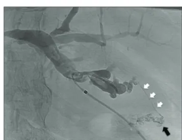
Figure 1: Cholangiography made through an angiographic catheter
show the fistula (white arrows) and the leakage (black arrow) at the entry site of previous PTBD
Figure 2: Selective catheterization of the fistula followed by injection
Bottari, et al.: Bilio‑cutaneous fistula obliteration with NBCA
312 Indian Journal of Radiology and Imaging / Volume 29 / Issue 3 / July - September 2019
given his/her/their consent for his/her/their images and
other clinical information to be reported in the journal. The
patients understand that their names and initials will not
be published and due efforts will be made to conceal their
identity, but anonymity cannot be guaranteed.
Financial support and sponsorship
Nil.
Conflicts of interest
There are no conflicts of interest.
References
1. Vu DN, Strub WM, Nguyen PM. Biliary duct ablation with N‑butyl cyanoacrylate. J Vasc Interv Radiol 2006;17:63‑9.
2. Görich J, Rilinger N, Sokiranski R, Siech M, Vogel J, Wikström M,
et al. Percutaneous transhepatic embolization of bile duct fistulas.
J Vasc Interv Radiol 1996;7:435‑8.
3. Seewald S, Groth S, Sriram PVJ, Xikun H, Akaraviputh T, Mendoza G, et al. Endoscopic treatment of biliary leakage with n‑butyl‑2 cyanoacrylate. Gastrointest Endosc 2002;56:916‑9.
4. Sharif K, de Ville de Goyet J, Bile duct of Luschka leading to bile leak after cholecystectomy‑‑revisiting the biliary anatomy. J Pediatr Surg 2003;38:E21‑3.
5. Minutoli F, Naso S, Visalli C, Iannelli D, Silipigni S, Pitrone A, et al. A new variant of cholecystohepatic duct: MR cholangiography demonstration. Surg Radiol Anat 2015;37:539‑41.
6. Smith AC, Schapiro RH, Kelsey PB, Warshaw AL. Successful treatment of nonhealing biliary‑cutaneous fistulas with biliary stents. Gastroenterology 1986;90:764‑9.
7. Lichtenstein S, Moorman DW, Malatesta JQ, Martin MF. The role of hepatic resection in the management of bile duct injuries following laparoscopic cholecystectomy. Am Surg 2000;66:372‑6; discussion 377.
8. Ryan ME, Geenen JE, Lehman GA, Aliperti G, Freeman ML, Silverman WB, et al. Endoscopic intervention for biliary leaks after laparoscopic cholecystectomy: A multicenter review. Gastrointest Endosc 1998;47:261‑6.
9. Kumar N, Thompson CC. Endoscopic therapy for postoperative leaks and fistulae. Gastrointest Endosc Clin N Am 2013;23:123‑36. 10. Crinò SF, Novel endoscopic management for pancreatic pseudocyst
with fistula to the common bile duct. World J Gastrointest Endosc 2014;6:620.
11. Brady AP, Malone DE, Deignan RW, O’Donovan N, McGrath FP. Fibrin sealant in interventional radiology: A preliminary evaluation. Radiology 1995;196:573‑8.
12. Matsumoto T, Iwaki K, Hagino Y, Kawano K, Kitano S, Tomonari KI, et al. Ethanol injection therapy of an isolated bile duct associated with a biliary‑cutaneous fistula. J Gastroenterol Hepatol 2002;17:807‑10.
13. Lauterio A, Slim A, Aseni P, Giacomoni A, Di Sandro S, Corso R,
et al. Percutaneous transhepatic bile duct ablation with n‑Butyl
cyanoacrylate in the treatment of a biliary complication after split liver transplantation. J Transplant 2009;2009:1‑3.
14. Carrafiello G, Piacentino F, Ierardi A, Cardim L. Percutaneous transhepatic embolization of biliary leakage with N‑butyl cyanoacrylate. Indian J Radiol Imaging 2012;22:19.
15. Mauri G, Pescatori LC, Mattiuz C, Poretti D, Pedicini V, Melchiorre F, et al. Non‑healing post‑surgical fistulae: Treatment with image‑guided percutaneous injection of cyanoacrylic glue. Radiol Med 2017;122:88‑94.
16. Kuran S, Disibeyaz S, Parlak E, Arhan M, Kacar S, Sahin B. Biliocutaneous fistula following alveolar hydatid disease surgery treated successfully with percutaneous cyanoacrylate. Dig Dis Sci 2006;51:18‑20.
17. Wajswol E, Jazmati T, Contractor S, Kumar A. Portal vein embolization utilizing N‑Butyl cyanoacrylate for contralateral lobe hypertrophy prior to liver resection: A systematic review and meta‑analysis. Cardiovasc Intervent Radiol 2018;41:1302‑12. 18. Caloggero S, Catanzariti F, Stagno A, Silipigni S, Bottari A. Use
of a mixture of lipiodol and cyanoacrylate in percutaneous embolization treatment of symptomatic renal Angiomyolipomas: Our experience. Radiol Case Rep 2019;14:343‑7.
19. Kim PH, Tsauo J, Shin JH, Yun S‑C. Transcatheter arterial embolization of gastrointestinal bleeding with N ‑butyl cyanoacrylate: A systematic review and meta‑analysis of safety and efficacy. J Vasc Interv Radiol 2017;28:522‑531.e5.
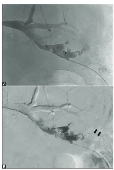
Figure 3 (A and B): The final cholangiography shows complete sealing
of the fistula (A); with digital subtraction the mold of glue (black arrows) along the path is better appreciable (B)
A