U
NIVERSITÀD
EGLIS
TUDID
IM
ESSINAT
ESI DID
OTTORATO DIR
ICERCA INB
IOLOGIAA
PPLICATAE
M
EDICINAS
PERIMENTALECURRICULUM IN MEDICINA SPERIMENTALE
XXXIII CICLO
SSD BIO/14
Neuroinflammation and Oxidative Stress in the Traumatic
Brain Injury and in the Pathogenesis of Neurodegenerative
Diseases: Modulation by Nutritional Supplementation
Candidata:D
OTT.
SSAR
OSALBAS
IRACUSACorrelatore: Relatore:
Ch.ma Prof.ssa Ch.mo Prof.
R
OSANNAD
IP
AOLAS
ALVATOREC
UZZOCREACoordinatore:
Ch.ma Prof.ssa NUNZIACARLA SPANÒ ANNO ACCADEMICO 2019-2020
CHAPTER 1: NEUROINFLAMMATION ... 4 1.1NEUROINFLAMMATION CELLS ... 5 1.1.1 Microglia ... 5 1.1.2 Astrocytes ... 6 1.2MOLECULES OF NEUROINFLAMMATION ... 7 1.2.1 Cytokines ... 7 1.2.2 Chemokines ... 11
CHAPTER 2: OXIDATIVE STRESS ... 15
2.1ENDOGENOUS ANTIOXIDANT SYSTEMS ... 17
CHAPTER 3: NEUROHORMESIS ... 20
CHAPTER 4: NUTRITIONAL SUPPLEMENTATION ... 22
4.1HERICIUM ERINACEUS ... 22
4.2MORINGA OLEIFERA ... 24
4.3HYDROXYTYROSOL ... 27
CHAPTER 5: TRAUMATIC BRAIN INJURY ... 29
5.1CLASSIFICATION OF TBI ... 30
5.2EPIDEMIOLOGY, INCIDENCE AND CAUSES ... 32
5.3THERAPEUTIC STRATEGIESOF TBI ... 33
CHAPTER 6: NEURODEGENERATIVE DISEASES ... 37
6.1PARKINSON'S DISEASE ... 38
6.1.1 Epidemiology and Etiology ... 39
6.1.2 Pathogenesis ... 41 6.1.3 Neuropathology ... 42 6.1.4 Symptomatology ... 43 6.1.5 Therapy ... 50 6.2ALZHEIMER’S DISEASE ... 54 6.2.1 Epidemiology ... 55
6.2.2 Clinical features and pathological changes ... 56
6.2.3 Pathogenesis ... 58
6.2.4 Therapy ... 62
CHAPTER 7: EXPERIMENTAL MODEL OF TRAUMATIC BRAIN INJURY ... 64
7.1MATERIALS AND METHODS ... 64
7.1.1 Animals ... 64
7.1.2 Controlled cortical impact (CCI) experimental TBI ... 64
7.1.3 Behavioral assessment ... 66
7.1.4 Histology ... 66
7.1.5 Western Blot Analysis for IkB-α, NF-kB, Bax, Bcl-2, GFAP and Iba-1 ... 67
7.1.6 Malondialdehyde Content, Superoxide Dismutase, and Glutathione Peroxidase Activity ... 67
7.1.7 Cytokines measurement ... 68
7.1.8 Materials ... 68
7.2.4 Effect of Hericium erinaceus or Moringa oleifera, or Hericium erinaceus plus Moringa oleifera
treatment on oxidative stress. ... 72
7.2.5 Effect of Hericium erinaceus or Moringa oleifera, or Hericium erinaceus plus Moringa oleifera treatment on astrocytes and microglial activation. ... 73
7.2.7 Effect of Hericium erinaceus or Moringa oleifera, or Hericium erinaceus plus Moringa oleifera treatment on apoptosis pathway. ... 74
CHAPTER 8: EXPERIMENTAL MODEL OF ALZHEIMER’S DISEASE ... 76
8.1MATERIALS AND METHODS ... 76
8.1.1 Animals ... 76
8.1.2 Behavioral assessment ... 77
8.1.3 Histopathological examination ... 78
8.1.4 Western Blot Analysis for IkB-α, NF-kB, Cox-2, iNOS and Nrf-2. ... 78
8.1.5 Immunohistochemical localization of Nitrotyrosine... 79
8.1.6 Malondialdehyde (MDA) levels ... 79
8.1.7 Cytokines measurement ... 80
8.1.8 Materials ... 80
8.1.9 Statistical evaluation ... 80
8.2RESULTS ... 80
8.2.1 Effects of Hericium erinaceus or Moringa oleifera, or Hericium erinaceus plus Moringa oleifera, treatment on body weight loss and behavioral alterations ... 80
8.2.2 Daily treatment with Hericium erinaceus or Moringa oleifera, or Hericium erinaceus plus Moringa oleifera, reduced histopathological modifications and b-amyloid depositions AlCl3-induced ... 82
8.2.3 Hericium erinaceus or Moringa oleifera, or Hericium erinaceus plus Moringa oleifera, decreased AlCl3-induced IκB-α degradation, NF-κB p65 nuclear translocation, COX-2 and iNOS expressions in rat brain tissue ... 85
8.2.4 Effects of Hericium erinaceus or Moringa oleifera, or Hericium erinaceus plus Moringa oleifera, treatment on Nrf-2 expression, MDA levels and TNF-a and IL-6 release. ... 86
8.2.5 Effects of Hericium erinaceus or Moringa oleifera, or Hericium erinaceus plus Moringa oleifera, treatment on nitrotyrosine expression ... 88
CHAPTER 9: EXPERIMENTAL MODEL OF PARKINSON’S DISEASE ... 90
9.1MATERIALS AND METHODS ... 90
9.1.1 Animals ... 90
9.1.2 Rotenone-induced PD and treatment ... 90
9.1.3 Behavioral testing ... 92
9.1.4 Histology ... 92
9.1.5 Stereological analysis ... 92
9.1.6 Immunohistochemical localization of tyrosine hydroxylase (TH), dopamine transporter (DAT) and α-synuclein (α-syn). ... 93
9.1.7 Immunofluorescence co-localization of TH/α-syn ... 93
9.1.8 Western blot analysis for IkB-α, NF-kB, Bax, Bcl-2, iNOS, NRLP3, ASC, Caspase-1, IL-18, IL-1β, Hsp70, Sirt-1 and HO1 ... 94
9.1.9 Protein Carbonyl Assay ... 95
9.1.10 Statistical Evaluation ... 95
9.2RESULTS ... 95
9.2.1 Effect of HD treatment on behavioral impairments and on the neuronal degeneration of dopaminergic tract induced by rotenone administration ... 95
9.2.2 HD treatment reduced loss of TH, DAT and α-synuclein expression in the SN induced by rotenone administration ... 97
9.2.3 Effect of HD on cellular stress response after rotenone administration ... 101
9.2.4 Modulation of protein carbonyls in mice brain after rotenone treatment and HD supplementation ... 104
9.2.5 Effect of HD treatment on NF-κB, IκB-α, iNOS expression and on apoptosis induced by rotenone administration ... 105
CHAPTER 10: DISCUSSIONS AND CONCLUSIONS ... 108 REFERENCES ... 115
Chapter 1: NEUROINFLAMMATION
Neuroinflammation is a "cytokine-mediated" inflammatory process that can be caused by systemic tissue damage or, more often, associated with direct damage to the central nervous system (CNS), both of traumatic and/or neurodegenerative origin [1-3]. Neuroinflammation differs from inflammation by the low presence of lymphatic vessels inside the cerebral parenchyma, by the absence of endogenous cells capable of presenting the antigen and by the presence of the blood-brain barrier (BBB), which reduces the exchange of immune cells and mediators of inflammation with the bloodstream. The persistence in the CNS of inflammatory processes can cause serious damage to the neuronal complex up to compromising its functional integrity.
1.1 Neuroinflammation cells 1.1.1 Microglia
Microglial cells represent 5-10% of the total brain cell population. It is a population of hematopoietic derivation: during embryogenesis, in fact, a subpopulation of monocytes migrates into the nervous system and differs into resident macrophages. Microglia cells have a small cell body and long tapered processes equipped with lamellipods that give it a branched morphology. They are spread homogeneously in the cerebral parenchyma and can be found attached to neurons, but also in correspondence with blood vessels and free in gray matter. These cells are internal to the BBB and are therefore ready to receive and respond to any damage to the barrier itself. Microglia are normally inactive in the CNS, the cellular soma remains immobile while the ramifications move continuously to monitor the surrounding environment. The occurrence of changes in homeostasis in the environment, such as increase in serum proteins, toxicity from glutamate, increase in purines (ATP, ADP) or the presence of lipopolysaccharide (molecule present on the membrane of GRAM-negative bacteria) are all stimuli capable of stimulate the microglial, through different receptors and signal pathways. The microglial cells present in the perivascular areas also exercise the function of antigen-presenting cells (APC) on myelin-specific T cells, which have infiltrated the CNS and can thus initiate inflammatory processes [4]. When the microglial is activated, it passes from a branched morphology to an amoeboid, the lamellipods retract and the cell assumes phagocytic capacity, aimed at eliminating any residual dead cells or engulfing bacteria and viruses. Activated microglia has the main role of promoting and supporting the inflammatory state through the production of cytokines, reactive oxygen intermediates, proteinases, complement factors and chemokines. These inflammatory mediators promote the infiltration of immune system cells from the bloodstream, the recall of other microglial cells from the surrounding areas and the activation of astrocytes. When the inflammatory stimulus that caused the activation is lacking,
the microglia participates in the processes of suppression of the inflammatory state with the production of immunomodulatory cytokines, such as Interleukin-15 (IL-15), and anti-inflammatory cytokines, such as Interleukin-10 (IL-10); it then returns to an inactivated state, or undergoes apoptosis [5]. Microglia also have a trophic property, useful for protecting neuronal cells. This action is mediated by the production of growth factors such as glial cell-derived neurotrophic factor (GDNF), brain cell-derived neurotrophic factor (BDNF) and nerve growth factor (NGF). Microglial activation and the neuroinflammatory events that follow are aimed at neuroprotection and elimination of the cause of the lack of homeostasis. In reality, both in chronic neurodegenerative diseases and in traumatic events, an uncontrolled and persistent microglial activation can have neurotoxic effects and contribute to exacerbate neuronal damage. The balance between neuroprotective and neurotoxic action of microglia is determined by many factors, including the nature of the activating stimulus and the interactions that are established between microglia, the other cells of the immune system and the neuronal network, so it is too simplistic to classify the role of microglia in an absolute way and additional studies are absolutely needed to shed light on the mechanisms that regulate this dual role [6]. Numerous evidences show that modulation of microglial activation, and in general of the inflammatory state in the brain, are able to improve the symptomatology of many pathological conditions and to decrease the phenomenon of neurodegeneration [7]. Based on these observations, microglial activation represents a potential pharmacological target for the treatment of traumatic brain injury and neurodegenerative diseases.
2. protoplasmic, mainly located in the gray matter and with short and branched processes; 3. radial, arranged perpendicular to the axis of the ventricles.
One of the main functions of astrocytes consists in the creation of the BBB by wrapping the cerebral capillaries with their processes. They contribute to the structural integrity of the barrier and participate in exchanges between the bloodstream and the cerebral parenchyma. Astrocytes are essential for interactions with adjacent neurons, in fact, they cover the synaptic terminations, safeguarding normal neuronal excitability thanks to the maintenance of extracellular ionic homeostasis [8]. Even astrocytes have the ability to respond to pathological conditions; in these conditions, in fact, they implement a series of functional and structural changes which are called astrogliosis. The astrocytes are activated by the cytokines produced by the microglia and are in turn capable of producing pro-inflammatory molecules.
1.2 Molecules of Neuroinflammation 1.2.1 Cytokines
Cytokines are a class of very small molecules (8-80 kDa) that act in an integrated way in cell communication. In addition to the main role of stimulating the inflammatory phenomenon, they play a role in promoting cell growth, survival and differentiation. Thanks to their biological activities it is therefore possible to group cytokines into different classes: there are 18 cytokines called interleukins (IL), some of these promote inflammation and are called pro-inflammatory cytokines such as IL1β and IL1α, IL6, IL8 and TNFα; while other cytokines suppress the activity of pro-inflammatory cytokines and are called anti-inflammatory cytokines such as IL-4, IL-10, TGFβ. Cytokines are secreted by a variety of immune cells such as T lymphocytes and macrophages, as well as by non-immune cells such as fibroblasts. They are synthesized from many cell types including monocytes, neutrophils, hepatocytes and tissue macrophages, and microglia. The main function of cytokines is to regulate the differentiation of T cells from undifferentiated T-helper 1 and 2 cells, regulatory T cells and T-helper 17 cells [9]. Many of
these cytokines have already been shown to be produced by neurons or glia in central nervous system disorders where they have increased significantly. In fact, it has been shown that after the ischemic insult, and the inflammatory state that follows, there is a significant increase in the concentration of pro and anti-inflammatory cytokines aimed at protecting neuronal networks from cell damage. However, their action is not always positive, and their role in this particular type of insult is still being studied [10]. Cytokines are among the main effector molecules of inflammation, in fact, they respond to various inflammatory stimuli and are also present in auto-inflammatory diseases. The Interleukin 1 family has 11 ligands active on different specific receptors. Among the different IL-1 there are some, such as IL-1beta, which act on trans membrane receptors and others, such as IL-33, which mediate nuclear responses by acting directly from transcription factors. The parent interleukins of this subfamily are IL1-α and IL1-β.
• IL1-α is synthesized in the cytoplasm starting from an active precursor, and once mature it remains mostly bound to the plasma membrane. A fraction is present at the nuclear level where it acts in an autocrine way.
• IL1-β, the most studied of this family, differs from the α type because it is synthesized as an inactive pro-peptide and stored in vesicles. Inflammatory stimuli, such as lipopolysaccharide (LPS) and ATP, determine the maturation of the pro-peptide by the enzyme ICE (interleukin converting enzyme, also called Caspase-1) and the secretion of IL-1β therefore plays a crucial role in the response rapid. 1β also undergoes significant induction of gene expression. IL-1β in the brain is involved in the induction mechanism of fever, together with IL-6, and in the activation of T cells, macrophages and astrocytes. In the CNS, IL-1β can mediate the neurotoxic
showed a reduction in the infarcted area in addition, mice with low production of this cytokine show a dramatic increase in ischemic damage [11, 12]. According to these data, the treatment with IL1-β induced a worsening of the damage. These observations clearly indicate that IL1-β has an important role in determining the severity of ischemic damage. These positive results allowed the start of clinical trials on the treatment with IL-1Ra of patients with cerebral ischemia [13].
• Tumor necrosis factor-α (TNF-α) is considered a fundamental cytokine in the inflammatory process; it is produced by monocytes-macrophages, dendritic cells and lymphocytes; in the brain it is produced by microglia and astrocytes and plays a fundamental role in directing the immune response. It exists both in transmembrane and soluble form, after cutting by the TACE enzyme (TNF-α converting enzyme). The balance between membrane and soluble forms depends on the activation state of the cell and is crucial for its activitỳ. Soluble TNF-α acts on transnmembrane receptors (TNFR1 and TNFR2) that activate different signal pathways, among which the main effector is the one that involves the transcription factor NF-kB, which positively regulates the transcription of numerous pro-inflammatory genes. In the TNF-α nervous system it mediates important functions including the activation of microglia and astrocytes, the regulation of the permeabilitỳ of the blood brain barrier, the induction of feverish state, glutamatergic transmission and synaptic plasticity [14]. TNF-α overexpression has neurotoxic effects: high levels of TNF-α have been measured in the serum of patients suffering from Alzheimer's Disease, Parkinson's disease and multiple sclerosis [15]. In cerebral ischemia TNF-α undergoes an increase in expression, but its role in this insult is still a matter of debate: evidence obtained in animal models shows that treatment with TNFR1 receptor inhibitor drugs determines a reduction in ischemic damage, while other studies have shown that treatment with anti-TNF-α antibodies, which do not discriminate activity on different receptors, leads to
reduced hippocampal neurogenesis. These observations suggest a different role of TNF-α receptors [14].
• IL-6 is also a pro-inflammatory cytokine produced by macrophages, microglia, astrocytes, T lymphocytes, fibroblasts, endothelial cells and keratinocytes. Among its main functions are the induction of feverish state, the generation and coordination of the immune response. In addition, IL-6 is able to activate B lymphocytes and induce them to synthesize antibodies. Unlike IL-1, IL-6 also has anti-inflammatory functions, in particular it inhibits the synthesis of TNF-α and induces the synthesis of soluble receptors for IL-1 and TNF-α, which decrease the share of cytokines available. High serum IL-6 levels have been measured in patients with acute ischemia, and in animal models, IL-6 is induced following ischemic insult in the CNS, particularly in the peripheral region of the ischemic zone. It has a dual role: in fact, it contributes both to brain damage and to repair mechanisms that are carried out through the binding of IL-6 with the gp130 receptor, common to other neurotrophic cytokines (LIF). Although a significant improvement in ischemic damage has not been observed in the animal KO for IL-6, other studies report that direct injection of IL-6 after induction of ischemia determines a reduction in damage.
• IL-10 is a powerful anti-inflammatory cytokine, mainly produced by monocytes-macrophages, microglia and, even if in smaller quantities, by lymphocytes. It is able to inhibit the expression of pro-inflammatory cytokines, such as TNF-α, INF-γ, IL-2 and IL-3. In the brain it plays an important role in controlling the neuroinflammatory state. It is up-regulated after ischemia, produced by the glia, and exerts a neuroprotective action. KO animals for IL-10 subjected to focal ischemia show a larger infarcted area, and other studies report that the
Another important class of mediators of the inflammatory response is prostanoids (prostaglandins, prostacycline and thromboassane), molecules that derive from arachidonic acid. Their synthesis takes place by cyclooxygenase (COX) or PGH-synthase. There are two isoforms of this enzyme: COX-1 and COX-2; they mediate the same catalytic function, but have different physiological roles. In fact, the COX-1 enzyme is constitutively expressed in many cells of the body, including neurons, microglia and lymphocytes and performs functions of maintaining homeostasis. COX-2 is instead inducibly expressed following a pro-inflammatory stimulus. These enzymes are the main target of nonsteroidal anti-pro-inflammatory drugs (NSAIDs). COX-2 expression is induced in many neuroinflammatory diseases. Also following the ischemic event, an increase in COX-2 expression is observed both in the area affected by heart attack and in distal regions. The role of these enzymes is mainly protective, but in the long term, the excessive production of prostaglandins has deleterious effects on the central nervous system. Recent studies suggest that the prostaglandin EP1 receptor has a role in ischemia-induced neurotoxicity. Furthermore, numerous evidences have shown that the treatment with COX-2 inhibitors improves the symptomatology after ischemic insult. COX activity leads to the production of reactive oxygen species, also accused of worsening the damage, although recent evidence has shown that the negative role of COX is mainly attributable to prostaglandins [10].
1.2.2 Chemokines
Chemokines (chemotactic cytokines) are cytokines with a predominantly chemotactic function; they are part of a superfamily of low MW proteins (6-14 kDa), active in the recall of various cell populations that participate in the immune response, such as neutrophil and eosinophil granulocytes, monocytes and lymphocytes, as well as in the cell migration processes that take place during embryogenesis. The chemokine family can be divided into three subfamilies characterized by 2-4 highly conserved cysteine residues in the sequence of the molecule. The
main families of chemokines are represented by: α-chemokines (or CXC not conserved (X);
β-chemokines (or CC β-chemokines), which have two juxtaposed cysteine residues; and δ-chemokines (CX3C δ-chemokines), which show the two cysteine residues separated by three
amino acid residues. Chemokines are produced by a large variety of cell types, generally involved in phlogistic responses. They act on multiple cell types, carrying out numerous actions described in vitro such as chemotaxis, the release of enzymes on cell deposits, the formation of oxygen radicals, the formation of lipid mediators and the induction of adhesion to the endothelium or to the extracellular matrix [17]. In ischemic tissues, different signal pathways (such as that of reactive oxygen species, that of cytokines, the complement cascade and the NF-kB system) can regulate the synthesis of chemokines, resulting in a rapid increase in their concentration, following white blood cell infiltration and immediate inflammatory response [18]. One of the most studied chemokines belongs to the family of CC chemokines and is the Monocyte Chemoattractant Protein-1 MCP-1 / CCL2. CCL2 has a fundamental role in the attraction of monocytes, T cells and NK cells; it is also implicated in diseases characterized by monocytic infiltration.
MCP-1 also has important effects in the heart attack, on the activation and recruitment of macrophages, on the synthesis of cytokines and on the accumulation of myofibroblasts. This chemokine can therefore exert its effects in the first hours after the infarction through distinct mechanisms, such as the recruitment of monocytes in the ischemic myocardium and the modulation of macrophage differentiation, activation of phagocytes and expression of cytokines.
influence of leukocytes, which occurs following axonal damage or in association with neuroinflammatory diseases, such as autoimmune encephalopathy, a model of sclerosis multiple induced in the rat [20].
The CXC chemokines family plays a fundamental role in the regulation of chemotaxis and in the activation of neutrophils in ischemic tissues; but they are still important in inflammation induced by Th1 cell infiltration. In addition, CXCs present angiostatic and inhibitory effects on fibroblast migration.
RANTES (Regulated upon Activation, Normal T-cell Expressed, and Secreted or CCL-5) is part of the CC-chemokines family, it is produced by circulating CD8+ T lymphocytes, endothelial cells, fibroblasts, platelets and in the brain by microglia and astrocytes. Its main function is the recruitment of leukocytes to inflammatory sites; it also activates the release by eosinophils of eosinophilic cationic proteins. RANTES also increases the adherence of monocytes to endothelial cells and selectively supports the migration of monocytes and T lymphocytes that express CD4 markers on the surface. Finally activates the basophils and causes the release of histamine.
Platelet Derived Growth Factor (PDGF) is a chemokine isolated from platelets and synthesized by megakaryocytes as a growth factor for connective tissue and glial cells; the biologically active protein is a dimer composed of two connected polypeptides called A and B. PDGF heterodimers are expressed by a variety of cell types, such as macrophages, endothelial cells, fibroblasts and smooth muscle cells. PDGF binds to plasma proteins and is involved in wound repair processes and angiogenesis. PDGF also stimulates the proliferation of astrocytes and inhibits the premature differentiation of progenitor cells; it is also involved in the development of the central nervous system as the PDGF-b isoform receptors are expressed in many areas of the brain. PDGF has a role in the activation of mesenchymal stem cells. PDGF-B and its PDGFR-β receptor are also expressed at the neuronal level, and an increase in
expression of these proteins has been detected in post mortem brains of patients with ischemia. PDGF has a role in addressing and regulating the differentiation of different stem cells, including neuronal stem cells present in the subventricular and sub-granular area [21].
Interferon Inducible Protein (IP-10 or CXCL10) is a member of the CXC family of chemokines. IFN-γ induces the expression of IP-10 in different cell types, such as monocytes, endothelial cells, keratinocytes, fibroblasts and microglia. IP-10 has chemoattractant activity for monocytes and T cells in humans and promotes the adhesion of T cells to endothelial cells; also in vivo it is able to inhibit angiogenesis and demonstrates anti-tumor activity. In the brain it is mainly produced by microglia and contributes to exacerbate the inflammatory state and its neutralization or depletion turns out to be neuroprotective.
Neural Regeneration Protein (NRP) is a chemokine expressed in stem cells and glial cells. It can induce migration and cell proliferation, promote neuronal survival and increase neurite development; it also induces the differentiation of neuronal stem cells into neurons. NRP exerts its effects on neuronal survival through the phosphorylation of ERK 1 and ERK 2, two cytosolic kinases [22].
Chapter 2: OXIDATIVE STRESS
The term oxidative stress represents the condition that is established when at the cellular level an imbalance is established between the production processes of reactive oxygen species (ROS) and those responsible for their removal [23]. The ROS production and removal processes are strictly regulated, so as to ensure that a reducing environment is preserved inside the cells. ROS represent a group of molecules characterized by a high tendency to react with substrates of various nature by oxidizing them. In physiological conditions, ROS are normally produced inside the cell. ROS have been shown to mediate numerous signaling pathways that take part in cell cycle control. Low or moderate intracellular levels of ROS are associated with the activation of signals involved in the processes of survival, growth, proliferation and cell migration. High quantities of ROS, on the other hand, can damage cellular structures and, consequently, activate cell death signaling pathways [24].
Proteins, nucleic acids and lipids represent the main targets of the oxidizing action of ROS. Proteins can undergo oxidation reactions especially at the level of the side chains of lysine, arginine, proline and threonine. Carbonylation is the consequence of protein oxidation and generally results in the loss of the functions of the protein itself. While moderately carbonylated proteins are directed to the proteasome and degraded, highly carbonylated proteins tend to form high molecular weight aggregates that accumulate inside the cell [25]. DNA is particularly vulnerable to oxidative damage which determines the modification of the bases, the oxidation of deoxyribose, the breakage of the filament, the formation of protein cross-links. The oxidation of the nitrogen bases can induce errors in the duplication process, which, if not corrected by the repair systems, can turn into a stable mutation [26, 27]. Lipids are much more reactive towards ROS than nucleic acids and proteins, because they have unsaturation sites in carbon-carbon double bonds. The hydroxyl radical (OH•) can react with the carbonaceous chain of the phospholipid by extracting a hydrogen atom and forming a carbon-centered radical in a reaction known as lipid peroxidation. Once irreversibly damaged, the lipid is enzymatically degraded
and resynthesized to repair the membrane. The greater the extent of peroxidative damage, the less the cell's ability to promptly repair the membrane.
The main source of intracellular ROS is the respiratory chain. It has been estimated that during breathing 1-2% of the oxygen is not completely converted to water, but only partially reduced producing the superoxide anion (O2• -), which can be converted into hydrogen peroxide (H2O2) and in hydroxyl radical (OH•). The main O2 production sites • - are located at the level of complexes I and III of the electron transport chain (ETC), from which electrons escape by directly reducing molecular oxygen. The ROS generated at the ETC level do not exert their toxic action only in the mitochondria, but also in the cytosolic compartment. In fact, hydrogen peroxide is a highly diffusible species and quickly leaves the matrix and also pours into the cytosol [27].
2.1 Endogenous Antioxidant Systems
Superoxide dismutase (SOD) (EC 1.15.1.1) is one of the most effective enzyme antioxidant systems; it catalyzes the conversion of the superoxide anion into molecular oxygen and hydrogen peroxide in the following dismutation reaction:
O2 • - + O2 • - + 2H + → O2 + H2O2
SOD is present in different isoforms, identifiable on the basis of the ions present in the active site, the amino acid composition and the distribution in the cellular compartments. Three isoforms have been identified in human cells: SOD1 (CuZn-SOD) represents the cytosolic isoform, SOD2 (Mn-SOD) is localized at the mitochondrial level, SOD3 (CuZn-SOD) is instead the extracellular isoform [28]. Hydrogen peroxide, deriving from the dismutation reaction, still represents a reactive species. The enzymes mainly responsible for its removal are catalase and glutathione peroxidase.
Catalase (Cat) (EC 1.11.1.6) is an enzyme present in peroxisomes and catalyzes the conversion of hydrogen peroxide to water and molecular oxygen in the following reaction:
H2O2 + H2O2 → 2H2O + O2
Cat is one of the enzymes with the highest turnover numbers: a catalase molecule can convert about six million hydrogen peroxide molecules in a minute [29].
Glutathione peroxidase (GPx) (EC 1.11.1.9) competes with catalase for the removal of hydrogen peroxide and represents the main source of protection against low levels of oxidative stress [30]. GPx catalyzes the conversion of hydrogen peroxide or an organic peroxide (ROOH) to water or alcohol (R-OH) by using glutathione as a reducing agent.
2GSH + H2O2 → GSSG + 2H2O
2GSH + R-OOH → GSSG + R-OH + H2O
Glutathione (GSH) is the most potent non-enzymatic endogenous antioxidant. It is a tripeptide (cysteine-glycine-glutamate) present in large quantities in the cytosol, in the nucleus and in the mitochondria. When glutathione is used during peroxide detoxification reactions, two
molecules of reduced GSH are oxidized to form a disulfide bridge (GS-SG) [31]. Under physiological conditions, oxidized glutathione makes up about 1% of total glutathione; variations in the GSH/GSSG ratio represent an index of the state of oxidative stress [32]. A recurrent theme is reactive redox signaling based on cysteine thiol. Under physiological conditions, reactive cysteine thiols exist as thiolate anions (S−) and are more reactive towards ROS than sulfhydryl groups (-SH). Reactive thiols interact with ROS to generate a range of cysteine oxidation products, including sulfuric, sulfinic and sulfonic acids; S-nitrosothiol and thionitrates; S-glutathiolation products; and inter and intraprotein disulfides [33]. The modification of the critical cysteine thiols of Keap1 and Nrf2 by oxidants and electrophiles is a main mechanism by which ARE inductors activate Nrf2 [34, 35].
Nrf2 directly influences ROS homeostasis by regulating antioxidant defense systems through different mechanisms. Among these we find:
1. induction of superoxide and peroxide catabolism through SOD, Prx and GPx; 2. regeneration of cofactors and oxidized proteins;
3. synthesis of reducing factors, such as GSH from GCLC and GCLM, and NADPH from G6PDH and 6PGD;
4. expression of the antioxidant protein Trx and inhibition of the expression of the Trx TXNIP inhibitor;
5. increase in oxide-reduction transport; 6. metal-chelation by MT1, MT2 and ferritin;
7. induction of stress response proteins, such as HO-1.
induced by oxidants through Nrf2, creating a positive feedback circuit with Nrf2. Among the proteins dependent on Nrf2 we have heme oxygenase 1 (HO-1), which in addition to removing excess toxic heme, produces iron, biliverdin and carbon monoxide ions. This protein, together with its products, have beneficial effects by protecting against oxidative lesions, regulating apoptosis, modulating inflammation and contributing to angiogenesis. Therefore, Nrf2 and HO-1 represent factors of protection from stress and anti-aging. Furthermore, recent discoveries have revealed that Nrf2 plays a role in regulating various processes such as autophagy; reporting to inflammasome; endoplasmic reticulum stress and unexplained protein response; mitochondrial biogenesis; stem cell function [36, 37].
Chapter 3: NEUROHORMESIS
The term hormesis refers to a process that places itself at the center of the responses of cells and organisms to environmental factors and biological organization [38]. This process is nothing more than the expression of integrative adaptive responses characterized by biphasic dose responses with specific quantitative response characteristics (maximum amplitude and width of the adaptive response) and induced by a direct stimulatory response or the result of compensatory biological processes following of an interruption of homeostasis [39, 40]. More simply, the term hormesis refers to those drugs, toxins or natural substances which, if administered in low doses, can give a positive response on the cell or organism, while at higher concentrations they are harmful and toxic [38].
At the cellular and molecular level, hormesis involves the activation of adaptive stress response pathways that involve, for example, a greater expression of heat shock proteins, antioxidant enzymes and anti-apoptotic proteins. All these phenomena act in most cases through hormonal mechanisms [41]. These hormone dose responses provide quantitative information on the limits of biological plasticity [42] and allow us to measure how much adaptive processes can be upregulated, which is particularly relevant for understanding the protective effects induced by plant and fungal species.
The hormonal concept is extremely important as it provides reliable estimates of the upper limit for the induction of potential therapeutic responses and should play a key role in carrying out experimental and clinical studies.
The hormesis has aroused particular interest in the toxicological community for the dose-response model, especially in vulnerable biological systems, such as the brain. We therefore
Neurohormesis affects the memory, learning, performance and neurodegenerative responses mediated by oxidative stress in cellular models for various diseases such as neurodegenerative ones [38, 43].
Common examples of neurohormesis are ischemic preconditioning [44] and adaptive responses of neurons to moderate intensity excitatory neurotransmission [45], exercise [46] and dietary restrictions [47]. Neurohormesis can also be induced by endogenous neurotoxic molecules such as nitric oxide [48], carbon monoxide [49], glutamate [50] and Ca2+ [51].
Both in vitro and in vivo studies have shown that the responses of neurohormesis to stressors can be mediated by several factors [52-55]:
- NF-kB;
- cAMP-response-element binding protein (CREB); - factor related to nuclear factor E2 (Nrf2);
- factor 1 inducible by hypoxia (HIF1).
Other mechanisms could include modulating the expression of proteins involved in the regulation of oxidative stress and Ca2+ cellular homeostasis [56].
Chapter 4: NUTRITIONAL SUPPLEMENTATION
Epidemiological studies and experiments on animal models have revealed that the phytochemicals present in fruit and vegetables can protect the nervous system from disease [57, 58]. In particular, it has been shown that the beneficial effects of phytochemicals on health are due to the antioxidant properties of many chemical products contained in them. For example, numerous studies have reported the neuroprotective effects of compounds found in natural products, including -tocopherol, lycopene, resveratrol, ginkgo biloba and ginsenosides [59]. Markers of oxidative stress have also been identified in the brains of patients with neurodegenerative diseases that support intervention with neuroprotective nutritional substances based on the antioxidant action and anti-inflammatory agents, such as polyphenols or fungi [60]. In fact, it is known that polyphenols and fungi activate the pathway of heat shock proteins (Hsp), which plays a crucial role in the response to cellular stress.
Therefore, the neuroprotective effects of these products are associated with their ability to reduce oxidative stress but, recently, other biological activities of phytochemicals are emerging that could be beneficial to human health [61].
4.1 Hericium erinaceus
Medicinal mushrooms have become a focus for researchers because the bioactive compounds they contain have many therapeutic properties. Hericium erinaceus has been commonly prescribed in traditional Chinese medicine (TCM), because its fruiting body and mycelia contain a high amount of structurally different bioactive and potentially bioactive components whose consumption has proven to be beneficial for human health. This mushroom species is
Figure 3. Hericium erinaceus
Fungus and fermented mycelia have been shown to produce numerous classes of bioactive molecules, including polysaccharides, proteins, lectins, phenols and terpenoids. The numerous bioactive compounds of Hericium erinaceus have been developed in food supplements and alternative medicines [62]. These compounds have been shown to have different properties such as antibiotic, antidiabetic, hypertensive, cardioprotective, anticarcinogenic, anti-hyperlipidemic, anti-aging, nefroprotective, hepatoprotective, neuroprotective, anti-depression and also improves anxiety and function cognitive [63]. The antioxidant property of Hericium erinaceus was shown in a diabetic rat model in which the intraperitoneal (ip) administration of an aqueous extract of Hericium erinaceus (100 and 200 mg/ kg) led to a significant reduction in serum glucose levels, increase in insulin level and attenuated serum lipid profiles compared to the control group. These results are associated with an increase in the activity of antioxidant enzymes such as catalase (CAT), superoxide dismutase (SOD) and glutathione peroxidase (GSH-Px), an increase in glutathione levels (GSH) and a reduction in malondialdehyde (MDA) in the liver. These data suggest that the health promoting mechanism appears to be the result of ROS inhibition [64]. Also interesting is the beneficial effect observed following the use of two
classes of terpenoid compounds, ericenones and erinacins, respectively isolated from fruiting bodies and mycelia. These compounds have been able to stimulate the synthesis of nerve growth factor (NGF) in cultured astrocytes [65]. The cholineacetyltransferase and acetyl cholinesterase enzymes are modulated by NGF which is therefore capable of influencing the cholinergic neurons of the basal midbrain. It is known that the first pathological events of Alzheimer's disease are precisely the loss and dysfunction of cholinergic neurons. Therefore, the administration of Hericium erinaceus improves cognitive dysfunction. Recent, both basic and clinical studies have shown that Alzheimer's disease is closely associated with beta amyloid-induced neuroinflammation (Aβ). This protein is responsible for activating the astrocytes and microglia that promote neuronal damage through the release of proinflammatory and cytotoxic factors, aggravating the course of the disease [66]. A new neuroprotective strategy is represented by the oral administration of a Hericium biomass preparation given for 3 months [67]. This type of intervention can represent a therapeutic goal to minimize the harmful effects related to the oxidative load, such as that which occurs in the brain during aging or neurodegenerative disorders. Hericium erinaceus treatment resulted in a significant increase in LXA4 production in most brain regions such as cortex, hippocampus, substantia Nigra, striatum and cerebellum and a modulated expression of cytoprotective proteins, such as HO-1, Hsp70 and Trx. These results are consistent with recent evidence from mice studies showing Hericium erinaceus neuroprotection on Aβ 25–35 peptide-induced cognitive dysfunction [68, 69].
4.2 Moringa oleifera
Figure 4. Moringa oleifera
Thanks to its high nutritional values, each part of the tree can be used for nutritional or commercial purposes. For example, the leaves are rich in minerals, vitamins and other essential compounds and for this reason their extracts are used for the treatment of malnutrition and to increase breast milk in breastfeeding mothers. As is known, most vegetables lose nutrients during cooking. However, it has been observed that Moringa leaves, fresh, cooked or stored as dried powder for months without refrigeration, have not lost their nutritional value [70]. Boiled leaves produced three times more bioavailable iron than raw leaves. These results were also observed in powdered Moringa leaves. In addition, Moringa was found to have a group of unique compounds containing sugar and rhamnose, which are unusual glucosinolates modified by sugar [71]. These compounds have been reported to exhibit some chemo preventive activity, inducing apoptosis [72].
Moringa oleifera has been used as a treatment in numerous diseases, such as obesity, diabetes, scurvy, hysteria and tumors [73, 74]. Moringa oleifera has anti-cancer [75], anti-inflammatory [76], anti-oxidant [77], hepatoprotective [78], anti-bacterial [79], cardioprotective [80], hypolipidemic [81], hypoglycemic [82] and antihypertensive [83] activity.
In a rat model of liver fibrosis, Moringa oleifera has been shown to reduce not only histopathological damage but also to modulate various biochemical markers such as aspartate aminotransferase (AST) and alanine aminotransferase (ALT), which are indicators of liver health conditions [84]. In another study, the protective ulcer property of 50% ethanolic leaf extract of Moringa oleifera was demonstrated in different induction models of gastric ulcers. The anti-inflammatory and anti-oxidant role of Moringa oleifera was initially observed in a model of diabetes induced by obesity. In this study, the ability of this natural compound to modulate cytokine levels such as tumor necrosis factor alpha (TNF-a), interleukin-1 beta (IL-1b), but also the inducible nitric oxide synthase (iNOS) and the nitric oxide (NO) [85-87]. Polyphenols represent the main natural compounds capable of reducing oxidative damage in tissues. Moringa oleifera leaves have a high number of polyphenols and therefore have an important antioxidant power [88]. Both mature and tender leaves of this plant have been shown to exhibit antioxidant activity against free radicals, preventing oxidative damage to major biomolecules and tissues [89]. In addition, the 50% ethanolic leaf extract has been tested to study the activities of LPO, CAT and SOD. The antioxidant properties of Moringa extract have been found to modulate the levels of SOD, CAT and LPO in the gastric mucosa of the rat [70]. In a recent study, based on the role of oxidative stress in stroke pathophysiology, it was determined whether Moringa oleifera protected against brain damage and oxidative stress in the animal model of focal stroke. The results obtained from this study showed that all the used doses of extract reduced the volume of the heart attack both in the cortex and in the sub-cortex. The protective effect of medium and low doses of extract in all areas occurred mainly through the reduction of oxidative stress [90]. Oxidative stress plays a crucial role in age-related
is capable of improving spatial memory and neurodegeneration in the areas CA1, CA2, CA3 and in the dentate gyrus of the hippocampus. It also decreases MDA levels and AChE activity but increases that of SOD and CAT. Therefore, the extract of Moringa oleifera leaves represents a therapeutic potential against cognitive disorders. The possible mechanism is probably related to the ability to reduce oxidative stress and increase cholinergic function [91].
4.3 Hydroxytyrosol
In recent years, numerous studies support the beneficial effects of the Mediterranean diet (MD) in preventing neurodegeneration. MD predicts a high intake of cereals, vegetables, fruit, olive oil, legumes, low meat consumption, and a moderate amount of fish and seafood and alcohol. This translates into a reduction in the consumption of saturated fat and an increase in the intake of foods containing high concentrations of bioactive compounds such as polyphenols and flavonoids [92, 93]. In this regard, one of the main phytochemicals present in oil and table olives is Hydroxytyrosol (HT).
Figure 5. Hydroxytyrosol
Its importance is supported by numerous studies that highlight the high antioxidant power of HT and its ability to eliminate free radicals [92]. Therefore, interest in HT has increased in
recent years and this is also due to the possession of other biological activities including anti-inflammatory, antimicrobial, anticarcinogenic and neuroprotective effects [92, 94-97]. All of these activities could give HT a central role in the prevention of neurodegenerative diseases. Although there are still few studies on the neuroprotective effects of HT in animal models, they provide important data that encourage the evaluation of this compound in the treatment of neurodegenerative disorders. In an animal model similar to Huntington's disease, the antioxidant effect of oral administration of extra virgin olive oil (EVOO) and HT has been shown in the brain. The results show a decrease in lipid peroxidation and an increase in cellular glutathione (GSH) levels. This displays that EVOO and HT act as brain antioxidants through a natural mechanism useful for protection from oxidative damage [98]. In another study, C57BL/6 mice were treated with oligomeric acid Aβ1–42 + ibotenic acid to induce neural behavioral dysfunction. After induction, a severe deficiency in the visuo-spatial and working memories took place. However, HT treatment appreciably ameliorated spatio-cognitive performances. Further research has shown that HT administration has been able to counteract dysregulation of signaling mechanisms in hippocampal neurons [99-101]. Recently, increasing evidence reports that HT and its derivates activate phase 2 response leading to the expression of nuclear factor erythroid 2-related factor (Nrf2) antioxidant pathway [102, 103]. Interestingly, Nrf2 represents a crucial mechanism of resistance to oxidative stress and inflammation in vitro and in vivo [104, 105]. In line with these observations, Nrf2 codifies the antioxidant pathway of vitagene that exists to counteract various forms of stress (eg oxidative, environmental and proteotoxic stress). The latter involves redox sensitive genes, such as γ-glutamyl cysteine synthetase (γ-GCS), HO-1, heat shock protein 70 (Hsp70), thioredoxin and sirtuin-1 (Sirt1),
Chapter 5: TRAUMATIC BRAIN INJURY
Traumatic brain injury (TBI) is a damage that does not occur due to a hereditary, congenital factor but can result from a wide variety of insults, such as external mechanical force. This can cause temporary or permanent impairment of both cognitive and physical functions [108]. Additionally, the TBI can lead to coma and even death. Cerebral lesions are divided into 2 subcategories: primary lesions, which occur at the time of the trauma, and secondary lesions, which occur immediately after the trauma and whose effects can be prolonged over time. What happens in the brain following TBI are a series of cellular and molecular interactions, which cause excitotoxic effects related to oxidative stress, inflammation, ionic and metabolic imbalance. The combination of these pathways induces progressive neuronal loss through necrosis and apoptosis [109, 110]. Also, very important are the changes that occur within the cells that are determined by the increased flow of calcium, which affects mitochondrial integrity. Lactate accumulation is also cytotoxic for cells as, together with the increase in vascular permeability, cerebral edema occurs, increase in intracranial pressure and decrease in brain perfusion [109-111]. People who have undergone a TBI also experience a number of stereotyped symptoms such as confusion, dizziness and sometimes loss of consciousness. In addition, about 70-80% of people with TBI, even after the resolution of the initial lesion, develop a series of long-lasting symptoms such as anxiety and depression that affect the patient's personality [112-115]. In addition, TBI may increase the risk of developing neurodegenerative diseases. An example is the repeated concussive TBI that appears to appear to lead to the development of chronic traumatic encephalopathy (CTE) in athletes. Furthermore, repeated or single TBI appears to be strongly associated with the risk of developing Alzheimer's disease, Parkinson's disease and amyotrophic lateral sclerosis [116].
Considering the high number of cases of TBI and its association with severe neurological problems and risk of neurodegenerative diseases, there is a strong interest in developing new
therapies that not only promote cell survival immediately after injury, but also address the development of secondary pathology [117-119].
Figure 6. Schematic representation of TBI
5.1 Classification of TBI
In the United States and many other countries, a score scale known as the Glasgow Coma Scale (GCS) is used to evaluate a patient's mental and neurological status following a brain injury [120]. The score is based on the sum of three components: eye opening response, verbal response and motor response (see the following table).
GCS can be further divided into minor injuries (13 to 15), moderate injuries (9 to 12) and serious injuries (3 to 8). The clinical features that allow us to define a mild injury are loss of consciousness for 20 minutes, absence of focal neurological changes, no intracranial injury and no intracranial surgery. The patient, on the other hand, falls into the moderate category when, regardless of mental state, he has a focal lesion of computed tomography (CT). Finally, the patient is defined as serious when, always regardless of mental state, he goes into a coma for at least 6 hours. The Glasgow scale is not the only one, however, the only scale used in terms of outcome. Another scale commonly used, especially by rehabilitation facilities, is the scale of cognitive functioning level of Rancho Los Amigos (see the following table) [120].
5.2 Epidemiology, incidence and causes
Each year, approximately 30 million visits, hospitalizations and injury deaths occur in the United States alone. Among hospitalizations, approximately 16% include TBI as a primary or secondary diagnosis. About a third of deaths include the TBI as a cause of direct or underlying death. To make an estimate, of all the people who are hospitalized for accidents, about 87% are treated and subsequently released, 11% are hospitalized and discharged, and about 2% die. These data, however, are not real as they do not take into account those people who have not needed medical treatment, who have made outpatient visits, or who have received treatment at a federal facility [121].
In the United States, children (0-4 years), teenagers (15-19 years) and adults (> 75 years) are the groups most likely to undergo a visit or be hospitalized for TBI [121]. In particular, the rates of hospitalizations and deaths following TBI are very high among adults aged ≥75 years. Overall, males account for approximately 59% of all TBI-related medical visits to the United States [121]. As can be seen from Table 4, in the period 2002-2010, the main causes of TBI-related visits were falls, road accidents and hits from or against an object.
Instead, the main causes of TBI-related deaths are traffic accidents, accidental falls and suicides [122]. Other data that are not estimated are the percentages of TBI that occur during sports and recreational activities. However, according to the National Electronic Injury Surveillance System - All Injury Program, in the period 2001-2009 (CDC, 2011) the activities associated with the highest estimated number of TBI-related hospital visits were cycling, soccer, game and basketball activities among people under the age of 19.
5.3 Therapeutic Strategies of TBI
Neuroprotection, neurovascular regeneration, and neuro-restoration have been proposed to be therapeutic strategies for TBI. As we have seen before, the TBI leads to a cascade of events and a loss of neurons, which clinically manifests itself with different degrees of neurological deficit depending on the site and the severity of the loss of neurons. Numerous studies have been conducted to reduce or prevent TBI-induced neuronal loss. Generally in in vivo studies a treatment to have neuroprotective effects on TBI must be carried out within a few hours from the first impact [123-125]. However, many clinical trials using neuroprotective treatment have not shown promising results. Therefore, even today, the TBI remains an unmet medical need and an important source of disability and mortality in developed and developing societies. Below we describe the current therapeutic strategies in use.
Acetylcholinesterase inhibitors: Central acetylcholinesterase (AChEI) inhibitors have the role
are normally used for the treatment of mild to moderate Alzheimer's disease but have also been used for other cognitive disorders, including TBI. The AChEIs most used in recent years in clinical trials for chronic TBI are donepezil [126-132] rivastigmine [131, 133-137] and galantamine. These studies have suggested that these compounds may have beneficial effects, especially in patients with moderate and severe chronic TBI who have persistent cognitive deficits, by increasing synaptic ACh levels. The benefits of these compounds have also been reported in preclinical studies of TBI. In particular, it was observed that AChEI had positive effects on acute injury processes with consequent reduction of TBI-induced neuronal death, on the conservation of neurons in the hippocampus region CA1, on the reduction of the interruption of the blood-brain barrier (BBB) , on the reduction of cerebral edema and on the reduction of neurological and motor deficits [126, 138].
Amantadine: Amantadine (1-adamantamine hydrochloride) is a tricyclic amine normally used
for the treatment of influenza A, and in recent years it has shown modest efficacy also for the treatment of Parkinson's disease. Studies have shown that amantadine increases extracellular dopamine (DA) concentrations by blocking the recovery of DA or facilitating the synthesis of DA [139]. It can also have post-synaptic effects, acting on the circuits of the DA and increasing the DA density of the receptor [140]. Amantadine has been studied little in the preclinical models of experimental TBI. One study showed that amantadine treatment, starting 1 day after a closed controlled cortical impact model of TBI in rats and continuing for 18 days after injury, resulted in modest improvement in Morris water maze (MWM) latencies [141]. However further studies are needed. Clinically, amantadine has been shown to be effective during the post-acute period in the patient with TBI. In another study, amantadine was shown to result in
Cyclosporine A/FK 506: Cyclosporin A (CsA) is capable of maintaining the potential of the
mitochondrial membrane by inhibiting the opening of the transition pore of the mitochondrial permeability after TBI. This action has been confirmed by in vivo studies that have shown that CsA is able to reduce reactive oxygen species thus preserving mitochondrial function [143, 144]. In addition, these compounds inhibit the calcineurin phosphatase protein, also having positive effects on axonal lesions and on learning and memory [145-149]. Another characteristic of CsA is their immunosuppressive power [150]. Not all studies have produced positive results. Both preclinical and clinical studies suggest that the acute treatment of patients with severe TBI is the most suitable route for the clinical development of CsA or FK 506.
Simvastatin/other statins: Statins, 3-hydroxy-3-methylglutaryl coenzyme A (HMGA)
reductase inhibitors, reduce serum cholesterol but also have potent effects in the brain relevant to mechanisms of TBI injury and recovery. Such effects target mechanisms that influence both the acute and chronic phases of TBI [151, 152]. There is pre-clinical evidence of beneficial effects including those on acute injury processes such as brain edema, BBB integrity, cerebral blood flow, neuroinflammation, axonal injury, and cell death, in addition to effects on key facets of regeneration such as trophic factor production. A variety of molecular outcomes are influenced including TUNEL staining, CREB, Akt, eNOS, FOXO1, NF-κB, GSK3, cytokines, BrdU labeling, blood vessel formation, and vascular endothelial growth factor [153].
N-acetylcysteine (NAC): NAC is FDA-approved as an antidote for acetaminophen overdose
and as a mucolytic for cystic fibrosis and other bronchopulmonary diseases. In animal models of TBI, NAC has shown strong antioxidant activity by increasing glutathione levels and decreasing markers of oxidative damage [154]. NAC also showed anti-inflammatory activity by decreasing the activation of NF-κB, while lowering interleukin (IL)-1β, tumor necrosis factor (TNF)-α, and intercellular adhesion molecule (ICAM)-1 levels [155, 156]. It is unclear how the anti-inflammatory action of NAC is related to its antioxidant activity. NAC has been
shown to reduce lesion volume while simultaneously reducing levels of the putative neuroprotective enzyme heme oxidase [155, 156].
Growth hormone (GH): GH is a polypeptide that is synthesized, stored, and secreted by
somatotrophic cells within the lateral wings of the anterior pituitary gland. GH deficiency/insufficiency (GHD/GHI) is the most common anterior pituitary abnormality after TBI. Manipulating the GH axis has been shown to improve motor function, enhance learning and memory retention after TBI in rats, and to improve spatial learning and memory in a mouse model of AD [157, 158].
Erythropoietin (EPO): EPO is a cytokine implicated in erythropoiesis and has a number of
beneficial effects that may be important in the treatment of TBI such as attenuation of glutamate and nitric oxide toxicity, anti-apoptotic, anti-oxidant and anti-inflammatory effects , stimulation of neurogenesis and angiogenesis, and protection of mitochondria [159-161]. Numerous studies have demonstrated the efficacy of EPO in rodent models of TBI [151, 162-167]. The route of administration, dosage and therapeutic window appear to be favorable. Research indicates that any parenteral route of administration appears to be effective for the treatment of TBI. In addition, the therapeutic window can be prolonged, with some studies suggesting a benefit with a first dose for up to 24 hours after injury [159]. Similarly, the administration of EPO in humans has been shown to be effective in chronic conditions [168]. EPO has been beneficial and has reduced gray matter loss in chronic schizophrenia [169].
Chapter 6: NEURODEGENERATIVE DISEASES
Neurodegenerative diseases represent a heterogeneous set of distinct nosographic entities, united by some pathogenic and clinical characteristics. From the point of view of pathogenesis, they are characterized by a chronic and selective process of cell death affecting neurons. The exact etiology behind this pathogenetic process is not yet defined. Although in some sporadic cases some genetic mutations responsible for the development of disease have been identified in families affected by some degenerative pathologies, in the etiopathogenesis of most of them numerous risk factors, of both genetic and environmental origin, seem to play a fundamental role.
The progressive degeneration, which precedes the appearance of symptoms for a few years, concerns, at least in the initial phase, a certain population of neurons. Subsequently, during the course of the disease, other neuronal systems may be damaged. From a clinical point of view, therefore, neurodegenerative diseases begin insidiously, generally in adulthood, and have a progressive and inexorable course that culminates in a serious disability, which often follows the patient's death [170].
Although in the early stages they can take on a focal character, these pathologies generally affect a specific neuronal system bilaterally, giving rise to an extremely varied clinical symptomatology. In fact, neuronal deterioration is the cause of irreversible and inevitable damage to brain functions that occurs, depending on the type of disease, with cognitive deficits, dementia, motor alterations and behavioral and psychological disorders, more or less serious. The definition and classification of neurodegenerative diseases, due to the overlapping of the symptoms and sometimes also the sharing of some phases of the pathogenetic process, continue to be the subject of a heated medical-scientific debate. However, it can now be said that several well-defined clinical entities are grouped under this name, of which the best known are Alzheimer's disease and Parkinson's disease. The other main neurodegenerative diseases are
Huntington's disease, amyotrophic lateral sclerosis, progressive supranuclear palsy, frontotemporal dementia and Lewy body dementia.
From a therapeutic point of view, although for some of these pathologies instruments are available which are able to delay or control, more or less effectively, the clinical symptoms, they still remain incurable diseases. Despite the significant progress made in recent years by biomedical research, in fact, there is still no therapeutic intervention that has proven capable of reversing or stopping the pathological process underlying these disorders. This situation largely depends on the fact that the cellular and molecular mechanisms underlying the neuronal damage observed in the various neurodegenerative diseases are still poorly understood [170].
6.1 Parkinson's disease
It is a chronic progressive disease, defined clinically by the association of tremor, rigidity, brady-akinesia and postural instability, and neuropathologically characterized by severe degenerative changes in the substantia nigra (SN) and in the pigmented nuclei of the brain stem, with the presence of specific cell inclusions (Lewy bodies) in residual neurons. Described initially as "agitating paralysis" by James Parkinson ("Essay on the Shaking Palsy", 1817) and later by Charcot (1872-1873) who specified its clinical characteristics, the disease found its first neuropathological identity with the studies by Tretiakoff (1919) who underlined the constancy of the alterations of the SN. Biochemical studies have made it possible to demonstrate the existence of a severe dopamine depletion of the striatum, as a consequence of the neuronal depletion of the SN, thus providing a pathophysiological interpretation of the disease [171].
Figure 7. Main feature of Parkinson’s disease.
6.1.1 Epidemiology and Etiology
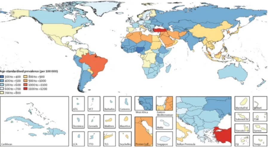
The disease begins insidiously in the second half of life (usually between 50-60 years); cases that begin before the age of 40 are relatively rare. The frequency is high: it is estimated that every year a new case appears for every 4000 inhabitants and, if referred to subjects over 50 years of age, a new case for every 1000. In Italy the prevalence has been indicated between 95 and 199 /100,000 in different epidemiological studies [172], with progressive growth over 50 years. It can be assumed that there are currently around 100,000 Parkinsonians in Italy. The disease affects males and females (with a slight preponderance for the male sex) and is ubiquitously widespread, albeit less frequently in China and Africa than in western nations. The cause of the disease is unknown, although studies on its etiology have intensified especially after the discovery of a toxic substance (1-methyl-4-phenyl-1,2,3,6-tetrahydropyridine or MPTP; rotenone) capable of reproducing, in humans and in some animal species, a clinical and neuropathological picture similar to that of Parkinson's [173]. The toxicity of MPTP develops as a result of its metabolization by glial monoamine oxidase B (MAO B) with the formation of the MPP + compound which is transported within dopaminergic neurons, where it accumulates in the mitochondria by inhibiting complex I of the respiratory chain and causing ATP depletion and therefore the onset of cell death.
The discovery of parkinsonism from MPTP or rotenone has supported the hypothesis that Parkinson's idiopathic disease may depend on exposure to toxic substances of environmental origin with variable distribution (the risk of disease would be higher in a rural environment, in relation to the use of pesticides and herbicides). However, research in this direction has not been conclusive and the hypothesis has been put forward that the responsible toxic substances are more widely distributed, but that only some individuals are vulnerable to potential toxic damage in relation to a genetic predisposition. Alternatively, it has been suggested that the toxic action on the nigral cells may be of endogenous origin: in particular, some products of the normal dopamine catabolism (similarly to any environmental toxins) would be responsible for the formation of free radicals and would induce oxidative stress damage. Free radical theory has been the subject of heated controversy [174, 175], but there is no doubt that a specific deficiency of mitochondrial complex I and an increase in iron levels have been documented in the SN of parkinsonian subjects, not compensated by increases in ferritin [176]. In addition, it has recently been shown that the toxic mechanisms mentioned above are capable of inducing the phenomenon of apoptosis which could represent the main factor responsible for neuronal death. The possible role of heredity in Parkinson's disease has also been the subject of conflicting opinions: the finding of a positive family history only in 10-15% of cases has suggested the existence of an autosomal dominant predisposition with reduced penetrance , which is difficult to highlight due to the marked variability of the preclinical phase of the disease [177]. The recent study by Tanner et al. (1999) on the degree of concordance between monozygotic and dizygotic twins suggests that early-onset parkinsonisms would be characterized by a strong genetic component, while in parkinsonisms with onset over 50 years
Figure 8. Diagnosed prevalent cases of PD
6.1.2 Pathogenesis
The consequence of the loss of dopaminergic neurons of the SN is the reduction of dopamine at the striatal level, the main biochemical alteration of Parkinson's disease [171]. There is a close parallel between the extent of neuronal depletion, the degree of depletion of striatal dopamine and the severity of clinical symptoms: the preclinical phase of disease has a variable duration, probably less than 7 years according to recent PET data [178], during which the loss of dopaminergic neurons and the reduction of the striatal dopamine content are accompanied by a minor reduction of the homovanillic acid (and of the enzymes tyrosine hydroxylase and dopa-decarboxylase) in relation to an increased dopaminergic turnover and to the compensatory development of receptor hypersensitivity from denervation (i.e. an increase in the number of dopaminergic receptors). Parkinsonian symptomatology occurs when the number of dopamine neurons and the content of striatal dopamine have fallen below a critical level (70-80%) [179]. As the disease progresses, the reduction of dopaminergic neurons is associated with a progressive reduction in the number of postsynaptic receptors. However, the biochemical alterations that characterize Parkinson's disease are much more complex and widespread,