69
Original Paper
Cell Physiol Biochem 2005;15:69-76 Accepted: November 02, 2004
Cellular Physiology
Cellular Physiology
Cellular Physiology
Cellular Physiology
Cellular Physiology
and Biochemistr
and Biochemistr
and Biochemistr
and Biochemistr
and Biochemistryyyyy
Copyright © 2005 S. Karger AG, Basel
Fax +41 61 306 12 34
© 2005 S. Karger AG, Basel 1015-8987/05/0154-0069$22.00/0
Thyroid Status Affects Rat Liver Regeneration After
Partial Hepatectomy by Regulating Cell Cycle and
Apoptosis Proteins
Anna Alisi
1,2, Ilaria Demori
3, Silvana Spagnuolo
4, Enrico Pierantozzi
1,
Emilia Fugassa
3and Silvia Leoni
11Department of Cellular and Developmental Biology, University “La Sapienza” Rome, 2Laboratory of
Gene Expression, Fondazione Andrea Cesalpino, I Clinica Medica, Policlinico Umberto I, 3Department of
Experimental, Enviromental and Applied Biology, University of Genoa, 4Department of Pharmacology of
Natural Substances and General Physiology, University “La Sapienza” Rome
Key Words
Thyroid hormones • Cyclins • p53 • p73
Abstract
In rats, various growth factors and hormones, as well as partial hepatectomy (PH) are able to trigger the proliferative response of hepatocytes. Although recent evidence highlights the important role of thyroid hormones and thyroid status in regulating the growth of liver cells in vitro and in vivo models, the mechanism involved in the pro-proliferative effects of thyroid hormones is still unclear. Here we have investigated how in rats made hypo- and hyperthyroid after prolonged treatment respectively with propylthiouracil (PTU) and triiodothyronine (T3), the thyroid status affects liver regeneration after PH by regulating cell cycle and apoptosis proteins. Our results show that both in control and partially hepatectomized animals hyperthyroidism increases the cyclin D1, E and A levels and the activity of cyclin-cdk complexes, and decreases the levels of cdk inhibitors such as p16 and p27. On the contrary hypothyroidism induces a down-regulation of the activity of cyclin cdk complexes decreasing cyclin levels. Thyroid hormones control
also p53 and p73, two proteins involved in apoptosis and growth arrest which are induced by PH. In particular, hypothyroidism increases and T3 treatment decreases p73 levels. The analysis of the phosphorylated forms of p42/44 and p38 MAPK revealed that they are induced during hepatic regeneration in euthyroid and hyperthyroid rats whereas they are negatively regulated in hypothyroid rats. In conclusion our data demonstrate that thyroid status can affects liver regeneration, altering the expression and the activity of the proteins involved in the control of cell cycle and growth arrest.
Introduction
Liver regeneration is a complex process controlled by multiple signaling pathways induced by a variety of hormones, growth factors and cytokines [1-3]. Differentiated hepatocytes rapidly re-enter the cell cycle after 2/3 partial hepatectomy (PH) and the restoration of the original and functional liver mass occurs within 2 weeks [4]. This regenerative response of liver, considered as compensatory hyperplasia, is a result of the action of
Silvia Leoni
Department of Cellular and Developmental Biology
different substances able to provoke a perfect balance between cell proliferation and apoptosis. During the first hours after PH, tumor necrosis factor-α (TNFα) and interleukin-6 (IL-6) activate the DNA-binding of transcription factors, such as signal transducer and activator of transcription 3 (STAT3) [5-7], and induce an increased expression of c-myc, c-fos, and c-jun [8, 9]. Besides the pivotal role of STAT3, also p42/44 mitogen-activated protein kinase (p42/44 MAPK) and p38 MAPK are able to affect cellular growth, transformation, differentiation and apoptosis in rat liver [10-12].
Hepatocytes can also be stimulated to proliferate by primary mitogens in the absence of liver injury or resection resulting in an increase of liver mass greater than the normal value. Liver mass subsequently returns to its normal value following removal of the mitogenic stimulus through apoptotic deletion of excess cells. One of the most relevant apoptotic pathways is regulated by proteins such as p53 and p73. Different roles for p53 related proteins have been postulated in regulating G1/S transition, growth arrest, apoptosis, differentiation, and DNA repair [13]. In particular, p53 and p73 activate similar target genes, including p21waf-1, [14, 15] and induce apoptosis and cell cycle arrest. p53, but not p73, is considered a tumor suppressor gene, in fact p73 is over-expressed by a subset of hepatocarcinoma and could serve as a useful indicator of prognosis in patients with this disease [16, 17].
It has been shown that several growth factors are able to induce liver hyperplasia by regulating the levels and the activities of different cyclin-dependent kinase complexes [18-20] In the mammalian cells the sequential activation of cyclin D1-cdk4/6, cyclin E-cdk2 and cyclin A-cdk2 is associated with the middle-G1 phase, G1/S transition and S phase respectively. The critical determinants for the activity of cyclin-cdk complexes are the tyrosine and threonine phosphorylation by cdk activating kinases and the interaction with cdk inhibitors [21, 22]. The proliferation of hepatocytes is often correlated with the expression of cyclin D1 suggesting that the regulation of cyclin D1 expression is critical for the proliferation and differentiation of hepatocytes [23, 24]. Furthermore, we have demonstrated that the treatment with EGF provokes an alteration in the timing of S phase, acting especially on the activity of cyclin E/ A-cdk2 complexes in regenerating liver [25].
It has been demonstrated that also triiodothyronine (T3) induces hepatocytes proliferation and gene expression in vivo and in vitro [26], and it is able to substitute PH in stimulating re-growth of transplanted
hepatocytes [27-28]. We have recently demonstrated that thyroid hormones stimulate DNA-synthesis by activating protein kinase C (PKC) and MAPK-dependent pathways that influence the expression and the activity of proteins controlling G1 and S phases of the cell cycle [29]. However, although it has been demonstrated that hypothyroidism and hyperthyroidism can influence liver regenerative response [30] and the course of some hepatic injuries [31, 32], the role of thyroid status and T3 in regulating liver growth remains unexplored. The comprehension of the mechanism that underlies the mitogenic capacity of T3 could represent an important advance in developing potentially useful methods to stimulate liver repopulation after transplantation.
In a previous work we have demonstrated that hypothyroidism delayed regeneration process [28], in fact 1 week after PH PTU treated animals recover only 46% of liver mass whereas control animals regained 78%. In this study, we investigate whether the effect of thyroid status on liver regeneration is obtained by altering the expression and the activity of cell cycle- or apoptosis-related proteins. Our findings demonstrated that the altered thyroid status affects the entry into S phase of hepatocytes and the activities of cyclin cdk complexes modifying the levels of D1, E and A cyclins and p16 and p27 inhibitors. Furthermore, interesting results indicate that the biochemical mechanisms by which thyroid status influences the regenerative ability of liver require the activation of different signaling pathways, such as p38 and p42/44 MAPKs.
Materials and Methods
Animal treatment
Male Wistar rats (Charles River, Milano, Italy) weighing 120-140 g at the start of the experiment were housed under conditions of controlled temperature and light. The animals’ maintenance and treatment were carried out according to national guidelines for animal care and use. Hypothyroidism was chemically induced by a 6-week treatment with 0.95% PTU added to the drinking water. Hyperthyroidism was induced by daily intraperitoneal injection of T3, given at the dose of 15 µg/ 100g body weight, for 1 week before PH. 70 % PH was performed under pentobarbital anaesthesia and aseptic conditions according to Higgins and Anderson [33]. The excised tissue was used as non-regenerating liver. Rats were killed by cervical dislocation at 12 and 24 hrs from surgery. Blood was collected and allowed to clot and serum was stored at -80°C. Serum T3 and thyroxine (T4) were evaluated by an enhanced chemiluminescence (ECL) enzyme immunoassay (Diagnostic Products Corporation, Los Angeles, CA). The detection limit of the assay was 0.35 ng/ml for T3 and 4 ng/ml for T4.
Immediately after death, livers were quickly frozen in liquid nitrogen and stored at -80°C until used.
Gel electrophoresis and Western blot
100 mg of liver tissues were minced and sonicated in 600 µl of lysis buffer (50 mM Tris/HCl [pH 7.5], 150 mM NaCl, 50 mM NaF, 5 mM EDTA, 1 % Triton X-100, 0.5 % sodium deoxycholate, 1 mM phenyl-methyl sulfonylfluoride (PMSF), 0.2 mM leupeptin, and 1 mM sodium orthovanadate). Lysates were then clarified by centrifugation for 1 minute at 13,000 rpm. Protein concentration was measured by Lowry’s procedure [34]. 200 µg of sample proteins were subjected to 7.5, 10 or 12.5 % SDS-PAGE and transferred onto nitrocellulose membranes (Bio-Rad Laboratories, Hercules, CA). The membranes were preincubated in Tris-buffer saline (TBS) containing 5 % BSA or defatted dried milk powder for 2 hrs at room temperature, and then probed with a specific primary antibody for 2-3 hrs (see below). Antibody reaction was revealed after incubation for 45 minutes with alkaline phosphatase (Sigma Chemical Co., St. Louis, Mo.) or with conjugated horseradish peroxidase (Santa Cruz Biotechnology, D.B.A. Italy) secondary antibody. Immunoelectrophoretic profile was visualised using NBT/BCIP reaction or a chemiluminescent method (ECL kit purchased from Amersham, Buckingamshire, U.K.).
Antibodies
Membranes were probed with mouse monoclonal anti-PCNA, rabbit policlonal anti-p42/44 MAPK and anti-p42/44 MAPK-activated all from Sigma. Mouse monoclonal anti-pp38 (sc-7973), rabbit polyclonal anti-cyclin A (sc-751), anti-cyclin E (sc-481), anti-cyclin D1 (sc-753), anti-p16 (sc-1623), anti-p27 (sc-528), anti-cdk4 (sc-260), anti-cdk2 (sc-163), and goat polyclonal anti-p21 397), anti-p53 6243), anti-p38 (sc-535) all from Santa Cruz Biotechnology. Mouse monoclonal anti-p73 ER15 antibody was obtained from Oncogene Science. Kinase activity experiments were performed using mouse monoclonal cyclin D1 (sc-450) and rabbit polyclonal anti-cyclin A (sc-751) and anti-anti-cyclin E (sc-481).
Immunoprecipitation and cyclin-cdk complex activity assay
100 mg of liver were homogenized in 1 ml of ice-cold Ripa buffer (50 mM Tris/HCl [pH 7.4], 1 mM phenyl-methylsulfonylfluoride, 40 mg of aprotinin per ml, 0.5 mM sodium orthovanadate, 1 % Triton X-100, 0.1 % [β] 2-mercaptoethanol. Homogenates were clarified by centrifugation (12,000 rpm, 5 min, 4°C). Clarified homogenates (0.5 ml; 2 mg of total proteins) were mixed with protein A-agarose and incubated overnight at 4°C with primary antibody. Protein A-agarose was recovered by centrifugation, the supernatant was discarded, and the immunocomplexes were washed and used for kinase assays or resuspended in sample buffer and prepared for SDS-PAGE. Briefly, for kinase assays, immunocomplexes were incubated, for 30 minutes at 30°C, with kinase reaction buffer [18], supplemented with 5µg Rb (retinoblastoma GST-conjugated) fusion protein (Santa Cruz Biotechnology) or 3µg histone H1 (Boerhinger, Mannheim, Germany), 20 µM adenosine triphosphate (ATP), and 5 µCi [γ-32 P]-ATP
(Amersham 3,000 Ci/mM). After 30 minutes of incubation at 30°C, the samples were separated by 10 % SDS-PAGE. The gels were dried and the phosphorylated proteins were detected by autoradiography. Coomassie staining of gel was used to control protein loading. Phosphorylated proteins were quantified by Phoretix 1D software.
Statistical Analysis
The values in the figures are means ± SD. Data of cyclin-cdk complex activity were analyzed by Student’s t test. A P value less than 0.05 was considered statistically significant.
Results
Thyroid hormone modulation of PCNA levels during hepatic regeneration
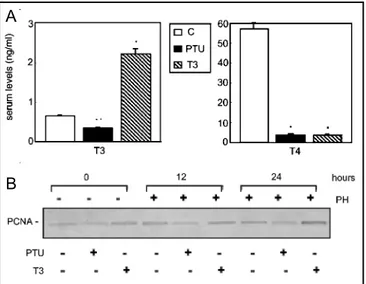
Firstly, we have examined serum thyroid hormone levels in rats as described in Materials and Methods section (fig. 1A). In euthyroid control rats, serum thyroid hormone levels are T3=0.65±0.02 ng/ml, T4=57.28±3.01 ng/ml. Daily T3 administration to euthyroid rats for 1 week before PH produces a hypertriiodothyroninemia with low T4 serum concentration (T3=2.02±0.31 ng/ml, T4<4 ng/ ml). The administration of PTU to euthyroid rats for 6 weeks induces hypothyroidism with low serum levels of thyroid hormones (T3=0.35±0.02 ng/ml, T4<4 ng/ml). The decrease in thyroid hormone levels results in impaired body growth and liver regeneration [28]. To assess the effects of thyroid hormones on S phase entry we analysed PCNA expression. PCNA is a nuclear protein expressed differentially during the cell cycle. Since we had previously demonstrated that PCNA levels increase rapidly in late-G1 (12 hrs) and S phase (24 hrs) during hepatic regeneration in rat [25], here we investigated whether rats with an altered thyroid status can show changes in the expression profile of PCNA. For the experiments we used rats, made hypo- and hyperthyroid by treatment with PTU for a month or T3 for one week. To analyse changes occurring during hepatic regeneration, the operated animals were killed at 12 and 24 hrs after PH. Sham operated rat livers showed the same results of the control (data not shown). As demonstrated in figure 1b, thyroid status influences the expression of PCNA, indeed, we observe that in non-regenerating livers, the treatment with PTU or T3 respectively, down-regulates or up-regulates the levels of PCNA with respect to controls. Interestingly, the hypo- and hyperthyroidism modify the profile of PCNA expression typical of partial hepatectomy. In particular, PCNA amount in PTU-treated is lower than in normal hepatectomized rats, both at 12 and 24 hrs, whereas in T3-treated rats it is higher at 24 hrs.
Fig. 1. Serum levels of thyroid hormones and the effects of
thyroid status on DNA-synthesis. A) Serum levels of thyroid hormones in euthyroid (C), hypothyroid (PTU) and hyperthyroid (T3) rats. Levels of thyroid hormones (T3 and T4) were evaluated by an enhanced chemiluminescence enzyme immunoassay. Data, expressed as ng/ml (means ± S.D.), were obtained from ten different experiments. * P< 0.001, ** P< 0.01 vs. C; Student’s t test. B) Time course of PCNA expression in non-regenerating and regenerating rat livers in the presence or absence of PTU or T3 treatment. PCNA levels were analysed by Western blotting. In the upper and lower panels we have described the treatment of animals: non-hepactemized (lanes 1, 2, 3) or hepatectomized (lanes 4-9) rats treated with PTU (lanes 2, 5, 8), or T3 (lanes 3, 6, 9) as described in Materials and Methods. The different time points after partial hepatectomy are also indicated (upper panel). The immunoblot is representative of three different experiments.
Thyroid hormone effects on the levels of cell cycle regulatory proteins
Our first aim was to determine the levels of the cyclins involved in the regulation of the late G1 phase G1/S transition and S phase, in the different experimental conditions analysed. When we compare the levels of all cyclins in rats with different thyroid status, we observe that the levels of cyclin D1, A and E are lower in hypothyroid rats and higher in hyperthyroid rats with respect to controls (fig. 2A). As reported in figure 2a, regeneration is able to increase cyclin D1, A and E at 12 and 24 hrs, hypothyroidism however keeps the expression levels of these proteins lower, whereas hyperthyroidism significantly increases their amount. On the contrary, the western blotting analysis of cdk4 and cdk2 (fig. 2B) shows that the levels of these proteins remain unaltered in all the experimental conditions. Finally we performed western blot analysis of some cdks inhibitors, such as p16, p21
Fig. 2. Effects of thyroid status on the expression of cell cycle
regulatory proteins. The expression of proteins in lysates was determined by Western blot analysis. The animals were treated in vivo as described in the legend to figure1. A) Expression profile of cyclin D1, cyclin E and cyclin A. B) Protein expression levels of cdk4 and cdk2. C) Expression profile of cdk inhibitors: p16, p21 and p27. Red Ponceau staining (data not shown) confirms equal loading of membranes. Representative immunoblots of three independent experiments are reported. and p27 (fig. 2C). In normal rats only p16 and p27 are detectable and their expression is reduced in hyperthyroid rats, whereas p21 levels are increased in both hyperthyroidism and hypothyroidism. During hepatic regeneration after PH, there is an evident upregulation of p21 expression, which is not influenced by thyroid conditions. Differently, p16, that is weakly down-regulated after 24 hrs from PH, is increased in hypothyroid rats. On the contrary the level of p27, that decreases during hepatic regeneration (especially at 12 hrs), is altered both in hypothyroidism and hyperthyroidism.
Thyroid hormone effects on cyclin-cdk complex activities
Changes in the levels of proteins is often unable to justify the activity of cyclin-cdk complexes. In order to A
B
A
B
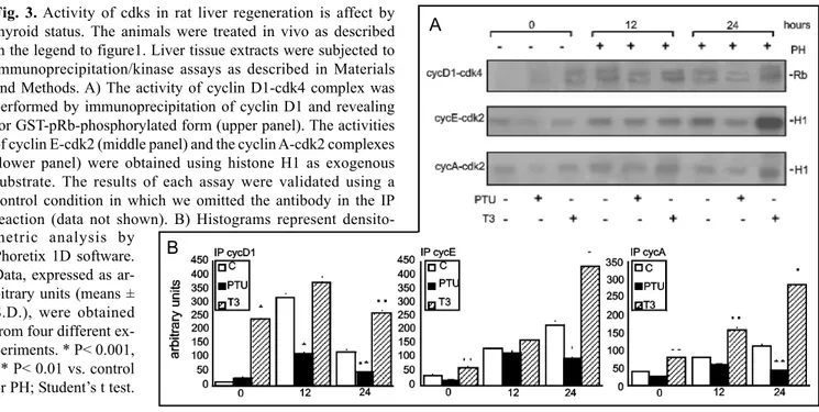
study the effect of thyroid status on the activity of cyclin-cdk complexes involved in control of G1/S transition, we performed different kinase assays as described in Material and Methods section. Figure 3 (A, upper panel) shows the results of the autoradiography of the cyclin D1-cdk4 activity. This activity, measured by retinoblastoma (Rb) phosphorylation assay, results unchanged in control and hypothyroid rats and it is enhanced in hyperthyroid rats. During hepatic regeneration the activity of cyclin D1-cdk4 complex is up-regulated, with a peak at 12 hrs after PH, both in euthyroid and in hyperthyroid animals, whereas hypothyroidism down-regulates the activity of this complex. To analyse the activity of the cyclin E/A-cdk2 complexes we performed a histone H1 phosphorylation assay. As shown in figure 3A the behaviour of these cyclin-cdk complexes resembles the regulation of the cyclin D1-cdk4 complex activity indicating that thyroid status influence also the activity of cyclin E/A-cdk2 complexes.
Thyroid hormone effects on p53 and p73 proteins Based on gene sequence homologies, a p53 gene family becomes apparent with the addition of the most recently identified p63 and p73 genes to the already known p53. These genes encode for different p53 proteins and multiple p63 and p73 isoforms. Actually, it is known that both p53 and p73 proteins elicit tumor suppressor gene properties that mediate cell cycle arrest or apoptosis after DNA damage [13].
We investigated the effect of thyroid status on p53 and p73, two major proteins that regulate cell growth arrest and apoptosis. Western blot analysis (fig. 4), demonstrates that the amount of both p53 and p73 increases during liver regeneration especially 12 hrs after PH. This result is consistent with the notion that these proteins are up-regulated during G1/S transition [35]. Interestingly, thyroid status radically influences the expression of p53 and p73 in control and partially hepatectomized rats. In particular, our results show that hyperthyroidism, both in non-hepactomized and hepatectomized rats, doesn’t change the expression of p53, whereas provokes a down-regulation of p73. On the other hand, hypothyroidism induces a down-regulation of the p53 expression levels whereas it up-regulates the p73 expression levels.
Thyroid hormone effects on p42/44 MAPK and p38 MAPK
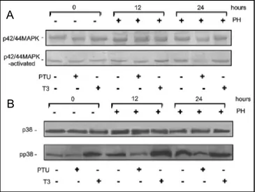
Since the activity of p42/44 MAPK and p38 MAPK often seems to be involved in regulating cell proliferation and growth arrest during hepatic regeneration, we analysed the effect of thyroid status on these two signal mediators. Changes in the levels of both unphosphorylated and phosphorylated (active) form of p42/44 MAPK and p38 MAPK were analysed by western blotting. As demonstrated in figure 5a, neither PH nor hyperthyroidism seem to affect the expression levels of p42/44 MAPK whereas hypothyroidism highly depresses the amount of this kinase especially at 24 hrs after PH. Conversely, the B
Fig. 3. Activity of cdks in rat liver regeneration is affect by
thyroid status. The animals were treated in vivo as described in the legend to figure1. Liver tissue extracts were subjected to immunoprecipitation/kinase assays as described in Materials and Methods. A) The activity of cyclin D1-cdk4 complex was performed by immunoprecipitation of cyclin D1 and revealing for GST-pRb-phosphorylated form (upper panel). The activities of cyclin E-cdk2 (middle panel) and the cyclin A-cdk2 complexes (lower panel) were obtained using histone H1 as exogenous substrate. The results of each assay were validated using a control condition in which we omitted the antibody in the IP reaction (data not shown). B) Histograms represent densito-metric analysis by
Phoretix 1D software. Data, expressed as ar-bitrary units (means ± S.D.), were obtained from four different ex-periments. * P< 0.001, ** P< 0.01 vs. control or PH; Student’s t test.
Fig. 4. Effects of thyroid status on the expression of
pro-apoptotic proteins. The expression of p53 and p73 was analysed by Western blotting in rats treated as described in the legend to figure1. Expression profile of p53 (upper panel) and p73 (lower panel) is reported. Red Ponceau staining (data not shown) confirms equal loading of membranes. Representative results of three experiments are reported.
Fig. 5. Effects of thyroid status on the activity of p42/44 and
p38 MAPKs. The expression of proteins in lysates was determined by Western blot analysis. The animals were treated in vivo as described in the legend to figure1. A) Expression profile of unphosphorylated form (upper panel) and phosphorylated form (lower panel) of p42/44 MAPK. B) Protein expression levels of unphosphorylated form (upper panel) and phosphorylated form (lower panel) of p38 MAPK. Representative immunoblots of three independent experiments are reported.
analysis of the phosphorylated form of p42/44 MAPK revealed that it increased during hepatic regeneration in euthyroid and hyperthyroid rats, whereas, according to previous results, it is negatively regulated in hypothyroid rats during PH. The same analysis was also performed to investigate p38 MAPK. As shown in figure 5B, in all experimental conditions there aren’t any relevant changes in the expression levels of the unphosphorylated form of p38 MAPK. On the contrary, the analysis of the phosphorylated form revealed that p38 is influenced by PH and thyroid status. In particular, we observed that hypothyroidism decreases whereas hyperthyroidism, as PH (especially at 12 hrs), increases the activated form of this kinase.
Discussion
As already described, even though differentiated hepatocytes in adult liver are quiescent, several stimuli (such as PH), induce their proliferation. The rapidly proliferating cells in a regenerating liver are extremely susceptible to carcinogenesis, as there is minimal time available for DNA repair between two consecutive cell cycles [36, 37]. It has been demonstrated that a single dose of T3 enhances DNA-synthesis and regenerative capacity of the liver after PH [38, 39]. Consistent with these findings, recently it has been reported that although hypothyroid rats are able to regenerate their liver mass completely, the recovery is significantly delayed; whereas T3 administration to hypothyroid rats restores the liver regenerative capacity [40].
Interestingly, Pibiri et al., [41] have found that when T3 is administered to rats it causes an accelerated onset
of DNA-synthesis associated with a more rapid increase of cyclin D1 and E expression, compared with partial hepatectomy.Furthermore, we have demonstrated that, during hepatic regeneration, thyroid status affects liver regeneration by altering: liver regeneration index, DNA-synthesis and poly (ADP-ribose) polymerase activity, which influence DNA replication and repair, cell differentiation and transformation [28]. Therefore, the evaluation of the possible effects of the thyroid status on liver ability to respond to the pro-proliferative stimuli of PH, could be a very relevant model for understanding the dynamics of the cell cycle.
In our study, we demonstrate that hypothyroidism and hyperthyroidism act on the entry in to S phase of hepatocytes as demonstrated by PCNA expression levels confirming previous data on the effect of T3 on thymidine incorporation [28]. This effect probably depends on the expression levels and activity of cell cycle proteins involved in G1 and S phases control, both in regenerating and non-regenerating livers. Our results indicate that hypothyroidism inhibits cell cycle progression depressing the activity of cyclin-cdks complexes involved in the control of G1/S transition, whereas hyperthyroidism seems to induce the S phase progression by a sustained
A
activation of cyclin-cdk complexes. These data confirm the results of Pibiri [41] on the levels of cyclin D1 and demonstrate that thyroid status may regulate cell cycle progression also acting on the expression of p16 and p27 cdk inhibitor and on cyclin E/A-cdk2 complex activities underlying that for liver re-growth, physiological levels of thyroid hormones are needed.
Liver re-growth that occurs in response to PH depends on a balance between proliferative and apoptotic processes [3]. Some authors have demonstrated that the expression of p53 is up-regulated in the regenerating liver after PH [42] and in this context it seems to act as a negative regulator of proliferation by providing cell cycle checkpoint activity [43]. As concerns p73, its behaviour and its role during liver regeneration is still unknown, but recently it has been shown that p73 is regulated during cell cycle and may be a target of the mitotic cyclin-cdk complex [35, 43]. Our results demonstrate an up-regulation of p73 confirming that apoptosis and growth events are well regulated during hepatic regeneration. Interestingly, this balance is altered by either thyroid hormone treatment or withdrawal. In fact, the levels of p73 slightly decrease in partially hepactomized hyperthyroid rats, whereas they are significantly up-regulated in partially hepatectomized hypothyroid rats with respect to controls. These data indicate that the reduced regenerative response occurring in hypothyroidism can be caused not only by a reduced activity of cell cycle regulating complexes, but also by an increased expression of p73 involved in growth arrest. On the contrary the data about p53 levels show only a little increase after PH and T3-treatment without any significant alteration in hypothyroidism.
Some reports suggest that p38 MAPK promotes DNA-synthesis in regenerating hepatocytes, whereas chronic activation of p42/44 MAPK has been shown to lead to cellular growth arrest [10, 11]. Moreover it has been demonstrated that thyroid hormones are also able to activate p42/44 MAPK-dependent signal transduction pathway [44-47]. Our results clearly indicate that thyroid hormones as partial hepatectomy influence the activity of these kinases, providing an additional clue to indicate that these pathways are both involved in the regulation of cell proliferation in liver. Furthermore, the negative regulation of the p42/44 MAPK expression and activity at 24 hrs after PH in hypothyroid rats, may suggest an interesting role of thyroid hormones in modulating the expression or degradation of this kinase.
In conclusion, we found that the thyroid status could affect the regenerative capacity of liver, altering the levels and the activities of proteins involved in the control of cell cycle progression and growth arrest. These changes in control liver cells homeostasis suggest that thyroid functionality could be an important parameter not only to determine the good-end of liver transplantation but also to evaluate the possible development of liver neoplasia during regenerative process, thus further genetic approaches (i.e. siRNA) could be useful to demonstrate a causal relation between cell cycle and/or apoptosis genes and the effect of thyroid hormones on liver.
Acknowledgements
This work was partially supported by ATENEO 40% grant 2002-3.
References
1 Michalopoulos GK: Liver regeneration: Molecular mechanism of growth control. FASEB J 1990;4:176-187.
2 Michalopoulos GK, DeFrances MC: Liver Regeneration. Science 1997;276:60-65. 3 Fausto N: Liver regeneration. J Hepatol
2000;32:19-31.
4 Higgins GM, Anderson RM: Experimental Pathology of the liver. Restoration of the liver of the white rat following partial surgical removal. Arch Pathol 1931;12:186-202.
5 Taub R: Liver regeneration 4: trans-criptional control of liver regeneration. FASEB J 1996;10:413-427.
6 Trautwein C, Rakemann T, Niehof M, Rose-John S, Manns MP: Acute phase response factor, increased binding, and target gene transcription during liver regeneration. Gastroenterology 1996;110:1854-1862.
7 Li W, Liang X, Kellendonk C, Poli V, Taub R: STAT3 contributes to the mito-genic response of hepatocytes during liver regeneration. J Biol Chem 2002;277:28411-28417.
8 Thompson NL, Mead JE, Braun L, Goyette M, Shank PR, Fausto N: Sequential protooncogene expression during rat liver regeneration. Cancer Res 1986;46:3111-3117.
9 Alcorn JA, Feitelberg SP, Brenner DA: Transient induction of c-jun during hepatic regeneration. Hepatology 1990;11:909-915.
1 0 Anderson P: Kinase cascades regulating entry into apoptosis. Microbiol Mol Biol Rev 1997;61: 33-46.
1 1 Woods D, Parry D, Cherwinski H, Bosch E, Lees E, McMahon M: Raf-induced proliferation or cell cycle arrest is determined by the level of Raf activity with arrest mediated by p21Cip1. Mol Cell Biol 1997;17:5598-5611.
1 2 Spector M, Auer K, Jarvis D, Ishac E, Gao B, Kunos G, Dent P: Differential regulation of the mitogen-activated protein and stress-activated protein kinase cascades by adrenergic agonists in quiescent and regenerating adult rat hepatocytes. Mol Cell Biol 1997;17:3556–3565.
1 3 Levrero M, De Laurenzi V, Costanzo A, Gong J, Wang JY, Melino G: The p53/ p63/p73 family of transcription factors: overlapping and distinct functions. J Cell Sci 2000;113:1661-1670.
1 4 Bénard J, Douc-Rasy S, Ahomadegbe JC: TP53 family members and human cancers. Hum Mutat 2003;21:182-191. 1 5 Alisi A, Giambartolomei S, Cupelli F, Merlo P, Fontemaggi G, Spaziani A, Balsano C: Physical and functional interaction between HCV core protein and the different p73 isoforms. Oncogene 2003;22:2573-2580.
1 6 Tannapfel A, Wasner M, Krause K, Geissler F, Katalinic A, Hauss J, Mossner J, Engeland K, Wittekind C: Expression of p73 and its relation to histopathology and prognosis in hepatocellular carcinoma. J Natl Cancer Inst 1991;91:1154-1158.
1 7 Zemel R, Koren C, Bachmatove L, Avigad S, Kaganovsky E, Okon E, Ben-Ari Z, Grief F, Ben-Yehoyada M, Shaul Y, Tur-Kaspa R: p73 overexpression and nuclear accumulation in hepatitis C virus-associated hepatocellular carcinoma. Dig Dis Sci 2002;47:716-22.
1 8 Albrecht JH, Poon RY, Ahonen CL, Rieland BM, Deng C, Crary G: Involvement of p21 and p27 in the regulation of CDK activity and cell cycle progression in the regenerating liver. Oncogene 1998;16:2141-2150. 1 9 Jeong JS, Lee JH, Kim HI, Park JI:
Changes in expression of cell cycle regulators and their hepatic lobular distribution in partial hepatectomy-induced regenerating rat liver. J Korean Med Sci 1999;14:635-642.
2 0 Weglarz TC, Sandgren EP: Timing of hepatocyte entry into DNA synthesis after partial hepatectomy is cell autonomous. Proc Natl Acad Sci U S A : 2000;97:12595-12600.
2 1 Sherr CJ, Roberts JM: Inhibitors of mammalian G1 cyclin-dependent kinases. Genes Dev 1995;9:1149-1163. 2 2 Sherr CJ: The 2000 Pezcoller lecture: cancer cell cycles revisited. Cancer Res 2000;60:3689-3695.
2 3 Albrecht JH, Hu MY, Cerra FB: Distinct patterns of cyclin D1 regulation in models of liver regeneration and human liver. Biochem Biophys Res Commun 1995;209:648-655.
2 4 Loyer P, Cariou S, Glaise D, Bilodeau M, Baffet G, Guguen-Guillouzo C: Growth factor dependence of progression through G1 and S phases of adult rat hepatocytes in vitro. Evidence of a mitogen restriction point in mid-late G1. J Biol Chem 1996;271:11484-11492. 2 5 Alisi A, Spagnuolo S, Leoni S: Treatment
with EGF increases the length of S-phase after partial hepatectomy in rat, changing the activities of cdks. Cell Physiol Biochem 2003;13:239-248. 2 6 Francavilla A, Carr BI, Azzarone A,
Polimeno L, Wang Z, Van Thiel DH, Subbotin V, Prelich JG, Starzl TE: Hepatocyte proliferation and gene expression induced by triiodothyronine in vivo and in vitro. Hepatology 1994;20:1237-1241.
2 7 Oren R, Dabeva MD, Karnezis AN, Petkov PM, Rosencrantz R, Sandhu JP: Role of thyroid hormone in stimulating liver repopulation in the rat by transplanted hepatocytes. Hepatology 1999;30:903-913.
2 8 Cesarone CF, Scarabelli L, Demori I, Balocco S, Fugassa E: Poly(ADP-ribose) polymerase is affected early by thyroid state during liver regeneration in rats. Am J Physiol Gastrointest Liver Physiol 2000;279:G1219-1225.
2 9 Alisi A, Spagnuolo S, Napoletano S, Spaziani A, Leoni S: Thyroid hormones regulate DNA-synthesis and cell-cycle proteins by activation of PKCα and p42/ 44 MAPK in chick embryo hepatocytes. J Cell Physiol 2004;201:259-265. 3 0 Cervinkova Z, Simek J: Effect of
propylthiouracil on liver regeneration in rats after partial hepatectomy. Physiol Res 1992;41:141-146.
3 1 Bruck R, Oren R, Shirin H, Aeed H, Papa M, Matas Z, Zaidel L, Avni Y, Halpern Z: Hypothyroidism minimizes liver damage and improves survival in rats with thioacetamide induced fulminant hepatic failure. Hepatology 1998;27:1013-1020. 3 2 Oren R, Resnick MB, Brill S, Dotan I, Zaidel L Lifshits-Mercer B, Halpern Z: Thyroxine accelerates proliferation of injured liver cells. J Hepatol 1998;29:634-637.
3 3 Higgins GM, Anderson RM: Experimental pathology of the liver. Restoration of the liver of the white rat following partial surgical removal. Arch Pathol 1931;12:186-202.
3 4 Lowry OH, Rosebrough NJ, Farr AL, Randall RJ: Protein measurement with the folin phenol reagent. J Biol Chem 195;193:265-275.
3 5 Gaiddon C, Lokshin M, Gross I, Levasseur D, Taya Y, Loeffler JP, Prives C: Cyclin-dependent kinases phosphorylate p73 at threonine 86 in a cell cycle-dependent manner and negatively regulate p73. J Biol Chem 2003;278:27421-27431.
3 6 Morin O, Normand C: Long-term maintenance of hepatocyte functional activity in co-culture: requirements for sinusoidal endothelial cells and dexa-methasone. J Cell Physiol 1986;129:103-110.
3 7 Ueno Y, Moriyama M, Uchida T, Arakawa Y: Irregular regeneration of hepatocytes is an important factor in the hepatocarcinogenesis of liver disease. Hepatology 2001;33:357-362. 3 8 Maliekal TT, Sudha B, Paulose CS:
Kinetic parameters of Thymidine kinase and DNA synthesis during liver regeneration: role of thyroid hormones. Life Sci 1997;60:1867-1874.
3 9 Malik R, Mellor N, Selden C, Hodgson H: Triiodothyronine enhances the regenerative capacity of the liver following partial hepatectomy. Hepatology 2003;37:79-86.
4 0 Moro L, Marra E, Captano F, Greco M: Thyroid hormone treatment of hypo-thyroid rats restores the regenerative capacity and the mitochondrial mem-brane permeability properties of the liver after partial hepatectomy. Endo-crinology 2004;145:5121-5128. 4 1 Pibiri M, Ledda-Columbano GM, Cossu
C, Simula G, Menegazzi M, Shinozuka H, Columbano A: Cyclin D1 is an early target in hepatocyte proliferation in-duced by thyroid hormone (T3). FASEB J 2001;15:1006-1013.
4 2 Kren BT, Trembley JH, Steer CJ: Alterations in mRNA stability during rat liver regeneration. Am J Physiol 1996;270:G763-777.
4 3 Arora V, Iversen PL: Antisense oligonucleotides targeted to the p53 gene modulate liver regeneration in vivo. Drug Metab Dispos 2000;28:131-138. 4 4 Fulco M, Costanzo A, Merlo P,
Mangiacasale R, Strano S, Blandino G, Balsano C, Lavia P, Levrero M: p73 is regulated by phosphorylation at the G2/M transition. J Biol Chem 2003;278:49196-49202.
4 5 Lin HY, Davis FB, Gordinier JK, Martino LJ, Davis PJ: Thyroid hormone induces activation of mitogen-activated protein kinase in cultured cells. Am J Physiol 1999;276:C1014-1024.
4 6 Davis PJ, Shih A, Lin HY, Martino LJ, Davis FB: Thyroxine promotes association of mitogen-activated protein kinase and nuclear thyroid hormone receptor (TR) and causes serine phosphorylation of TR. J Biol Chem 2000;275:38032-38039.
4 7 Shih A, Lin HY, Davis FB, Davis PJ: Thyroid hormone promotes serine phosphorylation of p53 by mitogen-activated protein kinase. Biochemistry 2001:40:2870-2878.