RESEARCH
Lymphopaenia in cardiac arrest patients
Paola Villois
1, David Grimaldi
1, Savino Spadaro
2, Claudia Righy Shinotsuka
1, Vito Fontana
1, Sabino Scolletta
3,
Federico Franchi
3, Jean‑Louis Vincent
1, Jacques Creteur
1and Fabio Silvio Taccone
1*Abstract
Background: A decrease in circulating lymphocytes has been described as a marker of poor prognosis after septic
shock; however, scarce data are available after cardiac arrest (CA). The aim of this study was to evaluate the impact of lymphopaenia after successful cardiopulmonary resuscitation.
Methods: This is a retrospective analysis of an institutional database including all adult CA patients admitted to the
intensive care unit (ICU) between January 2007 and December 2014 who survived for at least 24 h. Demographic, CA‑ related data and ICU mortality were recorded as was lymphocyte count on admission and for the first 48 h. A cerebral performance category score of 3–5 at 3 months was considered as an unfavourable neurological outcome.
Results: Data from 377 patients were analysed (median age: 62 [IQRs: 52–75] years). Median time to return of spon‑
taneous circulation (ROSC) was 15 [8–25] min and 232 (62%) had a non‑shockable initial rhythm. ICU mortality was 58% (n = 217) and 246 (65%) patients had an unfavourable outcome at 3 months. The median lymphocyte count on admission was 1208 [700–2350]/mm3 and 151 (40%) patients had lymphopaenia (lymphocyte count <1000/mm3). Predictors of lymphopaenia on admission were older age, a shorter time to ROSC, prior use of corticosteroid therapy and high C‑reactive protein levels on admission. ICU non‑survivors had lower lymphocyte counts on admission than survivors (1100 [613–2317] vs. 1316 [891–2395]/mm3; p = 0.05) as did patients with unfavourable compared to those with favourable neurological outcomes (1100 [600–2013] vs. 1350 [919–2614]/mm3; p = 0.003). However, lympho‑ paenia on admission was not an independent predictor of poor outcomes in the entire population, but only among OHCA patients.
Conclusions: A low lymphocyte count is common in CA survivors and is associated with poor outcome after OHCA. Keywords: Lymphopaenia, Cardiac arrest, Outcome, Prognosis
© The Author(s) 2017. This article is distributed under the terms of the Creative Commons Attribution 4.0 International License (http://creativecommons.org/licenses/by/4.0/), which permits unrestricted use, distribution, and reproduction in any medium, provided you give appropriate credit to the original author(s) and the source, provide a link to the Creative Commons license, and indicate if changes were made.
Background
Despite the high mortality rates still associated with sudden cardiac arrest (CA), advances have been made in recent years, in particular to improve the proportion of patients achieving return of spontaneous circula-tion (ROSC) after cardiopulmonary resuscitacircula-tion (CPR) [1]. Although the implementation of high-quality CPR and early defibrillation has increased the number of CA patients being admitted to the hospital, there has been no associated increase in survival rates and neurologi-cal recovery among CA survivors [2, 3]. The high mor-tality rate observed in this patient population is related
to the consequences of the global ischaemia–reperfu-sion process, the so-called post-cardiac arrest syndrome (PCAS), which is characterised by a systemic inflamma-tory response that may be involved in the development of myocardial dysfunction, brain injury and multiple organ failure [3, 4].
The PCAS is a complex pathophysiological process, which encompasses a generalised activation of immuno-logical and coagulation pathways and has many features common with sepsis [4]. Adrie et al. [4] reported a sig-nificant increase in the concentrations of various serum cytokines in CA patients in the first hours after the initial injury, which was more significant in non-survivors than in survivors. Other studies have also shown impaired microcirculatory perfusion and increased plasma endo-toxin levels in CA patients, similar to findings in sepsis
Open Access
*Correspondence: ftaccone@ulb.ac.be
1 Department of Intensive Care, Erasme Hospital, Université Libre de
Bruxelles (ULB), Route de Lennik, 808, 1070 Brussels, Belgium Full list of author information is available at the end of the article
[5, 6]. Interestingly, sepsis is rapidly accompanied by a state of relative immunosuppression, characterised by a reduction in the number of functional immune cells and T-cell dysfunction [7, 8], which predicts a poor outcome [9].
Among the different components of the immune response, lymphocytes are essential in the organism’s defence against external aggression, by interaction (T cells) with antigen-presenting cells and subsequent activation of the immune response (B and T cells), in a complex interplay of cell-to-cell interactions, produc-tion of adhesion molecules and modulaproduc-tion of growth factors [10]. Severe lymphopaenia has been described in almost 30% of patients admitted to the intensive care unit (ICU) with severe sepsis or septic shock and was associ-ated with high plasma levels of tumour necrosis factor-α, interleukin (IL)-6 and IL-10 [11]. Lymphopaenia dur-ing sepsis was also associated with features of immuno-suppression, such as spontaneous hypothermia and an increased risk of hospital-acquired infections, and was an independent predictor of poor outcome [9, 12]. Per-sistent lymphopaenia has also been associated with poor outcomes in trauma patients, in particular when an infec-tion occurred during the hospital stay [13]. Although sur-vivors from CA are at high risk of secondary infections, in particular early-onset pneumonia [14], no data are available on the relationship between lymphocyte count and the risk of infection or patient outcome. Thus, the aim of our study was to evaluate the prevalence of lym-phopaenia among CA survivors and its association with ICU survival and long-term neurological outcome. We hypothesised that lymphopaenia could be a significant predictor of poor outcome in this setting.
Methods
Study population
This study was performed in the Department of Intensive Care at Erasme Hospital. All comatose patients (Glasgow Coma Scale [GCS] score <9) admitted after in-hospital (IHCA) or out-of-hospital (OHCA) CA and surviving for at least 24 h after the arrest were included in an institu-tional database (January 2007–December 2014) and con-sidered as eligible for the study. Exclusion criteria were missing data for blood count or lymphocyte count on admission.
Post‑resuscitation care
All comatose CA patients were treated with targeted temperature management (TTM; target body tempera-ture 32–34 °C) for 24 h, according to a standardised insti-tutional protocol. Cooling was started immediately after hospital admission using a bolus of cold fluid (in general saline solutions, given as a dose of 20–30 mL/kg over
30 min) and a water-circulating blanket device (Medi-Therm II, Gaymar, USA, or Arctic Sun, Bard, France). Sedation and analgesia consisted of midazolam and mor-phine, which were adjusted to obtain deep sedation (e.g. severe depression of consciousness during which patients cannot be aroused with repeated or painful stimulation); cisatracurium was administered to control shivering in the induction phase (bolus of 0.15 mg/kg) and then given, if needed, by continuous infusion (1–3 mcg/kg/ min). Rewarming (<0.5 °C/h) was achieved passively, and sedation/analgesia was discontinued at normothermia (>37 °C).
Patients were kept in a 30° semi-recumbent posi-tion; ventilation was set to target PaCO2 between 35
and 45 mmHg and SpO2 >94%. Blood glucose was kept
between 110 and 150 mg/dL using a local protocol for continuous insulin infusion; enteral nutrition was allowed. Mean arterial pressure was maintained at least above 65 mmHg using volume resuscitation, noradrena-line and/or dobutamine, when needed. Intra-aortic balloon counterpulsation (IABP) or extracorporeal mem-brane oxygenation (ECMO) was also used in severe car-diogenic shock.
Data collection
We collected data on demographics, pre-existing chronic diseases and immunosuppressive treatment, and CPR (initial rhythm, bystander CPR, time to ROSC, total adrenaline dose) in all patients. A complete blood count, including neutrophil and lymphocyte cell counts, and C-reactive protein (CRP) concentration were recorded on ICU admission and during the first 48 h. We also recorded the use of any other immunosuppressive drugs, vasopressors, mechanical ventilation and continuous renal replacement therapy (CRRT) during the ICU stay. Lactate concentrations were also collected on admission. The development of infections during the ICU stay was recorded; survival was recorded at ICU discharge and ICU length of stay was noted. Neurological evaluation at 3 months after CA was assessed using the cerebral per-formance category score (CPC; 1 = no or mild neuro-logical disability, 2 = moderate neuroneuro-logical disability, 3 = severe neurological impairment, 4 = vegetative state, 5 = death). The CPC evaluation was assessed dur-ing follow-up visits or by telephone interview with the general practitioner.
Definitions
Lymphopaenia was defined as an absolute lymphocyte count of <1000/mm3 cells; severe lymphopaenia was
defined as an absolute lymphocyte count of <500/mm3
cells [15]. Patients were subsequently divided into four groups according to the lymphocyte levels during the
study period: “group 1” included patients with persistent lymphopaenia throughout the 48 h; “group 2” included patients with lymphopaenia on admission but with nor-mal lymphocyte counts at 48 h; “group 3” included those who had normal lymphocyte counts throughout the 48-h study period; and “group 4” included those with normal lymphocyte counts on admission but who had developed lymphopaenia by 48 h.
Favourable 3-month neurological outcome was defined as a CPC of 1–2 at 3 months and unfavourable neurologi-cal outcome as a CPC of 3–5. The diagnosis of infection was made according to the CDC/NHSN criteria [16]. Acute kidney injury (AKI) was defined as a daily urine output <0.5 mL/kg/h and/or an increase in serum cre-atinine level by at least 0.3 mg/dL or >1.5 times increase from baseline values, as previously reported [17]. Shock was defined as the need for vasopressor agents for more than 6 h.
Statistical analysis
Statistical analyses were performed using the SPSS 24.0 for Windows NT software package (SPSS Inc, Chicago, IL, USA). Descriptive statistics were computed for all study variables. The Kolmogorov–Smirnov test was used and histograms and normal-quantile plots were exam-ined to verify the normality of distribution of continuous variables. Discrete variables were expressed as counts (percentage) and continuous variables as means ± SD or median (25th to 75th percentiles). Demographics and clinical differences between groups were assessed using the Chi-square test, Fisher’s exact test, Student’s t test or Mann–Whitney U test, as appropriate. The signifi-cance of differences among the four groups was analysed using two-way (time and group) analysis of variance for repeated measures (ANOVA), followed by Bonferroni post hoc analysis. Multivariable logistic regression anal-ysis with lymphopaenia on admission as the depend-ent variable was performed in all patidepend-ents; co-linearity between variables was excluded prior to modelling; only variables associated with a higher risk of ICU mortal-ity (p < 0.2) on a univariate basis were introduced in the multivariable model. Odds ratios (OR) with 95% confi-dence intervals (CI) were computed. The same analysis was then performed to identify independent predictors of survival and neurological outcome and for all meas-ured outcome considering only IHCA or OHCA patients. Also, an additional analysis on the independent predic-tors of lymphopaenia on admission with the exclusion of patients under corticosteroids or other immunosuppres-sive therapies was performed. A p value <0.05 was con-sidered as statistically significant.
Results
Of the 424 eligible patients admitted during the study period, 23 died within the first 24 h and 24 were excluded for missing data, so that 377 were included in the final analysis. The main characteristics of the study population (age 62 [52–72] years—OHCA 56%) are given in Table 1. Median time to ROSC was 15 min and 232 (62%) patients had a non-shockable initial rhythm; 150 (40%) patients had a non-cardiac origin of arrest. ICU mortality was 58% (n = 217) and 131 patients (38%) had a favourable neurological outcome.
Lymphocyte count
The median white blood cell count on the day of admis-sion was 12,200 [8700–16,775]/mm3, including a
lymphocyte count of 1100 [600–2208]/mm3. The
lym-phocyte count was lower in patients previously treated with immunosuppressive agents (n = 88) compared to the other patients (800 [445–1989]/mm3 vs. 1290 [765–
2439]/mm3; p = 0.004), in patients with IHCA compared
to those with OHCA (946 [583–1450]/mm3 vs. 1637
[900–3120]/mm3; p < 0.001) and in patients with
non-shockable rhythms compared to those with non-shockable rhythms (1055 [601–2078]/mm3 vs. 1520 [950–3060]/
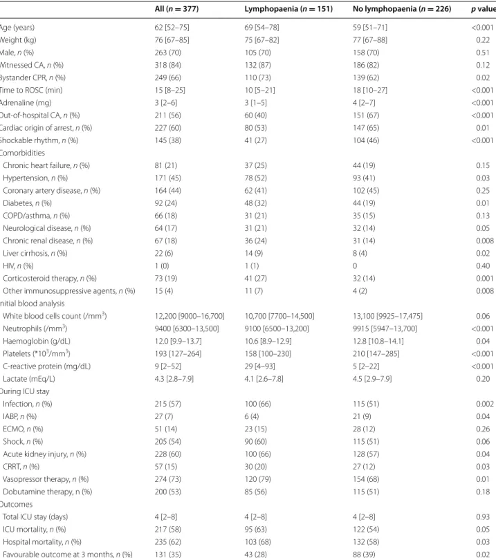
mm3; p < 0.001—Fig. 1).
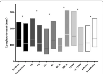
When grouped according to the change in lympho-cyte count during the 48-h period, 112 patients (30%) were in group 1, 39 (25%) in group 2, 86 (23%) in group 3 and 140 (37%) in group 4. The proportions of patients receiving immunosuppressive therapy, of non-survivors, of patients with unfavourable outcome and of patients developing an infection during the ICU stay were signifi-cantly higher in group 1 than in the other groups (Fig. 2).
Lymphopaenia
A total of 151 (40%) patients had lymphopaenia, which was severe in 48 patients. Patients with lymphopaenia on admission were older, had a shorter time to ROSC and were more likely to have had IHCA, a non-cardiac aetiology of CA, bystander CPR and a non-shockable initial rhythm than patients without lymphopaenia (Table 1). Comorbid hypertension, diabetes and immu-nosuppressive therapy were more common in patients with lymphopaenia than in those without. Patients with lymphopaenia on admission also had a lower neutrophil count and haemoglobin concentration on admission and higher CRP concentrations. Lymphopaenic patients more often developed an infection and AKI during the study period and more frequently required vasopressors and CRRT.
Lymphopaenia, survival and neurological outcome
ICU non-survivors had a lower lymphocyte count on admission than ICU survivors (1100 [613–2317]/mm3
vs. 1316 [891–2395]/mm3; p = 0.048). The non-survivors
were older, more frequently had unwitnessed CA, had a longer time to ROSC and had a higher occurrence of
Table 1 Characteristics of study population according to the presence of lymphopaenia on admission
CA cardiac arrest, CPR cardiopulmonary resuscitation, ROSC return of spontaneous circulation, COPD chronic obstructive pulmonary disease, IABP intra-aortic
balloon pump counterpulsation, ECMO extracorporeal membrane oxygenation, CRRT continuous renal replacement therapy, ICU intensive care unit, HIV human immunodeficiency virus
All (n = 377) Lymphopaenia (n = 151) No lymphopaenia (n = 226) p value
Age (years) 62 [52–75] 69 [54–78] 59 [51–71] <0.001
Weight (kg) 76 [67–85] 75 [67–82] 77 [67–88] 0.22
Male, n (%) 263 (70) 105 (70) 158 (70) 0.51
Witnessed CA, n (%) 318 (84) 132 (87) 186 (82) 0.12
Bystander CPR, n (%) 249 (66) 110 (73) 139 (62) 0.02
Time to ROSC (min) 15 [8–25] 10 [5–21] 18 [10–27] <0.001
Adrenaline (mg) 3 [2–6] 3 [1–5] 4 [2–7] <0.001
Out‑of‑hospital CA, n (%) 211 (56) 60 (40) 151 (67) <0.001
Cardiac origin of arrest, n (%) 227 (60) 80 (53) 147 (65) 0.01
Shockable rhythm, n (%) 145 (38) 41 (27) 104 (46) <0.001
Comorbidities
Chronic heart failure, n (%) 81 (21) 37 (25) 44 (19) 0.15
Hypertension, n (%) 171 (45) 78 (52) 93 (41) 0.03
Coronary artery disease, n (%) 164 (44) 62 (41) 102 (45) 0.25
Diabetes, n (%) 92 (24) 48 (32) 44 (19) 0.01
COPD/asthma, n (%) 66 (18) 31 (21) 35 (15) 0.13
Neurological disease, n (%) 64 (17) 31 (21) 32 (14) 0.05
Chronic renal disease, n (%) 67 (18) 36 (24) 31 (14) 0.008
Liver cirrhosis, n (%) 22 (6) 14 (9) 8 (4) 0.02
HIV, n (%) 1 (0) 1 (1) 0 0.40
Corticosteroid therapy, n (%) 73 (19) 41 (27) 32 (14) 0.001
Other immunosuppressive agents, n (%) 15 (4) 11 (7) 4 (2) 0.008
Initial blood analysis
White blood cells count (/mm3) 12,200 [9000–16,700] 10,700 [7700–14,500] 13,100 [9925–17,475] 0.06
Neutrophils (/mm3) 9400 [6300–13,500] 9100 [6500–13,200] 9915 [5947–13,700] <0.001
Haemoglobin (g/dL) 12.0 [9.9–13.7] 10.6 [8.9–12.9] 12.8 [10.8–14.1] 0.04 Platelets (*103/mm3) 193 [127–264] 158 [100–230] 210 [147–285] <0.001
C‑reactive protein (mg/dL) 9 [2–52] 29 [4–93] 5 [2–22] <0.001
Lactate (mEq/L) 4.3 [2.8–7.9] 4.1 [2.6–7.8] 4.5 [2.9–7.9] 0.20
During ICU stay
Infection, n (%) 215 (57) 100 (66) 115 (51) 0.002
IABP, n (%) 27 (7) 6 (4) 21 (9) 0.04
ECMO, n (%) 51 (14) 23 (15) 28 (12) 0.26
Shock, n (%) 205 (54) 90 (60) 115 (51) 0.06
Acute kidney injury, n (%) 228 (60) 100 (66) 128 (57) 0.04
CRRT, n (%) 57 (15) 30 (20) 27 (12) 0.03
Vasopressor therapy, n (%) 274 (73) 120 (79) 154 (68) 0.01
Dobutamine therapy, n (%) 200 (53) 85 (56) 115 (51) 0.18
Outcomes
Total ICU stay (days) 4 [2–8] 4 [2–8] 4 [2–8] 0.93
ICU mortality, n (%) 217 (58) 95 (63) 122 (54) 0.05
Hospital mortality, n (%) 235 (62) 103 (68) 132 (58) 0.03
non-cardiac origin of arrest and non-shockable rhythms than survivors (Table 2). Non-survivors had higher lactate levels on admission and more frequently had shock or AKI during the ICU stay than survivors. These
patterns were similar in patients with unfavourable com-pared to those with favourable outcomes (Table 2).
Multivariable analyses to predict lymphopaenia
In a multivariable logistic regression analysis, older age, a shorter time to ROSC, comorbid use of corticosteroid therapy and high CRP levels on admission were inde-pendently associated with the presence of lymphopae-nia on admission (Table 3); IHCA was not associated with the occurrence of lymphopenia (OR 1.741 [−3.032 to 49.710]; p = 0.083). In IHCA patients, older age, a shorter time to ROSC, high white blood cells count and CRP levels on admission and the use of corticosteroids were independently associated with the presence of lym-phopaenia on admission (Additional file 1: Supplemen-tal Tables 1 and 5). In OHCA patients, a shorter time to ROSC, high white blood cells and CRP levels as well as low platelet count on admission were independently associated with the presence of lymphopaenia on admis-sion (Additional file 1: Supplemental Tables 1 and 5). Finally, older age, a shorter time to ROSC, the presence of a non-shockable rhythm, high white blood cells count and CRP levels or low platelets count on admission and the use of corticosteroids were independently associated with the presence of lymphopaenia on admission in those patients without corticosteroids or immunosuppressive therapy (Additional file 1: Supplemental Tables 4 and 8).
Multivariable analyses to predict ICU mortality
Using the same statistical approach, older age, the absence of bystander CPR, a non-shockable initial rhythm, a non-cardiac aetiology of the arrest, high blood lactate levels on admission and the use of vasopressors were independent predictors of ICU mortality (Table 4), but lymphopaenia was not (OR 1.367 [0.787–2.567; p = 0.26). In IHCA patients, the absence of bystander CPR, a non-shockable rhythm, high epinephrine dose and the development of shock were independently asso-ciated with ICU mortality (Additional file 1: Supplemen-tal Tables 2, 6 and 7), but not lymphopaenia (OR 1.319 [0.708–2.458]; p = 0.38). In OHCA patients, older age, the absence of bystander CPR, a non-cardiac origin of the arrest, a non-shockable rhythm and high blood lactate levels on admission were independent predictors of ICU mortality (Additional file 1: Supplemental Tables 3, 6 and 7); lymphopaenia was not associated with ICU mortality (OR 1.912 [0.876–3.621]; p = 0.09).
Multivariable analyses to predict poor long‑term neurological outcome
In the multivariable analysis, older age, the absence of bystander CPR and a non-shockable initial rhythm were independent predictors of an unfavourable neurological
Fig. 1 Differences in lymphocyte counts on admission among
survivors versus non‑survivors; patients with favourable (FO) versus unfavourable neurological outcome (UO); patients receiving immu‑ nosuppressive agents (IS+) versus others (IS−); patients with in‑ hospital (IHCA) versus out‑of‑hospital cardiac arrest (OHCA); patients with shockable rhythms (VF/VT) versus others (no VF/VT); patients who developed infection versus those without infection. Data are presented as median and 25th (lower limit) and 75th percentiles (upper limit). *p < 0.05 for lymphocyte count
Fig. 2 Proportion of patients on immunosuppressive therapy, devel‑
oping infections, non‑survivors and with favourable neurological outcome (FO) according to the different groups of lymphocyte levels over the first 48 h after arrest: “group 1” included patients with persis‑ tent lymphopaenia throughout the 48 h; “group 2” included patients with lymphopaenia on admission but with normal lymphocyte counts at 48 h; “group 3” included those who had normal lymphocyte counts throughout the 48‑h study period; and “group 4” those with normal lymphocyte counts on admission but who had developed lymphopaenia by 48 h
outcome (Table 4); lymphopaenia was not significantly associated with unfavourable outcome either (OR 1.228 [0.723–3.167; p = 0.28). In IHCA patients, the absence of bystander CPR and a non-shockable rhythm were inde-pendently associated with UO (Additional file 1: Sup-plemental Tables 2, 6 and 7), but not lymphopaenia (OR 1.219 [0.719–2.973]; p = 0.36). In OHCA patients, older age, a non-cardiac origin of the arrest, lymphopaenia on
admission and high epinephrine doses were independ-ent predictors of UO (Additional file 1: Supplemental Tables 3, 6 and 7).
Discussion
In this study, 40% of post-CA patients had lymphopae-nia, and in one-third of them, this persisted through the first 48 h after admission. Predictors of lymphopaenia on
Table 2 Characteristics of study population according to ICU survival and long-term neurological outcome
CA cardiac arrest, CPR cardiopulmonary resuscitation, ROSC return of spontaneous circulation, COPD chronic obstructive pulmonary disease, IABP intra-aortic
balloon pump counterpulsation, ECMO extracorporeal membrane oxygenation, CRRT continuous renal replacement therapy, ICU intensive care unit, HIV human immunodeficiency virus
* p < 0.05 in survivors versus non-survivors OR favourable versus unfavourable outcome
ICU survivors (n = 160) ICU non‑survivors (n = 217) Favourable outcome
(n = 131) Unfavourable outcome (n = 246) Age (years) 59 [49–71] 66 [54–78]* 58 [50–70] 66 [53–77]* Weight (kg) 77 [70–85] 75 [65–85] 78 [70–85] 75 [65–85] Male, n (%) 118 (74) 145 (67) 97 (74) 166 (67) Witnessed CA, n (%) 144 (90) 174 (80)* 118 (90) 200 (80)* Bystander CPR, n (%) 122 (76) 127 (59)* 101 (77) 148 (60)*
Time to ROSC (min) 12 [5–20] 18 [10–25]* 12 [5–20] 17 [10–25]*
Adrenaline (mg) 2 [1–4] 4 [2–7]* 2 [1–4] 4 [2–6]*
Out‑of‑hospital CA, n (%) 86 (54) 125 (57) 74 (56) 137 (56)
Cardiac origin of arrest, n (%) 110 (69) 117 (54)* 102 (78) 133 (54)*
Shockable rhythm, n (%) 89 (56) 56 (26)* 82 (63) 63 (26)*
Comorbidities
Chronic heart failure, n (%) 33 (21) 48 (22) 27 (21) 54 (22)
Hypertension, n (%) 76 (48) 95 (44) 60 (46) 111 (45)
Coronary artery disease,
n (%) 67 (42) 97 (45) 54 (41) 110 (45)
Diabetes, n (%) 35 (22) 57 (26) 25 (19) 67 (27)
COPD/asthma, n (%) 26 (16) 40 (18) 19 (15) 47 (19)
Neurological disease, n (%) 22 (14) 42 (19) 13 (10) 51 (21)*
Chronic renal disease, n (%) 26 (16) 41 (19) 20 (15) 47 (19)
Liver cirrhosis, n (%) 7 (4) 15 (7) 4 (3) 18 (7) HIV, n (%) 1 (1) – 1 (1) – Corticosteroid therapy, n (%) 25 (16) 48 (22) 20 (15) 53 (22) Immunosuppressive agents, n (%) 5 (3) 10 (5) 3 (2) 12 (5) Lactate on admission (mEq/L) 3.8 [2.4–5.7] 4.9 [3.2–8.7]* 3.9 [2.5–5.7] 4.6 [3–8.5]*
During ICU stay
Infection, n (%) 105 (66) 110 (51)* 83 (63) 132 (54)
IABP, n (%) 9 (6) 18 (8) 7 (5) 20 (8)
ECMO, n (%) 19 (12) 32 (15) 18 (14) 33 (13)
Shock, n (%) 68 (43) 137 (63)* 58 (44) 147 (60)*
Acute kidney injury, n (%) 81 (51) 147 (68)* 65 (50) 163 (66)*
CRRT, n (%) 23 (14) 34 (16) 19 (15) 38 (15)
Vasopressor therapy, n (%) 99 (62) 175 (81)* 81 (62) 193 (78)*
Dobutamine therapy, n (%) 81 (51) 119 (55) 66 (50) 134 (54)
admission were older age, a shorter resuscitation time, a history of corticosteroid therapy and high levels of the inflammatory marker (e.g. CRP). Lymphopaenia was more frequent in patients with poor outcome but was not an independent predictor of mortality or unfavourable neuro-logical outcome, unless in the subgroup of OHCA patients. The occurrence of lymphopaenia and its impact on sur-vivors of CA has not been well studied. In a pig model of prolonged CA and CPR, Gu et al. reported a high degree of splenic lymphocyte apoptosis, which was initiated by activation of the Bcl-2/Bax mitochondrial pathway [18].
These authors also described a reduction in CD4+/CD8+
T lymphocytes and a shift in these cells from an anti-inflammatory (Th-2) to a pro-anti-inflammatory (T-helper [Th]-1) state in the myocardium [19, 20]. In mice submit-ted to global cerebral ischaemia secondary to CA within 3 h after resuscitation, Deng et al. [21] showed the pres-ence of infiltrating peripheral CD4+ T lymphocytes in
the brain. These experimental data indicate that early disturbances of immunological function after ROSC are associated with an increased production of inflamma-tory mediators and lymphocyte apoptosis, as well as an intense cardiac and neuroimmune response in which infiltrating T cells may play a key role. Few data are avail-able in humans. In 50 OHCA patients, Venet et al. [22] reported a moderate decrease in the number of circu-lating CD4+ T lymphocytes although total lymphocyte
count was normal. Nevertheless, the high mortality rate in that study (90%) prevented a comparison of lymphocyte levels in survivors and non-survivors. Indeed, as clinical studies have demonstrated that PCAS is characterised by high plasma cytokine and endotoxin levels, future studies should better characterise how these abnormalities may alter lymphocyte sub-populations and function in CA survivors and whether these changes are reversible or can be influenced by therapeutic interventions [23, 24].
Lymphopaenia has been widely described in patients with sepsis [12, 25, 26]. After an initial predominant
Table 3 Multivariable regression analysis to identify inde-pendent predictors of lymphopaenia on admission
Hosmer and Lemeshow goodness-of-fit test Chi-squared = 4.93 (p = 0.76). This model has a 69% correct classification (48% for lymphopenia and 83% for non-lymphopenia)
ROSC return of spontaneous circulation, CRP C-reactive protein
Lymphopaenia on admission p value OR 95% CI for OR
Lower Upper
Age (years) 0.012 1.023 1.005 1.042 Time to ROSC (min) 0.001 0.960 0.936 0.984 Previous corticosteroid therapy 0.048 2.040 1.007 4.131 CRP (mg/dL) 0.007 1.005 1.002 1.009
Table 4 Multivariable regression analysis to identify independent predictors of ICU mortality and unfavourable neuro-logical outcome at 3 months after cardiac arrest
ICU mortality: Hosmer and Lemeshow goodness-of-fit test Chi-squared = 5.16 (p = 0.23). This model has a 71% correct classification (57% for survivors and 81% for non-survivors)
Neurological outcome: Hosmer and Lemeshow goodness-of-fit test Chi-squared = 9.18 (p = 0.33). This model has a 73% correct classification (48% for good neurological outcome and 82% for poor neurological outcome)
CPR cardiopulmonary resuscitation ICU mortality p value OR 95% CI for OR Lower Upper Age (years) 0.001 1.042 1.033 1.055 Bystander CPR 0.006 0.441 0.218 0.755 Non‑cardiac aetiology 0.015 2.069 1.345 4.568 Non‑shockable rhythm 0.001 2.687 1.876 4.325 Vasopressor use 0.02 2.142 1.356 8.567
Lactate on admission (mEq/L) 0.003 1.113 1.096 1.301
Unfavourable neurological outcome
p value OR 95% CI for OR
Lower Upper
Age (years) 0.001 1.032 1.024 1.057
Bystander CPR 0.008 0.337 0.219 0.785
pro-inflammatory phase, many septic patients develop persistent immunosuppression, characterised by increased inhibitory receptors on T cells and antigen-pre-senting cells, decreased production of pro-inflammatory cytokines, expansion of myeloid-derived suppressor cells, and apoptosis-related loss of T and B-lymphocytes and dendritic cells [7, 27]. Induction of lymphocyte apoptosis increased mortality and prevented bacteraemic control in septic animals [28]. The detrimental effects of apoptosis are not only related to the severe loss of immune cells but also the impact that apoptotic cell uptake has on the sur-viving immune cells. As such, uptake of apoptotic cells by monocytes, macrophages and dendritic cells results in immune tolerance and cellular anergy, which is associ-ated with increased IL-10 production and the induction of a Th-2 cell immune phenotype; the net result of these changes is that the surviving phagocytic cells cannot pro-vide adequate defence against infection [29].
In our study, patients with lymphopaenia were older than those without. Lymphopaenia is a common find-ing in elderly hospitalised patients and has been associ-ated with poor outcome in these patients [30]. There is a significant interaction between the immune system and the ageing process, which may also influence the occur-rence of chronic diseases; in particular, thymic demise represents an important phenomenon that can cause a reduction in T-cell count and peripheral proliferation of pre-existing T-cell clones, which can trigger limited immune reactivity damage-associated molecular patterns [31]. As expected, previous therapy with corticosteroids was also an independent determinant of lymphopaenia [32]. Data on the prevalence of lymphopaenia among cor-ticosteroid users are scarce and biased by the presence of concomitant malignancies, administration of chemother-apy or other immunosuppressive drugs. The mechanisms include increased Th-2 cells with reduction in pro-inflam-matory circulating cytokines or externalisation of phos-phatidylserine, which may trigger cellular apoptosis, in particular of CD4+ cells [33]. Nevertheless, we found an
association between increasing CRP levels and the risk of lymphopaenia. This is probably due to increased produc-tion of IL-6, which has been shown to be an independent predictor of poor outcome in CA patients [34]. In a recent study, the injection of endotoxin in healthy volunteers was associated with systemic inflammation, which triggered the occurrence of lymphopaenia, by an increase in anti-inflammatory regulatory T cells and a relative functional impairment of T-cell cytokine production, despite detect-able levels of plasma pro-inflammatory cytokines [35]. Predictors of lymphopaenia in patients without previous immunosuppressive therapy were similar to those of the entire cohort, underlying again their relevance indepen-dently of patients’ immune status.
Interestingly, a shorter time to ROSC was another variable associated with lymphopaenia. We may have expected that the severity of the ischaemic injury, which is related to the longer duration of CPR, would have been associated with more severe apoptosis of T cells and have induced a low lymphocyte count in this setting. In stroke, although ischaemic cerebral damage may lead to suppres-sion of peripheral immune responses, which predisposes to infection, no association was found between the exten-sion of the infarct area and functional immune alterations [36]. The higher prevalence of IHCA in the lymphopae-nia group with a shorter intervention time and CPR may also explain this finding. Additional studies should evalu-ate the complex interplay between pre-existing comorbid conditions and acute cerebral injury and the development of altered immune responses in CA patients.
There was no independent association between lympho-paenia and poor outcome. In patients with sepsis and sep-tic shock, a low lymphocyte count was a strong predictor of poor outcome, even better than increased neutrophil cells [12]. Although sepsis has many common features with the PCAS, lymphocyte count and function may play a marginal prognostic role in CA survivors. One possible explanation could be that the severity of the initial anoxic brain injury (i.e. bystander CPR and time to ROSC) was not correlated with the occurrence of lymphopaenia. Moreover, many patients died within the first 2–3 days after arrest because of significant cardiovascular impair-ment or extended post-anoxic brain injury [37], limiting the impact of lymphopaenia on the occurrence of second-ary infections or delayed organ dysfunction. Neverthe-less, we observed that in the subgroup of OHCA patients, lymphopaenia on admission was associated with UO. This suggests that factors as the underlying different causes of the arrest between IHCA or OHCA or the management of CPR according to arrest location (i.e. different response time and quality of CPR) are an important determinant of biological biomarkers of poor prognosis and that routine monitor of the lymphocyte count in OHCA patients could be considered. However, future research should focus on a better characterisation of lymphocytes sub-populations, in relationship with other biomarker of “immunosuppres-sion” (i.e. HLA-DR on monocytes) to better understand the potential prognostic and therapeutic role of lympho-paenia in this patients’ population.
Our study has some limitations. Firstly, it was a retro-spective, single-centre study, which may limit the gen-eralisability of our conclusions; this can, however, also be an advantage, as patients were treated according to a local protocol of PCAS management, thus reducing het-erogeneity. Secondly, we did not record the exact time of no flow or the quality of CPR, which may influence the inflammatory response after reperfusion. Thirdly, all
patients were treated with TTM so that we cannot draw any conclusions on lymphopaenia in normothermic CA patients or on the effects of TTM use, which may blunt the inflammatory response after rewarming, on the impact of lymphopaenia. Forth, causes of CA and lym-phopaenia may differ between IHCA and OHCA. Also, it is possible that lymphopaenia may have preceded the arrest in some CA victims, such as in case of IHCA. Although location of arrest was not a significant predic-tor of lymphopaenia in our study, this might also be due to a lack of power and this question should be further addressed in future larger cohorts. Fifth, some patients, especially if suffering from IHCA, may have sepsis prior to ICU admission, and this could have been a significant trigger to lymphopaenia. Unfortunately, sepsis is widely under-recognised outside the ICU, so that this variable could not be reliably assessed in our database. Finally, we perform a multivariate model to identify variables associated with the occurrence of lymphopaenia, but we did not specifically investigate the lymphocyte count as a continuous variable. Although this might appear more appropriate, clinicians would be more interested in the presence of a “lymphopaenia”, its related risk factors and potential consequences rather than considering the abso-lute lymphocytes count.
Conclusions
Lymphopaenia is a common finding in CA survivors, in particular among those with an initial non-shockable rhythm, in-hospital CA and previous immunosuppres-sive therapy. A lower lymphocyte count on admission was associated with poor outcome, but was not an inde-pendent predictor of mortality or neurological recovery. Future studies should better characterise the immune response in patients resuscitated after CA to improve understanding of the pathophysiology of these findings and their potential therapeutic interventions.
Additional file
Additional file 1. This documents includes comparisons between patients with in‑hospital (IHCA) and out‑of‑hospital cardiac arrest (OHCA—Suppl Table 1); analysis of mortality and neurological outcome in IHCA (Suppl Table 2) and OHCA (Suppl Table 3) patients; the characteristics of lymphopenic patients after having excluded those receiving immuno‑ suppressive therapies (Suppl Table 4); a multivariable regression analysis to identify independent predictors of lymphopenia on admission in in‑hospital (IHCA) or out‑of‑hospital (OHCA) cardiac arrest (Suppl Table 5); a multivariable regression analysis to identify independent predictors of ICU outcome in in‑hospital (IHCA) or out‑of‑hospital (OHCA) cardiac arrest (Suppl Table 6); a multivariable regression analysis to identify independ‑ ent predictors of long‑term neurological outcome in in‑hospital (IHCA) or out‑of‑hospital (OHCA) cardiac arrest (Suppl Table 7); a multivariable regression analysis to identify independent predictors of lymphopenia on admission in patients without therapy with corticosteroids or other immunosoppressive drugs (Suppl Table 8).
Abbreviations
CA: cardiac arrest; CPR: cardiopulmonary resuscitation; CPC: cerebral perfor‑ mance category score; CRRT: continuous renal replacement therapy; ECMO: extracorporeal membrane oxygenation; IABP: intra‑aortic balloon counterpul‑ sation; ICU: intensive care unit; IHCA: in‑hospital cardiac arrest; OHCA: out‑of‑ hospital cardiac arrest; PCAS: post‑cardiac arrest syndrome; ROSC: return of spontaneous circulation; TTM: targeted temperature management. Authors’ contributions
PV, SSc and FST conceived the study protocol; PV, DG, VF, CRS and FST participated in the design and coordination of the study; PV, VF, CRS and FF were responsible for data collection; DG, JC, JLV and JLV supervised data collection; SSp, SSc, FF, DG and VF participated in data interpretation; FF, PV, DG and SSc carried out the literature search; PV, DG, FST and JLV drafted the present manuscript; FST, DG, JC, JLV and SSc and AK revised the manuscript. All authors read and approved the final manuscript.
Authors’ information
PV, DG, CRS, VF, JLV, JC and FST are from the Department of Intensive Care, Erasme Hospital in Brussels (Belgium). SS is from the Department of Morpho‑ logical Surgery and Experimental Medicine, Arcispedale Sant’Anna, University of Ferrara (Italy). SS and FF are from the Department of Anesthesia and Inten‑ sive Care, Policlinico Santa Maria alle Scotte, University of Siena (Italy). Author details
1 Department of Intensive Care, Erasme Hospital, Université Libre de Bruxelles
(ULB), Route de Lennik, 808, 1070 Brussels, Belgium. 2 Department of Morpho‑
logical Surgery and Experimental Medicine, Arcispedale Sant’Anna, Università di Ferrara, Via AldoMoro, 8, 44121 Ferrara, Italy. 3 Department of Anesthesia
and Intensive Care, Policlinico Santa Maria alle Scotte, Universitá di Siena, Viale Bracci, 14, 53100 Siena, Italy.
Acknowledgements
We thank all our clinical and nurse staff for the great support in our daily practice.
Competing interests
The authors declare that they have no competing interests. Availability of data and materials
Data supporting the study are available and can be requested from the cor‑ responding author.
Consent for publication
All authors agreed with the final version of the manuscript. Ethics approval and consent to participate
The local ethical committee (Comité d’Ethique Hospitalo‑Facultaire Erasme‑ULB) approved the study, but waived the need for informed consent because of its retrospective nature.
Source of funding
This study was supported by no funding. Publisher’s Note
Springer Nature remains neutral with regard to jurisdictional claims in pub‑ lished maps and institutional affiliations.
Received: 3 January 2017 Accepted: 6 August 2017
References
1. Nadkarni VM, Larkin GL, Peberdy MA, et al. National Registry of Cardiopul‑ monary Resuscitation Investigators. First documented rhythm and clinical outcome from in‑hospital cardiac arrest among children and adults. JAMA. 2006;295:50–7.
2. Stiell IG, Wells GA, Field B, et al. Advanced cardiac life support in out‑of‑ hospital cardiac arrest. N Engl J Med. 2004;351(7):647–56.
3. Neumar RW, Nolan JP, Adrie C, et al. Post‑cardiac arrest syndrome: epide‑ miology, pathophysiology, treatment, and prognostication: a consensus statement from the International Liaison Committee on Resuscitation (American Heart Association, Australian and New Zealand Council on Resuscitation, European Resuscitation Council, Heart and Stroke Foundation of Canada, Inter American Heart Foundation, Resuscitation Council of Asia, and the Resuscitation Council of Southern Africa); the American Heart Association Emergency Cardiovascular Care Committee; the Council on Cardiovascular Surgery and Anesthesia; the Council on Cardiopulmonary, Perioperative, and Critical Care; the Council on Clinical Cardiology; and the Stroke Council. Circulation. 2008;118:2452–83. 4. Adrie C, Adib‑Conquy M, Laurent I, et al. Successful cardiopulmonary
resuscitation after cardiac arrest as a “sepsis‑like” syndrome. Circulation. 2002;106(5):562–8.
5. Donadello K, Favory R, Salgado‑Ribeiro D, et al. Sublingual and muscular microcirculatory alterations after cardiac arrest: a pilot study. Resuscita‑ tion. 2011;82(6):690–5.
6. Grimaldi D, Sauneuf B, Guivarch E, et al. High level of endotoxemia follow‑ ing out‑of‑hospital cardiac arrest is associated with severity and duration of postcardiac arrest shock. Crit Care Med. 2015;43(12):2597–604. 7. Hotchkiss RS, Swanson PE, Freeman BD, et al. Apoptotic cell death in
patients with sepsis, shock, and multiple organ dysfunction. Crit Care Med. 1999;27(7):1230–51.
8. Grimaldi D, Louis S, Pène F, et al. Profound and persistent decrease of circulating dendritic cells is associated with ICU‑acquired infection in patients with septic shock. Intensive Care Med. 2011;37(9):1438–46. 9. Drewry AM, Samra N, Skrupky LP, Fuller BM, Compton SM, Hotchkiss
RS. Persistent lymphopenia after diagnosis of sepsis predicts mortality. Shock. 2014;42(5):383–91.
10. Bermejo‑Martin JF, Andaluz‑Ojeda D, Almansa R, et al. Defining immu‑ nological dysfunction in sepsis: a requisite tool for precision medicine. J Infect. 2016;72(5):525–36.
11. Chung KP, Chang HT, Lo SC, et al. Severe lymphopenia is associated with elevated plasma interleukin‑15 levels and increased mortality during severe sepsis. Shock. 2015;43(6):569–75.
12. Drewry AM, Fuller BM, Skrupky LP, Hotchkiss RS. The presence of hypo‑ thermia within 24 hours of sepsis diagnosis predicts persistent lympho‑ penia. Crit Care Med. 2015;43(6):1165–9.
13. Menges T, Engel J, Welters I, et al. Changes in blood lymphocyte popula‑ tions after multiple trauma: association with posttraumatic complica‑ tions. Crit Care Med. 1999;27:733–40.
14. Kakavas S, Mongardon N, Cariou A, Gulati A, Xanthos T. Early‑onset pneu‑ monia after out‑of‑hospital cardiac arrest. J Infect. 2015;70(6):553–62. 15. Le Tulzo Y, Pangault C, Gacouin A, et al. Early circulating lymphocyte
apoptosis in human septic shock is associated with poor outcome. Shock. 2002;18(6):487–94.
16. Horan TC, Andrus M, Dudeck MA. CDC/NHSN surveillance definition of health care‑associated infection and criteria for specific types of infec‑ tions in the acute care setting. Am J Infect Control. 2008;36:309–32. 17. Mehta RL, Kellum JA, Shah SV, et al. Acute Kidney Injury Network: report
of an initiative to improve outcomes in acute kidney injury. Crit Care. 2007;11:R31.
18. Gu W, Zhang Q, Yin W, Li C. Caspase‑3‑mediated splenic lymphocyte apoptosis in a porcine model of cardiac arrest. Am J Emerg Med. 2014;32(9):1027–32.
19. Gu W, Li CS, Yin WP, et al. Expression imbalance of transcription factors GATA‑3 and T‑bet in post‑resuscitation myocardial immune dysfunction in a porcine model of cardiac arrest. Resuscitation. 2013;84(6):848–53.
20. Gu W, Li CS, Yin WP, Hou X, Zhang D. Effects of Shen‑Fu injection on the expression of T‑cell specific transcription factors T‑bet/GATA‑3 in porcine postresuscitation lung injury. Evid Based Complement Altern Med. 2013;2013:464650.
21. Deng G, Carter J, Traystman RJ, Wagner DH, Herson PS. Pro‑inflammatory T‑lymphocytes rapidly infiltrate into the brain and contribute to neuronal injury following cardiac arrest and cardiopulmonary resuscitation. J Neuroimmunol. 2014;274(1–2):132–40.
22. Venet F, Cour M, Demaret J, Monneret G, Argaud L. Decreased monocyte HLA‑DR expression in patients after non‑shockable out‑of‑hospital cardiac arrest. Shock. 2016;46(1):33–6.
23. Timmermans K, Kox M, Gerretsen J, et al. The involvement of danger‑ associated molecular patterns in the development of immunoparalysis in cardiac arrest patients. Crit Care Med. 2015;43(11):2332–8.
24. Beurskens CJ, Horn J, de Boer AM, et al. Cardiac arrest patients have an impaired immune response, which is not influenced by induced hypo‑ thermia. Crit Care. 2014;18(4):R162.
25. Heffernan DS, Monaghan SF, Thakkar RK, Machan JT, Cioffi WG, Ayala A. Failure to normalize lymphopenia following trauma is associated with increased mortality, independent of the leukocytosis pattern. Crit Care. 2012;16(1):R12.
26. de Jager CP, van Wijk PT, Mathoera RB, de Jongh‑Leuvenink J, van der Poll T, Wever PC. Lymphocytopenia and neutrophil‑lymphocyte count ratio predict bacteremia better than conventional infection markers in an emergency care unit. Crit Care. 2010;14(5):R192.
27. Boomer JS, To K, Chang KC, et al. Immunosuppression in patients who die of sepsis and multiple organ failure. JAMA. 2011;306(23):2594–605. 28. Hotchkiss RS, Chang KC, Swanson PE, et al. Caspase inhibitors improve
survival in sepsis: a critical role of the lymphocyte. Nat Immunol. 2000;1(6):496–501.
29. Hotchkiss RS, Monneret G, Payen D. Sepsis‑induced immunosuppres‑ sion: from cellular dysfunctions to immunotherapy. Nat Rev Immunol. 2013;13(12):862–74.
30. Rubio‑Rivas M, Formiga F, Grillo S, Gili F, Cabrera C, Corbella X. Lympho‑ penia as prognostic factor for mortality and hospital length of stay for elderly hospitalized patients. Aging Clin Exp Res. 2016;28:271–7. 31. Youm YH, Kanneganti TD, Vandanmagsar B, et al. The Nlrp3 inflamma‑
some promotes age‑related thymic demise and immunosenescence. Cell Rep. 2012;1(1):56–68.
32. Yao Z, DuBois DC, Almon RR, Jusko WJ. Pharmacokinetic/pharmacody‑ namic modeling of corticosterone suppression and lymphocytopenia by methylprednisolone in rats. J Pharm Sci. 2008;97(7):2820–32.
33. Jetzek‑Zader M, Gudowius S, Feyen O, Stevens M, Lipfert P, Niehues T. A single intravenous dose of prednisolone induces phosphatidylser‑ ine externalization, loss of surface marker expression and a 24‑h net increase in human peripheral blood lymphocytes ex vivo. Rheumatol Int. 2007;27(7):667–73.
34. Vaahersalo J, Skrifvars MB, Pulkki K, et al. Admission interleukin‑6 is associated with post resuscitation organ dysfunction and predicts long‑ term neurological outcome after out‑of‑hospital ventricular fibrillation. Resuscitation. 2014;85(11):1573–9.
35. Ronit A, Plovsing RR, Gaardbo JC, et al. Inflammation‑induced changes in circulating t‑cell subsets and cytokine production during human endo‑ toxemia. J Intensive Care Med. 2017;32(1):77–85.
36. Klehmet J, Harms H, Richter M, et al. Stroke‑induced immunodepression and post‑stroke infections: lessons from the preventive antibacterial therapy in stroke trial. Neuroscience. 2009;158(3):1184–93.
37. Lemiale V, Dumas F, Mongardon N, et al. Intensive care unit mortality after cardiac arrest: the relative contribution of shock and brain injury in a large cohort. Intensive Care Med. 2013;39:1972–80.