Università degli Studi di Ferrara
DOTTORATO DI RICERCA IN
"BIOCHIMICA, BIOLOGIA MOLECOLARE E BIOTECNOLOGIE"
CICLO XXVI
COORDINATORE Prof. Bernardi Francesco
EDENTULISM PROBLEM:
USE OF STEM CELLS FOR BONE REGENERATION AND BONE INFLAMMATION
Settore Scientifico Disciplinare MED/04
Dottorando Tutore
Dott. Bressan Eriberto Prof. Pinton Paolo
Indice
Summary... 5 Introduction... 7 Edentulous patients...7 Tooth anatomy... 14 Dentin...14 Pulp... 15 Alveolar bone... 15 Odontogenesis... 16 Bone tissue...17Morphology of bone tissue...17
Bone extracellular matrix... 18
Bone cells... 21 Bone histogenesis... 23 Implant therapy...25 Peri-implantitis... 28 Bone regeneration... 31 Tissue engineering... 32 Biomaterials...32
Cell therapy applications for craniofacial regeneration...38
Stem cells... 41
Adipose tissue-derived stem cells...43
Dental pulp stem cells... 45
Scaffolds for cell therapy delivery to oral and craniofacial defects...46
Aim... 49
Materials and methods...50
In Vitro... 50
Isolation and culture of adult stem cells...50
Biomaterials:... 51
Differentiation...52
Proliferation tests... 53
Gene expression by Real-Time PCR...56
Cytogenetic analyses...59
Use of CGH array for perimplantitis...61
In Vivo...62
In vivo rat model... 62
In vivo sheep model...64
In vivo dog model...68
Results... 72
In Vitro... 72
Culture of adult stem cells... 72
In vitro hard tissue reconstruction...73
Donor age-related biological properties of human dental pulp stem cells...78
Perimplantitis and CGH analysis: chromosomal aberration...88
In Vivo...89
In vivo rat implantation of the construct with ADSCs...89
In vivo rat implantation of the construct with DPSCs...91
Sheep cellular response to scaffolds...93
Dogs cellular response to scaffolds...96
Discussion...99
Summary
In this study we have started from a real odontoiatric clinic problem, which affects a high percentage of patients: edentulism.
Implant therapy, started by Prof. Branemark in the seventies thanks to the discovery of osseointegration, has developed in the last 15 years.
This therapy represents a valid solution for edentulous patients, although it cannot always be used.
Severe osseous resorption or anatomical limitations hinder the use of implant and force the employ of biomaterials and/or autologous bone, which, according to the latest scientific research, represents the gold standard.
However, intraoral bone graft is not always well tolerated by patients and, in cases of severe atrophy, the quantity of bone that can be taken from the oral cavity is not enough for complete regeneration.
Although implant therapy represents the best solution for edentulous patients, in the latest years a biological complication has become more and more frequent: peri-implantitis.
It represents a serious problem since it begins with an inflammation of peri-implant hard tissues that clinically leads to a loss of peri-implant alveolar bone and, eventually, to the loss of the implant as a whole.
According to the most recent literature, this serious problem affects 10% of the patients and 4% of the implant sites after 10 years of follow-up.
To make this pathology even more serious is the little knowledge that clinicians and researchers have about early diagnosis, etiopathogenesis and therapy.
The purpose of this study is to address both edentulism and consequent osseous regeneration, as well as the problem of peri-implantitis from a biological point of view and with the help of tissue engineering.
In particular, we wanted to test the ability of stem cells taken from adult tissue to favor - in shorter time - osseous regeneration and implant osseointegration both in
vitro and on small and large animals.
Furthermore, we have tested the anti-inflammatory ability of stem cells in bone tissue. Finally, with regard to the problem of peri-implantitis, we have searched for predictive genetic factors in order to possibly identify patients at risk.
state that:
It is possible to isolate stem cells from different adult tissues (adipose
tissue, dental pulp) and test their genetic stability.
For the cells isolated from dental pulp, we could identify an important
connection between patient’s age and ability to differentiate and proliferate. Stem cells combined with different scaffolds are able to foster
osseous regeneration faster.
It has been recognized an actual anti-inflammatory ability of stem
cells in bone tissue.
Preliminary results on the use of CGH as genetic predictive technique for peri-implantitis are encouraging since they foreground a correlation between the genetic alteration of some chromosomal tracts and clinical onset of the disease.
Introduction
Edentulous patients
The absence of good oral health in adults can manifest into social, physical, and emotional health issues. Poor oral health, therefore, negatively impacts on quality of life. Oral health is instrumental to older people’s health, life satisfaction, quality of life and their perception of self. [Mitchell et al. 2013; Mariño et al. 2013]
In addition, clinical data demonstrates that poor oral health increases the risks to health in the same way as any disease of the body system [health 2001]. The interconnections between poor oral health, in particular periodontal disease, and other acute and chronic medical conditions (e.g. pneumonia, cerebrovascular and cardiovascular disease as acute myocardial infarction stroke and coronary heart disease [Emingil et al. 2000; Elter et al. 2003; Hung et al. 2007] diabetes, nutritional deficiencies are now established [Genco and Van Dyke 2010; LOESCHE et al. 1998].
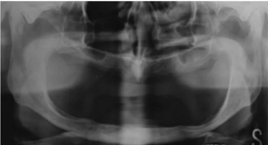
The ability to chew and swallow food comfortably, to speak and to interact socially, can be compromised by common oral diseases and partial or full edentulism conditions. (Figure 1, Figure 2)
Partial edentulsim is the absence of at least one natural tooth, and full edentulism is the complete absence of all natural teeth.
Thorstensson H. and Johansson B. [Thorstensson and Johansson 2010] suggests that the main cause of tooth loss is due to caries in about 55% of swedish individuals of the study, up to the oldest age substantial increase in frequency to 75%. Periodontitis, as a reason for tooth loss, is of minor importance compared with caries but increase steadily over the lifespan from 18 to 33%. Other reasons are toothache, endodontics and tooth/root fractures.
Figure 1: edentulous patient with severe atrophy: lateral view, frontal view
Figure 2: orthopantomography Oral health status and quality of life are strictly related.
The ability to chew food may affect dietary choices and nutritional intake and have consequences for general health [Joshipura et al. 1996a; KRALL et al. 1998]. Edentates have been shown to have a significantly lower fruit and vegetable intake than the fully dentate. Tsakos G et al.[Tsakos et al. 2010] suggests that edentate individuals consumed 50.7 g (27.0, 74.3) fewer fruits/vegetables per day than the dentate. Joshipura et al. [Joshipura et al. 1996b] investigated the association between tooth loss and diet. The edentulous had a higher intakes of total fat and saturated fat and a lower intake of non-starch polysaccharide (NSP) (dietary fiber), β-carotene, and fruits and vegetables than subjects with 25 or more teeth. Consumption of fruit and vegetables is also positively related with the number of natural teeth. Patients with loss of functional dentition and denture-wearing result in selective food intake, hard foods and foods cointaining seeds and pips are
avoided (such as tomatoes, grapes and whole grain bread) with the effects of material deprivation on nutrient intake [Moynihan and Bradbury 2001].
Oral health is not only related to masticatory function and chew ability. Esthetic and psycological problems are often related with loss of teeth. Missing teeth can have negative consequences on self-image, social interaction and psychological health. Interviews done on edentulous patients by Fiske J et al. [Fiske et al. 1998] suggests the main themes identified in reaction to tooth loss were: lowered self-confidence, altered self-image, dislike of appearance, an inability to discuss this taboo subject, a concern about prosthodontic privacy, behaving in a way that keeps the tooth loss secret, altered behaviour in socialising and forming close relationships, premature ageing, and lack of preparation. Active ageing requires maintenance of oral health status: desire for physical attractiveness and interest in appearance does not decline with age [Xiaoxian Meng et al. 2007]; consequently in edentates patients complete prosthetic treatment contributes to maintaining aesthetic appearance, fluent speech and suitable occlusal arrangements for masticatory efficiency [Quran et al. 2001].
Papadaki E. and Anastassiadou V. [Papadaki and Anastassiadou 2012] correlate emotional reactions to tooth loss with denture satisfaction attributes in elderly complete denture wearers. Questionnaire for Emotional reactions to tooth loss showed that 60% of patients had not only difficulties in accepting their tooth loss, but 65% of the younger participants and 47% of the older ones required more than 6 months to come to terms with it. The same pattern was revealed regarding time of acceptance. Four key feelings associated with losing the last tooth/teeth identify sensation of relieved in 1/4 of the subjects, sadness in 1/4 and resignation and oldness in 1/5. Older subjects more often felt relieved with tooth clearance, in contrast to the younger subjects who were more likely to develop negative feelings of bereavement. The study suggests that a substantial proportion of patients were satisfied with their complete dentures rehabilitation but some patients experienced increased social and psychological problems related to their edentulousness and the wearing of complete dentures. The aesthetic and functional aspects of complete dentures affected both patients’ social behaviour and self-confidence as going out and laughing in public, speaking difficulties correlated with avoiding social interaction.
The edentoulus patient has a resorption of alvelar bones. This condition in association with the absence of teeth create physiognomy changes as leak of intraoral volume and deflation of perioral tissues (lips and cheeks). Esthetics changes could create psychological problems and loss of self-esteem.
Carossa S. et al [Carossa et al. 2000] investigated the correlation between edentulism, sleep disorders and arterial hypertension. Respiratory disturbances during sleep are considered risk factors for arterial hypertension and cardiovascular diseases. Edentulism, by decreasing retro-pharyngeal space, may favor upper airway occlusion during sleep. In edentulous subjects, removing dentures during sleep may favor respiratory disorders, and increase the risk for hypertension and cardiovascular disease.
Different studies demonstrate that oral health conditions and edentulism is strictly related to age, education, socio-economic status, ethnicity and smoking [Thorstensson and Johansson 2010; Wu et al. 2012; Kim et al. 2012; Brennan et al. 2008; Elani et al. 2012].
In economically developed countries, the trend of edentulism has declined consistently. In England and Wales, the prevalence of edentulism for the adult population declined from 37% in 1968 to 12% in 1998 [Kelly et al. 1998]. In Australia, the prevalence of edentulism for the adult population declined from 20.5% in 1979 to 8% in 2002. Among older adults aged 65 and above, the reduction for males was from 59.7% to 26.5% and for females was from 71.5% to 40.3% [Sanders et al. 2004]. Similarly, in the United States, the few studies available on middle-aged and older adults have shown that edentulism in these age groups has been dropping for the past several decades. One study revealed that within the period of 1971 and 2001, for those in a low socioeconomic position (SEP), the prevalence of edentulism declined from 50% to 32% in adults age 55– 64 and 58–43% in age 65–74; the comparable declines for these age groups for individuals in a high SEP were 22–6% and 30–9%,respectively [Cunha-Cruz et al. 2007]. A report conducted by the National Centers for Health Statistics using the National Health and Nutrition Surveys of 1988–1994 (NHANES III) and NHANES 1999–2004 [Kim et al. 2012] found that the prevalence of edentulism declined in the United States over these two-time periods from 34% to 27% among adults aged 65 and older over [BA et al. 2007]. Thanks to improvements in oral health,
the proportion of the population who are edentulous has declined over the past 20 years [Sanders et al. 2004]. However, the number of people requiring complete dentures has been predicted to increase over the next 20 years in the United States. Moreover, although implant treatment is reportedly increasing, the need for complete denture treatment is likely to remain substantial in the future rise [Starr and Hall 2009].
Musacchio et al. [Musacchio et al. 2007] reported the prevalence of edentulism in north-est of Italy was 43.8%; this was more pronounced in women and increased with age. It was 31.8% in the 65-69 years age group and more than twice (63.9%) in the 90/ years group. The prevalence of edentulous subjects was much higher in heavy smokers than in non-smokers (55.6% versus 26%) and in subjects with 0-3 years of education (52.4%) than 4-8 years (44.3%) and >8 years (3.3%).
Douglass et al. [Douglass et al. 2002] indicated that edentulism has declined by 10% every decade and that only 90% of edentulous adults obtain and wear complete dentures. However, when the number of adults in each specific age group is multiplied by the percentage who need a complete maxillary or mandibular denture, the results suggest that the adult population in need of 1 or 2 complete dentures will increase from 33.6 million adults in 1991 to 37.9 million adults in 2020. The 10% decline in edentulism experienced each decade for the past 30 years will be more than offset by the 79% increase in the adult population older than 55 years. the number of people in the United States who need complete dentures will increase over the next 20 years despite an anticipated decline in the age-specific rates of edentulism.
The assumption that the most of edentulous persons wear and utilize dentures was proven Redford et al and Marcus et al. [Redford et al. 1996; Marcus et al. 1996] in their analysis of denture use in the United States and New England. These 2 research groups found that 89.6% [Redford et al. 1996] and 89.9% [Marcus et al. 1996], respectively, of the edentulous population used dentures. For past decades, conventional dentures were the only available treatment for edentulism. This treatment relies on the retention and support provided by remaining bone ridge, but many denture-wearing patients have a poor diet and cannot speak clearly due to lack of denture retention and stability [Sánchez-Ayala
et al. 2010]. Inevitable ridge resorption, at the onset of edentulism or over time, may further decrease oral function if dentures do not remain retentive and stable. Ill-fitting dentures can prevent enjoyment of food and affect overall nutrition [Marcus et al. 1996]. The success of conventional complete dentures treatment is variable and depends on a patient’s adaptive capacity to overcome the limitations of complete dentures by an habituation process. The inadequacy of conventional treatment makes implant therapy an alternative to provide significant improvement in stability, retention and quality of life in denture-wearing patients. [Sánchez-Ayala et al. 2010]. Moreover implant-supported fixed dental prosthesis could be considered a restitutio ad integrum of oral health problems.
The introduction of osseointegrated implants has opened new possibilities for improving chewing capacity of edentulous subjects wearing removable dentures. In addition to fixed dental prosthesis, implants can also be used to improve the function of removable prosthesis by the use of various retention systems.
The limit of implant therapy is inadequate quantity of bone: resorption and remodelling of the alveolar ridge is a process that occurs especially after tooth extraction and results in a decrease ridge dimension [Schropp et al. 2003; Araujo and Lindhe 2005]. The volume and rate of bone loss depends by different factors such as gender, hormones, general disease, denture rehabilitation and metabolism [Güler et al. 2005]. The greater amount of resorption occurs during the first 3 months of healing with a significative loss of height and width of the alveolar bone [Schropp et al. 2003; Kerr et al. 2008]. Horizontal dimensional changes consist of 50% alveolar ridge reduction after 1 year from tooth extraction [Schropp et al. 2003]. The resorption occurs primarily from the buccal aspect, with significantly less resorption from the lingual aspect [Araujo and Lindhe 2005]. Alveolar ridge resorption is usually more rapid in the premolar and molar region than the anterior region af the mandible [AA 2002].
Furthermore after teeth extraction in the anterior mandibular region there is higher basal bone disponibility and there are not anatomical landmarks to be compromised for implants insertion such as in the maxillary anterior region. Oikarinen et al suggest that implants of 8mm or longer could be inserted in the anterior maxilla in more than 50% of patients. The mandible showed that implantation was possible in almost every jaw with fixture of 8mm or longer in the canine regions [Oikarinen et al. 1995]. Instead in the maxillary posterior region the
proportion of sites with bone height ≥8mm and bone width ≥6mm was 28.3%, 18.4%, 8.0% and 18.2% at first premolar, second premolar, first molar and second molar sites, respectively [Pramstraller et al. 2011] The use of removable dentures significantly increases the severity of alveolar bone resorption in edentulous areas. Xie et al. reported that the edentulous maxilla has a much greater reduction in radiologic heights compared to the edentulous mandible for both sexes. The finding is considered to be in agreement with the study of Tallgren [Xie et al. 1997; Tallgren 1972].
In addition to the problem of edentulism, Injuries caused by trauma, tumor or cyst resection, infectious diseases, and also congenital and developmental conditions (i.e., cleft palate de- fects) may result into serious functional, aesthetical and psychological sequelae [Cohen 1995; Hunt and Hobar 2003]. In such situations, absence of hard and soft tissues can be disfiguring and often compromise basic functions such as mastication, speech, swallowing, and also lead to limited thermal and physical protection of important anatomical structures (i.e. brain, nerves, arteries, veins) [Davis and Telischi 1994; Kadota et al. 2008; Curtis et al. 1997; Urken et al. 1991]. The progression of certain oral conditions may also result in craniofacial defects of difficult resolution. For istance periodontitis is a chronic inflammatory disease of bacterial etiology, characterized by the loss of support around teeth, including alveolar bone resorption and soft tissue alterations [Genco 1992; Kinane and Bartold 2007; Feng and Weinberg 2006].
Achieving predictable regeneration in the treatment of craniofacial defects is remarkably challenging in most clinical scenarios
Another current cause of bone resorption is peri-implantitis. Peri-implantitis is defined as inflammation of peri-implant tissues accompanied with changes in the level of crestal bone and with the presence of bleeding on probing and/or suppuration, with or without concomitant deepening of peri-implant pockets [Lang et al. 2011].
Peri-implantitis is a serious current problem because neither the causes neither therapies are currently unclear.
Regenerative procedures, applying the concept of guided bone regeneration, use of bone grafts, and membranes, are implemented to rebuild peri-implant supporting bone, rebuild bone after implant-extraction for peri-implant inflammation and for alveolar ridge regeneration.
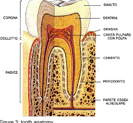
Tooth anatomy
Each tooth consists of three main parts: the crown, the neck and the root, that we can define with anatomic o clinical criteria. Here follow a brief review of the tooth anatomy (Figure 3) involved on stem cells-tissue engineering field.
Dentin
Dentin is a mesenchymal derived tissue lying between enamel or cementum and dental pulp (pulp chamber and root canal). It is a mineralized connective tissue with an organic matrix. It is made up of 70% inorganic materials (especially hydroxyapatite crystals), 20% organic materials and 10% water by weight. The bulk of organic matrix (85-90%) consists of type I collagen, there is also a minor amount of type V and VI collagen. Noncollagenous molecules of dentin are dentin phosphorines, Gla proteins, acidic glycoproteins, growthrelated factors, serum derived proteins, lipids and proteoglycans. Dentin has microscopic channels (0,5-3
μm), called dentinal tubules, radiating outward from pulp cavity to dentinoenamel or dentino-cementum junction. These tubules contain projections of cells secreting dental matrix, known as odontoblasts. The most peripheral aspect of the pulp is lined by the body of these odontoblasts [Yoshiba et al. 2002]
Pulp
Pulp consists of a loose connective tissue enclosed by rigid predentin and dentin. Along the border between the dentin and the pulp are odontoblasts. The thickness of dentinal layer increases with age due to the deposition of secondary and tertiary dentin, reducing the volume of the pulp chamber and the root canals. The most peripheral aspect of the pulp contains four layers of cells: the odontoblastic layer (the most external one), the cell-free zone, the cell-rich zone and the parietal plexus of nerves. Deep inside is the pulp proper, composed of a great amount of fibroblasts and ECM. Blood vessels and nerves enter the tooth mostly through the apical foramen. Other cells in the pulp include undifferentiated mesenchymal cells, deriving from dental papilla, fibrocytes, macrophages and lymphocytes.
Alveolar bone
The bone that supports the teeth is called alveolar bone. It is composed of compact bone and trabecular or spongy bone. The outside wall of the bone is compact bone, such as the thin layer that lines the socket known as lamina dura. The spongy bone is inside and contains bone marrow. The number and the size of the trabeculae in this bone are determined by the function activity of the organ. Alveolar bone proper is the part just around the tooth and it gives attachment to the PDL fibres (bundle bone). The alveolar bone proper is also called cribiform plate, due to the presence of perforation for the entry of vessels and nerves.
Bone is made of 65% inorganic material (mainly hydroxyapatite) by weight, 15% water, 20% organic material. The organic matrix is composed of collagen type I (90-95%), Gla proteins, glycoproteins, phosphorines, proteoglycans, growth factors and bone morphogenetic proteins (e.g. osteogenin) [Lindhe et al. 2009a].
Odontogenesis
During the sixth week of embryogenesis, after the migration of neural crest cells into head and neck mesenchyme, the ectoderm of the first brachial arch begins to proliferate giving rise to the vestibular lamina and the dental lamina.
Dental lamina is a band of ectodermic cells growing from the epithelium of the stomodeum into the underlying mesenchyme and giving rise to the enamel organs of teeth, along the horse shoe shaped dental arches.
Several transcription factors are implicated in odontogenesis, including Pax9, Pitx2, Runx2, Msx1, Msx2, Bmp2, Bmp4, Fgf8 and Fgf9 [Bei and Maas 1998]. The development is commonly divided into the following stages: the bud stage, the cap stage,the bell stage.
The early bell stage of odontogenesis is characterized by epithelial expansion and differentiation into the inner and outer enamel epithelium, stratum intermedium and stellate reticulum.
During the late bell stage, two tooth specific cell types are formed: ameloblasts, which derive from the inner enamel epithelium and produce enamel, and odontoblasts, which differentiate from dental papilla and synthesize dentin.
Dentinogenesis starts before enamel formation with the secretion of an organic matrix in the area directly adjacent to the inner enamel epithelium. Dentin is formed by the production of organic matrix (predentin) and the simultaneous mineralization of this matrix [Hao et al. 2009].
After crown formation, root development begins. The cells of the inner and of the outer enamel epithelium become in contact and give rise to the cervical loop at the base of enamel organ.
The cells of the cervical loop continue to grow away from the crown and become Hertwig’s epithelial root sheath. It induce the adjacent cells of dental papilla to differentiate into odontoblasts and produce dentin. Once this structure fragments, the dentin of the root comes in contact with the dental follicle and stimulates the cementoblasts to begin cementum secretion.
The dental follicle also gives rise to the other supporting structure of the tooth: the periodontal ligament and the alveolar bone proper [Luan et al. 2006].
Bone tissue
Bone tissue is a connective tissue specialized in providing support. Affinity of bone tissue with connective tissues is confirmed by both its origin from mesenchyme, the embryonic tissue where connective tissues is derived from, and its composition since the extracellular matrix is made up of collagen fibers and amorphous fundamental substance.
Bone tissue is characterized by mineralization. The presence of minerals and the peculiar distribution of organic components in the extracellular matrix lend to this tissue remarkable mechanic properties: hardness, resistance to pressure, traction and torsion. Thanks to these properties, bone tissue represents an ideal material for the formation of skeletal bones, which as a whole can be seen as a supportive scaffolding of the organism. Furthermore, given the relevant amount of calcium salts, bone tissue represents the principal store of calcium ions for the metabolic needs of the entire organism.
Calcium deposition in the bone and its mobilization, finely controlled by endocrine mechanisms, provide a crucial contribution to the regulation of plasma levels of this ion.
Morphology of bone tissue
From a macroscopic point of view, two varieties of bone can be distinguished: compact bone and spongy bone (Figure 4)
Compact bone
Compact bone is found in the outer layer of short, flat and long bones and constitutes the diaphysis of the latter as well.
The bone matrix of the compact bone is organized in lamellae which form cavities named bone lacunae containing osteocytes. Compact bone lamellae usually arrange themselves in three different ways:
- Concentrically around vascular channels to form cylindrical structures,
na-med Haversian systems or osteons, oriented along the major axis of the bone;
- In the spaces around osteons, taking on various dimensions and irregular
- Under the periosteum (a layer of connective tissue on the outer surface of the bone) and over the endostium (connective tissue on the inner surface of the bone) to form inner and outer circumferential lamellae.
In the compact bone two categories of vascular channels can be identified:
- Harvesian canals: longitudinal canals located at the center of osteons
containing one or two vessels;
- Volkmann’s canals: transverse canals interconnecting Harvesian canals
Spongy bone
Spongy bone can be found primarily inside short bone, flat bones and epiphysis of long bones.
It is made up by a tridimensional network of branched bone spicules named
trabeculae, that limit a labyrinth of interconnected spaces occupied by hemopoietic
bone marrow.
Figure 4: Distribution of compact and spongy bones in the bone.
Bone extracellular matrix
Being a connective tissue, bone tissue contains a relevant amount of organic extracellular matrix, composed of connective fibers and amorphous substance enriched by the mineral component.
Organic matrix
It favors resistance to traction and pressure. It consists of:
- connective fibers: almost totally made up of Type I collagen fibers, are
cha-racterized by a great number of crossed bundles that maintain united the si-gle molecules of tropocollagen. The abundant presence of collagen is the
main cause of the marked acidophilia that characterizes the intracellular substance of the bone.
Collagen fibers aggregate to form remarkably thick collagen fibers (5-10 µm) only in the fibrous bone tissue, whereas in the lamellar bone tissue col-lagen microfibrils group to form a homogeneous tridimensional network. Connective fibers are particularly abundant in the periosteum therefrom thick bundles of collagen fibers start to penetrate into the cortical bone tis-sue and get lost in the intracellular substance of the bone. These bundles constitute Sharpey’s perforating fibers, which fasten the periosteum to the surface of the bone.
- elastic fibers: virtually absent from the bone tissue, reticular fibers are
loca-ted at the level of the basal membrane that surrounds intraosseous blood vessels;
- amorphous substance: characterized by a peculiar and relevantly different
composition in respect to other connective tissues, it is composed of va-rious macromolecules:
Proteoglycans: made up of sulfated glycosaminoglycan acids
(chera-tan sulfate, chondroitin sulfate) which are kept together by short pro-tein chains.
o Glycoproteins: include several molecules, some of which are belie-ved to play a fundamental role in the mineralization processes. Among these:
a) Osteonectin: the most abundant protein. It possesses high af-finity for calcium, both as a free ion and as an associated ion in crystal-like complexes. It is believed that it works as nuclea-tion agent for mineral crystals since it is considered capable of concentrating calcium located nearby, thus creating the con-ditions to start the precipitation of calcium phosphatase.
b) Fibronectin: is an adhesion molecule located primarily in the pericelluar matrix and characterized by a portion capable of binding with collagen. It is believed that fibronectin is involved in the following processes: migration, adhesion to the matrix and organization of the bone cells.
c) Alkaline phosphatase: is an enzyme capable of hydrolyzing phosphate groups of organic substrates in a basic
environ-ment. It is involved in the processes of mineralization, provi-ding phosphate ions to form mineral crystals.
o Sialoproteins (BSP): specific glycoproteins containing traces of sialic acid which possess the RGD (Arg-Gly-Asp) aminoacid se-quences responsible for the adhesion of the cells to the bone ma-trix. They include osteopontin.
o Proteins containing gamma linolenic acid (GLA): thanks to GLA they are able to chelate bivalent cations such as calcium ion. Two proteins containing GLA can be identified in the bone:
a) Osteumcalcin: a small protein containing 3 or 5 GLA residues. It is involved in the inhibition of the matrix mineralization since it binds calcium ions and makes them available for the combi-nation with phosphate ions thus inhibiting the dimensional gro-wth of crystal minerals. It is abundant in the mature bone tis-sue whereas is scarce in the developing bone tistis-sue (osteoid tissue).
b) GLA protein of the matrix: it has a higher molecular weigh than osteumcalcin and can be found both in the mature bone and in the osteoid tissue as well as in the cartilage which is about to be replaced by osseous tissue.
Inorganic matrix
The mineral component represents 65% of the dry weight of the bone. Its function is to provide hardness and rigidity to the bone tissue.
It is made up of calcium crystals - mainly calcium phosphate - and of calcium carbonate, calcium fluoride and magnesium phosphate in smaller quantities. Calcium phosphate can be found in the form of apatite crystals (Ca10(PO4)62+)
whose positive charges are normally neutralized by the binding with two hydroxide ions thus forming hydroxyapatite.
Hydroxyapatite crystals appear like long and thin needles about 2 nm thick and 20-40 nm long. They tend to arrange themselves parallel to each other and to collagen microfibrils, covering their surface and permeating their porosities.
During the mineralization process of the bone, calcium phosphate precipitates at first in the form of tiny amorphous aggregates.
These initial nuclei of mineral concretion are rapidly replaced by very thin crystals positioned parallel to the filamentous molecules of the fundamental substance
called axial filaments.These crystals assume the typical shape of apatite crystals, progressively occupying most space inbetween collagen microfibrils and permeating the microfibrils themselves. Once the apatite crystals have formed, the deposition of new mineral can occur both through the formation of new crystals and through apposition on pre-existing crystals. This phenomenon is finely regulated by bone cells thanks to the production of specific molecules of the bone matrix.
Bone cells
Four types of cells can be identified in the bone tissue:
Osteoprogenitors (also called pre-osteoblasts), osteoblasts, osteocytes and
osteoclasts. Among these, osteoprogenitors, osteoblasts and osteocytes are in fact subsequent functional phases of the same cell type and are derived from the pluripotent mesenchymal cell of connective tissues. Osteoclasts, instead, are derived from precursors migrated from the blood to the bone tissue, the so called pre-osteoclasts, which are derived from the stem cells of the hematopoietic bone marrow.
Osteoprogentitor cells
Pre-osteoblasts have a spindle or oval shape, a dispersed chromatin nucleus (euchromatic) with a large nucleolus and scarce and basophilic cytoplasm because of the presence of a number of free polyribosomes, whereas other granules are scantily represented.
Osteoprogenitor cells place themselves on the free surfaces of the bones: the can be recognized at level of the inner layer of the periosteum, the so called Ollier’s osteogenic layer, rich in vessels and at the level of the endostium close to capillaries.
Osteoprogenitor cells are able to proliferate, a feature which is shown especially when their body is growing but can be observed also during adult life. They can produce and secrete growth and differentiaton factors, the so called bone
morphogenetic proteins (BMP). When they start the differentiation process,
Osteoblasts
Osteoblasts are primarily responsible for synthesizing the extracellular bone matrix and for its mineralization. They have a spherical or polyhedric shape and tend to align to form epithelious laminae by the developing bone surfaces. In the active phase of the deposition of the bone matrix, osteoblasts have relevant dimensions (about 20 µm), a euchromatic spherical nucleus with a large nucleolus, a basophilic abundant cytoplasm with PAS-positive granulations, a well-developed Golgi’s apparatus and a number of long-shaped mitochondria. Osteoblasts are characterized by the positivity for alkaline phosphatase and by the presence of small cytoplasmatic granules pink in colour containing the precursors of the bone matrix glycoproteins. On the side facing the mineralizing bone matrix, they show several vescicles rich in proteoglycan which, once expelled, will form the mineralization nuclei.
Osteoclasts are interconnected with each other and with nearby osteocytes by means of gap junctions to exchange signal molecules which coordinate metabolic activity and bone matrix deposition.
Osteocytes
Osteocytes are cells which are typically present in the mature bone and are responsible for its maintenance and turnover. They are terminal cells with a finite lifespan.
An osteocyte is a star-shaped cell, with a cellular body resembling a biconvex lens and with several cytoplasmatic extensions.
It presents a heterochromatic nucleus (with condensed chromatin) a small nucleolus and a perinuclear cytoplasm rather scarce and basophilic. Cytoplasmatic organules, RER and Golgi’s apparatus tend to decrease their dimension as the cell grows older until it dies due to apoptosis.
In the bone tissue, the osteocyte is enclosed in a niche carved in the bone matrix, called bone lacuna, whose shape replicates that of the cell, whereas its extensions reach thin canals called bone canaliculi.
Each osteocyte is in contact with surrounding osteocytes through gap junctions at the extremities of their extensions. Water and metabolites can reach all osteocytes, even the farthest away from blood vessels, through non-mineralized osteoid tissue which covers the inner communicating surface of lacunae and canaliculi. Moreover, metabolites and signal molecules dissolved in the cytoplasm can be exchanged between osteocytes through the gap junctions.
Osteoclasts
Osteoclasts are cells specialized in bone resorption. They derive from pre-osteoclasts originating in the hematopoietic bone marrow and are carried by the blood stream up to the sites of bone resorption, where they melt together generating active osteoclasts, i.e. syncytial elements capable of dissolve and digest organic components of the bone tissue.
Mature osteoclasts are giant plurinucleated cells (100-200 µm) with acidophilic cytoplasm; in a single osteoclast can be detected up to 50 nuclei with lax chromatin and clearly visible nucleolus. When a osteoclasts is activated it adheres to the mineralized matrix and due to its erosive action a cavity called Howship’s
lacuna is formed. The side of the cell which is clinged to the bone is characterized
by the so called ruffled border, a thickening of the cell surface made up of a large number of cytoplasmatic lamellae, having different sizes and lengths, which considerably widen the extension of the plasmalemma.
Through the ruffled border osteoclasts adhere tightly to the surface of the bone to be resorbed, delimiting the extracellular environment where ostoelytic substances are released, an area called sealing zone.The resorbtion of the bone matrix begins with the dissolution of the mineral component due to the acidification of the microenvironment in the sealing zone, followed by exocytosis of lysosomal enzymes which digest the organic components of the bone matrix. Furthermore, osteoclasts stimulate osteobalsts to release collagenase enzyme which contributes, through its lytic activity, to the digestion of the organic matrix of the bone. Once the first lacuna has been formed, the osteoclast separates from the bone matrix and migrates with amoeboid movement to a portion of an adjacent bone, adhering again and forming a new lacuna. Osteoclastic function is finely regulated by hormonal and local factors.
Bone histogenesis
Bone always develops replacing a pre-existing tissue, be it mesenchyme or a differentiated connective tissue. The processes which lead to the genesis of the bone tissue within another tissue are called ossification or osteogenesis.
These processes are maximized during prenatal life and continue to maintain a high pace throughout the period of the somatic development.
Two types of ossification can be identified:
- Direct or intramembranous ossification;
- Indirect or chondral ossification.
Direct ossification
Direct or intramembranous ossification is typical of flat bones. It starts from ossification centers which develop in the mesenchyme at early stages of foetal life or in membranes of fibrous, dense connective tissue derived from mesenchyme at later stages of intrauterine and postnatal life.
Direct ossification begins when, beside a rich vascular network, mesenchymal cells differentiate into osteoprogenitor cells which, in turn, change into osteoblasts. Through gap junctions osteoblasts align themselves in epithelial-like rows and start depositing the organic matrix of the bone, i.e. osteoid tissue. When the osteoid tissue undergoes mineralization, it changes into spongy bone and osteoblasts remain enclosed in bone lacunae transforming into osteocytes. Afterwards preosteoclasts differentiate into osteoclasts which dissociate fibrous bone, later replaced by lamellar bone.
Indirect ossification
Indirect or chondral ossification is the most widespread variant. Typically, the bone is preceded by a cartilage scaffold with about the same shape of the bone segment to be and which is later reabsorbed and replaced by bone tissue. Bone can develop both inside the cartilage scaffold (endochondral ossification) and on the outer surface of the cartilage in contact with the perichondrium (perichondral ossification).
Perichondrial ossification starts in the perichondrium where osteoprogenitor cells differentiate into osteoblasts which deposit osteoid tissue. This tissue is committed to become fiber bone through mineralization and is later rearranged by osteoclasts with subsequent deposition of lamellar bone. The bone thus generated binds to the surface of the cartilage, under the perichondrium which then develops into periosteum.
Endochondrial ossification starts when the chondrocytes of the cartilage scaffold undergo hypertrophy. In their cytoplasm drops of glycogen accumulation and PAS-positive granulations resembling the calcifying globules of the osteoclasts can be detected. They release vesicles inducing the calcification of the cartilage matrix and, finally, are destined to undergo apoptosis.
Calcified cartilage matrix is partially subject to erosion thanks to the intervention of cells of osteoclastic nature, coming from the nearby already-formed bone and generating wide cartilage lacunae which merge into each other and where blood vessels, departing from the perichondrium and accompanied by mesenchymal cells, penetrate. Mesenchymal cells differentiate into osteoprogenitor cells and afterwards in osteoblasts which deposit fibrous bone by the remnants of the calcified cartilage matrix. Finally, osteoclasts intervene to resorb both the fiber bone and the mineralized bone matrix, whereas new osteoblasts deposit lamellar bone. Some of the mesenchymal cells, penetrated with blood vessels, originate new vessels and hematopoietic bone marrow. [Zallone 2007; Capitani et al. 1996]
Implant therapy
Missing teeth and supporting oral tissues have traditionally been replaced with removable dentures or fixed bridges permitting restoration of masticatory, phonetic function, and aesthetics.
Replacement of lost dentition has been traced to ancient Egyptian and South American civilizations. In ancient Egyptian writings implanted animal and carved ivory teeth were the oldest examples of primitive implantology. In eighteenth and nineteenth century England and colonial America, poor individuals sold their teeth for extraction and transplantation to wealthy recipients. The clinical outcomes of these transplanted dentitions were either ankylosis or root resorption. Continued research prolonged allotransplant survival but did not appreciably improve predictability.
In 1809 Maggiolo placed an immediate single-stage gold implant in a fresh extraction site with the coronal aspect of the fixture protruding just above the gingiva. Postoperative complications included severe pain and gingival inflammation. Since then various implant materials were used ranging from roughened lead roots holding a platinum post to tubes of gold and iridium. Adams in 1937 patented a submergible threaded cylindrical implant with a ball head screwed to the root for retention for an overdenture in a fashion similar to that done today.
Up to this point implant success was marginal with a maximum longevity of only a few years. Strock placed the first long-term endosseous implant at Harvard in
1938. This implant was a threaded cobalt-chrome-molybdenum screw with a cone-shaped head for the cementation of a jacket crown. The implant remained stable and asymptomatic until 1955, at which time the patient died in a car accident. Strock wrote, “The histological sections of implants in the dog study showed remarkable complete tolerance of the dental implant and the pathologist report so indicated to our gratification.” Strock demonstrated for the first time that metallic endosteal dental implants were tolerated in humans, with a survival rate of up to 17 years.
Due to inadequate alveolar bone height in certain sites of the jaws, subperiosteal implants were developed. In 1943 Dahl placed a metal structure on the maxillary alveolar crest with four projecting posts. Multiple variations to this initial design were fabricated but these devices often resulted in wound dehiscence. Blade implants were introduced by Linkow and by Roberts and Roberts. There were numerous configurations with broad applications, and the implants became the most widely used device in implantology in the United States and abroad.
A two-staged threaded titanium root-form implant was first presented in North America by Brånemark in 1978. He showed that titanium oculars, placed in the femurs of rabbits, osseointegrated in the femurs of rabbits after a period of healing.
Two-staged titanium implants were first placed in patients in 1965 and studies showed prolonged survival, free-standing function, bone maintenance, and significant improvement in benefit-to-risk ratio over all previous implants. This breakthrough has revolutionalized maxillofacial reconstruction. Subsequently, various implant designs have been manufactured and research in implantology has grown exponentially. The frontiers of implantology are rapidly being advanced and esthetics continue to be an integral part of this progress [Brånemark et al. 1985].
In 1977, Brånemark presented his research work carried out over 10 years showing that bone can grow intimately onto the surface of titanium implants (Brånemark 1977). The now well-accepted concept, termed osseointegration, has undoubtedly been one of the most significant scientific breakthroughs in dentistry over the past 30 years. A multitude of implant designs have been marketed since, and the clinical situations in which osseointegrated implantretained prostheses are used have expanded enormously.
maintenance of healthy tissues around them. A cause-effect relationship between bacterial plaque accumulation and the development of inflammatory changes in the soft tissues surrounding dental implants has been shown [Pontoriero et al. 1994]. If this reversible condition, called ’peri-implant mucositis’, is left untreated, it may lead to the progressive destruction of the tissues supporting an implant (peri-implantitis) and ultimately to its failure. [Mombelli 1999] [Esposito et al. 2011]
The 20-year cumulative survival rates of short and standard implants were 92.3 and 95.9%, respectively. The cumulative success rates were 78.3 and 81.4%. The survival rates of short implants in posterior and anterior regions were comparable: 95 and 96.4%, respectively.[Lops et al. 2012]
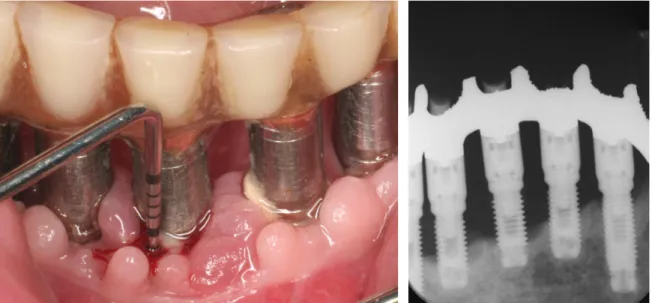
Implant therapy nowadays is a major branch of dentistry that is constantly evolving also thanks to the scientific interest of many companies. The patients themselves requiring implant therapy to get a fixed prosthesis that mimics the most of the natural tooth (Figure 5, Figure 6). Implant therapy can also be used to stabilize dentures and thus ensuring greater comfort to the patient both masticatory that phonetic and aesthetic (Figure 7).
Figure 7: patient before and after treatment
Peri-implantitis
Implant therapy is a well established method of replacing missing teeth. Excellent long- term results can be achieved, but biologic complications may occur.
The most common biological complications were hygiene-related: 30.2% of patients displayed peri-implant mucositis and 10.4% peri-implantitis [Francetti et al. 2013].
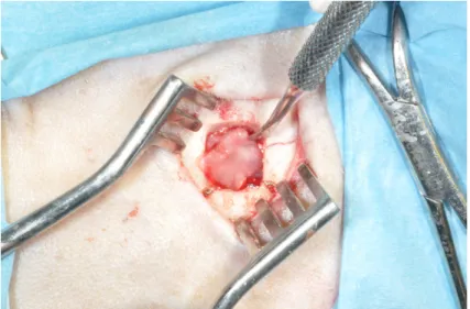
Perimplantitis is defined as: plaque-induced progressive marginal bone loss observed on radiographs with clinical signs of infection of the peri-implant soft tissues (Figure 8).
Figure 8: patient with severe peri-implantitis: presence of suppuration and bone loss
The occurrence of peri-implantitis is not rare. In a single-cohort study [Roos-Jansåker et al. 2006] peri-implantitis, defined as a marginal bone loss of 3 mm or more in combination with bleeding on probing or pus or both, was diagnosed in 16% of patients treated with turned (machined) Brånemark implants 9 to 14 years after loading. The occurrence of peri-implantitis around implants with roughened surfaces is likely to be even higher, since it was observed in another Cochrane systematic review [Esposito et al. 2007] that statistically significantly more peri-implantitis occurred at 3 years of loading around implants with roughened surfaces when compared to turned (machined) Brånemark implants [Esposito et al. 2011]. In a recent consensus conference Klinge et Meyle reported the prevalence of peri-implantitis over a 5-10 year period following implant placement has been in the order of 10% of implants and 20% of patients [Klinge et al. 2012].
Cecchinato showed that during the 10-year follow-up period, 12% of patients and 5% of implants displayed signs of peri-implantitis (bone loss >0.5 mm, BoP+, PPD ≥6 mm) [Cecchinato et al. 2013].
Keratinized gingiva has been shown to promote soft tissue health around teeth. However, around dental implants, the presence of keratinized gingiva may or may not be important for preservation of crestal bone. Krekeler and colleagues suggested that there is a strong correlation of keratinized gingiva with implant failure and the absence of an adequate band of keratinized mucosa surrounding the abutment. This suggested relationship was based on the ability of the keratinized mucosa to withstand bacterial insult and ingression, which can lead to
peri-implantitis [Krekeler et al. 1985].
The keratinized gingiva allows a better marginal seal, mantaining a mucosal collar that prevents the access of bacteria into the underlying tissues. These bacteria can maintain inflammatory conditions harmful to the peri implant soft and hard tissues. The Brito’s systematic review concludes that the presence of an adequate zone of keratinized tissue may be necessary because it was shown to be related to better peri-implant tissue health [Brito et al. 2013].
From studies of Lang, Lindhe and Schou conducted on animal models in which they were created peri-implantitis and experimental periodontitis, is evident that to support peri-implant pathology plaque is a key factor. Plaque is a biofilm rich in bacteria. The bacteria around teeth and implants have very similar characteristics: sites showing periodontal and peri-implant inflammation with diffuse biofilms, contain a significant amount of gram-negative bacteria.
The main treatment of peri-implantitis is based on the resolution of the inflammatory lesion. In experiments on animals conducted by Ericsson, Persson and Lindhe, peri-implant experimentally induced lesions were subsequently subjected to therapy. The animals were administered systemic antibiotics, while the local treatment was performed only on some of the implant sites affected. After several months of healing, the implant sites were also receiving local therapy, ie the subgingival mechanical cleaning, inflammatory lesions had resolved, While in non-exposed to local treatment sites had been maintained the inflammatory infiltrate in the mucosa and bone.
Peri-implantitis antibiotics treatment must be combined with a meticulous removal of biofilm from contaminated implant surface.
To conclude it can be stated that lesions caused by peri-implantitis are little encapsulated, extend inside the marginal bone tissue and can, if left to progress, lead to loss of the implant. Symptoms of peri-implantitis are related to infectious/inflammatory lesion. Therefore you are having radiographic signs of bone loss, which looks lijke a crater. Swelling and redness of the mucosa occur with frequency, but also bleeding after slight probing and suppuration. However the implant may still remain stable for extended periods of time [Lindhe et al. 2009b].
Bone regeneration
Autogenous tissues have been widely used and are still considered as the gold standard to which all other biomaterials are compared [Dimitriou et al. 2011a]. Nevertheless, even the most advanced reconstructive techniques using autologous materials are often insufficient to restore extensive or complex maxillofacial defects [Susarla et al. 2011]. Autografts contain all of the basic elements necessary to induce effective tissue regeneration, provided cells, extracellular matrix and cytokines [Pape et al. 2010; Khan et al. 2005]. However, the use of autogenous tissue involves the need of harvesting it from a donor site, with the consequent drawbacks in terms of costs, procedure time, patient discomfort and possible complications.
Additionally, oftentimes the volume of harvested tissues is not sufficient to fill or cover a defect, given the limited availability of autogenous tissues [Dimitriou et al. 2011b; Zouhary 2010]. To overcome these limitations, a variety of exogenous substitute materials, including allografts, xenografts and alloplasts, have been introduced in clinical practice over the last three decades [Bauer and Muschler 2000; De Long et al. 2007]. These materials primarily act as scaffolds, supporting the migration of cells from the periphery of the grafted area. Substitutes are indicated in the treatment of cases where the application of autografts alone may not be possible [Finkemeier 2002]. Unfortunately, when comparing these biomaterials to autografts other limitations emerge.
The presence of cellular populations, orchestrate the release of growth factors, maintenance of a stable scaffold, and stimulate angiogenesis and are key for successful tissue regeneration as they play a fundamental role on the healing process [Taba et al. 2005]. Controlling the dynamics of these elements allows for a more predictable treatment of challenging alveolar bone loss or extremily atrophy. Novel tissue engineering therapies aimed at enabling clinicians to achieve predictable regeneration have been recently developed.
Tissue engineering has a great potential in the clinical area for the regeneration of both hard and soft tissues and could represent a new important instrument to enhance wound healing in different scenarios.
Tissue engineering
The great progress in the knowledge in the field of cellular biology and of biotechnologies has led to the development of technologies aimed at the in vitro growing and reconstruction of tissues or organs, thus defining a new branch of biomedical sciences known as tissue engineering. The combination of these technologies make possible the ex vivo expansion of autologous cells and their employment in the repair of lesions and in the regeneration of tissues through the use of biocompatible three-dimensional matrices. By suitably modulating the chemical, mechanical and physical characteristics of such matrices, it is possible to use them as supports for the inoculation, growth and differentiation of autologous cells for the in vitro regeneration of specialized tissues.
Tissue engineering allows to combine the potentials of living cell transplantation with the technology of artificial organs for the realization of functional structures. For this reason, tissue engineering, with the introduction of bioartificial structures, represents an evolution with respect to the substitutes of first generation: traditional artificial organs like heart valve prostheses, pacemakers and orthopaedic prostheses, whose clinical alternative was represented by the transplantation of organs from donors.
The in vitro reconstruction of a tissue or of a whole organ, however, requires not only an in-depth study of the composition and structure of the three-dimensional matrices, but also of the physical forces acting on it. Furthermore, it requires knowledge about the chemical and molecular factors that regulate the growth and differentiation of cells and tissues.
Therefore, it can be reasonably stated that tissue engineering is based on two main components: three-dimensional support and biological component. The three-dimensional support is represented by biomaterials, whereas the biological component comprises both the cells and the molecular factors.
Biomaterials
A biomaterial is the three-dimensional support necessary for cells to be distributed in the three dimensions and to lay the extracellular matrix. Therefore, a biomaterial is an element or a combination of several elements used in the treatment, improvement or replacement of a tissue or of a whole organ.
Biomaterials, in order to be suitable for tissue engineering, must possess the following characteristics:
- Biocompatibility with the receiving tissue or organ;
- Biodegradability: the ideal degradation speed corresponds to the speed of
formation of the new tissue;
- Non-toxicity;
- Non-immunogenicity;
- Optimal mechanical properties to be inserted in the surrounding tissue; - Suitable porosity and morphology for the transport of cells, gases,
metabol-ites, nutrients and molecules both within the biomaterial and between the biomaterial and the surrounding environment.
In general, biomaterials are designed and built drawing inspiration from the extracellular matrix, as this guarantees communication between the cells and the stability of the tissues by means of the adhesion molecules. In general, the extracellular matrix is made up of the ground substance, a very hydrous gel that provides the matrix with compressive strength, of fibres that provide tensile strength, and of water that favours the rapid diffusion of substances. These elements, common to all extracellular matrices, are combined with different ratios in each tissue. Natural, synthetic or semi-synthetic biomaterials were used in order to obtain three-dimensional structures compatible with the extracellular matrix of the tissue to be regenerated. Natural biomaterials are, for example, collagen, gelatin, fibrin, hyaluronic acid, cellulose, chitin, alginates, hydroxyapatite and materials from cadavers or from animals. They provide several advantages, such as selective adhesion (collagen), biodegradability (gelatin and chitin) and mechanical properties similar to those of natural tissues (heart valves and blood vessels from animals). Natural biomaterials have some disadvantages as well: the possibility to transmit viral infections, antigenicity and instability. On the other hand, synthetic biomaterials are: polyglycolic acid (PGA), polylactic acid (PLA), polytetrafluoroethylene (PTFE), ceramic and alloys. They feature a satisfactory three-dimensional architecture but questionable biocompatibility; furthermore, most of the synthetic biomaterials have a poor content of information and signals for the cells. Finally, semi-synthetic biomaterials derive from alterations of the natural biomaterials for the purpose of improving their performance. Some examples are modified hyaluronic acid and hydroxyapatite derivatives.
reconstruction of soft tissue or for the reconstruction of hard tissue [Bressan et al. 2011] [Gardin et al. 2011].
Soft Tissue
At present, the strategies for the reconstruction of soft tissue are based on grafts of autologous tissue or on bioengineered implants made up of cells inoculated in biocompatible supports. To this purpose, different natural biomaterials are used, also in association with synthetic or organic materials.
Some examples of biomaterials used in soft tissue regeneration are:
- Collagen: it is the main component of the extracellular matrix of the con-nective tissue. Thanks to its biocompatibility, strength and flexibility, it is widely used to generate dermal substitutes. Several scaffolds based on col-lagen are currently marketed as dermal substitutes, in particular in the form of hydrogel. These substitutes are usually obtained through the suspension of dermal fibroblasts in a collagen hydrogel. However, their use as a per-manent graft is limited due to their low resistance to degradation, especially if the hydrogel is a low-concentration hydrogel [Helary et al. 2011]. Dermal substitutes made up of a collagen layer associated with dermal glycosa-minoglycans superimposed to a silicone layer are available on the market. These substitutes were successfully used in the treatment of chronic cu-taneous wounds [Kahn et al. 2011].
- Gelatin: it is produced from the hydrolysis of collagen, it has high haemo-static power and does not cause antigenicity. Thanks to its properties, gelat-in was widely used as a tissue adhesive for the closure of wounds [Dhan-dayuthapani et al. 2010].
- Fibrin: it derives from the polymerization of fibrinogen in the presence of thrombin. It is not part of the extracellular matrix but is temporarily present during the healing of wounds as it is involved in blood clotting. In the field of regenerative medicine, fibrin glue was widely used as a tissue adhesive. Fibrin glue is marketed in the form of two separate solutions: one of fibrino-gen and the other of thrombin, applicable by means of a double syringe or a spray. When they are mixed together, they mime the last phases of the clot-ting cascade and form a fibrin clot [Thompson et al. 1988]. Fibrin glue was used, as an alternative to clips, in burn patients subjected to the removal of the wound and to skin graft. It has proven to be safe and efficient, giving better results than clipping. Furthermore, the use of fibrin glue has obtained
compliance from patients, since the removal of clips is often painful and re-quires more time [Foster et al. 2008].
- Hyaluronic acid: it is a polysaccharide of the extracellular matrix diffused in a ubiquitous way. In an aqueous environment, it generates viscous matrices guaranteeing tissue hydration, it regulates the organization of matrix pro-teoglycans, and it is also involved in cellular adhesion, proliferation, migra-tion and differentiamigra-tion. In vitro and in vivo studies have widely demon-strated its potentials in the construction of three-dimensional supports use-ful for tissue engineering [Solchaga et al. 1999] . Hyaluronic acid derivatives are available on the market, such as HYAFF® 11 (Fidia, Italy), a linear
deriv-ative of hyaluronic acid modified by complete esterification of the carboxyl groups of glucuronic acid with benzyl groups. This modification determines lower hydrophilicity and degradation by the hyaluronidases. The esterified derivatives maintain the same biological characteristics as hyaluronic acid, but they have a longer permanence time when they are implanted and they can be processed by means of various weaving techniques producing vari-ous articles like gauzes, sponges, microspheres, granules, membranes and non-woven felts, with characteristics that can be adapted to many clinical applications [Rastrelli et al. 1990].
- Laminin: it is a glycoprotein of the extracellular matrix generally present in the basement membrane. It promotes cellular adhesion, migration, growth and differentiation. In the field of tissue engineering laminin is used to im-prove the functionalities of three-dimensional supports and to prevent or minimize transplant rejection. It has been demonstrated that associating laminin with a chitin-based support (a structural element of the exoskeleton of crustaceans) promotes the healing of wounds by fastening re-epithelializ-ation through the reduction of inflammatory infiltrates and the higher prolif-eration of fibroblasts [Min et al. 2010].
Hard Tissue
In general, by speaking of hard tissue reconstruction one means the regeneration of the hard tissue par excellence: bone tissue. At present, progresses in the field of nanotechnologies have allowed to produce bone substitutes alternative to bone tissue grafts deriving from the same patient (autotransplantation) or from donors (allotransplantation). Today autotransplantation is still the “gold standard” in bone reconstruction as it features some important qualities:
- Osteoconductivity: ensuring cellular adhesion and proliferation in a structure that allows for cellular migration and for the formation of new vessels;
- Osteogenicity: ensuring the presence of osteoblasts in the graft adhesion
seat to favour the generation of new bone tissue;
- Osteoinductivity: capability to stimulate the surrounding stem or
osteopro-genitor cells to differentiate in osteoblasts.
Despite these characteristics, autotransplantation is associated with several collateral effects, among which: post-surgery pain, formation of hematomas, haemorrhages, nervous lesions, infections and aesthetic defects.
Allotransplantation, on the other hand, implies a high risk of disease transmission
and immune responses; for this reason, bone grafts, before being implanted, are frozen or freeze-dried. An alternative to allotransplantation is the demineralized
bone matrix, produced from decalcified cortical bone treated with radiation and
chemical agents. The result, a bone matrix made up of denatured proteins only, is not a stable structure but an osteoconductive substrate to be used in case of structurally stable bone lesions. Although there exist different forms of bone transplantation, they all share reduced osteoinductivity and the absence of a cellular component, as the donor grafts are devitalized through radiation or freeze-drying [Finkemeier 2002].
A way of getting round the drawbacks related to bone tissue transplantation is to treat the bone defects with bioengineered substitutes. In addition to the already mentioned characteristics of osteoconductivity, osteogenicity and osteoinductivity, the ideal bone substitute must possess other precise characteristics [Gardin et al. 2012b]:
- Osseointegration: the capability to establish connections with the original
bone tissue;
- Biodegradability;
- Morphology similar to the human bone;
- Easy clinical use;
- Economical.
A great number of bone substitutes, of natural and synthetic origin, which promote the proliferation, differentiation and migration of cells, are available on the market. Among these there are biomaterials made up of:
- Metals: they were widely used, especially titanium, as bone substitutes