Case Report Veterinarni Medicina, 57, 2012 (11): 618–621
618
Hypertrophic osteopathy associated with a bronchial
foreign body (grass awn) in a dog: a case report
A. Palumbo Piccionello, F. Dini, A.M. Tambella, M. Cerquetella, C. Vullo
Faculty of Veterinary Medicine, University of Camerino, Matelica, Italy
ABSTRACT: A five-year-old dog was referred with a five-month history of lethargy, decreased appetite, cough and intermittent forelimb lameness. Radiographs revealed an intra-thoracic lesion and a marked periosteal bone apposition of the second digit on the left forelimb. As it was palisading and circumferential, the latter appeared typical of hypertrophic osteopathy (HO). A grass awn in a sub-lobar ramification of the right caudal bronchus was identified and removed by bronchoscopy. At three months follow-up, the digit appeared clinically normal. On radiographs the periosteal bone reaction had decreased, indicative of resolving hypertrophic osteopathy. Thoracic radiographs showed no abnormalities five months after foreign body removal and the bone lesion on the digit had disappeared. Successful treatment of the pulmonary foreign body abscess led to spontaneous regression of HO and eventually to complete resolution of clinical signs. To the authors’ knowledge, this is the first reported case of HO secondary to a bronchial-pulmonary grass an abscess.
Keywords: hypertrophic osteopathy; grass awn; foreign body; dog
Hypertrophic osteopathy (HO) is an uncommon disorder characterised by a painful periosteal re-action and oedematous soft tissue swelling of the limbs usually associated with intrathoracic disease (Dunn et al. 2007). Radiographically, HO is charac-terised by new periosteal bone formation starting in the digits and extending toward the axial skeleton (Caywood et al. 1985; Dunn et al. 2007).
Broncho-pulmonary inflammatory diseases such as abscesses rarely cause HO (Caywood et al. 1985). On the other hand, pyogranulomatous inflamma-tion or abscessainflamma-tion in various body sites due to migration of grass awns, also referred to as “grass awn disease” is a rather common occurrence in Italy (Brennan and Ihrke 1983; Lotti and Niebauer 1992). The characteristic barbed shape of Hordeum spp. explains their tendency toward tissue migra-tion. The most commonly affected sites include the external ear canal, the interdigital area, the nasal cavity and the third eyelid. There is also a rela-tively high incidence of intra-thoracic abscessation caused by awn inhalation followed by long-lasting broncho-pleural migration. Despite the fact that such chronic intra-thoracic foreign body abscesses may be seen relatively often in endemic areas an association with HO has not been described (Lotti
and Niebauer 1992).We hereby report a case of HO secondary to bronchial grass awn inhalation, its successful treatment by bronchoscopic extraction and the ensuing spontaneous regression of HO. Case description
A five-year-old, male, German Shepherd dog, weighing 41 kg was referred with a five-month history of lethargy, decreased appetite, cough and intermit-tent forelimb lameness. A progressively increasing firm swelling of the distal thoracic extremities had developed over the course of the last five months. For one week before presentation the dog was treated with antibiotics and corticosteroids (enrofloxacine 5 mg/kg orally once a day and prednisone 0.5 mg/kg orally twice a day) with some clinical improvement.
On physical examination, the dog was normo-thermic with crackles and wheezes audible in the right dorso-caudal lung fields and a grade one lame-ness present on the left forelimb. On haematology a leucocytosis (18 900/ml), with a mature neutrophilia (11 718/ml), eosinophilia (2268/ml) and monocyto-sis (3024/ml) were found; RBC parameters as well as serum chemical analysis were normal.
Veterinarni Medicina, 57, 2012 (11): 618–621 Case Report
619 On orthopaedic examination, both distal thoracic
extremities were swollen, but there was a clearly visible oedema and pain only in the first phalanx of the second digit of the left forelimb. Long-bone palpation was unremarkable.
Thoracic radiographs revealed a radio-dense area in the right caudal lung lobe (Figure 1). Radiographs of the forelimbs showed a marked circumferential palisading periosteal reaction in the second digit of the left forelimb (Figure 2). These lesions were compatible with hypertrophic osteopathy (HO). A computed tomography (CT) of the thorax and dig-its showed a 2.3 cm diameter mass within the right caudal lung lobe and a focal interstitial pneumonia (Figure 3 and 4). The pulmonary lesion was nega-tive for contrast uptake (Iomeprolo, 600 mg/kg i.v.). A fine needle aspirate yielded reactive macrophages and normal pulmonary cells.
The left front paw CT confirmed the circumfer-ential palisading periosteal reaction of the phalan-ges of the second digit (Figure 5).
A tracheo-bronchoscopy revealed the presence of a grass awn at a sub-lobar ramification of the right caudal bronchus (right caudal lung lobe) which was successfully extracted.
The dog was discharged with antibiotic therapy. At a one week follow-up, pulmonary signs had disappeared and the lameness had completely re-solved. The digit was still oedematous, however.
At three months follow-up, the digit appeared normal and no lameness was present. On
radio-Figure 1. Lateral thoracic radiograph. A well-defined cir-cular lesion is visible in the caudal thorax
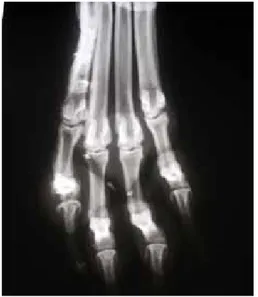
Figure 2. Dorsopalmar radiograph of the left forelimb extremities. Soft tissue swelling at the second digit and marked circumferential palisading periosteal reaction of the second digit bones
Figure 4. CT scan of the chest, mediastinal window. Focal bronchiectasis and tissue reaction at the right caudal lung lobe
Figure 3. CT scan of the chest, chest window. 2, 05* 2, 3 cm diameter mass at the right caudal lung lobe
Case Report Veterinarni Medicina, 57, 2012 (11): 618–621
620
graphs of the extremities the decreased thickness of the palisading periosteal reaction was indicative of resolving Hypertrophic Osteopathy (Figure 6). Thoracic radiographs were normal.
DISCUSSION AND CONCLUSIONS
The mechanisms of HO development are not com-pletely understood (Caywood et al. 1985); several theories have been proposed to explain the pathol-ogy (Dunn et al. 2007). For example, increased blood flow to the distal extremities may occur, with sub-sequent stimulation of connective tissue apposition and local osteogenesis that typically starts in the digits and extends toward the axial skeleton (Allan 2002); alternatively, a neural autonomic reflex that originates in the thorax and modulates the connec-tive tissue and periosteum growth of the limbs may be involved. Furthermore, the metabolites produced by tumour cells might stimulate new bone formation at distant sites or megacariocytes originating from the bone marrow might migrate to pulmonary capil-lary beds and give rise to mature platelets by budding (Hancey and Pass 1972; Lavi et al. 1982;Caywood et al. 1985; Barrand and Scudamore 2001; Peeters et al. 2001; Dunn et al. 2007).
Hypertrophic osteopathy has been reported in different species and is generally associated with primary or secondary neoplasms in the lung or tho-racic cavity (Hancey and Pass 1972; Lavi et al. 1982; Caywood et al. 1985; Hirakata and Kitamura 1995; Amstrong et al. 2007; Dunn et al. 2007; Fridlington et al. 2007), as well as occasionally also by
intra-abdominal lesions (Halliwell and Ackerman 1974; Peeters et al. 2001). Reports of HO associated with non-neoplastic thoracic lesions are less frequent and may include inflammatory lung disease, heart-worm disease, bacterial endocarditis, oesophageal disease secondary to Spirocerca lupi infestation, eosino-philic bronchitis, pulmonary abscess and congenital megaesophagus (Murray 1968; Caywood et al. 1985; Watrus and Blumenfeld 2002). Only one case of a dog with hypertrophic osteopathy associated with a bronchial foreign body is reported (a nail; Caywood et al. 1985). Hypertrophic osteopathy with bronchial grass awn abscesses had not yet been reported.
In certain geographical areas, grass awn inhala-tion and migrainhala-tion has a considerable importance in small animal practice (Brennan and Ihrke 1983; Dobbie et al. 1986; Lotti and Niebauer 1992). The sharp anterior end of the floret of the awn can easily penetrate the skin or any of the body orifices; they can also be inhaled. The barbed nature of the awn prevents retrograde migration, and consequently, progressive forward migration occurs in conjunc-tion with any movement of the surrounding tissues or by air movement within the respiratory tract (Shults and Zwingenberger 2007). Thus, the awns gain access to a variety of sites, even the thoracic cavity, by broncho-pulmonary abcessation ((Shults and Zwingenberger 2007). Despite the relatively high incidence of grass awn-related thoracic disease in endemic areas, HO seems to be an unusual result.
Figure 6. Dorsopalmar radiograph of extremities (three months follow up). Decreased thickness of the palisad-ing periosteal reaction of the second digit bones Figure 5. CT scan of the left forelimb fingers, bone
window. Marked circumferential palisading periosteal reaction of the first phalanx of the second digit
Veterinarni Medicina, 57, 2012 (11): 618–621 Case Report
621 The chronicity of the inflammatory reaction due
to long-standing foreign body migration may have enhanced the development of HO in this case.
The reversibility of HO after removal of the for-eign body and the healing of the lung lesion is a remarkable finding, although regression of HO after resolution of intra-thoracic disease has been reported (Hancey and Pass 1972). Lameness as a clinical sign with hypertrophic osteopathy as the underlying lesion should be included as possible complications of chronic broncho-pneumonia due to grass awn migration in the dog.
Acknowledgement
Thanks to Professor Gert Niebauer (DVM, PhD, Dipl. ECVS, Full Professor in Surgery at Ecole Veterinaire, Paris, EAEVE executive director) for his help in reviewing this manuscript.
RefeReNCeS
Allan G (2002): Radiographic signs of joint disease. In: Thrall’s Textbook of Veterinary Radiology. 4th ed. W.B. Saunders, Philadelphia. 202 pp.
Amstrong DJ, McCausland EMA, Wright GD (2007): Hypertrophic pulmonary osteoarthropathy (HPOA) (Pierre Marie-Bamberger syndrome): two cases pre-senting as acute inflammatory arthritis. Description and review of the literature. Rheumatology Interna-tional 27, 399–402.
Barrand KR, Scudamore CL (2001): Canine hypertrophic osteoarthropathy associated with a malignant Sertoli cell tumour. Journal of Small Animal Practice 42, 143–145. Brennan KE, Ihrke PJ (1983): Grass awn migration in
dogs and cats: a retrospective study of 182 cases. Jour-nal of American Veterinary Medical Association 182, 1201–1204.
Caywood DD, Kromek BA, Feeney DA, Johnston GR (1985): Hypertrophic osteopathy associated with a bronchial for-eign body and lobar pneumonia in a dog. Journal of Amer-ican Veterinary Medical Association 186, 698–700. Dobbie GR, Darke PGG, Head KW (1986):
Intrabron-chial foreign bodies in dogs. Journal of Small Animal Practice 27, 227–238.
Dunn ME, Blond L, Letard D, Di Fruscia R (2007): Hy-pertrophic osteopathy associated with infective endo-carditis in an adult boxer dog. Journal of Small Animal Practice 48, 99–103.
Fridlington J., Weaver J., Kelly B, Kelly E (2007): Second-ary hypertrophic osteoarthropathy associated with solitary fibrous tumour of the lung. Journal of Amer-ican Academy of Dermatology 57, 106–110.
Halliwell WH, Ackerman N (1974): Botryoid rhabdo-myosarcoma of the urinary bladder and hypertrophic osteoarthropathy in a young dog. Journal of American Veterinary Medical Association 165, 911–913. Hancey JB, Pass MA (1972): Hypertrophic pulmonary
osteoarthropathy in a Great Dane. Canadian Veteri-nary Journal 5, 118–120.
Hirakata Y, Kitamura S (1995): Pulmonary Hypertrophic osteoarthropathy and clubbing of digits in patients with lung cancer. Japanese Journal of Thoracic Disease 33, 1080–1085.
Lavi Y, Paladugu RR, Benfield JR (1982): Hypertrophic pulmonary osteoarthropathy in experimental canine lung cancer. Journal of Thoracic Cardiovascular Sur-geon 84, 373–376.
Lotti U, Niebauer G (1992): Tracheobronchial foreign bodies of plant origin in 153 hunting dogs. Compen-dium on Continuing Education for the Practicing Vet-erinarian 14, 900–904.
Murray M (1968): Incidence and pathology of Spirocerca lupi in Kenya. Journal of Comparative Pathology 78, 401–405.
Peeters D, Clercx C, Thiry A , Hamaide A, Snaps F, Hen-roteaux M, Olgive GK, Day MJ (2001): Resolution of paraneoplastic leucocytosis and hypertrophic osteopa-thy after resection of a renal transitional cell carcinoma producing granulocyte-macrophage colony-stimulating factor in a young bull terrier. Journal of Veterinary In-ternal Medicine 15, 407–411.
Schultz RM, Zwingenberger A (2007): Radiographic com-puted tomographic and ultrasonographic findings with migrating intrathoracic grass awns in dogs and cats. Veterinary Radiology and Ultrasound 49, 249–255. Watrus BJ, Blumenfeld B (2002): Congenital
megae-sophagus with hypertrophic osteopathy in a 6-year-old dog. Veterinary Radiology and Ultrasound 6, 545–549.
Received: 2012–08–24 Accepted after corrections: 2012–11–13 Corresponding Author:
Angela Palumbo Piccionello, DVM, PhD, University of Camerino, Faculty of Veterinary Medicine, Clinical Department, Via Circonvallazione 93/95, 62024 Matelica, Italy