Morphology and Molecular Composition of Sarcoplasmic Reticulum
Surface Junctions in the Absence of DHPR and RyR in Mouse
Skeletal Muscle
Edward Felder,* Feliciano Protasi,
†Ronit Hirsch,
†Clara Franzini-Armstrong,* and Paul D. Allen
†*Department of Cell and Developmental Biology, University of Pennsylvania, Philadelphia, Pennsylvania 19104-6058; and†Department of
Anesthesiology, Brigham and Woman’s Hospital, Boston, Massachusetts 02115 USA
ABSTRACT Calcium release during excitation-contraction coupling of skeletal muscle cells is initiated by the functional interaction of the exterior membrane and the sarcoplasmic reticulum (SR), mediated by the “mechanical” coupling of ryanodine receptors (RyR) and dihydropyridine receptors (DHPR). RyR is the sarcoplasmic reticulum Ca2⫹release channel and DHPR is an L-type calcium channel of exterior membranes (surface membrane and T tubules), which acts as the voltage sensor of excitation-contraction coupling. The two proteins communicate with each other at junctions between SR and exterior membranes called calcium release units and are associated with several proteins of which triadin and calsequestrin are the best characterized. Calcium release units are present in diaphragm muscles and hind limb derived primary cultures of double knock out mice lacking both DHPR and RyR. The junctions show coupling between exterior membranes and SR, and an apparently normal content and disposition of triadin and calsequestrin. Therefore SR-surface docking, targeting of triadin and calsequestrin to the junctional SR domains and the structural organization of the two latter proteins are not affected by lack of DHPR and RyR. Interestingly, simultaneous lack of the two major excitation-contraction coupling proteins results in decrease of calcium release units frequency in the diaphragm, compared with either single knockout mutation.
INTRODUCTION
In muscle cells depolarization of the cell membrane results
in rapid release of Ca
2⫹from the sarcoplasmic reticulum
(SR) and subsequent contraction of the myofibrils. The
series of events linking these two steps is called
excitation-contraction (e-c) coupling. Specialized junctions (calcium
release units (CRUs)), formed by a close apposition of the
junctional SR (jSR) to either the plasmalemma or the
trans-verse (T) tubules, constitute the sites of calcium release via
the ryanodine receptor (RyR) or SR Ca
2⫹release channel.
The apposition of exterior and interior membranes at CRUs
allows functional and structural interaction between two
essential contributors to e-c coupling: the voltage sensing,
dihydropyridine sensitive, L-type Ca
2⫹channel
(dihydro-pyridine receptors (DHPR)) of the cell membrane/T-tubules
and the RyR (Franzini-Armstrong and Protasi, 1997).
Within each CRU there may be either one or two
function-ally interacting clusters of RyRs and DHPRs called
cou-plons (Stern et al., 1997). In skeletal muscle, conversion of
the T-tubule depolarization into Ca
2⫹release from the SR is
thought to occur via a micromechanical coupling between
specific domains of type 1 RyR (RyR1) and of the II-III
loop of the skeletal muscle specific
␣1subunit (␣1s) of
DHPR (Schneider and Chandler, 1973; Rios and Brum,
1987; Tanabe et al., 1990).
Mild detergent extraction of junctional SR cisternae
de-rived from CRUs leaves behind a supramolecular complex
of several proteins associated with RyRs (Caswell and
Brunswick, 1984; Costello et al., 1986). Of these,
calseques-trin, junctin, and triadin are best characterized and also have
a structural signature defined below. Calsequestrin is a
luminal SR protein (Jorgensen et al., 1983), which is
spe-cifically targeted to the jSR by its acidic carboxy-terminal
end (Nori et al., 1997, 1999). It is visible as an electron
dense network in the lumen of the SR (Meissner, 1975;
Jones et al., 1998), and the network is connected to the
luminal side of the SR membrane, predominantly in the
region associated with feet by elongated links
(Franzini-Armstrong et al., 1987). Junctin and triadin are two intrinsic
membrane proteins of the SR membrane. Overexpression
experiments have shown that junctin (Zhang et al., 2000)
and triadin (Tijskens, Franzini-Armstrong, and Jones,
un-published observations) have similar roles in inducing tight
periodic clustering of calsequestrin in proximity of the jSR
membrane, perhaps in association with RyRs. Thus,
al-though triadin and/or junctin cannot be directly visualized in
thin section electron microscopy, their presence is
unequiv-ocally put in evidence by the clustering of calsequestrin.
The question arises whether the close linkage between
DHPR and RyR not only allows the two proteins to interact
functionally but also represents the structural framework
that keeps the two membranes attached to each other and
organizes other jSR proteins. Available null mutations that
result in lack of either RyR1 or
␣1s subunit of DHPR have
been used to determine the role of these two proteins in the
function and formation of CRUs. Observations on a RyR1
knock out mutation (“dyspedic”) and on a spontaneous
Submitted October 2, 2001, and accepted for publication February 20, 2002.
Address reprint requests to Edward Felder, Department of Cell and De-velopmental Biology, Anatomy-Chemistry Bldg./B1, University of Penn-sylvania, Philadelphia, PA 19104-6058. Tel.: 215-898-3345; Fax: 215-573-2170; E-mail: [email protected].
clearly demonstrate the functional importance of both
pro-teins in e-c coupling. Muscle activation is not possible in the
absence of either RyR1 or
␣1s DHPR (Takeshima et al.,
1994; Buck et al., 1996; Beam et al., 1986; Knudson et al.,
1989; Adams et al., 1990), but SR/T-tubule junctions are
present in both types of mutations (Franzini-Armstrong et
al., 1991; Takekura et al., 1995; Takekura and
Franzini-Armstrong, 1999; Flucher et al., 1993). These findings
demonstrate that the link between DHPR and RyR1 is not
an essential prerequisite for the assembly of CRUs.
How-ever, based on the published results it is not clear whether or
not the presence of at least one of the two proteins is
necessary for the formation of the junction. That is, we do
not know whether the RyR-DHPR link stabilizes the
junc-tion after its formajunc-tion and whether either protein is
neces-sary for the triadin/calsequestrin association with the jSR
membrane.
The present study compares the structure of SR/T-tubule
junctions and the frequency of couplons in the diaphragm
muscle and primary cultures of skeletal muscle from
wild-type, dyspedic, dysgenic, and new double knockout mutant
mice that lack both RyR1 and
␣1s DHPR. We show that
(necessarily nonfunctional) CRUs with a similar general
architecture to wild type are formed in the double knock out,
despite the lack of these two major e-c coupling components
and that the luminal calsequestrin association with the jSR
membrane is unaltered. Interestingly, we also find that the
frequency of double knock out CRUs is reduced in
dia-phragm in vivo but seems to be unaffected in cultured
primary myotubes.
MATERIALS AND METHODS
Double null mice breeding
Double null mice were obtained by breeding of previously described dyspedic (RyR null) and dysgenic (DHPR null) mice (Buck et al., 1996; Beam et al., 1986). Genotype was determined by polymerase chain reaction of tail DNA. The number of double mutant embryos obtained in these breedings was 5 of 145.
Preparation of primary cultures
Forelimbs and hind limbs muscle were removed from wild-type, dyspedic, dysgenic, and the same double knock out neonatal mice that were used for diaphragm electron microscopy. Satellite cells were selected by the method of Rando and Blau (1994). Briefly, cells were enzymatically dissociated from minced muscle by the addition of 2 ml/g of tissue of a solution of dispase (grade II, 2.4 U/ml, Boehringer Mannheim Corp., Indianapolis, IN) and collagenase (class II, 1% Boehringer Mannheim Corp.) supplemented with CaCl2to final concentration of 2.5 mM. The slurry was maintained at 37°C for 30 to 45 min and triturated every 15 min with a 5-ml plastic pipette and then passed through 80-m nylon mesh (NITEX; Tetko Inc., Monterey Park, CA). The filtrate was spun at 350⫻ g to sediment the dissociated cells, the pellet resuspended in growth medium, and the sus-pension was plated on collagen-coated dishes. During the first several passages of the primary cultures, myoblasts were enriched by preplating (Richler and Yaffe, 1970).
The cells were expanded at 37°C in low glucose Dulbecco’s modified Eagle medium (GIBCO, Invitrogen Corp., Grand Island, NY) containing 20% fetal bovine serum, 100 U/ml penicillin, 100g/ml streptomycin, and additional 2 mML-glutamine (growth medium), 20 nM basic fibroblast growth factor (Promega, Madison, WI). After⬃36/48 h the cells were replated in 35-mm dishes containing THERMANOX coverslips (Nunc Inc., Naperville, IL) coated with MATRIGEL (Collaborative Biomedical Products, Bedford, MA). When cells reached⬃40% confluence, growth medium was replaced with differentiation medium (containing 5% heat inactivated horse serum instead 20% of fetal bovine serum with no bFGF) to induce differentiation. The medium was changed every day, and the cells were either fixed or imaged 4 to 5 d later.
Immunolabeling
The cells were fixed in methanol for a minimum of 20 min at⫺20°C, blocked in phosphate-buffered saline containing 1% bovine serum albumin and 10% goat serum for 1 h, incubated first with primary antibodies and then with secondary antibodies (cyanine 3-CY3 conjugated, Jackson Im-munoResearch Laboratories, Lexington, KY) respectively for 2 h and 1 h at room temperature. Code, specificity, working dilution, original refer-ence, and the sources of primary antibodies are as follows: 34C, mouse monoclonal anti-RyR antibody not type-specific, 1:20 (Airey et al., 1990) (Developmental Studies Hybridoma Bank, The University of Iowa); 21A6, mouse monoclonal anti-␣1DHPR, 1:250 (Morton and Froehner, 1987) (Chemicon International, Temecula, CA); GE4.90, mouse monoclonal anti-triadin, 1:500 (Caswell et al., 1991) (gift of Dr. A.H. Caswell); CSQpAb (rabbit polyclonal, anti-dog cardiac calsequestrin, 1:500) (Jones et al., 1998). The specimens were viewed in an inverted fluorescence microscope Olympus IX70 with a 100X oil immersion lens (UplanFI 110X/1.30 n.a.).
Electron microscopy
Diaphragm muscles from one wild-type, one dysgenic, one dyspedic, and two double mutant mice at a late embryonic stage (18 –19 d of gestation, E18 –19) from the same litter were dissected and prefixed in 5% glutaraldehyde in 0.1 M cacodylate buffer (Franzini-Armstrong et al., 1991) and postfixed in 2% OsO4in 0.1 M cacodylate buffer. After an overnight en bloc stain with saturated uranyl acetate the specimens were dehydrated and embedded in Epon for thin sectioning. The diaphragm from one wild-type mouse at E 18 was from a different litter. Thin sections were examined in a Philips 410 electron microscope at 80 kV. Counting of couplons, measurements of the reference area (myofibril occupied area), and of the fiber diameter were performed on digitized micrographs using NIH image software.
RESULTS AND DISCUSSION
As all of the mutations are birth lethal, but allow embryonic
muscle differentiation, we have focused on the late
embry-onic stage of the diaphragm, which is the most differentiated
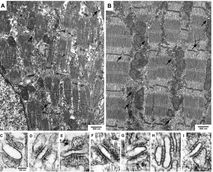
muscle at birth. Fig. 1, A through I compare the overall
structure of diaphragm muscle fibers and the disposition of
CRUs in double knock out and wild-type mice. CRUs are
present in all muscles (some junctions marked with arrows,
Fig. 1, A and B) and have the same general architecture: one
or two dilated SR cisternae with a dense content are apposed
to a T tubule that has an apparently empty lumen (Fig. 2).
The random orientation of the junctions relative to the fiber
long axis as show in Fig. 1 (C–I) is typical of diaphragm
muscle at late embryonal age (Takekura et al., 2001).
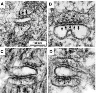
Fig. 2 shows the structure of CRUs in wild-type (Fig. 2 A),
dysgenic (missing DHPR, Fig. 2 B), dyspedic (missing RyR1;
Fig. 2 C), and double knock out (missing both DHPR and
RyR1, Fig. 2 D) fibers. The structure of the junctional SR
cisternae and of the junctional gap between SR and T tubules
are indistinguishable from each other in thin sections of normal
and dysgenic CRUs (Fig. 2, A and B). In both types of muscles
the apposed SR and T-tubule membranes are separated by a
junctional gap distance of approximately 10 to 12 nm; and the
gap is occupied by an evenly spaced row of feet, representing
the cytoplasmic domains of RyRs (arrows) (see also
Franzini-Armstrong et al., 1991). CRUs in dyspedic and double knock
out mice (Fig. 2, C and D), on the other hand, show two clear
differences from wild-type and dysgenic diaphragms: the
junc-tional gap distance is smaller and no feet are visible in the gap
between the two membranes. Thus, lack of RyR is clearly
detectable in thin sections, whereas lack of DHPRs is not. The
latter is put in evidence by freeze-fracture (Franzini-Armstrong
et al., 1991), but this technique was not used in the present
study.
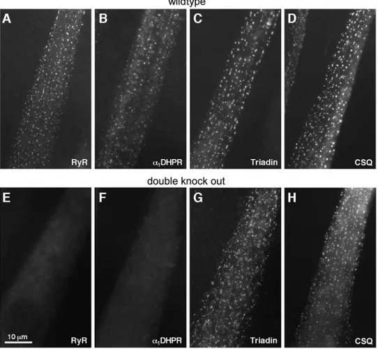
To detect the presence of triadin and calsequestrin in
CRUs and to confirm their interaction, we combined
im-munolocalization at the light microscope level with electron
microscopy. This allowed us to detect the presence of
tria-din and calsequestrin in appropriately located foci by
im-munolabeling. Presence of calsequestrin and its clustering
in the jSR cisternae are directly visible in the electron
micrographs. For technical ease, immunolabeling was not
done on diaphragm but was performed on myotubes derived
from cultured primary myoblasts. Labeling for DHPR, RyR,
calsequestrin (CSQ), and triadin shows that all four proteins
form peripherally located foci, representing CRU sites, in
FIGURE 1 Overview of late embryonic (E18 –19) diaphragm muscle of double knock out (A) and wild-type (B) mice and images of single CRUs of double knock out (C–F), dyspedic (G), dysgenic (H), and wild-type (I) mice. Muscle fibers in double knock out mutant mice (A) show smaller and less well aligned myofibrils than in wild type (B). Arrows point to junctional complexes (CRUs). The myofiber long axis in images (C–I) is vertical and the orientation of junctions relative to the fiber axis is varied at this developmental stage in all mutants as well as in wild-type mice.
wild-type myotubes (Fig. 3, A–D; compare with Protasi et
al., 1997, 1998, 2000). In the double mutant, peripheral foci
are present at approximately equal density as in the
wild-type myotubes and contain calsequestrin and triadin (Fig. 3,
G and H) but lack
␣1s DHPR and RyR1 (Fig. 3, E and F).
In the electron micrographs, the content of the jSR
cis-ternae is remarkably similar in wild type and in all three
mutants (Fig. 2, asterisks). In all cases the electron density
due to CSQ is present and it is also clearly periodically
clustered in proximity of the junctional membrane, an effect
traceable to the presence and appropriate localization of
triadin. This confirms that CSQ and either triadin or junctin
are present in the junctional SR and that CSQ is linked to at
least one of them.
We conclude that not only SR-T tubule docking but also
targeting of calsequestrin and triadin and the formation of a
complex between them is independent of the presence of
either RyRs or DHPR. This strengthens previous results
from single mutations (Takekura et al., 1995; Takeshima et
al., 1995; Takekura and Franzini-Armstrong, 1999; Flucher
et al., 1993) and extends them to include the other
junction-specific proteins. During normal differentiation of cardiac
and skeletal muscle, SR docking and the presence of a
luminal jSR densities due to CSQ clearly precede clustering
of RyR (Protasi et al., 1996; Flucher and
Franzini-Arm-strong, 1996; Takekura et al., 2001), in agreement with the
above observations. Thus junctophilin, the docking protein
that is required for specific SR-surface membrane junction
formation, acts in the absence of RyRs and DHPRs, a
conclusion supported by the ability of this protein to induce
junctions between the endoplasmic reticulum and the
sur-face membrane in a nonmuscle cell (Takeshima et al., 2000;
Ito et al., 2001).
Morphometry reveals an important difference in the
abundance of SR/T-tubule junctions in the diaphragm
be-tween the wild type, the single null, and the double null
mutations. Despite the fact that a limited sample size was
available to obtain our data, the trend is very clear. Table 1
compares the frequency of couplons in the diaphragms of
wild-type, dyspedic, dysgenic and double knock out mice,
expressed as the number of couplons per
myofibril-occu-pied area of a fiber section. This number is proportional to
the ratio of couplons to myofibril-occupied fiber volume,
assuming that the CRUs are of the same size on the average.
The frequency of couplons in diaphragms of dyspedic and
dysgenic mice is reduced to
⬃50% compared with the wild
type, and the frequency of couplons in the two mutant
muscle types are not different from each other. A
consid-erable further reduction (again by
⬃50%) is seen between
double null mice and the single null dyspedic/dysgenic
mice.
The fiber cross-sectional area is not reduced in mutated
versus normal fibers. The mean cross-sectional area is
135.0
⫾ 58.6
m
2, n
⫽ 106 (mean ⫾ 1 SD, n ⫽ number of
fibers) for wild-type fibers; 179.4
⫾ 100.7
m
2, n
⫽ 92 for
dysgenic; 148.1
⫾ 71.0
m
2, n
⫽ 54 for dyspedic; and
187.1
⫾ 98.6
m
2, n
⫽ 120 for double null animals. It
should be noted however that diaphragms of null animals
show a considerable reduction in the total number of fibers
relative to the wild-type diaphragm, probably indicating a
high level of apoptosis, a phenomenon previously seen in
dyspedic muscle ( P.D. Allen and J. Sommer, unpublished
observations). Therefore it is possible that the measured
fiber diameters compare a population of primary and
sec-ondary fibers in the wild type versus only one generation of
fibers in the mutated diaphragms.
In all null mutants the myofibrils fill most of the available
space but show defects: changes in orientation and splitting
(see Fig. 1 A). All three mutations result in muscle paralysis
due to absence of excitation-contraction coupling, and all
have similar overall fiber morphology with some
myofibril-lar defects. The lack of activity might be the cause of the
observed defects as it was shown that muscles paralyzed
with tetrodotoxin exhibit a similar disordered alignment of
filaments (Houenou et al., 1990), indicating the importance
of muscle activity for an adequate alignment of myofibrils.
CRU frequency, on the other hand, is more strongly affected
in the diaphragm of double null than in either single
muta-tion. A possible explanation is that some movements of
Ca
2⫹into the cytoplasm may occur in muscles with single
mutations, either by leak through the RyR in the dysgenic
FIGURE 2 Images of single CRUs showing the presence of feet (on opposite side of the jSR membrane from the tips of arrows) in wild-type (A) and dysgenic mice (B) and their absence in dyspedic (C) and double knock out (D) mice. The junctional gap between the sarcoplasmic mem-brane and the memmem-brane of the T-tubule is slightly narrower in dyspedic and double knock out mutants. Calsequestrin is visible in the SR sacs (asterisk) of wild-type and all mutant mice. Note: the CRUs in Fig. 2 are all presented in the same orientation, even though their positioning relative to the myofibrils varied in the original micrographs.
mouse or through voltage gated activation of the DHPR in
the dyspedic mouse. In the absence of either source of
cytoplasmic Ca
2⫹movement, formation of the membrane
system might be affected. Indeed, it is known that lack of
functional DHPR causes altered transcription levels of
cer-tain genes during muscle cell development (Chaudari and
Beam, 1997; Luo et al., 1996), and a further reduction in
calcium movements are most likely to have more serious
effects. The fact that only the diaphragm muscle showed
this defect but both cultured myotubes (present study) and
dyspedic hind limb muscle of the same age (Takekura et al.,
1995) do not suggests that this effect is somehow related to
the degree of development/differentiation. This is because
the last two types of muscles are less differentiated than the
diaphragm at this stage of development. Interestingly, fiber
diameters in dyspedic leg muscles are also considerably
smaller than in the wild type (Takekura et al., 1995), which
was not found in diaphragm muscle. A lower degree of
apoptosis in the leg muscle (again due to a lower degree of
differentiation) could explain this fact as well.
The major conclusions of this work are that neither RyR
nor DHPR, alone or separately, are necessary for T-SR
docking and for the targeting and/or association of
calse-questrin and triadin in the junctional SR. Both proteins
however are needed for appropriate muscle development.
This work was supported by the National Institutes of Health Grant PO1 535849.
FIGURE 3 Immunolabeling images of cultured myotubes from wild-type (A–D) and double knock mice (E and F). Labeling for RyR and DHPR proves that neither is expressed in the mutants (E and F). Calsequestrin (CSQ) and triadin are located in distinct foci (G and H) in cells of in double knock out mice and show the same pattern as that of cells from wild-type mice.
TABLE 1. Frequency of couplons in wild-type and mutant mice diaphragm muscle
Mouse Type
Number of couplons/100m2 of myofibril occupied area
(Mean⫾ SD)
Wildtype 6.1⫾ 2.2*
Dysgenic 3.6⫾ 2.0*
Dyspedic 3.3⫾ 1.8*
Double knock out 1.7⫾ 1.1† *One animal.
†Two animals.
Four different parts from each diaphragm were sectioned. 15–20 randomly chosen areas were counted per section (⬃20,0002m per animal in total).
REFERENCES
Adams, B. A., T. Tanabe, A. Mikami, S. Numa, and K. G. Beam. 1990. Intramembrane charge movement restored in dysgenic skeletal muscle by injection of dihydropyridine receptor cDNAs. Nature. 346:569 –572. Airey, J. A., C. F. Beck, K. Murakami, S. J. Tanksley, T. J. Deerinck, M. H. Ellisman, and J. L. Sutko. 1990. Identification and localization of two triad junctional foot protein isoforms in mature avian fast-twitch skeletal muscle. J. Biol. Chem. 265:14187–14194.
Beam, K. G., J. M. Knudson, and J. A. Powell. 1986. A lethal mutation in mice eliminates the slow calcium current in skeletal muscle cells. Na-ture. 320:168 –170.
Buck, E. D., H. T. Nguyen, I. N. Pessah, and P. D. Allen. 1996. Dyspedic mouse skeletal muscle expresses major elements of the triadic junction but lacks detectable ryanodine receptor protein and function. J. Biol. Chem. 272:7360 –7367.
Caswell, A. H., N. R. Brandt, J. P. Brunschwig, and S. Purkerson. 1991. Localization and partial characterization of the oligomeric disulfide-linked molecular weight 95,000 protein (triadin) which binds the ryan-odine and dihydropyridine receptors in skeletal muscle triadic vesicles. Biochemistry. 30:7507–7513.
Caswell, A. H., and J-P. Brunswick. 1984. Identification and extraction of proteins that compose the triad junction of skeletal muscle. J. Cell Biol. 99:929 –939.
Chaudari, N., and K. G. Beam. 1997. mRNA for cardiac calcium channel is expressed during development of skeletal muscle. Dev. Biol. 155: 507–515.
Costello, B. R., A. Saito, A. Maurer, and S. Fleischer. 1986. Characteri-sation of the junctional face membrane from terminal cisterne of sarco-plasmic reticulum. J. Cell Biol. 103:741–753.
Flucher, B. E., S. B. Andrews, S. Fleischer, A. R. Mark, A. Caswell, and J. A. Powell. 1993. Molecular organization of transverse tubule/ sarcoplasmatic reticulum junctions during development of excitation-contraction coupling in skeletal muscle. J. Cell Biol. 123:1161–1174. Flucher, B. E., and C. Franzini-Armstrong. 1996. Formation of junctions
involved in excitation-contraction coupling in skeletal and cardiac mus-cle. Proc. Natl. Acad. Sci. U. S. A. 93:8101– 8106.
Franzini-Armstrong, C., L. Kenney, and E. Varriano-Marston. 1987. The structure of calsequestrin in triads of vertebrate muscle. J. Cell Biol. 105:49 –56.
Franzini-Armstrong, C., M. Pincon-Raymond, and F. Rieger. 1991. Muscle fibers from dysgenic mouse in vivo lack a surface component of periph-eral couplings. Dev. Biol. 146:364 –376.
Franzini-Armstrong, C., and F. Protasi. 1997. Ryanodine receptors of striated muscles: a complex channel capable of multiple interactions. Physiol. Rev. 3:699 –729.
Houenou, L. J., M. Pincon-Raymond, L. Garcia, A. J. Harris, and F. Rieger. 1990. Neuromuscular development following tetrodotoxinduced in-activity in mouse embryos. J. Neurobiol. 21:1249 –1261.
Ito, K., S. Komazaki, K. Sasamoto, M. Yoshida, M. Nishi, K. Kitamura, and H. Takeshima. 2001. Deficiency of triad junction and contraction in mutant skeletal muscle lacking junctophilin type 1. J. Cell Biol. 154: 1059 –1067.
Jones, L. R., Y. J. Suzuki, W. Wang, Y. M. Kobayashi, V. Ramesh, C. Franzini-Armstrong, L. Cleemann, and M. Morad. 1998. Regulation of Ca2⫹ signaling in transgenic mouse cardiac myocytes overexpressing calsequestrin. J. Clin. Invest. 101:1385–1393.
Jorgensen, A. O., A. C. Shen, K. P. Campell, and D. H. Mac Lennan. 1983. Ultrastructural localization of calsequestrin in rat skeletal muscle by immunoferritin labeling of ultrathin frozen sections. J. Cell. Biol. 97: 1573–1581.
Knudson, C. M., N. Chaudhari, A. H. Sharp, J. A. Powell, K. G. Beam, and K. P. Campbell. 1989. Specific absence of the alpha 1 subunit of the dihydropyridine receptor in mice with muscular dysgenesis. J. Biol. Chem. 264:1345–1348.
Luo, Z. D., M. Pincon-Raymond, and P. Taylor. 1996. Acetylcholinester-ase and nicotinic acetylcholin receptor expression diverge in muscular
dysgenic mice lacking the L-type calcium channel. J. Neurochem. 67: 111–117.
Meissner, G. 1975. Isolation and characterization of two types of sarco-plasmic reticulum vesicles. Biochim. Biophys. Acta. 389:51– 68. Morton, M. E., and S. C. Froehner. 1987. Monoclonal antibody identifies
a 200-kDa subunit of the dihydropyridine-sensitive calcium channel. J. Biol. Chem. 262:11904 –11907.
Nori, A., E. Gola, S. Tosato, M. Cantini, and P. Volpe. 1999. Targeting of calsequestrin to sarcoplasmic reticulum after deletions of its acidic carboxy terminus. Am. J. Physiol. 277:C974 –981.
Nori, A., K. A. Nadalini, A. Martini, R. Rizzuto, A. Villa, and P. Volpe. 1997. Chimeric calsequestrin and its targeting to the junctional sarco-plasmic reticulum of skeletal muscle. Am. J. Physiol. 272:C1421–1428. Protasi, F., C. Franzini-Armstrong, and P. D. Allen. 1998. Role of ryano-dine receptors in the assembly of calcium release units in skeletal muscle. J. Cell. Biol. 140:831– 842.
Protasi, F., C. Franzini-Armstrong, and B. E. Flucher. 1997. Coordinated incorporation of skeletal muscle dihydropyridine receptors and ryano-dine receptors in peripheral couplings of BC3H1 cells. J. Cell Biol. 137:859 – 870.
Protasi, F., X. Sun, and C. Franzini-Armstrong. 1996. Formation and maturation of the calcium release apparatus in developing and adult avian myocardium. Dev. Biol. 173:265–278.
Protasi, F., H. Takekura, Y. Wang, S. R. W. Chen, G. Meissner, P. D. Allen, and C. Franzini-Armstrong. 2000. RyR1 and RyR3 have different roles in the assembly of calcium release units of skeletal muscle. Biophys. J. 79:2494 –2508.
Rando, T. A., and H. M. Blau. 1994. Primary mouse myoblast purification, characterization and transplantation for cell-mediated gene therapy. J. Cell Biol. 125:1275–1287.
Richler, C., and D. Yaffe. 1970. The in vitro cultivation and differentiation capacities of myogenic cell lines. Dev. Biol. 23:1–22.
Rios, E., and G. Brum. 1987. Involvement of dihydropyridine receptors in excitation-contraction coupling in skeletal muscle. Nature. 325: 717–720.
Schneider, M. F., and W. K. Chandler. 1973. Voltage dependent charge movement in skeletal muscle: a possible step in excitation-contraction coupling. Nature. 242:244 –246.
Stern, M. D., G. Pizarro, and E. Rios. 1997. Local control model of excitation-contraction coupling in skeletal muscle. J. Gen. Physiol. 110:415– 440.
Takekura, H., B. E. Flucher, and C. Franzini-Armstrong. 2001. Sequential docking, molecular differentiation and positioning of T tubules/SR junc-tions in developing mouse skeletal muscle. Dev. Biol. 239:204 –214. Takekura, H., and C. Franzini-Armstrong. 1999. Correct targeting of
dihydropyridine receptors and triadin in dyspedic mouse skeletal muscle in vivo. Dev. Dyn. 214:373–380.
Takekura, H., M. Nishi, T. Noda, H. Takeshima, and C. Franzini-Armstrong. 1995. Abnormal junctions between surface membrane and sarcoplasmic reticulum in skeletal muscle with a mutation targeted to the ryanodine receptor. Proc. Natl. Acad. Sci. U. S. A. 92:3381–3385. Takeshima, H., S. Komazaki, M. Nishi, M. Lino, and K. Kengawa. 2000.
Junctophilins: a novel family of junctional membrane complex proteins. Mol. Cell. 6:11–22.
Takeshima, H., M. Lino, H. Takekura, M. Nishi, J. Kuno, O. Minowa, H. Takano, and T. Noda. 1994. Excitation-contraction uncoupling and muscular degeneration in mice lacking functional skeletal muscle ryan-odine-receptor gene. Nature. 369:556 –559.
Takeshima, H., T. Yamazawa, T. Ikemoto, H. Takekura, M. Nishi, T. Noda, and M. Iino. 1995. Ca2⫹ induced Ca2⫹ release in myocytes from dyspedic mice lacking the type 1 ryanodine receptor. EMBO J. 14: 2999 –3006.
Tanabe, T., K. G. Beam, B. A. Adams, T. Niidome, and S. Numa. 1990. Regions of the skeletal muscle dihydropyridine receptor critical for excitation-contraction coupling. Nature. 346:567–569.
Zhang, L., C. Franzini-Armstrong, V. Ramesh, and L. Jones. 2000. Struc-tural alterations in cardiac calcium release units resulting from overex-pression of junctin. J. Mol. Cell. Cardiol. 33:233–247.