ORIGINAL ARTICLE
Five-year retrospective clinical study of indirect
composite restorations luted with a light-cured composite
in posterior teeth
Camillo D’Arcangelo&Maciej Zarow&Francesco De Angelis&
Mirco Vadini&Michele Paolantonio&Mario Giannoni&
Maurizio D’Amario
Received: 2 October 2012 / Accepted: 8 May 2013 # Springer-Verlag Berlin Heidelberg 2013
Abstract
Objectives The purpose of this study was to evaluate the clinical performance of indirect composite onlays–overlays bonded with a light-cured composite on vital molars. Materials and methods Forty-one patients were restored with 79 indirect composite restorations. The restorations were studied for an observation time of 5 years. Marginal adaptation, marginal discolouration, secondary caries, col-our match and anatomic form were clinically examined following modified United States Public Health Service criteria. Each restoration was also examined for fractures and debonding. Endodontic complications were registered. Survival rate, based just on failures that required a replace-ment, and success rate that included also failures that re-quired a repair intervention were statistically determined using a restoration and a patient-related analysis.
Results After 5 years, using each restoration as a statistical unit, the survival rate was 91.1 % and the success rate
84.8 %, with a high Kaplan–Meier estimated success prob-ability of 0.852. Using the patient as the statistical unit, the survival rate was 90.2 % and the success rate 85.4 %, corresponding to a Kaplan–Meier estimated success proba-bility of 0.857. On the basis of the criteria used, most of the restorations rated Alpha. Regarding marginal adaptation and marginal discolouration, 5 and 10.1 % of the restorations, respectively, revealed Bravo ratings
Conclusions Indirect composite restorations offer a predict-able and successful treatment modality giving an optimal preservation of sound tooth tissue.
Clinical relevance The preparation, cementation and finishing procedures are considered key factors for the long-term success of the indirect composite restorations.
Keywords Adhesive luting . Clinical evaluation . Indirect composite . Onlay . Overlay
Introduction
Greater demand for aesthetic restorations has revolutionised modern dentistry and brought about the widespread use of resin composites. In accordance with respective clinical in-dications, resin composite materials are suitable to be used for both direct and indirect restorations [1]. The rehabilita-tion of decayed or fractured posterior teeth using an indirect technique was introduced to overcome some of the prob-lems associated with direct restorative techniques, amongst others, polymerization shrinkage and insufficient wear re-sistance or mechanical properties of directly placed filling materials [2]. Moreover, the achievement of proper
C. D’Arcangelo (*)
:
F. De Angelis:
M. VadiniDepartment of Restorative Dentistry, School of Dentistry,
University G. D’Annunzio, Via dei Vestini 31,
66100 Chieti, Italy e-mail: [email protected] M. Zarow
Private practice in Krakow, Krakow, Poland M. Paolantonio
Department of Periodontology, School of Dentistry,
University G. D’Annunzio, Chieti, Italy
M. Giannoni
:
M. D’AmarioDepartment of Life, Health and Environmental Sciences, School
of Dentistry, Dental Clinic, University of L’Aquila, L’Aquila, Italy
interproximal contact and occlusal morphology and the com-plete cure of composite resins in the deepest regions of a cavity are other challenges related to direct composite resto-rations. The achievement of a proper degree of conversion is of paramount clinical interest for resin composite materials as unreacted monomers and inadequate polymerization may compromise mechanical behaviour under masticatory loads leading to increased wear [3], solubility, dimensional instabil-ity, colour change and reduced biocompatibility [4] which may consequently affect the restorations longevity and ulti-mate clinical success [5]. Various approaches have been de-veloped to improve some of the deficiencies of direct-placement composites [6, 7]. However, no method has completely eliminated the problem of marginal microleakage associated with direct composite [8,9].
For these reasons, indirect resin composite (IRC) resto-rations, including inlays (no cuspal coverage), onlays (at least one cuspal coverage) and overlays (full cuspal cover-age), constitute today a substantial portion of contemporary aesthetic restorative treatments for large restorations of se-verely destroyed teeth. Among the parameters that may influence the clinical success of indirect restorations, a proper degree of polymerization of the resin luting agent should be taken into account [10]. Indirect restorations adhesive luting could be performed employing both dual-curing and light-dual-curing cements [11,12]. Dual-curing ma-terials are advantaged by their self-curing component, which favours the conversion even in the presence of scarce radiant energy, but have the disadvantages of being considerably fluid and requiring a mixture of two elements, arising in the probable formation of porosities or voids and incorporation of bubbles. On the other hand, light-curing materials used as luting agent are easily handled and are characterised by controllable hardening times that create high quality mar-gins; with no time restriction, it is easier to achieve a good positioning of the inlay/onlay and to accurately remove all the excess cement, in this way improving all restoration quality [13,14]. Only their light activation can constitute a disadvantage, since light polymerisation of all portions of
the cement may not always be possible because the substan-tial thickness and opacity of the inlay restoration may block the light [10].
The purpose of this study was to evaluate the clinical performance of 79 IRC restorations bonded with a light-cured composite over a period of 5 years.
Materials and methods
Study design
This is a retrospective non-interventional study based on the evaluation of patient records and on the clinical follow-up examination of patients treated with IRC restorations. An institutional review board approval was obtained from the local ethic committee (protocol number 0126128/12). Registration of the records of all patients (n=41) who had received IRC onlays or overlays (n=79) on molars during the period April 2005 to January 2007 was performed from May 2010 to February 2012 by the authors. The patients belonged to the ordinary clientele of the dental clinic of the Department of Life, Health and Environmental Sciences—University of L'Aquila (Italy) and were treated by one dentist with over 15 years experience of restorative dentistry and a long interest in metal-free restorations. Indication for treatment was the replacement of failed res-torations or primary caries in stress-bearing class II prepa-rations on first and second molars. A comprehensive list of the inclusion and exclusion criteria adopted in the study is provided in Table 1. All materials used in this study were standard restorative materials in the dental clinic at the time when the restorations were placed. The patients were treated with a minimum of one, up to a maximum of three IRC restorations. All the patients were males or females at least 18 years of age who were regular dental attendees of the clinic and with a good level of oral hygiene. The patients included in the study were scheduled for annual check-ups and were asked to contact the clinic whenever they
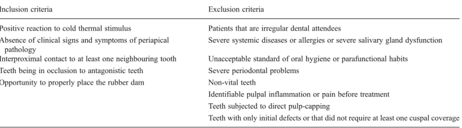
Table 1 List of inclusion and exclusion criteria
Inclusion criteria Exclusion criteria
Positive reaction to cold thermal stimulus Patients that are irregular dental attendees
Absence of clinical signs and symptoms of periapical pathology
Severe systemic diseases or allergies or severe salivary gland dysfunction
Interproximal contact to at least one neighbouring tooth Unacceptable standard of oral hygiene or parafunctional habits
Teeth being in occlusion to antagonistic teeth Severe periodontal problems
Opportunity to properly place the rubber dam Non-vital teeth
Identifiable pulpal inflammation or pain before treatment Teeth subjected to direct pulp-capping
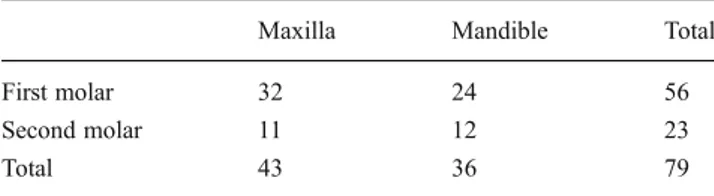
experienced problems with their restorations or abutment teeth. The procedures carried out during each recall exam-ination are outlined in a following paragraph. Once the patient was entered into the clinic, his or her teeth were cleaned to remove extrinsic stains and dental calculus. Patients were informed about the need for good gingival health and were educated in effective plaque control. Both preoperative and post-operative photographs were taken for each patient in order to evaluate the change in appearance (Fig.1). The distribution of IRC restorations by tooth type is presented in Table 2. The restorations included 57 onlays and 22 overlays (Table3).
Tooth preparation and laboratory procedures
A local anaesthetic was used for all patients. Teeth were cleaned first with a fluoride-free prophylaxis paste and a rubber cup. All cavities were prepared according to common principles for adhesive onlays/overlays. After caries and/or failed restorations removal, the preparations were performed
with 80-μm grit cylindrical round-ended diamond burs (Intensiv, Viganello-Lugano, Switzerland). The preparation designs were based on class II or MOD cavities with an occlusal box of at least 2 mm depth and an isthmus width of at least 2 mm. Convergence angles of 10–12° between opposing walls were prepared. At least one cuspal coverage was required for each restoration. The occlusal tooth sur-face was reduced by 2 mm following the occlusal anatomy. Preparation margins were not bevelled but prepared butt joint. After cavity preparation and before cavity finishing, adhesive procedures (EnaEtch, EnaBond; Micerium, Genova, Italy) were performed [15] under rubber dam in order to achieve an immediate dentin sealing [16]. A light-curing composite filling material (Enamel Plus HFO; Micerium, Avegno, Genova, Italy) was, then, used to block out defect-related undercuts and to maintain a standardised prepa-ration protocol. When prepaprepa-ration margins extended into the dentin, the teeth were only included in the study
Fig. 1 A mandibular first molar, with caries recurrence on an occlusal composite restoration (a). Cavity preparation (b). Cementation of an indirect composite
restoration (c). The outcome after 5 years of clinical service shows the wearing out of the composite at the margin (Bravo score for marginal adaptation) (d)
Table 2 Distribution of restored teeth
Maxilla Mandible Total
First molar 32 24 56
Second molar 11 12 23
Total 43 36 79
Table 3 Number of composite indirect restorations in relation to restored surfaces
Maxilla Mandible Total
Onlays 29 28 57
Overlays 14 8 22
when rubber dam placement for subsequent restoration placement was still possible. The finishing phases were performed with 25-μm grit diamond burs with a slight taper (Intensiv, Viganello-Lugano, Switzerland) and with silicone points (Enhance; Dentsply DeTrey GmbH, Konstanz, Germany) under a stereomicroscope (SOM 32; Karl Kaps GmbH & Co.KG, Asslar/Wetzlar, Germany) magnification. After preparation, complete arch impressions were taken with a polyether material (Impregum, 3M ESPE, St. Paul, MN, USA). The teeth were protected with temporary eugenol-free temporary restorations (Cavit, 3M-Espe).
The IRC restorations were made by a dental technician who was experienced in fabricating composite resin onlays, strictly following manufacturer’s instructions (Enamel Plus HFO, Micerium).
Placements of the IRC restorations
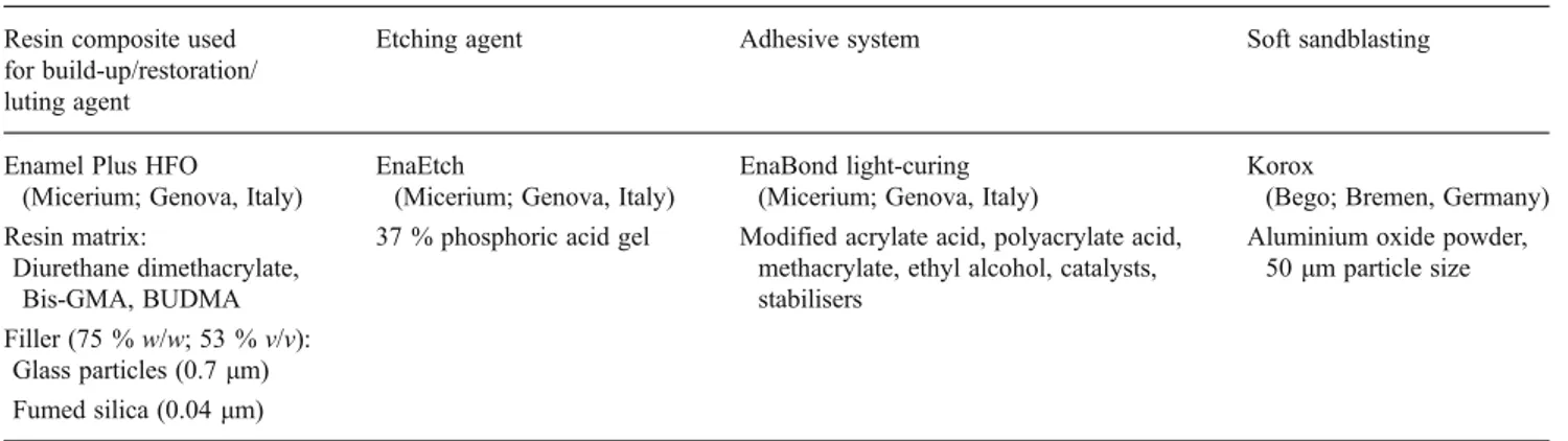
All restorations were definitively inserted within 2 weeks after impression. Provisional restorations were removed using an ultrasonic handpiece. A try-in of the onlays/overlays was performed to check proximal con-tacts and marginal fit. Minor corrections of the restora-tions during their initial try out were made chair-side. IRC restorations requiring major corrections or needing complete revisions were sent to the dental laboratory. Before cementation, the internal surfaces of indirect restorations were abraded with 50 μm aluminium oxide (Korox, Bego; Bremen, Germany), using an intraoral sandblasting device (Micerium, Avegno; Genova, Italy). The tip of the microetcher was kept 5 cm away from the surfaces and applied for 10 s at 2.0 bar pressure [12]. The restorations were subsequently rinsed under running water to remove the debris (20 s), cleaned in an ultrasonic device (2 min) and air-dried. All the bonding procedures were carried out using rubber dental dam. A
two-step total etch technique (EnaBond, Micerium) was used for onlay/overlay cementation [17]. To avoid inac-curacies of fit, the adhesive was not light-polymerized before restoration placement. An adhesive layer was also applied to the abraded restoration surface without light-curing. A light-curing composite (Enamel Plus HFO; Micerium) was warmed up, using a heater for composite preset to 55 °C (Ena Heat; Micerium), put on the cementation surface of each onlay/overlay and used as luting agent. The restoration was lightly pressed into place with a silicon tip obturator and a thin explor-er was used to remove excess luting matexplor-erial extruded from the margins. The pressure was stopped when no more excess of luting material extruded from the mar-gins. Six to eight seconds of light-polymerizing at the occlusal surface ensured stabilisation of the restoration (Optilux 501; Demetron/Kerr Co., Orange, CA, USA). Residual cement was removed under a stereomicroscope (SOM 32; Karl Kaps GmbH & Co.KG) magnification with explorer, scalpel and Superfloss (Oral-B, London, UK) for interproximal sides. Each restoration surface was then light-polymerized in two sessions of 40 s each with a light intensity of at least 1,000 mW/cm2(Optilux 501; Demetron/Kerr Co.). Restoration margins were then checked again under a stereomicroscope and using a dental probe. Residual excess cement was further re-moved with a 15c scalpel (#371716, Bard-Parker; Becton-Dickinson, Dr. Franklin Lakes, NJ, USA). No diamond burs, polishing discs or silicone polishers were used to finish the restoration; interproximal floss was used for interproximal sides. All the materials employed for the cementation are summarised in Table 4. After removal of rubber dam, static and dynamic occlusions were checked and eventually adjusted using fine grit diamond burs. The patients were initially recalled after 1 week for checking occlusion, proximal contact re-lationships, marginal integrity and gingival margin
Table 4 Materials employed for the adhesive procedures Resin composite used
for build-up/restoration/ luting agent
Etching agent Adhesive system Soft sandblasting
Enamel Plus HFO (Micerium; Genova, Italy)
EnaEtch
(Micerium; Genova, Italy)
EnaBond light-curing (Micerium; Genova, Italy)
Korox
(Bego; Bremen, Germany)
Resin matrix: 37 % phosphoric acid gel Modified acrylate acid, polyacrylate acid,
methacrylate, ethyl alcohol, catalysts, stabilisers
Aluminium oxide powder,
50μm particle size Diurethane dimethacrylate, Bis-GMA, BUDMA Filler (75 % w/w; 53 % v/v): Glass particles (0.7μm) Fumed silica (0.04μm)
Bis-GMA isopropylidene-bis[2(3)-hydroxy-3(2)-(4-phenoxy) propyl]-bis(methacrylate), BUDMA 1,4 butandioldimethacrylat, w/w weight/weight ratio, v/v volume/volume ratio
health. In the same visit, the clinical evaluation of each restoration at baseline was performed.
Assessment of the restorations
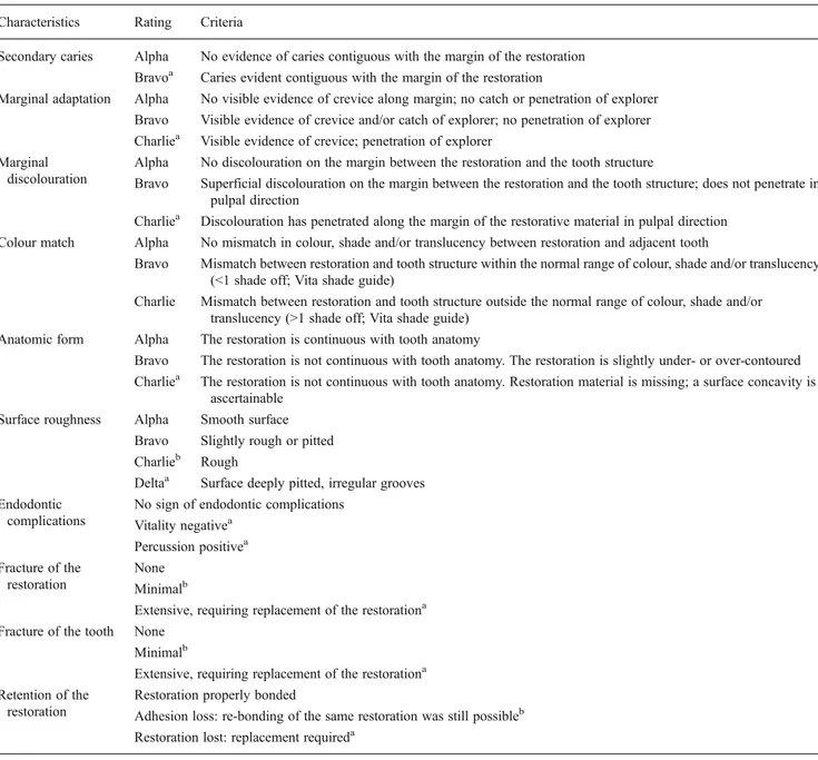
At the baseline recall and at every next annual check-up, the IRC restorations were classified according to the modified United States Public Health Service (USPHS) criteria [14,
18,19]. Recall assessments were always performed by the same single evaluator, different from the clinician who had placed the restorations. The restorations were visually inspected with dental mirror and probe, and clinically
examined with wax-free dental floss. The list of the evalu-ation criteria is provided in Table 5. Deviations in colour match and anatomic form were recorded. Each restoration was examined for cracks, fractures and debonding. Pulp vitality was verified with CO2 test. The patients were
questioned about possible post-operative complaints [14,
20]. The involved teeth were photographed preoperatively and postoperatively, as well as at recall appointments.
It was established to consider as absolute failure (Table5) any occurrence that required the replacement of the entire restoration (Bravo score for secondary caries; any endodon-tic complications; Charlie score for marginal adaptation,
Table 5 USPHS criteria and clinical parameters used for the evaluation of the indirect resin composite restorations
Characteristics Rating Criteria
Secondary caries Alpha No evidence of caries contiguous with the margin of the restoration
Bravoa Caries evident contiguous with the margin of the restoration
Marginal adaptation Alpha No visible evidence of crevice along margin; no catch or penetration of explorer
Bravo Visible evidence of crevice and/or catch of explorer; no penetration of explorer
Charliea Visible evidence of crevice; penetration of explorer
Marginal discolouration
Alpha No discolouration on the margin between the restoration and the tooth structure
Bravo Superficial discolouration on the margin between the restoration and the tooth structure; does not penetrate in
pulpal direction
Charliea Discolouration has penetrated along the margin of the restorative material in pulpal direction
Colour match Alpha No mismatch in colour, shade and/or translucency between restoration and adjacent tooth
Bravo Mismatch between restoration and tooth structure within the normal range of colour, shade and/or translucency
(<1 shade off; Vita shade guide)
Charlie Mismatch between restoration and tooth structure outside the normal range of colour, shade and/or
translucency (>1 shade off; Vita shade guide)
Anatomic form Alpha The restoration is continuous with tooth anatomy
Bravo The restoration is not continuous with tooth anatomy. The restoration is slightly under- or over-contoured
Charliea The restoration is not continuous with tooth anatomy. Restoration material is missing; a surface concavity is
ascertainable
Surface roughness Alpha Smooth surface
Bravo Slightly rough or pitted
Charlieb Rough
Deltaa Surface deeply pitted, irregular grooves
Endodontic complications
No sign of endodontic complications
Vitality negativea Percussion positivea Fracture of the restoration None Minimalb
Extensive, requiring replacement of the restorationa
Fracture of the tooth None
Minimalb
Extensive, requiring replacement of the restorationa
Retention of the restoration
Restoration properly bonded
Adhesion loss: re-bonding of the same restoration was still possibleb
Restoration lost: replacement requireda
a
Absolute failure (the replacement of the entire restoration was required) b
marginal discolouration, anatomic form, fracture of the res-toration, fracture of the tooth, retention of the restoration; Delta score for surface roughness). Failures that required any other type of repair intervention, excluding the replace-ment (Bravo score for restoration or tooth fractures; Charlie score for surface roughness; adhesion loss, when re-bonding of the same restoration was still possible), were seen as relative failures. Any other score amongst those indicated in Table5 was not considered as a failure because judged clinically acceptable. For statistical evaluation, the number of failures that required a replacement (absolute) was used to calculate a survival rate. The number of both relative (with repair intervention) and absolute (requiring replacement) failures was summarised in a success rate, which was also calculated according to the Kaplan–Meier analysis and graphically depicted [14,21]. The beginning of the obser-vation interval started with the incorporation of the
restoration and the end of the interval was defined by the incidence of an absolute failure.
Two different approaches were used for the analysis [14]: & A restoration-related analysis, using each restoration as a
statistical unit;
& A patient-related analysis that considered the patient as the statistical unit. In this case, according to Roulet [22], where more than one restoration was placed in a patient, the evaluated restoration was selected by random, using a random table.
Results
The study population consisted of 41 patients: 23 (56 %) women (mean age 35 years, range 20–48 years) and 18
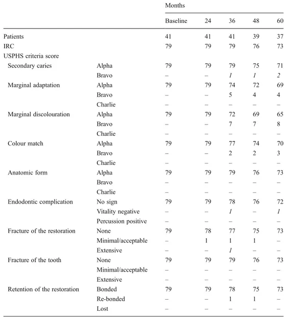
Table 6 Results of the clinical evaluation at baseline and after 24, 36, 48 and 60 months
Absolute failures are indicated in italics
IRC indirect resin composite
Months
Baseline 24 36 48 60
Patients 41 41 41 39 37
IRC 79 79 79 76 73
USPHS criteria score
Secondary caries Alpha 79 79 79 75 71
Bravo – – 1 1 2
Marginal adaptation Alpha 79 79 74 72 69
Bravo – – 5 4 4
Charlie – – – – –
Marginal discolouration Alpha 79 79 72 69 65
Bravo – – 7 7 8
Charlie – – – – –
Colour match Alpha 79 79 77 74 70
Bravo – – 2 2 3
Charlie – – – – –
Anatomic form Alpha 79 79 79 76 73
Bravo – – – – –
Charlie – – – – –
Endodontic complication No sign 79 79 78 76 72
Vitality negative – – 1 – 1
Percussion positive – – – – –
Fracture of the restoration None 79 78 77 75 73
Minimal/acceptable – 1 1 1 –
Extensive – – 1 – –
Fracture of the tooth None 79 79 79 76 73
Minimal/acceptable – – – – –
Extensive – – – – –
Retention of the restoration Bonded 79 79 78 75 73
Re-bonded – – 1 1 –
(44 %) men (mean age 36 years, range 18–51 years). In 45 teeth (57 %), the placement of an IRC restoration was required as a replacement of a failed restoration. The num-ber of patients and restorations examined at the baseline and at each of the following annual check-ups are summarised in Table6. The failures recorded in the whole 5-year period are shown in Fig. 2. Up to the 24th month, no failures were recorded and all restorations in all categories were rated Alpha. Two patients showed negative vitality at the 36th and 60th month recalls. Four further absolute failures in the form of secondary decay at the restoration–tooth interface were observed: one at the 40th, one at 48th and two at 60th month recalls. One extensive restoration fracture was recorded at the 36th month recall. As a consequence of the seven absolute failures, after 5 years, the restoration-related
survival rate was 91.1 %, whilst the patient-related survival rate was 90.2 %. Concerning relative failures, three IRC restorations showed minimal composite cohesive fractures (chippings); the restorations were finished and polished and remained in situ. Two restorations lost adhesion and were successfully re-bonded between the 36th and the 48th month of service (Table6); they were not excluded from the sub-sequent follow-ups for success probability calculation. No tooth fracture was observed during the whole follow-up period. As a consequence of the seven absolute and the five relative failures, after 5 years, the restoration-related success rate was 84.8 %, corresponding to an estimated success probability of 0.852, according to Kaplan–Meier estimation
Fig. 2 Graphical representation summarising the different time points of the failures recorded during the whole 5-year follow-up. Each line represents 1 of the 15 restorations that experienced a failure or that were lost from the follow-up. The first line on top represents all the remaining 64 restorations that were censored after 60 months showing no failure. The occurrence of a relative failure did not exclude the restoration from the subsequent follow-ups. All the numbers in figure represent months. IRC = indirect resin composite
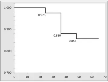
Fig. 3 Kaplan–Meier success probability, stating the time interval
[months]. Each restoration was seen as a statistical unit
Fig. 4 Kaplan–Meier success probability, stating the time interval
[months]. One restoration from each patient was selected by random and evaluated over time. As a consequence, the patient was seen as a statistical unit
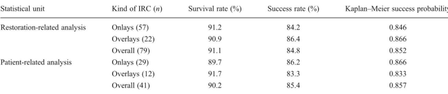
method (Fig.3); the patient-related success rate was 85.4 %, corresponding to an estimated success probability of 0.857 (Fig.4). The results of the statistical analysis that was also performed differentiating the 57 onlays and 22 overlays are summarised in Table7.
A total of two patients were lost to follow-up: the first one (two restorations) between the 36th and the 48th month; the second one (one restoration) between the 48th and the 60th month (Fig.2).
Discussion
In the present study, the longevity of 79 IRC onlays/overlays, placed by one operator in a general practice setting and bonded with a light-cured composite, was eval-uated over a period of 5 years. According to a recent research [14], in the present study, the Kaplan–Meier
anal-ysis was done in two ways: a patient-related analanal-ysis, which is the very strict approach, fully respecting the statistical independence of the data; the other analysis was performed looking at the restoration as an independent data point, despite the fact that Kaplan–Meier statistics were initially designed to deal with individuals. Using the tooth as a unit instead of the mouth can be justified [22]. Since this was not a prospective study, it seemed reasonable not to restrict the data to one tooth per patient, which may reduce the power of the study [14,22].
The present study found satisfactory 5-year results of IRC restorations. All the IRC restorations evaluated were onlays or overlays and showed promising survival rate of 91.1 % (restoration related) to 90.2 % (patient related). In other studies, IRC restorations achieved success rates of 87.2 and 76.6 [17], 89.8 and 84.1 % [23], 93 % [24] and 97.4 % [2] after 3 years, depending on the materials tested. Dukic et al. [25] reported a success rate of 100 % at 36 months for indirect composite restorations based on two different resin composites. However, the success rates of these previous studies need to be considered with regard to the period of only 3 years of clinical service. This rela-tively short period results only in a limited validity of the
data, which needs to be confirmed by longer follow-up investigations. There is a lack of studies in the literature that can give clinically pertinent information about long-term performance of IRC adhesive restorations. Huth et al. [19] reported failure rates of 12.8 and 23.4 % for Artglass and Charisma inlays at the 4-year recall. In the present study, four caries lesions contiguous with the IRC restoration margins (5.1 %) and two endodontic complications (2.5 %) were detected after the total observation period of 60 months. Since secondary caries is the most frequently cited reason for failure of dental restorations in general practice [26] and represents up to 50 % of all operative dentistry procedures delivered to adults [27], a strict clinical follow-up over time seems advisable also for IRC restora-tions, in order to allow an early interception of caries re-currences. All the secondary lesions were recorded on the interproximal side of the restorations. Donly et al. [28] reported failures due to secondary caries and fractures pre-dominantly in molar restorations. However, Manhart et al. [23] reported no significant differences between premolars and molars.
Also bulk fracture is considered to be one of the most frequent causes for restoration failure. It can be caused by weak material properties, such as insufficient polymeriza-tion of the inlay composite resin material or insufficient material thickness [19,29]. In the present study, only one (1.3 %) extensive fracture of the restoration was detected after an observation period of 36 months.
Marginal integrity is one of the most important criteria for the success of a restoration [24]. The results of the present study regarding marginal adaptation and marginal discolouration (5 and 10.1 % Bravo rating, respectively, after 5 years) were particularly satisfying, especially consid-ering the fact that only large restorations on molars were included. In fact, a premolar restoration is usually subjected to less occlusal stress than a molar restoration, the access for dental treatment is easier and oral hygiene measures are more easily controlled by the patient. Huth et al. [19] reported that both the inlay systems evaluated experienced significant deterioration of marginal integrity and significant increase of marginal discolouration comparing baseline and
Table 7 Survival rates, success rates and Kaplan–Meier success probabilities observed after 60 months
Statistical unit Kind of IRC (n) Survival rate (%) Success rate (%) Kaplan–Meier success probability
Restoration-related analysis Onlays (57) 91.2 84.2 0.846
Overlays (22) 90.9 86.4 0.866
Overall (79) 91.1 84.8 0.852
Patient-related analysis Onlays (29) 89.7 86.2 0.866
Overlays (12) 91.7 83.3 0.833
Overall (41) 90.2 85.4 0.857
4-year data. A significantly better marginal integrity was found in small restorations inserted in premolars [24]. Because of the elastic behaviour and fatigue of the compos-ite and bonding agent, the negative influences of occlusal stresses show more effect on large restorations and on molars, which are usually subjected to higher occlusal load-ing at the restoration–tooth interface. The authors believe that the favourable quality of restoration margins obtained in this study can be correlated to the preparation, cementation and finishing procedures adopted. The satisfactory clinical results for IRC restorations were achieved using a light-cured composite used as luting agent. Moreover, to avoid compromising restorations’ marginal accuracy, no diamond burs, polishing discs or interproximal polishing strips were used to finish the restorations. Residual cement was re-moved only with an explorer, scalpel and floss, checking restorations’ margins under a stereomicroscope magnifica-tion. The necessary working time for positioning the indirect restorations and removing the excess cement was conve-niently extended at the discretion of the clinician using a light-curing composite as luting agent, overcoming the rel-atively restricted working time allowed by dual-cure ce-ments. Marginal adaptation of indirect composite restorations before cementation has been reported to widely range between approximately 70 and 130 μm [30]. Imperfections in the marginal adaptation could be future sites for staining and caries in clinical service; consequently, the material that fills this gap exposed to the oral environ-ment should preferably have high mechanical properties and wear resistance. A filler content of at least 70 % would be necessary, whilst common dual-curing cements and light-activated flowable resins have reduced filler contents.
In the present study, only three restorations (3.8 %) demonstrated small cohesive fractures (chipping), though remaining clinically acceptable after a new finishing and polishing procedure. This is in accordance with Tsitrou et al. [31] that found that resin composites have a lower tendency for marginal chipping than ce-ramics. In the current study, two IRC restorations debonded and were re-bonded, respectively, after 36 and 48 months. The high retention rate of the present study could be correlated to the improved success rate of adhesive procedures through the constant use of rubber dam, which is permitted by the supragingival preparations. Moreover, the treatment of the intaglio surface of indirect restorations determines the bonding of the restoration to the tooth. The use of hydrofluoric acid for surface treatment causes microstructural alter-ation of the composite because of the dissolution of the inorganic particles [32, 33]; the risks involved in its handling and the poor outcomes obtained [34, 35] may make it less than ideal as a composite surface prepara-tion agent [32]. The best alternative method to raise the
surface energy is by a “soft-sandblasting technique” with 50 μm aluminium oxide particles for 10 s [12,
36,37]. This method causes a non-selective degradation of the resin and promotes better adhesion.
The IRC restorations showed very good results for colour match and anatomic form. Colour match showed only 3.8 % Bravo rating after 5 years, whereas a decline in anatomic appearance during follow-up period was not found.
Conclusions
Longevity of dental restorations is dependent upon many different factors, including materials-, patient- and dentist-related factors. Within the methodological limitations of the present clinical study, principal reasons for failure or IRC restorations were secondary caries, endodontic complications and fractures. However, it can be observed that consistently following a protocol of cementation technique using a light-cured composite with the constant use of rubber dam isolation, sandblasting treatment of the IRC area of adhesion and a careful hand finishing was associated with high survival rates of IRC restorations after 5 years of service.
Conflict of interest The authors declare that they have no conflict
of interest.
References
1. Thordrup M, Isidor F, Hörsted-Bindslev P (2006) A prospective clinical study of indirect and direct composite and ceramic inlays:
ten-year results. Quintessence Int 37:139–144
2. Barone A, Derchi G, Rossi A, Marconcini S, Covani U (2008)
Longitudinal clinical evaluation of bonded composite inlays: a
3-year study. Quintessence Int 39:65–71
3. Manhart J, Kunzelmann KH, Chen HY, Hickel R (2000) Mechanical properties and wear behavior of light-cured packable
composite resins. Dent Mater 16:33–40
4. Geurtsen W (2000) Biocompatibility of resin-modified filling
ma-terials. Crit Rev Oral Biol Med 11:333–355
5. Hadis M, Leprince JG, Shortall AC, Devaux J, Leloup G, Palin WM (2011) High irradiance curing and anomalies of exposure
reciprocity law in resin-based materials. J Dent 39:549–557
6. Carvalho RM, Pereira JC, Yoshiyama M, Pashley DH (1996) A review of polymerization contraction: the influence of stress
de-velopment versus stress relief. Oper Dent 21:17–24
7. Davidson CL, Feilzer AJ (1997) Polymerization shrinkage and polymerization shrinkage stress in polymer-based restoratives. J
Dent 25:435–440
8. Thonemann B, Federlin M, Schmalz G, Grundler W (1999) Total bonding vs selective bonding: marginal adaptation of class 2
composite restorations. Oper Dent 24:261–271
9. Loguercio AD, Alessandra R, Mazzocco KC, Dias AL, Busato ALS, Singer JDM, Rosa P (2002) Microleakage in class II com-posite resin restorations: total bonding and open sandwich tech-nique. J Adhes Dent 4:137–144
10. D'Arcangelo C, De Angelis F, Vadini M, Carluccio F, Vitalone LM, D'Amario M (2012) Influence of curing time, overlay material and thickness on three light-curing composites used for luting
indirect composite restorations. J Adhes Dent 14:377–384
11. Bott B, Hannig M (2003) Effect of different luting materials on the marginal adaptation of class I ceramic inlay restorations in vitro.
Dent Mater 19:264–269
12. D'Arcangelo C, Vanini L (2007) Effect of three surface treatments on the adhesive properties of indirect composite restorations. J
Adhes Dent 9:319–326
13. Acquaviva PA, Cerutti F, Adami G, Gagliani M, Ferrari M, Gherlone E, Cerutti A (2009) Degree of conversion of three composite materials employed in the adhesive cementation of
indirect restorations: a micro-Raman analysis. J Dent 37:610–615
14. D'Arcangelo C, De Angelis F, Vadini M, D'Amario M (2012) Clinical evaluation on porcelain laminate veneers bonded with light-cured composite: results up to 7 years. Clin Oral Investig
16:1071–1079
15. D'Arcangelo C, Vanini L, Prosperi GD, Di Bussolo G, De Angelis F, D'Amario M, Caputi S (2009) The influence of adhesive thick-ness on the microtensile bond strength of three adhesive systems. J
Adhes Dent 11:109–115
16. Magne P (2005) Immediate dentin sealing: a fundamental proce-dure for indirect bonded restorations. J Esthet Restor Dent 17:144– 154, discussion 155
17. D'Arcangelo C, De Angelis F, D'Amario M, Zazzeroni S, Ciampoli C, Caputi S (2009) The influence of luting systems on the microtensile bond strength of dentin to indirect resin-based com-posite and ceramic restorations. Oper Dent 34:328–336
18. Cvar JF, Ryge G (2005) Reprint of criteria for the clinical evaluation of dental restorative materials. 1971. Clin Oral Investig 9:215–232 19. Huth KC, Chen HY, Mehl A, Hickel R, Manhart J (2011) Clinical
study of indirect composite resin inlays in posterior stress-bearing cavities placed by dental students: results after 4 years. J Dent 39:478–488
20. Bayne SC, Schmalz G (2005) Reprinting the classic article on USPHS evaluation methods for measuring the clinical research
performance of restorative materials. Clin Oral Investig
9:209–214
21. Guess PC, Stappert CF (2008) Midterm results of a 5-year pro-spective clinical investigation of extended ceramic veneers. Dent Mater 24:804–813
22. Roulet JF (1997) Longevity of glass ceramic inlays and
amalgam—results up to 6 years. Clin Oral Investig 1:40–46
23. Manhart J, Chen HY, Mehl A, Hickel R (2010) Clinical study of indirect composite resin inlays in posterior stress-bearing
preparations placed by dental students: results after 6 months and
1, 2, and 3 years. Quintessence Int 41:399–410
24. Manhart J, Neuerer P, Scheibenbogen-Fuchsbrunner A, Hickel R (2000) Three-year clinical evaluation of direct and indirect
com-posite restorations in posterior teeth. J Prosthet Dent 84:289–296
25. Dukic W, Dukic OL, Milardovic S, Delija B (2010) Clinical evaluation of indirect composite restorations at baseline and
36 months after placement. Oper Dent 35:156–164
26. Mjör IA, Moorhead JE, Dahl JE (2000) Reasons for replacement of restorations in permanent teeth in general dental practice. Int Dent
J 50:361–366
27. Mjör IA, Toffenetti F (2000) Secondary caries: a literature review
with case reports. Quintessence Int 31:165–179
28. Donly KJ, Jensen ME, Triolo P, Chan D (1999) A clinical com-parison of resin composite inlay and onlay posterior restorations
and cast-gold restorations at 7 years. Quintessence Int 30:163–168
29. Pallesen U, Qvist V (2003) Composite resin fillings and inlays. An
11-year evaluation. Clin Oral Investig 7:71–79
30. Fonseca RB, Correr-Sobrinho L, Fernandes-Neto AJ, Quagliatto PS, Soares CJ (2008) The influence of the cavity preparation design on marginal accuracy of laboratory-processed resin
com-posite restorations. Clin Oral Investig 12:53–59
31. Tsitrou EA, Northeast SE, van Noort R (2007) Brittleness index of machinable dental materials and its relation to the marginal chipping factor. J Dent 35:897–902
32. Lucena-Martín C, González-López S, Navajas-Rodríguez de Mondelo JM (2001) The effect of various surface treatments and bonding agents on the repaired strength of heat-treated composites. J Prosthet Dent 86:481–488
33. Hummel SK, Marker V, Pace L, Goldfogle M (1997) Surface treatment of indirect resin composite surfaces before cementation. J Prosthet Dent 77:568–572
34. Crumpler DC, Bayne SC, Sockwell S, Brunson D, Roberson TM (1989) Bonding to resurfaced posterior composites. Dent Mater 5:417–424
35. Brosh T, Pilo R, Bichacho N, Blutstein R (1997) Effect of combi-nations of surface treatments and bonding agents on the bond strength of repaired composites. J Prosthet Dent 77:122–126 36. Cho S, Rajitrangson P, Matis B, Platt J (2012) Effect of Er,
Cr:YSGG laser, air abrasion, and silane application on repaired
shear bond strength of composites. Oper Dent 38(3):E1–E9
37. Caneppele TM, de Souza AC, Batista GR, Borges AB, Torres CR (2012) Influence of Nd:YAG or Er:YAG laser surface treatment on microtensile bond strength of indirect resin composites to resin cement. Lasers surface treatment of indirect resin composites. Eur