molecules
ArticleRational Design of Nucleoside–Bile Acid Conjugates
Incorporating a Triazole Moiety for Anticancer
Evaluation and SAR Exploration
Maria Luisa Navacchia1,* ID, Elena Marchesi2 ID, Lara Mari2, Nicola Chinaglia2,
Eleonora Gallerani3, Riccardo Gavioli3, Massimo Luigi Capobianco1and Daniela Perrone2 ID
1 Consiglio Nazionale delle Ricerche, Istituto per la Sintesi Organica e la Fotoreattività (CNR-ISOF), via P. Gobetti 101, 40129 Bologna, Italy; [email protected]
2 Dipartimento di Scienze Chimiche e Farmaceutiche, Università degli studi di Ferrara, via L. Borsari 46, 44121 Ferrara, Italy; [email protected] (E.M.); [email protected] (L.M.); [email protected] (N.C.); [email protected] (D.P.)
3 Dipartimento di Scienze della Vita e Biotecnologie, Università degli studi di Ferrara, via L. Borsari 46, 44121 Ferrara, Italy; [email protected] (E.G.), [email protected] (R.G.)
* Correspondence: [email protected]; Tel.: +39-051-639-8291
Received: 27 September 2017; Accepted: 10 October 2017; Published: 12 October 2017
Abstract: Herein we report a study on the synthesis and biological evaluation of a library of nucleoside-bile acid conjugates prepared by combining 20-deoxyadenosine, 20-deoxyguanosine, 20-deoxyuridine as well as adenosine and guanosine derivatives with cheno-, urso-, nor-cheno-, nor-urso- and taurourso-desoxycholic acid derivatives by means of the click reaction. The new nucleoside-bile acid conjugates incorporating a triazole moiety were tested in vitro against leukemic K562 and HCT116 colon carcinoma, as well as on normal fibroblast cells. Six compounds displayed interesting anti-proliferative activity against the selected cancer lines and no cytotoxic effects against normal fibroblasts. A possible structure activity relationship was also investigated.
Keywords:bioconjugates; bile acids; nucleosides; click chemistry; cytoselectivity; anticancer activity
1. Introduction
Important traditional chemotherapeutic drugs or anticancer agents were mostly derived from natural sources through synthetic structural modifications. Successful examples of this approach are represented, among others, by the anthracyclines, taxanes and camptothecins that are still considered a structural platform for discovering new anticancer drugs [1].
Nucleosides and nucleotides—endogenous small molecules that can be chemically fine-tuned leading to the corresponding analogues—can behave as antimetabolites and can inhibit the cellular division and viral replication by incorporation into DNA or RNA, resulting in potential therapeutic benefits. They can also act as inhibitors of essential enzymes such as DNA polymerases, kinases and so on. In such a way, they would operate by stopping the synthesis of pre-DNA molecule building blocks or by direct damage of the DNA in the nucleus of the cell or by effecting the synthesis or by breakdown of the mitotic spindles. Currently, several nucleoside and nucleotide analogues derived from 20-deoxycitidine, 20-deoxyadenosine and 20-deoxyguanosine have been approved by the FDA as anti-cancer drugs or anti-viral agents [2].
Despite their therapeutic potential, the bioavailability of hydrophilic nucleoside-based drugs remains a critical negative feature since they do not readily cross the plasma membrane by passive diffusion, and accordingly, their clinical efficacy also depends on nucleoside delivery systems [3,4].
With the aim to discover new nucleoside analogues with anticancer activity we consider conjugation as a powerful approach. In principle, a targeted conjugation can be helpful to tune
the cytotoxicity, for instance by coupling a 20-deoxyadenosine derivative with a NO photodonor unit the intrinsic cytotoxicity of the bioconjugate combined upon light irradiation with that of the photogenerated NO leads to an interplay of anticancer mechanisms of action [5]. Furthermore, 20-deoxyadenosine derivatives conjugated with cheno- and urso-deoxycholic acids through a triazole or a thioalkyl unit tested on four cancer cell lines (K562, Jurkat, HCT116 and A2780) showed interesting antiproliferative activity selectively towards leukemic T-cells whereas no cytotoxicity against the solid tumors HCT116 and A2780 was found [6]. In our studies, bile acids (BAs) were chosen as combination partners by virtue of their biological as well as physico-chemical properties. For instance, the cytotoxic activity of certain BAs and BA-derivatives is well recognized, including the potential of several unconjugated BAs to induce cell death in a wide range of cells, through their non-specific ability to disrupt cell membranes (biological surfactant feature) or receptor-mediated interactions and DNA oxidative damage [7]. On the other hand, the conjugation with hydrophilic glycine and taurine can dramatically decreases BA cytotoxicity while enhancing the neuroprotective effects [8,9]. Moreover, taking advantage of their organotropism in the enterohepatic circulation mediated by the BA transport systems, the presence of BA units can be helpful in targeting a drug conjugate to the liver or to improve its metabolic stability [10]. It has been also reported that the conjugation of zidovuidine (AZT), a nucleoside analogue-based drug, with ursodesoxycholic acid increases the poor permeability of AZT through the intracellular departments [11]. Thanks to their intrinsic chemical features BAs can be fine tailored.
We present herein a study on the synthesis and biological evaluation of nucleoside-BA conjugates obtained by combining a selection of nucleoside analogues and bile acid derivatives. For this purpose, 20-deoxyadenosine (dA), 20-deoxyguanosine (dG), 20-deoxyuridine (dU) as well as adenosine (A) and guanosine (G) modified at a suitable position with an alkynyl chain containing an acetylenic bond were conjugated by means of the click reaction with cheno- (CDC), urso- (UDC), taurourso- (TUDCA), nor-cheno (nor-CDC) and nor-urso- (nor-UDC) deoxycholic acid derivatives equipped with the azido group at the head or the tail position. The new nucleoside-BA conjugates were characterized and tested in vitro against two types of cancer cell lines: leukemic K562, a hematological cancer, and the solid tumor HCT116 colon carcinoma, as well as on normal fibroblast cells.
2. Results
The click chemistry approach, being a specific and high yield reaction, was considered a good synthetic approach for the preparation of our target nucleoside-BA hybrids. Moreover, the triazole moiety resulting from the 1,3-cycloaddition is biologically relevant, being able to improve the biostability, bioavailability and also the anticancer activity of bioactive compounds [12–15].
Click chemistry requires the presence of a terminal alkyne moiety and an azido group. To provide those features the nucleoside units were modified with an alkynyl moiety at C-8 position in the case of the purine bases and at the C-6 position in the case of the pyrimidine one in order to keep unchanged their intrinsic characteristic of recognition of natural nucleic acids through specific hydrogen bond patterns (Watson-Crick and Hoogsteen). 8-(1,7-Octadynyl)-20-deoxyadenosine (ALK-dA) was prepared as previously reported [6]. Similarly, the 8-(1,7-octadynyl)-derivative of A, dG and G, as well as 6-(1,7-octadynyl)-20-deoxyuridine (namely ALK-A, ALK-dG, ALK-G and ALK-dU, respectively) were synthesized through a standard palladium catalyzed cross-coupling reaction starting from commercially available 8-bromo-A, 8-bromo-dG, 8-bromo-G and 6-iodo-dU [16] (Scheme1).
The azido-BA derivatives 3α-N3-CDC and 3α-N3-UDC were synthesized starting from commercially available BAs, using a synthetic approach that we described previously [6,17] (Scheme2). The 3α-N3 derivative of TUDCA was prepared in three steps in 75% overall yield from the
corresponding 3α-N3-UDC: the methyl ester was hydrolyzed with a 1.5 M LiOH in MeOH to the corresponding acid, which in turn was coupled with the aminoethanesulfonic acid taurine after activation of the free acid with ethyl chloroformate (Scheme2).
Molecules 2017, 22, 1710 3 of 18
Molecules 2017, 22, 1710 3 of 17
Scheme 1. Synthesis of the alkyne-nucleoside intermediates. Reagents and conditions: (PPh3)2PdCl2, CuI, DMF, TEA, 50 °C, 2.30 h; (a): 8-Br-2′-deoxyadenosine or 8-Br-adenosine; (b): 8-Br-2′-deoxyguanosine or 8-Br-guanosine; (c): 6-I-2′-deoxyuridine.
Finally, the 23-N3-nor-CDC and 23-N3-nor-UDC were synthesized starting from the corresponding
bile acids following a recently reported metal free iodo-decarboxylation method [18]. Accordingly, the C3 and C7 free hydroxyl groups of CDCA and UDCA were firstly protected as formyloxy derivatives by using formic acid at 55 °C for 24 h, then concentrated in vacuo and the residues irradiated for 2 h in presence of 1,3-diiodo-5,5-dimethylhydantoin (DIH) as a sole reagent. After purification by flash chromatography the 23-I-nor-CDC and -UDC derivatives (85–90% yield) were converted into the target compounds through a nucleophilic substitution with sodium azide in DMF at room temperature, followed by hydrolysis of the formate esters with 25% NaOH which allowed the precipitation of 23-N3-nor BAs derivatives as pure compounds in satisfactory yields (75–78%
after two steps; Scheme 2).
Scheme 2. Synthesis of BA-azide intermediates. Reagents and conditions: (a): HCOOH, 55 °C, 24 h; (b): DIH,
DCE, hν, reflux 2 h; (c): NaN3, DMF, r.t., 6 h; (d): NaOH 25%, MeOH, r.t., 2 h; (e): see reference [6]; (f): 1.5 M LiOH in MeOH, r.t., 21 h; (g): TEA, ClCOOEt, THF, 0 °C to r.t., then taurine, NaOH 10%, r.t., 12 h.
The click chemistry was performed via a Cu (I)-mediated 1,3-dipolar cycloaddition reaction under commonly used conditions: a (1:1:1.5) H2O/tert-BuOH/THF (v/v) solution of the appropriate alkyne–nucleoside derivative and of the BA-azide in the presence of the CuSO4 catalyst and sodium Scheme 1.Synthesis of the alkyne-nucleoside intermediates. Reagents and conditions: (PPh3)2PdCl2, CuI, DMF, TEA, 50◦C, 2.30 h; (a): 8-Br-20-deoxyadenosine or 8-Br-adenosine; (b): 8-Br-20-deoxyguanosine or 8-Br-guanosine; (c): 6-I-20-deoxyuridine.
Scheme 1. Synthesis of the alkyne-nucleoside intermediates. Reagents and conditions: (PPh3)2PdCl2, CuI, DMF, TEA, 50 °C, 2.30 h; (a): 8-Br-2′-deoxyadenosine or 8-Br-adenosine; (b): 8-Br-2′-deoxyguanosine or 8-Br-guanosine; (c): 6-I-2′-deoxyuridine.
Finally, the 23-N3-nor-CDC and 23-N3-nor-UDC were synthesized starting from the corresponding
bile acids following a recently reported metal free iodo-decarboxylation method [18]. Accordingly, the C3 and C7 free hydroxyl groups of CDCA and UDCA were firstly protected as formyloxy derivatives by using formic acid at 55 °C for 24 h, then concentrated in vacuo and the residues irradiated for 2 h in presence of 1,3-diiodo-5,5-dimethylhydantoin (DIH) as a sole reagent. After purification by flash chromatography the 23-I-nor-CDC and -UDC derivatives (85–90% yield) were converted into the target compounds through a nucleophilic substitution with sodium azide in DMF at room temperature, followed by hydrolysis of the formate esters with 25% NaOH which allowed the precipitation of 23-N3-nor BAs derivatives as pure compounds in satisfactory yields (75–78%
after two steps; Scheme 2).
Scheme 2. Synthesis of BA-azide intermediates. Reagents and conditions: (a): HCOOH, 55 °C, 24 h; (b): DIH, DCE, hν, reflux 2 h; (c): NaN3, DMF, r.t., 6 h; (d): NaOH 25%, MeOH, r.t., 2 h; (e): see reference [6]; (f): 1.5 M LiOH in MeOH, r.t., 21 h; (g): TEA, ClCOOEt, THF, 0 °C to r.t., then taurine, NaOH 10%, r.t., 12 h. The click chemistry was performed via a Cu (I)-mediated 1,3-dipolar cycloaddition reaction under commonly used conditions: a (1:1:1.5) H2O/tert-BuOH/THF (v/v) solution of the appropriate
alkyne–nucleoside derivative and of the BA-azide in the presence of the CuSO4 catalyst and sodium
Scheme 2. Synthesis of BA-azide intermediates. Reagents and conditions: (a): HCOOH, 55◦C, 24 h; (b): DIH, DCE, hν, reflux 2 h; (c): NaN3, DMF, r.t., 6 h; (d): NaOH 25%, MeOH, r.t., 2 h; (e): see reference [6]; (f): 1.5 M LiOH in MeOH, r.t., 21 h; (g): TEA, ClCOOEt, THF, 0◦C to r.t., then taurine, NaOH 10%, r.t., 12 h.
Finally, the 23-N3-nor-CDC and 23-N3-nor-UDC were synthesized starting from the corresponding bile acids following a recently reported metal free iodo-decarboxylation method [18]. Accordingly, the C3 and C7 free hydroxyl groups of CDCA and UDCA were firstly protected as formyloxy derivatives by using formic acid at 55◦C for 24 h, then concentrated in vacuo and the residues irradiated for 2 h in presence of 1,3-diiodo-5,5-dimethylhydantoin (DIH) as a sole reagent. After purification by flash chromatography the 23-I-nor-CDC and -UDC derivatives (85–90% yield) were converted into the target compounds through a nucleophilic substitution with sodium azide in DMF at room temperature, followed by hydrolysis of the formate esters with 25% NaOH which allowed the precipitation of 23-N3-nor BAs derivatives as pure compounds in satisfactory yields (75–78% after two steps; Scheme2).
The click chemistry was performed via a Cu (I)-mediated 1,3-dipolar cycloaddition reaction under commonly used conditions: a (1:1:1.5) H2O/tert-BuOH/THF (v/v) solution of the appropriate
alkyne–nucleoside derivative and of the BA-azide in the presence of the CuSO4catalyst and sodium
ascorbate was stirred at room temperature for 18 h. The target conjugates were obtained in yields ranging from 60% to 90% after purification. In Scheme3is depicted for example, the synthesis of the conjugate compounds of adenosine with all the BA selected (namely A-CDC, A-UDC, A-TUDCA, A-nor-CDCand A-nor-UDC).
Molecules 2017, 22, 1710 4 of 17
ascorbate was stirred at room temperature for 18 h. The target conjugates were obtained in yields ranging from 60% to 90% after purification. In Scheme 3 is depicted for example, the synthesis of the conjugate compounds of adenosine with all the BA selected (namely A-CDC, A-UDC, A-TUDCA,
A-nor-CDC and A-nor-UDC).
Scheme 3. Synthetic scheme for the preparation of A-CDC, A-UDC, A-nor-CDC, A-nor-UDC and A-TUDCA conjugates. Reagents and conditions: (a): CuSO4·5H2O, sodium ascorbate, 1:1:1.5 H2O/tert-BuOH /THF (v/v), 25 °C, 18 h.
All conjugated compounds prepared via click chemistry and listed in Table 1 were evaluated in vitro for their cytotoxic activity against K562 leukemia cells and the colon cancer cell line HCT116. Normal human skin fibroblast cells were chosen as a control and cisplatin served as a reference compound. The cytotoxicity was evaluated using the MTT assay (details are reported in the Materials and Methods section). In all experiments the cell growth inhibition of K562 and HCT116 was determined for each compound at concentrations of 10, 25 and 50 μM after 72 h of treatment and up to 100 μM in the case of fibroblasts. Figure 1 shows the antiproliferative activity of the most active conjugates against K562 and HCT116 cancer cells whereas in Table 1 the IC50 values are reported for
all the compounds tested, including the alkyne-nucleoside and BA-azide building blocks.
With regard to the 2′-deoxyadenosine derivatives, dA-nor-CDC was found cytotoxic against both K562 and HCT116, with comparable IC50 values (16.2 and 17.0 μM, respectively). On the other
hand, dA-nor-UDC showed a preferential cytotoxicity against HCT116 cancer cells (IC50 = 44.8 μM
vs. 87.1 μM extrapolated value; Table 1).
In the case of the adenosine derivatives, both A-CDC and A-nor-CDC were found active against K562 and HCT116 but with an opposite cytoselectivity. Indeed, A-CDC showed a higher cytotoxicity against HCT116, whereas A-nor-CDC was found more toxic towards K562. None of the bioconjugates of 2′-deoxyguanosine and guanosine series showed any cytotoxicity, with the only exception of
G-CDC which was found to be selectively cytotoxic against HCT116 cancer cells (IC50 = 25.3 μM).
Scheme 3. Synthetic scheme for the preparation of A-CDC, A-UDC, A-nor-CDC, A-nor-UDC and A-TUDCA conjugates. Reagents and conditions: (a): CuSO4·5H2O, sodium ascorbate, 1:1:1.5 H2O/tert-BuOH /THF (v/v), 25◦C, 18 h.
All conjugated compounds prepared via click chemistry and listed in Table1were evaluated in vitro for their cytotoxic activity against K562 leukemia cells and the colon cancer cell line HCT116. Normal human skin fibroblast cells were chosen as a control and cisplatin served as a reference compound. The cytotoxicity was evaluated using the MTT assay (details are reported in the Materials and Methods section). In all experiments the cell growth inhibition of K562 and HCT116 was determined for each compound at concentrations of 10, 25 and 50 µM after 72 h of treatment and up to 100 µM in the case of fibroblasts. Figure1shows the antiproliferative activity of the most active conjugates against K562 and HCT116 cancer cells whereas in Table1the IC50values are reported for
all the compounds tested, including the alkyne-nucleoside and BA-azide building blocks.
With regard to the 20-deoxyadenosine derivatives, dA-nor-CDC was found cytotoxic against both K562 and HCT116, with comparable IC50values (16.2 and 17.0 µM, respectively). On the other hand,
dA-nor-UDCshowed a preferential cytotoxicity against HCT116 cancer cells (IC50= 44.8 µM vs. 87.1
µM extrapolated value; Table1).
In the case of the adenosine derivatives, both A-CDC and A-nor-CDC were found active against K562 and HCT116 but with an opposite cytoselectivity. Indeed, A-CDC showed a higher cytotoxicity against HCT116, whereas A-nor-CDC was found more toxic towards K562. None of the bioconjugates of 20-deoxyguanosine and guanosine series showed any cytotoxicity, with the only exception of G-CDC which was found to be selectively cytotoxic against HCT116 cancer cells (IC50= 25.3 µM).
Molecules 2017, 22, 1710 5 of 18
Table 1. IC50values were determined from the dose–response curves using MTT assay after 72 h incubation time. Results are expressed as the mean of three independent experiments±SD. Cisplatin was used as a reference compound. Where indicated IC50> 50 inhibition found was <20% at 50 µM; where indicated IC50> 100 inhibition found was <10% at 100 µM.
Molecular Structure Compound Name IC50
µM
K562 HCT116 FIBRO
Molecules 2017, 22, 1710 5 of 17
Table 1. IC50 values were determined from the dose–response curves using MTT assay after 72 h incubation time. Results are expressed as the mean of three independent experiments ± SD. Cisplatin was used as a reference compound. Where indicated IC50 > 50 inhibition found was <20% at 50 μM; where indicated IC50 > 100 inhibition found was <10% at 100 μM.
Molecular Structure Compound Name IC50 μM
K562 HCT116 FIBRO R=H, (7α): dA-CDC1 8.5 ± 4.0 >200 >200 R=H, (7β): dA-UDC1 >100 >200 >200 R=OH, (7α): A-CDC 43.5 ± 1.3 23.1 ± 1.7 >100 R=OH, (7β): A-UDC >50 >50 >100 R=H, (7α): dA-nor-CDC 16.2 ± 2.2 17.0 ± 2.5 91.5 ± 3.5 R=H, (7β): dA-nor-UDC >50 44.8 ± 3.5 >100 R=OH, (7α): A-nor-CDC 23.6 ± 1.2 44.1 ± 2.5 >100 R=OH, (7β): A-nor-UDC >50 >50 >100 R=H: dA-TUDCA >50 >50 >100 R=OH, A-TUDCA >50 >50 >100 R=H, (7α): dG-CDC >50 >50 >100 R=H, (7β): dG-UDC >50 >50 >100 R=OH, (7α): G-CDC >50 25.3 ± 3.8 >100 R=OH, (7β): G-UDC >50 >50 >100 R=H, (7α): dG-nor-CDC >50 >50 >100 R=H, (7β): dG-nor-UDC >50 >50 >100 R=OH, (7α): G-nor-CDC >50 >50 >100 R=OH, (7β): G-nor-UDC >50 >50 >100 R=H: dG-TUDCA >50 >50 >100 R=OH, G-TUDCA >50 >50 >100 (7α): dU-CDC >50 >50 >100 (7β): dU-UDC 21.5 ± 2.0 23.5 ± 1.6 91.5 ± 4.5 (7α): dU-nor-CDC 42.9 ± 1.9 >50 >100 (7β): dU-nor-UDC 24.8 ± 1.5 43.0 ± 3.5 >100 NH N N N O O R OH HO NNN OH O O NH2 R=H, (7α): dA-CDC1 8.5 ± 4.0 >200 >200 R=H, (7β): dA-UDC1 >100 >200 >200 R=OH, (7α): A-CDC 43.5 ± 1.3 23.1 ± 1.7 >100 R=OH, (7β): A-UDC >50 >50 >100
Table 1. IC50 values were determined from the dose–response curves using MTT assay after 72 h incubation time. Results are expressed as the mean of three independent experiments ± SD. Cisplatin was used as a reference compound. Where indicated IC50 > 50 inhibition found was <20% at 50 μM; where indicated IC50 > 100 inhibition found was <10% at 100 μM.
Molecular Structure Compound Name
IC50 μM K562 HCT116 FIBRO R=H, (7α): dA-CDC1 8.5 ± 4.0 >200 >200 R=H, (7β): dA-UDC1 >100 >200 >200 R=OH, (7α): A-CDC 43.5 ± 1.3 23.1 ± 1.7 >100 R=OH, (7β): A-UDC >50 >50 >100 R=H, (7α): dA-nor-CDC 16.2 ± 2.2 17.0 ± 2.5 91.5 ± 3.5 R=H, (7β): dA-nor-UDC >50 44.8 ± 3.5 >100 R=OH, (7α): A-nor-CDC 23.6 ± 1.2 44.1 ± 2.5 >100 R=OH, (7β): A-nor-UDC >50 >50 >100 R=H: dA-TUDCA >50 >50 >100 R=OH, A-TUDCA >50 >50 >100 R=H, (7α): dG-CDC >50 >50 >100 R=H, (7β): dG-UDC >50 >50 >100 R=OH, (7α): G-CDC >50 25.3 ± 3.8 >100 R=OH, (7β): G-UDC >50 >50 >100 R=H, (7α): dG-nor-CDC >50 >50 >100 R=H, (7β): dG-nor-UDC >50 >50 >100 R=OH, (7α): G-nor-CDC >50 >50 >100 R=OH, (7β): G-nor-UDC >50 >50 >100 R=H: dG-TUDCA >50 >50 >100 R=H, (7α): dA-nor-CDC 16.2 ± 2.2 17.0 ± 2.5 91.5 ± 3.5 R=H, (7β): dA-nor-UDC >50 44.8 ± 3.5 >100 R=OH, (7α): A-nor-CDC 23.6 ± 1.2 44.1 ± 2.5 >100 R=OH, (7β): A-nor-UDC >50 >50 >100 Molecules 2017, 22, 1710 5 of 17
Table 1. IC50 values were determined from the dose–response curves using MTT assay after 72 h incubation time. Results are expressed as the mean of three independent experiments ± SD. Cisplatin was used as a reference compound. Where indicated IC50 > 50 inhibition found was <20% at 50 μM; where indicated IC50 > 100 inhibition found was <10% at 100 μM.
Molecular Structure Compound Name IC50 μM
K562 HCT116 FIBRO R=H, (7α): dA-CDC1 8.5 ± 4.0 >200 >200 R=H, (7β): dA-UDC1 >100 >200 >200 R=OH, (7α): A-CDC 43.5 ± 1.3 23.1 ± 1.7 >100 R=OH, (7β): A-UDC >50 >50 >100 R=H, (7α): dA-nor-CDC 16.2 ± 2.2 17.0 ± 2.5 91.5 ± 3.5 R=H, (7β): dA-nor-UDC >50 44.8 ± 3.5 >100 R=OH, (7α): A-nor-CDC 23.6 ± 1.2 44.1 ± 2.5 >100 R=OH, (7β): A-nor-UDC >50 >50 >100 R=H: dA-TUDCA >50 >50 >100 R=OH, A-TUDCA >50 >50 >100 R=H, (7α): dG-CDC >50 >50 >100 R=H, (7β): dG-UDC >50 >50 >100 R=OH, (7α): G-CDC >50 25.3 ± 3.8 >100 R=OH, (7β): G-UDC >50 >50 >100 R=H, (7α): dG-nor-CDC >50 >50 >100 R=H, (7β): dG-nor-UDC >50 >50 >100 R=OH, (7α): G-nor-CDC >50 >50 >100 R=OH, (7β): G-nor-UDC >50 >50 >100 R=H: dG-TUDCA >50 >50 >100 R=OH, G-TUDCA >50 >50 >100 (7α): dU-CDC >50 >50 >100 (7β): dU-UDC 21.5 ± 2.0 23.5 ± 1.6 91.5 ± 4.5 (7α): dU-nor-CDC 42.9 ± 1.9 >50 >100 (7β): dU-nor-UDC 24.8 ± 1.5 43.0 ± 3.5 >100 NH N N N O O R OH HO NNN OH O O NH2 R=H: dA-TUDCA >50 >50 >100 R=OH, A-TUDCA >50 >50 >100 Molecules 2017, 22, 1710 5 of 17
Table 1. IC50 values were determined from the dose–response curves using MTT assay after 72 h incubation time. Results are expressed as the mean of three independent experiments ± SD. Cisplatin was used as a reference compound. Where indicated IC50 > 50 inhibition found was <20% at 50 μM; where indicated IC50 > 100 inhibition found was <10% at 100 μM.
Molecular Structure Compound Name IC50 μM
K562 HCT116 FIBRO R=H, (7α): dA-CDC1 8.5 ± 4.0 >200 >200 R=H, (7β): dA-UDC1 >100 >200 >200 R=OH, (7α): A-CDC 43.5 ± 1.3 23.1 ± 1.7 >100 R=OH, (7β): A-UDC >50 >50 >100 R=H, (7α): dA-nor-CDC 16.2 ± 2.2 17.0 ± 2.5 91.5 ± 3.5 R=H, (7β): dA-nor-UDC >50 44.8 ± 3.5 >100 R=OH, (7α): A-nor-CDC 23.6 ± 1.2 44.1 ± 2.5 >100 R=OH, (7β): A-nor-UDC >50 >50 >100 R=H: dA-TUDCA >50 >50 >100 R=OH, A-TUDCA >50 >50 >100 R=H, (7α): dG-CDC >50 >50 >100 R=H, (7β): dG-UDC >50 >50 >100 R=OH, (7α): G-CDC >50 25.3 ± 3.8 >100 R=OH, (7β): G-UDC >50 >50 >100 R=H, (7α): dG-nor-CDC >50 >50 >100 R=H, (7β): dG-nor-UDC >50 >50 >100 R=OH, (7α): G-nor-CDC >50 >50 >100 R=OH, (7β): G-nor-UDC >50 >50 >100 R=H: dG-TUDCA >50 >50 >100 R=OH, G-TUDCA >50 >50 >100 (7α): dU-CDC >50 >50 >100 (7β): dU-UDC 21.5 ± 2.0 23.5 ± 1.6 91.5 ± 4.5 (7α): dU-nor-CDC 42.9 ± 1.9 >50 >100 (7β): dU-nor-UDC 24.8 ± 1.5 43.0 ± 3.5 >100 NH N N N O O R OH HO NNN OH O O NH2 R=H, (7α): dG-CDC >50 >50 >100 R=H, (7β): dG-UDC >50 >50 >100 R=OH, (7α): G-CDC >50 25.3 ± 3.8 >100 R=OH, (7β): G-UDC >50 >50 >100 Molecules 2017, 22, 1710 5 of 17
Table 1. IC50 values were determined from the dose–response curves using MTT assay after 72 h incubation time. Results are expressed as the mean of three independent experiments ± SD. Cisplatin was used as a reference compound. Where indicated IC50 > 50 inhibition found was <20% at 50 μM; where indicated IC50 > 100 inhibition found was <10% at 100 μM.
Molecular Structure Compound Name IC50 μM
K562 HCT116 FIBRO R=H, (7α): dA-CDC1 8.5 ± 4.0 >200 >200 R=H, (7β): dA-UDC1 >100 >200 >200 R=OH, (7α): A-CDC 43.5 ± 1.3 23.1 ± 1.7 >100 R=OH, (7β): A-UDC >50 >50 >100 R=H, (7α): dA-nor-CDC 16.2 ± 2.2 17.0 ± 2.5 91.5 ± 3.5 R=H, (7β): dA-nor-UDC >50 44.8 ± 3.5 >100 R=OH, (7α): A-nor-CDC 23.6 ± 1.2 44.1 ± 2.5 >100 R=OH, (7β): A-nor-UDC >50 >50 >100 R=H: dA-TUDCA >50 >50 >100 R=OH, A-TUDCA >50 >50 >100 R=H, (7α): dG-CDC >50 >50 >100 R=H, (7β): dG-UDC >50 >50 >100 R=OH, (7α): G-CDC >50 25.3 ± 3.8 >100 R=OH, (7β): G-UDC >50 >50 >100 R=H, (7α): dG-nor-CDC >50 >50 >100 R=H, (7β): dG-nor-UDC >50 >50 >100 R=OH, (7α): G-nor-CDC >50 >50 >100 R=OH, (7β): G-nor-UDC >50 >50 >100 R=H: dG-TUDCA >50 >50 >100 R=OH, G-TUDCA >50 >50 >100 (7α): dU-CDC >50 >50 >100 (7β): dU-UDC 21.5 ± 2.0 23.5 ± 1.6 91.5 ± 4.5 (7α): dU-nor-CDC 42.9 ± 1.9 >50 >100 (7β): dU-nor-UDC 24.8 ± 1.5 43.0 ± 3.5 >100 NH N N N O O R OH HO NNN OH O O NH2 R=H, (7α): dG-nor-CDC >50 >50 >100 R=H, (7β): dG-nor-UDC >50 >50 >100 R=OH, (7α): G-nor-CDC >50 >50 >100 R=OH, (7β): G-nor-UDC >50 >50 >100 Molecules 2017, 22, 1710 5 of 17
Table 1. IC50 values were determined from the dose–response curves using MTT assay after 72 h incubation time. Results are expressed as the mean of three independent experiments ± SD. Cisplatin was used as a reference compound. Where indicated IC50 > 50 inhibition found was <20% at 50 μM; where indicated IC50 > 100 inhibition found was <10% at 100 μM.
Molecular Structure Compound Name IC50 μM
K562 HCT116 FIBRO R=H, (7α): dA-CDC1 8.5 ± 4.0 >200 >200 R=H, (7β): dA-UDC1 >100 >200 >200 R=OH, (7α): A-CDC 43.5 ± 1.3 23.1 ± 1.7 >100 R=OH, (7β): A-UDC >50 >50 >100 R=H, (7α): dA-nor-CDC 16.2 ± 2.2 17.0 ± 2.5 91.5 ± 3.5 R=H, (7β): dA-nor-UDC >50 44.8 ± 3.5 >100 R=OH, (7α): A-nor-CDC 23.6 ± 1.2 44.1 ± 2.5 >100 R=OH, (7β): A-nor-UDC >50 >50 >100 R=H: dA-TUDCA >50 >50 >100 R=OH, A-TUDCA >50 >50 >100 R=H, (7α): dG-CDC >50 >50 >100 R=H, (7β): dG-UDC >50 >50 >100 R=OH, (7α): G-CDC >50 25.3 ± 3.8 >100 R=OH, (7β): G-UDC >50 >50 >100 R=H, (7α): dG-nor-CDC >50 >50 >100 R=H, (7β): dG-nor-UDC >50 >50 >100 R=OH, (7α): G-nor-CDC >50 >50 >100 R=OH, (7β): G-nor-UDC >50 >50 >100 R=H: dG-TUDCA >50 >50 >100 R=OH, G-TUDCA >50 >50 >100 (7α): dU-CDC >50 >50 >100 (7β): dU-UDC 21.5 ± 2.0 23.5 ± 1.6 91.5 ± 4.5 (7α): dU-nor-CDC 42.9 ± 1.9 >50 >100 (7β): dU-nor-UDC 24.8 ± 1.5 43.0 ± 3.5 >100 NH N N N O O R OH HO NNN OH O O NH2 R=H: dG-TUDCA >50 >50 >100 R=OH, G-TUDCA >50 >50 >100 Molecules 2017, 22, 1710 5 of 17
Table 1. IC50 values were determined from the dose–response curves using MTT assay after 72 h incubation time. Results are expressed as the mean of three independent experiments ± SD. Cisplatin was used as a reference compound. Where indicated IC50 > 50 inhibition found was <20% at 50 μM; where indicated IC50 > 100 inhibition found was <10% at 100 μM.
Molecular Structure Compound Name IC50 μM
K562 HCT116 FIBRO R=H, (7α): dA-CDC1 8.5 ± 4.0 >200 >200 R=H, (7β): dA-UDC1 >100 >200 >200 R=OH, (7α): A-CDC 43.5 ± 1.3 23.1 ± 1.7 >100 R=OH, (7β): A-UDC >50 >50 >100 R=H, (7α): dA-nor-CDC 16.2 ± 2.2 17.0 ± 2.5 91.5 ± 3.5 R=H, (7β): dA-nor-UDC >50 44.8 ± 3.5 >100 R=OH, (7α): A-nor-CDC 23.6 ± 1.2 44.1 ± 2.5 >100 R=OH, (7β): A-nor-UDC >50 >50 >100 R=H: dA-TUDCA >50 >50 >100 R=OH, A-TUDCA >50 >50 >100 R=H, (7α): dG-CDC >50 >50 >100 R=H, (7β): dG-UDC >50 >50 >100 R=OH, (7α): G-CDC >50 25.3 ± 3.8 >100 R=OH, (7β): G-UDC >50 >50 >100 R=H, (7α): dG-nor-CDC >50 >50 >100 R=H, (7β): dG-nor-UDC >50 >50 >100 R=OH, (7α): G-nor-CDC >50 >50 >100 R=OH, (7β): G-nor-UDC >50 >50 >100 R=H: dG-TUDCA >50 >50 >100 R=OH, G-TUDCA >50 >50 >100 (7α): dU-CDC >50 >50 >100 (7β): dU-UDC 21.5 ± 2.0 23.5 ± 1.6 91.5 ± 4.5 (7α): dU-nor-CDC 42.9 ± 1.9 >50 >100 (7β): dU-nor-UDC 24.8 ± 1.5 43.0 ± 3.5 >100 NH N N N O O R OH HO NNN OH O O NH2 (7α): dU-CDC >50 >50 >100 (7β): dU-UDC 21.5 ± 2.0 23.5 ± 1.6 91.5 ± 4.5
Molecules 2017, 22, 1710 6 of 18
Table 1. Cont.
Molecular Structure Compound Name IC50µM
K562 HCT116 FIBRO
Table 1. IC50 values were determined from the dose–response curves using MTT assay after 72 h incubation time. Results are expressed as the mean of three independent experiments ± SD. Cisplatin was used as a reference compound. Where indicated IC50 > 50 inhibition found was <20% at 50 μM; where indicated IC50 > 100 inhibition found was <10% at 100 μM.
Molecular Structure Compound Name IC50 μM
K562 HCT116 FIBRO R=H, (7α): dA-CDC1 8.5 ± 4.0 >200 >200 R=H, (7β): dA-UDC1 >100 >200 >200 R=OH, (7α): A-CDC 43.5 ± 1.3 23.1 ± 1.7 >100 R=OH, (7β): A-UDC >50 >50 >100 R=H, (7α): dA-nor-CDC 16.2 ± 2.2 17.0 ± 2.5 91.5 ± 3.5 R=H, (7β): dA-nor-UDC >50 44.8 ± 3.5 >100 R=OH, (7α): A-nor-CDC 23.6 ± 1.2 44.1 ± 2.5 >100 R=OH, (7β): A-nor-UDC >50 >50 >100 R=H: dA-TUDCA >50 >50 >100 R=OH, A-TUDCA >50 >50 >100 R=H, (7α): dG-CDC >50 >50 >100 R=H, (7β): dG-UDC >50 >50 >100 R=OH, (7α): G-CDC >50 25.3 ± 3.8 >100 R=OH, (7β): G-UDC >50 >50 >100 R=H, (7α): dG-nor-CDC >50 >50 >100 R=H, (7β): dG-nor-UDC >50 >50 >100 R=OH, (7α): G-nor-CDC >50 >50 >100 R=OH, (7β): G-nor-UDC >50 >50 >100 R=H: dG-TUDCA >50 >50 >100 R=OH, G-TUDCA >50 >50 >100 (7α): dU-CDC >50 >50 >100 (7β): dU-UDC 21.5 ± 2.0 23.5 ± 1.6 91.5 ± 4.5 (7α): dU-nor-CDC 42.9 ± 1.9 >50 >100 (7β): dU-nor-UDC 24.8 ± 1.5 43.0 ± 3.5 >100 NH N N N O O R OH HO NNN OH O O NH2 (7α): dU-nor-CDC 42.9 ± 1.9 50 100 (7β): dU-nor-UDC 24.8 ± 1.5 43.0 ± 3.5 100 Molecules 2017, 22, 1710 6 of 17 dU-TUDCA >0 >50 >100 ALK-dA 1 >50 >50 >100 ALK-A >50 >50 >100 ALK-dG >50 >50 >100 ALK-G >50 >50 >100 ALK-dU >50 >50 >100 3α-N3-CDC 23.0 ± 2.0 31.0 ± 2.3 >100 3α-N3-UDC 25.0 ± 1.0 22.0 ± 1.5 >100 3α-N3-TUDCA >50 >50 >100 23-N3-nor-CDC 21 ± 1.2 25 ± 2.0 79 ± 3.1 23-N3-nor-UDC 15 ± 1.0 22 ± 1.4 81 ± 2.0 CISPLATIN 5.4 ± 1.0 8.5 ± 1.2 23.6 ± 3.5
1 data from ref. [6].
Figure 1. Antiproliferative activity of the most active conjugates against K562 and HCT116 cancer
cells at 10, 25, 50 μM after 72 h.
In the 2′-deoxyuridine series dU-UDC was found cytotoxic towards both K562 and HCT116, with comparable IC50 values (21.5 and 23.5 μM, respectively), whereas dU-nor-UDC showed cytoselectivity towards K562, with IC50 = 24.8 μM vs. HCT116 IC50 = 43.0 μM (Figure 1, Table 1). No cytotoxic activity was found for the conjugates with TUDCA.
In this study we also tested in vitro the nucleoside-alkyne derivatives (namely ALK-A, ALK-G, ALK-dG and ALK-dU) and the N3-BA building blocks (namely 3α-N3-CDC, 3α-N3-UDC, 3α-N3-TUDCA, 23-N3-nor-CDC and 23-N3-nor-UDC). The nucleoside-alkyne derivatives were found
to consistently not be cytotoxic towards any of tested cell lines with the results previously reported for ALK-dA [6]. As far as for the 3α-N3-BA is concerned, we found that 3α-N3-CDC and 3α-N3-UDC
are active against both cancer cell lines indiscriminately and not active towards the fibroblast cells up to 100 μM, whereas 3α-N3-TUDCA was found to not be cytotoxic against any of the cell lines at
the concentrations tested. On the other hand, the 23-N3-nor-BA series showed cytotoxicity against
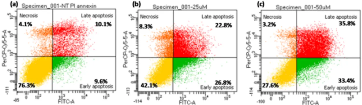
the cancer cell lines, with IC50 values ≤ 25 μM, and, to a minor extent, also towards the fibroblasts. (Table 1). To determine whether the antiproliferative activity induced by dA-nor-CDC was related to apoptosis, as previously reported for the corresponding dA-CDC conjugate [6], K562 cells were treated with compound dA-nor-CDC (25 and 50 μM) for 24 h, then assayed by flow cytometry analysis with Annexin V-FITC staining. The results of the cell apoptosis assay indicated that compound dA-nor-CDC induced apoptosis in a dose dependent manner (Figure 2).
0 20 40 60 80 100 10 µ M 25 µ M 50 µM 10 µ M 25 µ M 50 µM 10 µ M 25 µ M 50 µM 10 µ M 25 µ M 50 µM 10 µ M 25 µ M 50 µM 10 µ M 25 µ M 50 µM
dA-nor-CDC A-CDC A-nor-CDC G-CDC dU-UDC dU-nor-UDC
% Gr
ow
th inhibition
K562
HCT116
dU-TUDCA >0 >50 >100 ALK-dA1 >50 >50 >100 ALK-A >50 >50 >100 ALK-dG >50 >50 >100 ALK-G >50 >50 >100 ALK-dU >50 >50 >100 3α-N3-CDC 23.0 ± 2.0 31.0 ± 2.3 >100 3α-N3-UDC 25.0 ± 1.0 22.0 ± 1.5 >100 3α-N3-TUDCA >50 >50 >100 23-N3-nor-CDC 21 ± 1.2 25 ± 2.0 79 ± 3.1 23-N3-nor-UDC 15 ± 1.0 22 ± 1.4 81 ± 2.0 CISPLATIN 5.4 ± 1.0 8.5 ± 1.2 23.6 ± 3.51data from ref. [6].
Molecules 2017, 22, 1710 6 of 17 dU-TUDCA >0 >50 >100 ALK-dA 1 >50 >50 >100 ALK-A >50 >50 >100 ALK-dG >50 >50 >100 ALK-G >50 >50 >100 ALK-dU >50 >50 >100 3α-N3-CDC 23.0 ± 2.0 31.0 ± 2.3 >100 3α-N3-UDC 25.0 ± 1.0 22.0 ± 1.5 >100 3α-N3-TUDCA >50 >50 >100 23-N3-nor-CDC 21 ± 1.2 25 ± 2.0 79 ± 3.1 23-N3-nor-UDC 15 ± 1.0 22 ± 1.4 81 ± 2.0 CISPLATIN 5.4 ± 1.0 8.5 ± 1.2 23.6 ± 3.5 1 data from ref. [6].
Figure 1. Antiproliferative activity of the most active conjugates against K562 and HCT116 cancer cells at 10, 25, 50 μM after 72 h.
In the 2′-deoxyuridine series dU-UDC was found cytotoxic towards both K562 and HCT116, with comparable IC50 values (21.5 and 23.5 μM, respectively), whereas dU-nor-UDC showed
cytoselectivity towards K562, with IC50 = 24.8 μM vs. HCT116 IC50 = 43.0 μM (Figure 1, Table 1). No
cytotoxic activity was found for the conjugates with TUDCA.
In this study we also tested in vitro the nucleoside-alkyne derivatives (namely ALK-A, ALK-G,
ALK-dG and ALK-dU) and the N3-BA building blocks (namely 3α-N3-CDC, 3α-N3-UDC,
3α-N3-TUDCA, 23-N3-nor-CDC and 23-N3-nor-UDC). The nucleoside-alkyne derivatives were found
to consistently not be cytotoxic towards any of tested cell lines with the results previously reported for ALK-dA [6]. As far as for the 3α-N3-BA is concerned, we found that 3α-N3-CDC and 3α-N3-UDC
are active against both cancer cell lines indiscriminately and not active towards the fibroblast cells up to 100 μM, whereas 3α-N3-TUDCA was found to not be cytotoxic against any of the cell lines at
the concentrations tested. On the other hand, the 23-N3-nor-BA series showed cytotoxicity against
the cancer cell lines, with IC50 values ≤ 25 μM, and, to a minor extent, also towards the fibroblasts.
(Table 1). To determine whether the antiproliferative activity induced by dA-nor-CDC was related to apoptosis, as previously reported for the corresponding dA-CDC conjugate [6], K562 cells were treated with compound dA-nor-CDC (25 and 50 μM) for 24 h, then assayed by flow cytometry analysis with Annexin V-FITC staining. The results of the cell apoptosis assay indicated that compound dA-nor-CDC induced apoptosis in a dose dependent manner (Figure 2).
0 20 40 60 80 100 10 µ M 25 µ M 50 µM 10 µ M 25 µ M 50 µM 10 µ M 25 µ M 50 µM 10 µ M 25 µ M 50 µM 10 µ M 25 µ M 50 µM 10 µ M 25 µ M 50 µM
dA-nor-CDC A-CDC A-nor-CDC G-CDC dU-UDC dU-nor-UDC
% Gr
ow
th inhibition
K562
HCT116
Figure 1.Antiproliferative activity of the most active conjugates against K562 and HCT116 cancer cells at 10, 25, 50 µM after 72 h.
In the 20-deoxyuridine series dU-UDC was found cytotoxic towards both K562 and HCT116, with comparable IC50values (21.5 and 23.5 µM, respectively), whereas dU-nor-UDC showed cytoselectivity
towards K562, with IC50= 24.8 µM vs. HCT116 IC50= 43.0 µM (Figure1, Table1). No cytotoxic activity
In this study we also tested in vitro the nucleoside-alkyne derivatives (namely ALK-A, ALK-G, ALK-dG and ALK-dU) and the N3-BA building blocks (namely 3α-N3-CDC, 3α-N3-UDC,
3α-N3-TUDCA, 23-N3-nor-CDC and 23-N3-nor-UDC). The nucleoside-alkyne derivatives were found to consistently not be cytotoxic towards any of tested cell lines with the results previously reported for ALK-dA[6]. As far as for the 3α-N3-BA is concerned, we found that 3α-N3-CDCand 3α-N3-UDC are active against both cancer cell lines indiscriminately and not active towards the fibroblast cells up to 100 µM, whereas 3α-N3-TUDCAwas found to not be cytotoxic against any of the cell lines at the concentrations tested. On the other hand, the 23-N3-nor-BA series showed cytotoxicity against the cancer cell lines, with IC50values≤25 µM, and, to a minor extent, also towards the fibroblasts.
(Table1). To determine whether the antiproliferative activity induced by dA-nor-CDC was related to apoptosis, as previously reported for the corresponding dA-CDC conjugate [6], K562 cells were treated with compound dA-nor-CDC (25 and 50 µM) for 24 h, then assayed by flow cytometry analysis with Annexin V-FITC staining. The results of the cell apoptosis assay indicated that compound dA-nor-CDC induced apoptosis in a dose dependent manner (FigureMolecules 2017, 22, 1710 2). 7 of 17
Figure 2. dA-nor-CDC induced apoptosis in K562 cells after 24 h treatment: (a) control: untreated
cells; (b) dA-nor-CDC 25 μM; (c) dA-nor-CDC 50 μM. 3. Discussion
The reported in vitro screening highlighted some compounds with an interesting anticancer activity, with IC50 values ≤ 25 μM, which are dA-nor-CDC, A-nor-CDC, dU-UDC and dU-nor-UDC with respect to K562 leukemia cells and dA-nor-CDC, A-CDC, G-CDC and dU-UDC with respect to HCT116 colon carcinoma (Table 1, Figure 1). Among them, only dA-nor-CDC and dU-UDC showed good anti-proliferative activity against both K562 and HCT116, with comparable IC50 values. It is worth noting that these two compounds showed also a higher IC50 value (around 100 μM) respect to the other conjugates towards the fibroblast cells therefore the lack of cytoselectivity among the selected cancer lines could be related to the greater activity of the compounds (Table 1).
In agreement with our previous data [6] the A/dA-based bioconjugates were confirmed to be potential active anticancer compounds. Besides, the present screening also evidenced G- and U-based conjugates with interesting cytotoxicity/cytoselectivity. Moreover, it can be observed that CDC/nor-CDC scaffolds conjugated with A/dA nucleosides showed in all cases a fair cytotoxic activity and cytoselectivity. Conversely, UDC/nor-UDC scaffolds showed cytotoxic activity only when coupled with 2′-deoxyuridine (Table 1).
In our previous paper on 2′-deoxyadenosine-BA conjugates, including dA-CDC and dA-UDC those IC50 values are also reported in Table 1 for comparison, we demonstrated that the conjugation of dA with CDC and UDC actually plays a crucial role in the cytotoxic/cytoselective process [6]. Starting from this point we would like to discuss the possible structure-activity relationship in the light of the biological evaluation of the nucleoside-BA conjugates incorporating a triazole moiety herein reported. A marked cytoselectivity trend among the selected cancer lines can be identified in the adenosine series. In fact, compound A-CDC is preferentially cytotoxic against HCT116 cells whereas the corresponding A-nor-CDC derivative showed cytoselectivity towards K562 cells. Therefore, CDC bile acid seems to address the cytoselectivity to HCT116 whereas nor-CDC bile acid does the same for K562 cells. This seems to be supported by the data of both the G and dU series where G-CDC was found highly cytoselective against the HCT116 and dU-nor-CDC cytoselective against the K562 as expected in the light of the previous consideration. However, this hypothesis is in contrast with the cytoselectivity of dA-CDC that is selective against the K562 unless it is a CDC derivative. The overall data also indicate that the CDC/nor-CDC derivatives are more active than the corresponding UDC/nor-UDC except for the conjugates with 2′-deoxyuridine (Figure 2). Therefore, the conjugation with a pyrimidine nucleoside seems to improve the anticancer activity of the UDC/nor-UDC series. Looking more deeply through the biological data it can be seen that also the sugar nature, deoxy- or ribo-, seems to influence the cytoselectivity being the ribo form more active against the HCT116 in the adenine series (comparison between dA-CDC and A-CDC) and also in the corresponding guanine series (G-CDC) (Table 1). Finally, in the case of TUDCA-conjugates no cytotoxic activity was found, as for the corresponding 3α-N3-TUDCA building block. The overall
data herein debated seems to indicate that the cytoselectivity is mainly driven by the BA and can be fine-tuned by the nucleoside nature, i.e., purine or pyrimidine, deoxy- or ribo-.
Figure 2. dA-nor-CDCinduced apoptosis in K562 cells after 24 h treatment: (a) control: untreated cells; (b) dA-nor-CDC 25 µM; (c) dA-nor-CDC 50 µM.
3. Discussion
The reported in vitro screening highlighted some compounds with an interesting anticancer activity, with IC50values≤25 µM, which are dA-nor-CDC, A-nor-CDC, dU-UDC and dU-nor-UDC
with respect to K562 leukemia cells and dA-nor-CDC, A-CDC, G-CDC and dU-UDC with respect to HCT116 colon carcinoma (Table1, Figure1). Among them, only dA-nor-CDC and dU-UDC showed good anti-proliferative activity against both K562 and HCT116, with comparable IC50 values. It is
worth noting that these two compounds showed also a higher IC50value (around 100 µM) respect to
the other conjugates towards the fibroblast cells therefore the lack of cytoselectivity among the selected cancer lines could be related to the greater activity of the compounds (Table1).
In agreement with our previous data [6] the A/dA-based bioconjugates were confirmed to be potential active anticancer compounds. Besides, the present screening also evidenced G- and U-based conjugates with interesting cytotoxicity/cytoselectivity. Moreover, it can be observed that CDC/nor-CDC scaffolds conjugated with A/dA nucleosides showed in all cases a fair cytotoxic activity and cytoselectivity. Conversely, UDC/nor-UDC scaffolds showed cytotoxic activity only when coupled with 20-deoxyuridine (Table1).
In our previous paper on 20-deoxyadenosine-BA conjugates, including dA-CDC and dA-UDC those IC50values are also reported in Table1for comparison, we demonstrated that the conjugation of
dA with CDC and UDC actually plays a crucial role in the cytotoxic/cytoselective process [6]. Starting from this point we would like to discuss the possible structure-activity relationship in the light of the biological evaluation of the nucleoside-BA conjugates incorporating a triazole moiety herein reported. A marked cytoselectivity trend among the selected cancer lines can be identified in the adenosine series. In fact, compound A-CDC is preferentially cytotoxic against HCT116 cells whereas the corresponding
A-nor-CDCderivative showed cytoselectivity towards K562 cells. Therefore, CDC bile acid seems to address the cytoselectivity to HCT116 whereas nor-CDC bile acid does the same for K562 cells. This seems to be supported by the data of both the G and dU series where G-CDC was found highly cytoselective against the HCT116 and dU-nor-CDC cytoselective against the K562 as expected in the light of the previous consideration. However, this hypothesis is in contrast with the cytoselectivity of dA-CDCthat is selective against the K562 unless it is a CDC derivative. The overall data also indicate that the CDC/nor-CDC derivatives are more active than the corresponding UDC/nor-UDC except for the conjugates with 20-deoxyuridine (Figure2). Therefore, the conjugation with a pyrimidine nucleoside seems to improve the anticancer activity of the UDC/nor-UDC series. Looking more deeply through the biological data it can be seen that also the sugar nature, deoxy- or ribo-, seems to influence the cytoselectivity being the ribo form more active against the HCT116 in the adenine series (comparison between dA-CDC and A-CDC) and also in the corresponding guanine series (G-CDC) (Table1). Finally, in the case of TUDCA-conjugates no cytotoxic activity was found, as for the corresponding 3α-N3-TUDCAbuilding block. The overall data herein debated seems to indicate that the cytoselectivity is mainly driven by the BA and can be fine-tuned by the nucleoside nature, i.e., purine or pyrimidine, deoxy- or ribo-.
4. Materials and Methods
4.1. General Information
Reactions were monitored by TLC on pre-coated silica gel plates (thickness 0.25 mm, Merck, Darmstadt, Germany), and phosphomolybdic acid solution was used as the spray reagent to visualize the steroids. Flash column chromatography was performed on silica gel 60 (230–400 mesh). HPLC-MS analyses were performed on an Agilent 1100 HPLC system (Agilent Tech. Inc., Santa Clara, CA, USA) and an Esquire 3000 Plus mass spectrometer (Bruker, Billerica, MA, USA) using a Zorbax C8 column (4.6 mm×150 mm, 5 µm) (linear gradient water/CH3CN at a 0.5 mL/min flow rate, detection at λ 260 nm). ESI-HRMS were
acquired on an Agilent Dual ESI Q TOF 6520 (Agilent Tech. Inc., Santa Clara, CA, USA), in positive-ion mode, using methanol. NMR spectra were recorded for DMSO-d6 solutions, unless
otherwise specified, with a Mercury Plus 400 MHz instrument (Varian, Palo Alto, CA, USA). IR spectra were recorded on a Spectrum 100 FT-IR spectrometer (Perkin–Elmer, Waltham, MA, USA). 8-Br-Adenosine, 8-Br-20-deoxyadenosine, 8-Br-20-deoxyguanosine, 8-Br-guanosine, 5-I-20-deoxyuridine, chenodeoxycholic, ursodeoxycholic, are commercially available compounds that were used without further purification. The corresponding azides, methyl 3α-azido-7α-hydroxy-5β-cholan-24-oate (3α-N3-CDC), and methyl 3α-azido-7β-hydroxy-5β-cholan-24-oate (3α-N3-UDC), were prepared according to the literature procedures [6,17].
4.2. General Procedure for the Synthesis of Alkynes
Alkynes were prepared following the procedure reported in the literature [16]. In all cases they were obtained in 70–80% yield and no chromatographic purification was necessary. An analytical sample for the characterization analyses was obtained after flash chromatography using CH2Cl2:MeOH = 9:1 as eluent in all cases.
8-(1,7-Octadynyl)-adenosine (ALK-A)1H-NMR δ = 8.18 (1H, s, H2), 7.56 (2H, br s; disappeared upon shaking with D2O; NH2), 5.90 (1H, d, J = 7.2 Hz; collapsing to s upon irradiation at δ 4.98; H10), 5.54
(1H, m; disappeared upon shaking with D2O; C50-OH), 5.39 (1H, d, J = 6.4 Hz; collapsing to s upon
irradiation at δ 4.98; disappeared upon shaking with D2O; C20-OH), 5.16 (1H, d, J = 3.6 Hz; collapsing
to s upon irradiation at δ 4.17; disappeared upon shaking with D2O; C30-OH), 4.98 (1H, m; collapsing
to dd, J1= 7.2 Hz and J2= 6.4 Hz, upon irradiation at δ 4.17; H20), 4.18 (1H, m; H30), 3.96 (1H, m; H40),
3.64 (1H, m; H50), 3.50 (1H, m; H500), 2.53 (3H, m), 2.37 (2H, m), 1.64 (4H, m).13C-NMR: δ = 153.7 (CH), 153.4 (q), 149.5 (q), 132.0 (q), 94.7 (q), 94.6 (q), 89.9 (CH), 87.2 (CH), 72.1 (CH), 71.6 (CH), 66.2 (q), 62.8
(CH), 55.5 (CH2), 27.5 (CH2), 27.3 (CH2), 18.7 (CH2), 18.5 (CH2). MS (ESI, ES+) m/z: 394 (M + 23), 372
(M + 1).
8-(1,7-Octadynyl)-guanosine (ALK-G) 1H-NMR: δ = 5.75 (1H, d, J = 7.2 Hz; collapsing to s upon
irradiation at δ 4.90; H10), 4.98 (1H, m; collapsing to dd, J1= 7.2 Hz and J2= 6.8 Hz, upon irradiation at
δ4.08; H20), 4.08–4.05 (1H, m; H30), 3.94–3.91 (1H, m; H40), 3.64–3.40 (2H, m; H50, H500), 2.75 (1H, t, J = 2.8 Hz), 2.58–2.50 (2H, m), 2.23–2.18 (2H, m), 1.65–1.53 (4H, m).13C-NMR: δ = 163.4 (q), 137.1 (q),
134.6 (q), 131.4 (q), 118.9 (q), 94.5 (q), 89.5 (q), 87.1 (CH), 84.9 (CH), 72.4 (q), 72.1 (CH), 72.1 (CH), 71.8 (CH), 63.3 (CH2), 27.8 (CH2), 26.7 (CH2), 18.8 (CH2), 17.9 (CH2). MS (ESI, ES+) m/z: 410 (M + 23), 388
(M + 1).
8-(1,7-Octadynyl)-20-deoxyguanosine (ALK-dG)1H-NMR: δ = 6.23–6.18 (1H, m; H10), 4.40–4.38 (1H, m;
H30), 3.90–3.83 (1H, m; H40), 3.68–3.60 (1H, m; H50), 3.58–3.42 (1H, m; H500), 3.02–2.95 (1H, m; H200), 2.75 (1H, t, J = 2.8 Hz ), 2.58–2.50 (2H, m), 2.25–2.19 (2H, m), 2.18–1.98 (1H, m; H20), 1.70–1.52 (4H, m).
13C-NMR: δ = 163.4 (q), 137.1 (q), 134.6 (q), 131.4 (q), 118.9 (q), 94.5 (q), 89.5 (q), 87.1 (CH), 84.9 (CH),
72.4 (q), 72.1 (CH), 71.8 (CH), 63.3 (CH2), 40.2 (CH2), 27.8 (CH2), 26.7 (CH2), 18.8 (CH2), 17.9 (CH2).
MS (ESI, ES+) m/z: 394 (M + 23); (ESI, ES−) m/z: 370 (M−1).
5-(1,7-Octadynyl)-20-deoxyuridine (ALK-dU)1H-NMR: δ = 8.10 (1H, s, H6), 6.08 (1H, dd, J1= 6.4 Hz;
collapsing to s upon irradiation at δ 2.08; H10), 5.22 (1H, m; disappeared upon shaking with D2O;
C50-OH), 5.07 (1H, m, disappeared upon shaking with D2O; C30-OH), 4.21–4.18 (1H, m; H30), 3.78 (1H,
m; collapsing to d, J = 2.8 Hz upon irradiation at δ 3.55; H40), 3.58–3.53 (2H, m; H50and H500), 2.74 (1H, t, J = 2.4 Hz; collapsing to s upon irradiation at δ 2.18), 2.362.32 (2H, m), 2.20–2.17 (2H, m), 2.11–2.06 (2H, m, H20and H200), 1.60–1.53 (4H, m).13C-NMR: δ = 163.2 (q), 150.1 (q), 143.4 (CH), 99.7 (q), 93.7 (q), 88.1 (CH), 85.3 (CH), 85.1 (q), 72.0 (q), 70.8 (CH), 61.5 (CH2), 46.4 (CH2), 27.9 (CH2), 27.7 (CH2),
19.0 (CH2), 17.9 (CH2). MS (ESI, ES+) m/z: 665 (2M + 1), 687 (2M + 23), 355 (M + 23), 333 (M + 1).
4.3. Synthesis of Diformyloxy-5β-23-iodio-24-norcholanes
3α,7β-Diformyloxy-5β-23-iodo-24-norcholane and 3α,7α-diformyloxy-5β-23-iodo-24-nor-cholane were prepared from the corresponding bile acid according to the literature procedure [18]. A mixture of bile acid (2.5 mmol) and formic acid (4 mL) was stirred at 55◦C for 24 h and concentrated in vacuo. The residue was crystallized by adding water to warm EtOH solution and used in the next step. A solution of diformyloxy bile acid (0.5 mmol) and DIH (228 mg, 0.6 mmol) in DCE (4 mL) was irradiated for 2 h under reflux conditions. After chromatography on silica gel (eluent, 0:100 to 50:50 EtOAc/hexane) 23-I-nor-UDC and 23-I-nor-CDC were obtained.
3α,7β-Diformyloxy-5β-23-iodio-24-norcholane (23-I-nor-UDC). Yield 85%1H-NMR (CDCl3): δ = 7.99
(s, 1H), 7.97 (s, 1H), 4.94–4.75 (m, 2H), 3.33–3.23 (m, 1H), 3.12–3.02 (m, 1H), 2.05–1.12 (m, 24H), 0.98 (s, 3H), 0.91 (d, J = 6.15 Hz, 3H), 0.64 (s, 3H);13C-NMR (CDCl3): δ = 161.0 (q), 160.6 (q), 73.5 (CH), 73.3
(CH), 55.2 (CH), 54.8 (CH), 43.7 (q), 42.0 (CH), 40.1 (CH2), 39.8 (CH2), 39.7 (CH), 39.4 (CH), 36.9 (CH),
34.3 (CH2), 33.9 (q), 32.8 (CH2), 32.7 (CH2), 28.3 (CH2), 26.3 (CH2), 25.8 (CH2), 23.2 (CH3), 21.2 (CH2),
17.86 (CH3), 12.0 (CH3), 5.3 (CH2). MS (ESI, ES+) m/z: 553 (M + 23).
3α,7α-Diformyloxy-5β-23-iodio-24-norcholane (23-I-nor-CDC). Yield 90%1H-NMR (CDCl3): δ = 8.08
(s, 1H), 8.02 (s, 1H), 5.03 (br s, 1H), 4.78–4.65 (m, 1H), 3.38–3.23 (m, 1H), 3.17–3.04 (m, 1H), 2.18–1.03 (m, 24H), 0.98 (s, 3H), 0.96 (d, J = 6.2 Hz, 3H), 0.64 (s, 3H);13C-NMR (CDCl3): δ = 160.8 (q), 74.1 (CH),
71.4 (CH), 55.6 (CH), 50.1 (CH), 42.8 (q), 40.9 (CH), 40.2 (CH2), 39.4 (CH2), 37.9 (CH), 37.1 (CH), 34.8
(CH2), 34.6 (CH), 34.0 (q), 33.8 (CH2), 31.5 (CH2), 28.0 (CH2), 26.8 (CH2), 23.5 (CH2), 22.7 (CH3), 20.6
(CH2), 17.9 (CH3), 11.8 (CH3), 5.2 (CH2). MS (ESI, ES+) m/z: 553 (M + 23).
4.4. General Procedure for the Synthesis of Nor-Azides
The 23-iodo derivative (0.5 mmol) was dissolved in DMF (3 mL) and NaN3(4 mmol) was added.
extracted twice with Et2O (12 mL). The combined organic layers were dried over anhydrous Na2SO4,
filtered and concentrated in vacuo to give the diformyloxy azido-compound. The pale yellow solid was treated with 25% NaOH in MeOH at room temperature monitoring by TLC (AcOEt/cyclohexane 1:1) until disappearing of the starting material (2 h for UDC, 12 h for CDC). The corresponding dihydroxy azido derivatives 23-N3-nor-UDCand 23-N3-nor-CDC were precipitated by adding water to the solution.
3α,7β-Dihydroxy-5β-23-azido-24-norcholane (23-N3-nor-UDC). Amorphous white solid, yield 75%; IR: ν (cm−1) 3593 (O-H), 3447 (O-H), 2970–2855 (C-H), 2086 (N3);1H-NMR: δ = 4.43 (d, J = 4.5 Hz, 1H), 3.86 (d, J = 6.8 Hz, 1H), 3.42–3.31 (m, 1H), 3.29–3.18 (m, 3H), 1.98–1.58 (m, 6H), 1.51–0.91 (m, 18H), 0.89 (d, J = 6.4 Hz, 3H), 0.82 (s, 3H), 0.59 (s, 3H);13C-NMR: δ = 69.7 (CH), 69.4 (CH), 55.8 (CH), 54.8 (CH), 48.3 (CH2), 43.1 (q), 42.9 (CH), 42.1 (CH), 39.7 (CH2), 38.7 (CH), 37.7 (CH2), 37.2 (CH2), 34.8 (CH2), 34.3 (CH2), 33.7 (q), 33.1 (CH), 30.2 (CH2), 28.2 (CH2), 26.7 (CH2), 23.3 (CH3), 20.8 (CH2), 18.4 (CH3), 11.9 (CH3). MS (ESI, ES+) m/z: 1191 (3M + 23).
3α,7α-Dihydroxy-5β-23-azido-24-norcholane (23-N3-nor-CDC). Amorphous white solid, yield 78%; IR: ν (cm−1) 3439 (O-H), 2970–2864 (C-H), 2091 (N3);1H-NMR: δ = 4.29 (d, J = 4.6 Hz, 1H), 4.11 (d, J = 3.2 Hz, 1H), 3.61 (br s, 1H), 3.44–3.38 (m, 1H), 3.29–3.08 (m, 2H), 2.22–2.09 (m, 1H), 1.95–1.58 (m, 8H), 1.48–0.96 (m, 15H), 0.89 (d, J = 6.5 Hz, 3H), 0.82 (s, 3H), 0.60 (s, 3H);13C-NMR: δ = 70.3 (CH), 66.1 (CH), 55.7 (CH), 50.0 (CH), 48.3 (CH2), 42.0 (q), 41.4 (CH), 39.6 (CH2), 39.3 (CH2), 39.1 (CH), 35.3 (CH2), 34.8 (CH2), 34.7 (q), 34.2 (CH2), 33.2 (CH), 32.3 (CH), 30.5 (CH2), 27.9 (CH2), 23.1 (CH2), 22.7 (CH3), 20.2 (CH2), 18.2 (CH3), 11.6 (CH3). MS (ESI, ES+) m/z: 1191 (3M + 23).
4.5. Synthesis of (3α-Azido-7β-hydroxy-5β-cholanoate) (3α-N3-UDCA)
LiOH (1.5 M, 15 mL, 23 mmol) was added to a solution of 3α-N3-UDC(1.0 g, 2.3 mmol) in methanol (10 mL). The mixture was stirred at room temperature for 21 h. Then 2N HCl was added until pH = 4–5 and the solution was extracted with ethyl acetate (2×20 mL). The combined organic phase was washed with water, dried over MgSO4, and concentrated in vacuo to afford a white powder.
Yield 94%1H-NMR: δ = 11.93 (br s, 1H), 3.91 (br s, 1H), 3.41–3.22 (m, 2H), 2.25–0.82 (m, 32H), 0.60 (s, 3H);13C-NMR: δ = 174.8 (q), 69.1 (CH), 60.0 (CH), 55.4 (CH), 54.5 (CH), 42.9 (CH), 42.8 (q), 42.0 (CH),
39.4 (CH2), 38.4 (CH), 37.2 (CH2), 34.7 (CH), 34.5 (CH2), 33.6 (q), 32.7 (CH2), 30.6 (CH2), 28.0 (CH2),
26.6 (CH2), 26.0 (CH2), 23.1 (CH3), 20.7 (CH2), 18.2 (CH3), 11.9 (CH3). MS (ESI, ES+) m/z: 440 (M + 23).
4.6. Synthesis of (3α-Azido-7β-hydroxy-5β-cholan-24-oyl)-2-aminoethanesulfonic Acid (3α-N3-TUDCA) To a solution of 3α-azido-7β-hydroxy-5β-cholanoate (500 mg, 1.19 mmol) in anhydrous THF (5 mL) stirred at 0◦C were added triethylamine (0.18 mL, 1.3 mmol) and ethyl chloroformate (0.13 mL, 1.3 mmol). After 2 h at room temperature a solution of taurine (136 mg, 1.3 mmol) in NaOH/H2O
(1 mL, 1.43 mmol) was added. The reaction mixture was stirred at room temperature overnight and then acidified with 5% HCl to pH 1. After evaporation of THF, the mixture was diluted with water and washed with EtOAc. The aqueous phase was extracted with n-butanol and the organic layer dried over anhydrous Na2SO4, filtered and concentrated under reduced pressure to gove the title compound
as an amorphous white solid, yield 80%; IR: ν (cm−1) 3309 (O-H), 2931–2866 (C-H), 2090 (N3), 1648
(C=O);1H-NMR: δ = 7.72 (br s, 1H), 6.83–6.80 (m, 1H), 3.98–3.88 (m, 2H), 3.28–3.17 (m, 2H), 3.15–3.01 (m, 2H), 2.68–2.75 (m, 1H), 2.57–2.48 (m, 2H), 2.08–0.93(m, 23H), 0.87 (s, 3H), 0.85 (d, J = 6.4 Hz, 3H), 0.59 (s, 3H);13C-NMR: δ = 171.9 (q), 69.0 (CH), 60.0 (q), 55.4 (CH), 54.5 (CH), 50.5 (CH2), 45.6 (CH2), 42.9 (CH), 42.8 (q), 42.1 (CH), 40.0 (CH), 38.4 (CH), 37.2 (CH2), 35.4 (CH2), 34.8 (CH), 34.5 (CH2), 33.6 (CH2), 32.5 (CH2), 31.4 (CH2), 28.0 (CH2), 26.6 (CH2), 26.0 (CH2), 23.1 (CH3), 20.7 (CH2), 18.4 (CH3), 11.9 (CH3). MS (ESI, ES+) m/z: 547 (M + 23).
4.7. General Procedure for the “Click” Reaction
To a solution of the appropriate alkyne ALK-dA, ALK-A, ALK-G, ALK-dG, ALK-dU (0.03 mmol) in 1.4 mL of a 1:1:1.5 mixture of H2O/tert-BuOH /THF (v/v), sodium ascorbate (0.06 mmol) and
copper(II) sulfate (0.012 mmol) were added. Then the appropriate azide 3α-N3-CDC, 3α-N3-UDC,
23-N3-nor-CDC, 23-N3-nor-UDC, 3α-N3-TUDCA(0.045 mmol) was added and the resulting solution was stirred at room temperature overnight.
4.7.1. Method A (Purification of Conjugates with 3α-Azides)
The mixture was concentrated under reduced pressure, added with water and extracted with dichloromethane. The organic layers was dried over Na2SO4, filtered and concentrated in vacuo.
The resulting crude solid was washed three times with Et2O.
4.7.2. Method B (Purification of Conjugates with nor-azides)
The mixture was concentrated in vacuo until the complete elimination of THF and tert-BuOH. The crude precipitated solid was filtered, washed with water, EtOH, EtOAc and finally dried with Et2O.
4.7.3. Method C (Purification of Conjugates with TUDCA-Azides)
The mixture was concentrated under reduced pressure, added with water and extracted with n-butanol. The organic layers was dried over Na2SO4, filtered and concentrated in vacuo. The crude
white solid was washed twice with EtOH (10 mL) and dried with Et2O.
A-CDC. Colourless syrup, yield 80%; IR: ν (cm−1) 3418–3315 (O-H), 2928–2866 (C-H), 2241 (C≡C), 1693 (C=O), 1665–1524 (C=C,C=N);1H-NMR: δ = 8.18 (br s, 1H), 7.88 (s, 1H), 7.58 (br s, 2H), 5.98 (d, J = 6.83 Hz 1H), 5.61–5.58 (m, 1H), 5.42 (d, J = 6.25 Hz 1H), 5.20 (d, J = 4.30 Hz, 1H), 5.12–4.98 (m, 1H), 4.32–4.16 (m, 3H), 3.98 (m, 1H), 3.72–3.42 (s, 7H), 2.74–2.59 (m, 5H), 2.38–2.16 (m, 3H), 2.01–0.95 (m, 25H), 0.90 (s, 3H), 0.86 (d, J = 6.44 Hz, 3H), 0.60 (s, 3H);13C-NMR: δ = 173.8 (q), 156.0 (q), 153.2 (CH), 148.3 (q), 146.1 (q), 134.0 (q), 120.0 (q), 119.9 (CH), 97.4 (q), 89.3 (CH), 86.6 (CH), 71.5 (CH), 71.08 (CH), 70.3 (q), 66.1 (CH), 62.2 (CH2), 60.1 (CH), 55.4 (CH), 51.2 (CH3), 49.9 (CH), 41.9 (CH), 41.7 (q), 40.1 (CH2), 37.7 (CH2), 35.4 (CH2), 34.9 (q), 34.8 (CH), 34.4 (CH2), 32.2 (CH), 30.6 (CH2), 30.4 (CH2), 28.2 (CH2), 27.7 (CH2), 27.2 (CH2), 27.1 (CH2), 24.6 (CH2), 23.1 (CH2), 22.6 (CH3), 20.3 (CH2), 18.4 (CH2),
18.1 (CH3), 11.6 (CH3). HRMS calculated for [C43H62N8O7+ H]+803.4814, found 803.4819.
A-UDC. Colourless syrup, yield 78%; IR: ν (cm−1) 3411 (O-H), 2926–2865 (C–H), 2240 (C≡C), 1693 (C=O), 1660–1524 (C=C,C=N); 1H-NMR: δ = 8.18 (br s, 1H), 8.02 (s, 1H), 7.58 (br s, 2H), 5.98 (d, J = 6.83 Hz, 1H), 5.61–5.58 (m, 1H), 5.42 (d, J = 6.23 Hz, 1H), 5.20 (d, J = 4.31 Hz, 1H), 5.12–4.98 (m, 1H), 4.38 (br, s, 1H), 4.18 (br, s, 1H), 3.98 (br, s, 1H), 3.91 (br, s, 1H), 3.71–3.62 (m, 1H), 3.58–3.45 (m, 4H), 2.74–2.59 (m, 5H), 2.38–2.11 (m, 5H), 2.01–0.95 (m, 25H), 0.92 (s, 3H), 0.84 (d, J = 6.44 Hz, 3H), 0.60 (s, 3H);13C-NMR: δ = 174.2 (q), 156.4 (q), 153.4 (CH), 148.7 (q), 142.8 (q), 133.9 (q), 120.4 (q), 120.2 (CH), 98.0 (q), 88.8 (CH), 85.6 (CH), 71.8 (q), 70.8 (CH), 66.5 (CH), 62.7 (CH2), 60.5 (CH), 55.9 (CH), 51.6 (CH3), 50.4 (CH), 42.4 (q), 42.1 (CH), 39.6 (CH), 38.0 (CH2), 37.7 (CH2), 35.9 (CH2), 35.3 (CH), 35.3 (CH2), 34.9 (q), 32.7 (CH2), 31.1 (CH), 30.8 (CH2), 28.7 (CH2), 28.2 (CH2), 27.6 (CH2), 27.5 (CH2), 25.0 (CH2), 23.5 (CH2), 23.1 (CH2), 20.7 (CH3), 18.8 (CH2), 18.6 (CH3), 12.1 (CH3); HRMS calculated for [C43H62N8O7+ H]+803.4814, found 803.4829.
dG-CDC. Colourless syrup, yield 76%; IR: ν (cm−1) 3327 (O-H), 2933–2865 (C-H), 2243 (C≡C), 1692 (C=O), 1629–1568 (C=C,C=N);1H-NMR: δ = 10.82 (br, s, 1H), 7.85 (s, 1H), 6.57 (br, s, 2H), 6.25–6.19 (m, 1H), 5.22 (br, s, 1H), 4.86 (br, s, 1H), 4.4–4.16 (m, 3H), 3.8 (m, 1H), 3.6–3.58 (m, 2H), 3.59 (s, 3H), 3.57–3.42 (m, 2H), 3.15–2.99 (m, 1H), 2.71–2.62 (m, 2H), 2.53 (m, 2H), 2.38–2.01 (m, 5H), 1.92–0.99 (m, 25H), 0.89 (s, 3H), 0.86 (d, J = 6.40 Hz, 3H), 0.60 (s, 3H);13C-NMR: δ = 173.6 (q), 156.5 (q), 153.6 (q), 150.5 (q), 146.3 (q), 130.1 (q), 121.6 (CH), 116.8 (q), 95.2 (q), 87.6 (CH), 84.6 (CH), 70.9 (q), 70.6 (CH), 70.5 (CH), 66.0 (CH), 62.0 (CH2), 60.0 (CH), 55.4 (CH), 51.1 (CH3), 49.8 (CH), 41.9 (CH), 41.6 (q), 40.0
(CH2), 39.6 (CH), 38.8 (CH2), 37.1 (CH2), 35.4 (CH2), 34.7 (q), 34.3 (CH2), 32.1 (CH), 30.6 (CH2), 30.3
(CH2), 28.1 (CH2), 27.7 (CH2), 27.1 (CH2), 24.5 (CH2), 23.0 (CH2), 22.6 (CH3), 20.2 (CH2), 18.3 (CH2),
18.2 (CH3), 11.6 (CH3). HRMS calculated for [C43H62N8O7+ H]+803.4814, found 803.4815.
dG-UDC. Colourless syrup, yield 78%; IR: ν (cm−1) 3312 (O-H), 2929–2860 (C-H), 2240 (C≡C), 1686 (C=O), 1601–1565 (C=C,C=N);1H-NMR: δ = 10.78 (br, s, 1H), 7.99 (s, 1H), 6.48 (br, s, 2H), 6.28–6.18 (m, 1H), 5.22 (br, s, 1H), 4.93–4.82 (m, 1H), 4,38 (br, s, 2H), 3.91 (d, J = 6.45 Hz, 1H), 3.79–3.75 (m, 1H), 3.62–3.41 (m, 5H), 3.19–2.98 (m, 1H), 2.68–0.99 (m, 36H), 0.97 (s, 3H), 0.86 (d, J = 6.44 Hz, 3H), 0.59 (s, 3H);13C-NMR: δ = 173.7 (q), 158.3 (q), 155.9 (q), 153.6 (q), 150.5 (q), 149.9 (q), 129.5 (q), 119.8 (CH), 94.0 (q), 87.6 (CH), 83.6 (CH), 71.8 (q), 71.0 (CH), 68.9 (CH), 68.0 (CH), 62.0 (CH2), 59.5 (CH), 55.2 (CH), 54.5 (CH), 51.1 (CH3), 42.9 (CH), 42.4 (CH), 41.8 (q), 40.0 (CH2), 37.2 (CH2), 36.8 (CH2), 34.9 (CH2), 34.7 (CH), 34.0 (q), 33.7 (CH2), 30.6 (CH2), 30.2 (CH2), 28.1 (CH2), 27.5 (CH2), 27.1 (CH2), 26.5 (CH2), 24.5 (CH2), 23.8 (CH2), 23.1 (CH3), 20.8 (CH2), 18.2 (CH2), 18.1 (CH3), 11.9 (CH3). HRMS calculated for [C43H62N8O7+ H]+803.4814, found 803.4816.
G-CDC. Colourless syrup, yield 80%; IR: ν (cm−1) 3330 (O-H), 2918–2850 (C-H), 2238 (C≡C), 1736 (C=O), 1645–1580 (C=C,C=N); 1H-NMR: δ = 10.78 (br, s, 1H), 7.88 (s, 1H), 6.48 (br, s, 2H), 5.78 (d, J = 6.44 Hz, 1H), 5.40 (d, J = 6.25 Hz, 1H), 5.11 (d, J = 4.88, 1H), 4.99–4.82 (m, 2H), 4,32–4.18 (m, 2H), 4.15 (br, s, 1H), 3.82 (br, s, 1H), 3.63–3.41 (m, 4H), 2.75–2.15 (m, 5H), 1.96–0.99 (m, 31 H), 0.88 (s, 3H), 0.84 (d, J = 6.44 Hz, 3H), 0.60 (s, 3H);13C-NMR: δ = 173.7 (q), 157.4 (q), 155.9 (q), 153.7 (q), 150.7 (q), 130.2 (q), 119.9 (CH), 116.8 (q), 95.2 (q), 88.3 (CH), 85.5 (CH), 70.9 (q), 70.6 (CH), 70.5 (CH), 66.0 (CH), 62.0 (CH2), 60.0 (CH), 55.4 (CH), 51.1 (CH3), 49.8 (CH), 41.9 (CH), 41.6 (q), 40.0 (CH2), 39.6 (CH), 38.8 (CH2), 37.1 (CH2), 35.4 (CH2), 34.8 (CH), 34.7 (q), 34.3 (CH2), 32.1 (CH), 30.6 (CH2), 30.3 (CH2), 28.1 (CH2), 27.7 (CH2), 27.1 (CH2), 24.5 (CH2), 23.0 (CH2), 22.5 (CH3), 20.2 (CH2), 18.3 (CH2), 18.1 (CH3),
11.6 (CH3). HRMS calculated for [C43H62N8O8+ H]+819.4763, found 819.4768.
G-UDC. Light yellow syrup, yield 78%; IR: ν (cm−1) 3327 (O-H), 2930–2875 (C-H), 2238 (C≡C), 1735 (C=O), 1645–1572 (C=C,C=N); 1H-NMR: δ = 10.78 (br, s, 1H), 8.02 (s, 1H), 6.48 (br, s, 2H), 5.78 (d, J = 6.43 Hz, 1H), 5.40 (d, J = 6.24 Hz, 1H), 5.11 (d, J = 4.88, 1H), 4.99–4.82 (m, 2H), 4.42–4.35(m, 1H), 4,18–4.12 (m, 1H), 3.91 (d, J = 6.64 Hz, 1H), 3.823.78 (m, 1H), 3.68–3.38 (m, 6H), 2.71–0.98 (m, 34H), 0.92 (s, 3H), 0.83 (d, J = 6.44 Hz, 3H), 0.58 (s, 3H);13C-NMR: δ = 173.5 (q), 156.0 (q), 153.7 (q), 150.7 (q), 146.3 (q), 130.2 (q), 121.6 (CH), 116.8 (q), 95.2 (q), 88.3 (CH), 85.5 (CH), 71.8 (q), 71.0 (CH), 68.9 (CH), 68.0 (CH), 62.0 (CH2), 59.5 (CH), 55.2 (CH), 54.5 (CH), 51.1 (CH3), 42.9 (CH), 42.4 (CH), 41.8 (q), 40.0 (CH2), 37.2 (CH2), 36.8 (CH2), 34.9 (CH2), 34.7 (CH), 34.0 (q), 33.7 (CH2), 30.6 (CH2), 30.2 (CH2), 28.1 (CH2), 27.5 (CH2), 27.1 (CH2), 26.5 (CH2), 24.5 (CH2), 23.8 (CH2), 23.1 (CH3), 20.8 (CH2), 18.2 (CH2),
18.1 (CH3), 11.9 (CH3). HRMS calculated for [C43H62N8O7+ H]+819.4763, found 819.4766.
dU-CDC. Amorphous white solid, yield 90%; IR: ν (cm−1) 3440–3310 (O-H), 2930–2861 (C-H), 2244 (C≡C), 1693 (C=O), 1633–1565 (C=C,C=N);1H-NMR: δ = 8.62 (s, 1H), 7.81 (s, 1H), 6.42 (s, 1H), 6.17–6.12 (m, 1H), 5.28 (d, J = 4.3 Hz, 1H), 5.14–5.09 (m, 1H), 4.24–4.18 (m, 3H), 3.88 (q, J = 3.71 Hz, 1H), 3.67–3.58 (m, 3H), 3.57 (s, 3H), 2.78–2.58 (m, 5H), 2.40–2.24 (m, 2H), 2.23–2.13 (m, 1H), 2.08–1.98 (m, 1H), 1.92–0.95 (m, 27H), 0.89 (s, 3H), 0.86 (d, J = 6.44 Hz, 3H), 0.60 (s, 3H);13C-NMR: δ = 173.8 (q), 171.2 (q), 158.1 (q), 153.8 (q), 146.1 (q), 136.8 (CH), 119.8 (CH), 106.4 (q) , 99.9 (q), 88.1 (CH), 87.4 (CH), 69.7 (CH), 66.1 (CH), 60.8 (CH), 60.1 (CH2), 55.5 (CH), 51.2 (CH), 49.9 (CH3), 42.0 (q), 41.7 (CH), 41.21 (CH), 37.3 (CH2), 35.4 (CH2), 34.9 (CH), 34.8 (CH2), 34.4 (q), 32.2 (CH), 30.7 (CH2), 30.4 (CH2), 28.3 (CH2), 27.8 (CH2), 27.2 (CH2), 27.1 (CH2), 26.0 (CH2), 24.7 (CH2), 23.1 (CH2), 22.6 (CH3), 20.3 (CH2), 18.2 (CH3), 11.7 (CH3);
HRMS calculated for [C42H61N5O8+ H]+764.4592, found 764.4602.
dU-UDC. Amorphous white solid, yield 70%; IR: ν (cm−1) 3447–3309 (O-H), 3016–2942 (C-H), 2254 (C≡C), 1690 (C=O), 1645–1522 (C=C,C=N);1H-NMR: δ = 11.58 (s, 1H), 8.18 (s, 1H), 7.98 (s, 1H), 6.12–6.07 (m, 1H), 5.28–5.21 (m, 1H), 5.18–5.15 (m, 1H), 4.42–4.28 (m, 1H), 4.21 (br s, 1H), 3.95 (d, J = 6.64 Hz, 1H), 3.78 (br s, 1H), 3.65–3.48 (m, 4H), 2.65–2.58 (m, 2H), 2.41–0.98 (m, 36H), 0.95 (s, 3H), 0.85 (d, J = 6.44 Hz, 3H), 0.62 (s, 3H);13C-NMR: δ = 175.4 (q), 171.8 (q), 162.2 (q), 149.9 (q), 146.7 (q),